Search This Blog

Wednesday, December 11, 2024

CICE (sea ice model)

From Wikipedia, the free encyclopedia
https://en.wikipedia.org/wiki/CICE_(sea_ice_model)

CICE (/ss/) is a computer model that simulates the growth, melt and movement of sea ice. It has been integrated into many coupled climate system models as well as global ocean and weather forecasting models and is often used as a tool in Arctic and Southern Ocean research. CICE development began in the mid-1990s by the United States Department of Energy (DOE), and it is currently maintained and developed by a group of institutions in North America and Europe known as the CICE Consortium. Its widespread use in Earth system science in part owes to the importance of sea ice in determining Earth's planetary albedo, the strength of the global thermohaline circulation in the world's oceans, and in providing surface boundary conditions for atmospheric circulation models, since sea ice occupies a significant proportion (4-6%) of Earth's surface. CICE is a type of cryospheric model.

Development

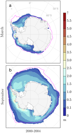
Depiction of Antarctic sea ice simulated by the Community Earth System Model
Output from CICE within a coupled climate model: Averaged 2000-2004 (a) March and (b) September Antarctic sea ice thickness and extent (sea ice with greater than 15% concentration) of five ensemble members from the Community Earth System Model (CESM) large ensemble. The magenta contour is the measured ice edge according to the NOAA Climate Data Record.

Development of CICE began in 1994 by Elizabeth Hunke at Los Alamos National Laboratory (LANL). Since its initial release in 1998 following development of the Elastic-Viscous-Plastic (EVP) sea ice rheology within the model, it has been substantially developed by an international community of model users and developers. Enthalpy-conserving thermodynamics and improvements to the sea ice thickness distribution were added to the model between 1998 and 2005. The first institutional user outside of LANL was Naval Postgraduate School in the late-1990s, where it was subsequently incorporated into the Regional Arctic System Model (RASM) in 2011. The National Center for Atmospheric Research (NCAR) was the first to incorporate CICE into a global climate model in 2002, and developers of the NCAR Community Earth System Model (CESM) have continued to contribute to CICE innovations and have used it to investigate polar variability in Earth's climate system. The United States Navy began using CICE shortly after 2000 for polar research and sea ice forecasting and it continues to do so today. Since 2000, CICE development or coupling to oceanic and atmospheric models for weather and climate prediction has occurred at the University of Reading, University College London, the U.K. Met Office Hadley Centre, Environment and Climate Change Canada, the Danish Meteorological Institute, the Commonwealth Science and Industrial Research Organisation, and Beijing Normal University, among other institutions. As a result of model development in the global community of CICE users, the model's computer code now includes a comprehensive saline ice physics and biogeochemistry library that incorporates mushy-layer thermodynamics, anisotropic continuum mechanics, Delta-Eddington radiative transfer, melt-pond physics and land-fast ice. CICE version 6 is open-source software and was released in 2018 on GitHub.

Keystone Equations

There are two main physics equations solved using numerical methods in CICE that underpin the model's predictions of sea ice thickness, concentration and velocity, as well as predictions made with many equations not shown here giving, for example, surface albedo, ice salinity, snow cover, divergence, and biogeochemical cycles. The first keystone equation is Newton's second law for sea ice:

where is the mass per unit area of saline ice on the sea surface, is the drift velocity of the ice, is the Coriolis parameter, is the upward unit vector normal to the sea surface, and are the wind and water stress on the ice, respectively, is acceleration due to gravity, is sea surface height and is internal ice the two-dimensional stress tensor within the ice. Each of the terms require information about the ice thickness, roughness, and concentration, as well as the state of the atmospheric and oceanic boundary layers. Ice mass per unit area is determined using the second keystone equation in CICE, which describes evolution of the sea ice thickness distribution for different thicknesses spread of the area for which sea ice velocity is calculated above:

where is the change in the thickness distribution due to thermodynamic growth and melt, is redistribution function due to sea ice mechanics and is associated with internal ice stress , and describes advection of sea ice in a Lagrangian reference frame. From this, ice mass is given by:

for density of sea ice.

Code Design

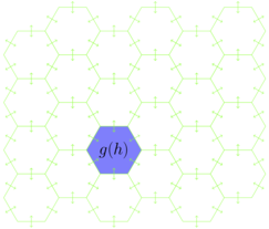
Icepack on an unstructured grid decor
Schematic demonstrating placement of Icepack, in which the thickness distribution is represented (blue), within the MPAS dycore (green) that solves for momentum evolution and horizontal sea ice advection on the E3SM unstructured grid (arrows)

CICE version 6 is coded in FORTRAN90. It is organized into a dynamical core (dycore) and a separate column physics package called Icepack, which is maintained as a CICE submodule on GitHub. The momentum equation and thickness advection described above are time-stepped on a quadrilateral Arakawa B-grid within the dynamical core, while Icepack solves diagnostic and prognostic equations necessary for calculating radiation physics, hydrology, thermodynamics, and vertical biogeochemistry, including terms necessary to calculate , , , , and defined above. CICE can be run independently, as in the first figure on this page, but is frequently coupled with earth systems models through an external flux coupler, such as the CESM Flux Coupler from NCAR for which results are shown in the second figure for the CESM Large Ensemble. The column physics were separated into Icepack for the version 6 release to permit insertion into earth system models that use their own sea ice dynamical core, including the new DOE Energy Exascale Earth System Model (E3SM), which uses an unstructured grid in the sea ice component of the Model for Prediction Across Scales (MPAS), as demonstrated in the final figure.

Stressor

From Wikipedia, the free encyclopedia

A stressor is a chemical or biological agent, environmental condition, external stimulus or an event seen as causing stress to an organism. Psychologically speaking, a stressor can be events or environments that individuals might consider demanding, challenging, and/or threatening individual safety.

Events or objects that may trigger a stress response may include:

Stressors can cause physical, chemical and mental responses internally. Physical stressors produce mechanical stresses on skin, bones, ligaments, tendons, muscles and nerves that cause tissue deformation and (in extreme cases) tissue failure. Chemical stresses also produce biomechanical responses associated with metabolism and tissue repair. Physical stressors may produce pain and impair work performance. Chronic pain and impairment requiring medical attention may result from extreme physical stressors or if there is not sufficient recovery time between successive exposures. Stressors may also affect mental function and performance. Mental and social stressors may affect behavior and how individuals respond to physical and chemical stressors.

Social and environmental stressors and the events associated with them can range from minor to traumatic. Traumatic events involve very debilitating stressors, and oftentimes these stressors are uncontrollable. Traumatic events can deplete an individual's coping resources to an extent where the individual may develop acute stress disorder or even post-traumatic stress disorder. People who have been abused, victimized, or terrorized are often more susceptible to stress disorders. Most stressor-stress relationships can be evaluated and determined - either by the individual or by a psychologist. Therapeutic measures are often taken to help replenish and rebuild the individual's coping resources while simultaneously aiding the individual in dealing with current stress.

Psychological stressors

Stressors occur when an individual is unable to cope with the demands of their environment (such as crippling debt with no clear path to resolving it). Generally, stressors take many forms, such as: traumatic events, life demands, sudden medical emergencies, and daily inconveniences, to name a few. There are also a variety of characteristics that a stressor may possess (different durations, intensity, predictability, and controllability).

Measuring psychological stress

Due to the wide impact and the far-reaching consequences of psychological stressors (especially their profound effects on mental well-being), it is particularly important to devise tools to measure such stressors. Two common psychological stress tests include the Perceived Stress Scale (PSS) devised by American psychologist Sheldon Cohen, and the Social Readjustment Rating Scale (SRRS) or the Holmes-Rahe Stress Scale. While the PSS is a traditional Likert scale, the SRRS assigns specific predefined numerical values to stressors.

Biological responses to stressors

Traumatic events or any type of shock to the body can cause an acute stress response disorder (ASD). The extent to which one experiences ASD depends on the extent of the shock. If the shock was pushed past a certain extreme after a particular period in time ASD can develop into what is commonly known as Post-traumatic stress disorder (PTSD). There are two ways that the body responds biologically in order to reduce the amount of stress an individual is experiencing. One thing that the body does to combat stressors is to create stress hormones, which in turn create energy reservoirs that are there in case a stressful event were to occur. The second way our biological components respond is through an individual's cells. Depending on the situation our cells obtain more energy in order to combat any negative stressor and any other activity those cells are involved in seize.

One possible mechanism of stressors influencing biological pathways involves stimulation of the hypothalamus, CRF (corticotropin release factor) causing the pituitary gland to releases ACTH (adrenocorticotropic hormone), which causes the adrenal cortex to secrete various stress hormones (e.g., cortisol). Stress hormones travel in the blood stream to relevant organs, e.g., glands, heart, intestines, triggering a flight-or-fight response. Between this flow there is an alternate path that can be taken after the stressor is transferred to the hypothalamus, which leads to the sympathetic nervous system; after which the adrenal medulla secretes epinephrine.

Predictability and controllability

When individuals are informed about events before they occur, the magnitude of the stressor is less than when compared to individuals who were not informed of the stressor. For example, an individual would prefer to know when they have a deadline ahead of time in order to prepare for it in advance, rather than find out about the deadline the day of. In knowing that there is a deadline ahead of time, the intensity of the stressor is smaller for the individual, as opposed to the magnitude of intensity for the other unfortunate individual who found out about the deadline the day of. When this was tested, psychologists found that when given the choice, individuals had a preference for the predictable stressors, rather than the unpredictable stressors. The pathologies caused by the lack of predictability are experienced by some individuals working in fields of emergency medicine, military defense, disaster response and others.

Additionally, the degree to which the stressor can be controlled plays a variable in how the individual perceives stress. Research has found that if an individual is able to take some control over the stressor, then the level of stress will be decreased. During this study, it was found that the individuals become increasingly anxious and distressed if they were unable to control their environment. As an example, imagine an individual who detests baths in the Middle Ages, taking a bath. If the individual was forced to take the bath with no control over the temperature of the bath (one of the variables), then their anxiety and stress levels would be higher than if the individual was given some control over the environment (such as being able to control the temperature of the water).

Based on these two principles (predictability and control), there are two hypotheses that attempt to account for these preferences; the preparatory response hypothesis and safety hypothesis attempt to accommodate these preferences.

Preparatory response hypothesis

The idea behind this hypothesis is that an organism can better prepare for an event if they are informed beforehand, as this allows them to prepare for it (biologically). In biologically preparing for this event beforehand, the individual is able to better decrease the event's aversiveness. In knowing when a potential stressor will occur (such as an exam), the individual could, in theory, prepare for it in advance, thus decreasing the stress that may result from that event.

Safety hypothesis

In this hypothesis, there are two time periods, one in which is deemed safe (where there is no stressor), and one which is deemed unsafe (in which the stressor is present). This is similar to procrastination and cramming; during the safe intervals (weeks before an exam) the individual is relaxed and not anxious, and during the unsafe intervals (the day or night before the exam) the individual most likely experiences anxiety.

Stellar engine

From Wikipedia, the free encyclopedia
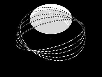
Diagram of a class-C stellar engine (to scale) built around a Sun-like star. It consists of a partial Dyson swarm composed of 5 Dyson rings of solar collectors (the class-B component), and a large statite Shkadov thruster (the class-A component). Perspective is from below the system's ecliptic at a distance of ~2.8 AU. The system's direction of acceleration is on a vector from the center of the star through the center of the Shkadov thruster, which is hovering over the star's north pole (with regards to the ecliptic), at a distance of 1 AU.

Stellar engines are a class of hypothetical megastructures which use the resources of a star to generate available work (also called exergy). For instance, they can use the energy of the star to produce mechanical, electrical or chemical work or they can use the impulse of the light emitted by the star to produce thrust, able to control the motion of a star system. The concept has been introduced by Bădescu and Cathcart. The variants which produce thrust may accelerate a star and anything orbiting it in a given direction. The creation of such a system would make its builders a type-II civilization on the Kardashev scale.

Classes

Three classes of stellar engines have been defined.

Class A (Shkadov thruster)

One of the simplest examples of a stellar engine is the Shkadov thruster (named after Dr. Leonid Shkadov, who first proposed it), or a class-A stellar engine. Such an engine is a stellar propulsion system, consisting of an enormous mirror/light sail—actually a massive type of solar statite large enough to classify as a megastructure—which would balance gravitational attraction towards and radiation pressure away from the star. Since the radiation pressure of the star would now be asymmetrical, i.e. more radiation being emitted in one direction as compared to another, the "excess" radiation pressure acts as net thrust, accelerating the star in the direction of the hovering statite. Such thrust and acceleration would be very slight, but such a system could be stable for millennia. Any planetary system attached to the star would be "dragged" along by its parent star. For a star such as the Sun, with luminosity 3.85×1026 W and mass 1.99×1030 kg, the total thrust produced by reflecting half of the solar output would be 1.28×10¹⁸ N. After a period of one million years this would yield an imparted speed of 20 m/s, with a displacement from the original position of 0.03 light-years. After one billion years, the speed would be 20 km/s and the displacement 34,000 light-years, a little over a third of the estimated width of the Milky Way galaxy.

Class B

A class-B stellar engine consists of two concentric spheres around a star. The inner sphere (which may be assimilated with a Dyson shell) receives energy from the star and becomes hotter than the outer sphere. The difference of temperature between the two spheres drives thermal engines able to provide mechanical work.

Unlike the Shkadov thruster, a class-B stellar engine is not propulsive.

Class C

A class-C stellar engine, such as the Badescu–Cathcart engine, combines the two other classes, employing both the propulsive aspects of the Shkadov thruster and the energy generating aspects of a class-B engine. A higher temperature Dyson shell partially covered by a mirror combined with an outer sphere at a lower temperature would be one incarnation of such a system. The non-spherical mirror ensures conversion of light impulse into effective thrust (like a class-A stellar engine) while the difference of temperature may be used to convert star energy into mechanical work (like a class-B stellar engine). Notice that such system suffers from the same stabilization problems as a non-propulsive shell, as would be a Dyson swarm with a large statite mirror (see image above). A Dyson bubble variant is already a Shkadov thruster (provided that the arrangement of statite components is asymmetrical); adding energy extraction capability to the components seems an almost trivial extension.

Caplan thruster

Astronomer Matthew E. Caplan of Illinois State University has proposed a type of stellar engine that uses concentrated stellar energy (repurposing the mirror statites from class A) to excite certain regions of the outer surface of the star and create beams of solar wind for collection by a multi-Bussard ramjet assembly. The ramjets would produce directed plasma to stabilize its orbit and jets of oxygen-14 to push the star. Using rudimentary calculations that assume maximum efficiency, Caplan estimates that the Bussard engine would use 1012 kg of solar material per second to produce a maximum acceleration of 10−9 m/s2, yielding a velocity of 200 km/s after 5 million years and a distance of 10 parsecs over 1 million years. While theoretically the Bussard engine would work for 100 million years, given the mass loss rate of the Sun, Caplan deems 10 million years to be sufficient for a stellar collision avoidance. His proposal was commissioned by the German educational YouTube channel Kurzgesagt.

Svoronos Star Tug

Alexander A. Svoronos of Yale University proposed the 'Star Tug', a concept that combines aspects of the Shkadov thruster and Caplan engine to produce an even more powerful and efficient mechanism for controlling a star's movement. Essentially, it replaces the giant parabolic mirror of the Shkadov thruster with an engine powered by mass lifted from the star, similar to the Caplan engine. However, instead of pushing a star from behind with a beam of thrust, as the Caplan engine does, it pulls the star from the front via its gravitational link to it, same as the Shkadov thruster. As a result, it only needs to produce a single beam of thrust (toward but narrowly missing the star), whereas the Caplan engine must produce two beams of thrust (one to push the star from behind and negate the force of gravity between the engine and the star, and one to propel the system as a whole forward). The result is that the Svoronos Star Tug is a much more efficient engine capable of significantly higher accelerations and max velocities. The Svoronos Star Tug can, in principle (assuming perfect efficiency), accelerate the Sun to ~27% the speed of light (after burning enough of the Sun's mass to transition it to a brown dwarf).

Bile acid

From Wikipedia, the free encyclopedia
https://en.wikipedia.org/wiki/Bile_acid

Bile acids are steroid acids found predominantly in the bile of mammals and other vertebrates. Diverse
bile acids are synthesized in the liver. Bile acids are conjugated with taurine or glycine residues to give anions called bile salts.

Primary bile acids are those synthesized by the liver. Secondary bile acids result from bacterial actions in the colon. In humans, taurocholic acid and glycocholic acid (derivatives of cholic acid) and taurochenodeoxycholic acid and glycochenodeoxycholic acid (derivatives of chenodeoxycholic acid) are the major bile salts. They are roughly equal in concentration. The salts of their 7-alpha-dehydroxylated derivatives, deoxycholic acid and lithocholic acid, are also found, with derivatives of cholic, chenodeoxycholic and deoxycholic acids accounting for over 90% of human biliary bile acids.

Bile acids comprise about 80% of the organic compounds in bile (others are phospholipids and cholesterol). An increased secretion of bile acids produces an increase in bile flow. Bile acids facilitate digestion of dietary fats and oils. They serve as micelle-forming surfactants, which encapsulate nutrients, facilitating their absorption. These micelles are suspended in the chyme before further processing. Bile acids also have hormonal actions throughout the body, particularly through the farnesoid X receptor and GPBAR1 (also known as TGR5).

Bile acid synthesis is the only manner in which humans or other mammals may excrete excess cholesterol, as the parent compound of all bile acids is cholesterol.

Structure of cholic acid showing relationship to other bile acids

Production

Primary bile acids

Bile acid synthesis occurs in liver cells, which synthesize primary bile acids (cholic acid and chenodeoxycholic acid in humans) via cytochrome P450-mediated oxidation of cholesterol in a multi-step process. Approximately 600 mg of bile salts are synthesized daily to replace bile acids lost in the feces, although, as described below, much larger amounts are secreted, reabsorbed in the gut and recycled.

The rate-limiting step in synthesis is the addition of a hydroxyl group of the 7th position of the steroid nucleus by the enzyme cholesterol 7 alpha-hydroxylase. This enzyme is down-regulated by cholic acid, up-regulated by cholesterol and is inhibited by the actions of the ileal hormone FGF15/19.

Prior to secreting any of the bile acids (primary or secondary, see below), liver cells conjugate them with either glycine or taurine, to form a total of 8 possible conjugated bile acids. These conjugated bile acids are often referred to as bile salts. The pKa of the unconjugated bile acids are between 5 and 6.5, and the pH of the duodenum ranges between 3 and 5, so when unconjugated bile acids are in the duodenum, they are almost always protonated (HA form), which makes them relatively insoluble in water. Conjugating bile acids with amino acids lowers the pKa of the bile-acid/amino-acid conjugate to between 1 and 4. Thus conjugated bile acids are almost always in their deprotonated (A-) form in the duodenum, which makes them much more water-soluble and much more able to fulfil their physiologic function of emulsifying fats.

Secondary bile acids

Once secreted into the lumen of the intestine, bile salts are modified by gut bacteria. They are partially dehydroxylated. Their glycine and taurine groups are removed to give the secondary bile acids, deoxycholic acid and lithocholic acid. Cholic acid is converted into deoxycholic acid and chenodeoxycholic acid into lithocholic acid. All four of these bile acids are recycled, in a process known as enterohepatic circulation.

Functions

Lipid digestion

As molecules with hydrophobic and hydrophilic regions, conjugated bile salts sit at the lipid/water interface and, above the right concentration, form micelles. The added solubility of conjugated bile salts aids in their function by preventing passive re-absorption in the small intestine. As a result, the concentration of bile acids/salts in the small intestine is high enough to form micelles and solubilize lipids. "Critical micellar concentration" refers to both an intrinsic property of the bile acid itself and amount of bile acid necessary to function in the spontaneous and dynamic formation of micelles. Bile acid-containing micelles aid lipases to digest lipids and bring them near the intestinal brush border membrane, which results in fat absorption.

Synthesis of bile acids is a major route of cholesterol metabolism in most species other than humans. The body produces about 800 mg of cholesterol per day and about half of that is used for bile acid synthesis producing 400–600 mg daily. Human adults secrete between 12 and 18 g of bile acids into the intestine each day, mostly after meals. The bile acid pool size is between 4–6 g, which means that bile acids are recycled several times each day. About 95% of bile acids are reabsorbed by active transport in the ileum and recycled back to the liver for further secretion into the biliary system and gallbladder. This enterohepatic circulation of bile acids allows a low rate of synthesis, only about 0.3 g/day, but with large amounts being secreted into the intestine.

Bile acids have other functions, including eliminating cholesterol from the body, driving the flow of bile to eliminate certain catabolites (including bilirubin), emulsifying fat-soluble vitamins to enable their absorption, and aiding in motility and the reduction of the bacteria flora found in the small intestine and biliary tract.

Cell signalling

Bile acids have metabolic actions in the body resembling those of hormones, acting through two specific receptors, the farnesoid X receptor and G protein-coupled bile acid receptor/TGR5. They bind less specifically to some other receptors and have been reported to regulate the activity of certain enzymes  and ion channels and the synthesis of diverse substances including endogenous fatty acid ethanolamides.

Structure and synthesis

Bile salts constitute a large family of molecules, composed of a steroid structure with four rings, a five- or eight-carbon side-chain terminating in a carboxylic acid, and several hydroxyl groups, the number and orientation of which is different among the specific bile salts. The four rings are labeled A, B, C, and D, from the farthest to the closest to the side chain with the carboxyl group. The D-ring is smaller by one carbon than the other three. The structure is commonly drawn with A at the left and D at the right. The hydroxyl groups can be in either of two configurations: either up (or out), termed beta (β; often drawn by convention as a solid line), or down, termed alpha (α; displayed as a dashed line). All bile acids have a 3-hydroxyl group, derived from the parent molecule, cholesterol, in which the 3-hydroxyl is beta.

IUPAC recommended ring lettering (left) and atom numbering (right) of the steroid skeleton. The four rings A-D form a sterane core.

The initial step in the classical pathway of hepatic synthesis of bile acids is the enzymatic addition of a 7α hydroxyl group by cholesterol 7α-hydroxylase (CYP7A1) forming 7α-hydroxycholesterol. This is then metabolised to 7α-hydroxy-4-cholesten-3-one. There are multiple steps in bile acid synthesis requiring 14 enzymes in all. These result in the junction between the first two steroid rings (A and B) being altered, making the molecule bent; in this process, the 3-hydroxyl is converted to the α orientation. The simplest 24-carbon bile acid has two hydroxyl groups at positions 3α and 7α. This is 3α,7α-dihydroxy-5β-cholan-24-oic acid, or, as more usually known, chenodeoxycholic acid. This bile acid was first isolated from the domestic goose, from which the "cheno" portion of the name was derived (Greek: χήν = goose). The 5β in the name denotes the orientation of the junction between rings A and B of the steroid nucleus (in this case, they are bent). The term "cholan" denotes a particular steroid structure of 24 carbons, and the "24-oic acid" indicates that the carboxylic acid is found at position 24, at the end of the side-chain. Chenodeoxycholic acid is made by many species, and is the prototypic functional bile acid.

An alternative (acidic) pathway of bile acid synthesis is initiated by mitochondrial sterol 27-hydroxylase (CYP27A1), expressed in liver, and also in macrophages and other tissues. CYP27A1 contributes significantly to total bile acid synthesis by catalyzing sterol side chain oxidation, after which cleavage of a three-carbon unit in the peroxisomes leads to formation of a C24 bile acid. Minor pathways initiated by 25-hydroxylase in the liver and 24-hydroxylase in the brain also may contribute to bile acid synthesis. 7α-hydroxylase (CYP7B1) generates oxysterols, which may be further converted in the liver to CDCA.

Cholic acid, 3α,7α,12α-trihydroxy-5β-cholan-24-oic acid, the most abundant bile acid in humans and many other species, was discovered before chenodeoxycholic acid. It is a tri-hydroxy-bile acid with 3 hydroxyl groups (3α, 7α and 12α). In its synthesis in the liver, 12α hydroxylation is performed by the additional action of CYP8B1. As this had already been described, the discovery of chenodeoxycholic acid (with 2 hydroxyl groups) made this new bile acid a "deoxycholic acid" in that it had one fewer hydroxyl group than cholic acid.

Deoxycholic acid is formed from cholic acid by 7-dehydroxylation, resulting in 2 hydroxyl groups (3α and 12α). This process with chenodeoxycholic acid results in a bile acid with only a 3α hydroxyl group, termed lithocholic acid (litho = stone) having been identified first in a gallstone from a calf. It is poorly water-soluble and rather toxic to cells.

Different vertebrate families have evolved to use modifications of most positions on the steroid nucleus and side-chain of the bile acid structure. To avoid the problems associated with the production of lithocholic acid, most species add a third hydroxyl group to chenodeoxycholic acid. The subsequent removal of the 7α hydroxyl group by intestinal bacteria will then result in a less toxic but still-functional dihydroxy bile acid. Over the course of vertebrate evolution, a number of positions have been chosen for placement of the third hydroxyl group. Initially, the 16α position was favored, in particular in birds. Later, this position was superseded in a large number of species selecting the 12α position. Primates (including humans) utilize 12α for their third hydroxyl group position, producing cholic acid. In mice and other rodents, 6β hydroxylation forms muricholic acids (α or β depending on the 7 hydroxyl position). Pigs have 6α hydroxylation in hyocholic acid (3α,6α,7α-trihydroxy-5β-cholanoic acid), and other species have a hydroxyl group on position 23 of the side-chain.

Many other bile acids have been described, often in small amounts, resulting from bacterial enzymatic or other modifications. The "iso-" epimers have the 3-hydroxyl group in the β position. The "allo-" epimers have the 5α configuration, which changes the relative position of the A and B rings.

Ursodeoxycholic acid was first isolated from bear bile, which has been used medicinally for centuries. Its structure resembles chenodeoxycholic acid but with the 7-hydroxyl group in the β position.

Obeticholic acid, 6α-ethyl-chenodeoxycholic acid, is a semi-synthetic bile acid with greater activity as an FXR agonist, which has been developed as a pharmaceutical agent in certain liver diseases.

Hormonal actions

Bile acids also act as steroid hormones, secreted from the liver, absorbed from the intestine and having various direct metabolic actions in the body through the nuclear receptor Farnesoid X receptor (FXR), also known by its gene name NR1H4. Another bile acid receptor is the cell membrane receptor known as G protein-coupled bile acid receptor 1 or TGR5. Many of their functions as signaling molecules in the liver and the intestines are by activating FXR, whereas TGR5 may be involved in metabolic, endocrine and neurological functions.

Regulation of synthesis

As surfactants or detergents, bile acids are potentially toxic to cells, and so their concentrations are tightly regulated. Activation of FXR in the liver inhibits synthesis of bile acids, and is one mechanism of feedback control when bile acid levels are too high. Secondly, FXR activation by bile acids during absorption in the intestine increases transcription and synthesis of FGF19, which then inhibits bile acid synthesis in the liver.

Metabolic functions

Emerging evidence associates FXR activation with alterations in triglyceride metabolism, glucose metabolism, and liver growth.

Other interactions

Bile acids bind to some other proteins in addition to their hormone receptors (FXR and TGR5) and their transporters. Among these protein targets, the enzyme N-acyl phosphatidylethanolamine-specific phospholipase D (NAPE-PLD) generates bioactive lipid amides (e.g. the endogenous cannabinoid anandamide) that play important roles in several physiological pathways including stress and pain responses, appetite, and lifespan. NAPE-PLD orchestrates a direct cross-talk between lipid amide signals and bile acid physiology.

Clinical significance

Hyperlipidemia

As bile acids are made from endogenous cholesterol, disruption of the enterohepatic circulation of bile acids will lower cholesterol. Bile acid sequestrants bind bile acids in the gut, preventing reabsorption. In so doing, more endogenous cholesterol is shunted into the production of bile acids, thereby lowering cholesterol levels. The sequestered bile acids are then excreted in the feces.

Cholestasis

Tests for bile acids are useful in both human and veterinary medicine, as they aid in the diagnosis of a number of conditions, including types of cholestasis such as intrahepatic cholestasis of pregnancy, portosystemic shunt, and hepatic microvascular dysplasia in dogs. Structural or functional abnormalities of the biliary system result in an increase in bilirubin (jaundice) and in bile acids in the blood. Bile acids are related to the itching (pruritus) which is common in cholestatic conditions such as primary biliary cirrhosis (PBC), primary sclerosing cholangitis or intrahepatic cholestasis of pregnancy. Treatment with ursodeoxycholic acid has been used for many years in these cholestatic disorders.

Gallstones

The relationship of bile acids to cholesterol saturation in bile and cholesterol precipitation to produce gallstones has been studied extensively. Gallstones may result from increased saturation of cholesterol or bilirubin, or from bile stasis. Lower concentrations of bile acids or phospholipids in bile reduce cholesterol solubility and lead to microcrystal formation. Oral therapy with chenodeoxycholic acid and/or ursodeoxycholic acid has been used to dissolve cholesterol gallstones. Stones may recur when treatment is stopped. Bile acid therapy may be of value to prevent stones in certain circumstances such as following bariatric surgery.

Bile acid diarrhea

Excess concentrations of bile acids in the colon are a cause of chronic diarrhea. It is commonly found when the ileum is abnormal or has been surgically removed, as in Crohn's disease, or cause a condition that resembles diarrhea-predominant irritable bowel syndrome (IBS-D). This condition of bile acid diarrhea/bile acid malabsorption can be diagnosed by the SeHCAT test and treated with bile acid sequestrants.

Bile acids and colon cancer

Bile acids may have some importance in the development of colorectal cancer. Deoxycholic acid (DCA) is increased in the colonic contents of humans in response to a high fat diet. In populations with a high incidence of colorectal cancer, fecal concentrations of bile acids are higher, and this association suggests that increased colonic exposure to bile acids could play a role in the development of cancer. In one particular comparison, the fecal DCA concentrations in Native Africans in South Africa (who eat a low fat diet) compared to African Americans (who eat a higher fat diet) was 7.30 vs. 37.51 nmol/g wet weight stool. Native Africans in South Africa have a low incidence rate of colon cancer of less than 1:100,000, compared to the high incidence rate for male African Americans of 72:100,000.

Experimental studies also suggest mechanisms for bile acids in colon cancer. Exposure of colonic cells to high DCA concentrations increase formation of reactive oxygen species, causing oxidative stress, and also increase DNA damage. Mice fed a diet with added DCA mimicking colonic DCA levels in humans on a high fat diet developed colonic neoplasia, including adenomas and adenocarcinomas (cancers), unlike mice fed a control diet producing one-tenth the level of colonic DCA who had no colonic neoplasia.

The effects of ursodeoxycholic acid (UDCA) in modifying the risk of colorectal cancer has been looked at in several studies, particularly in primary sclerosing cholangitis and inflammatory bowel disease, with varying results partly related to dosage. Genetic variation in the key bile acid synthesis enzyme, CYP7A1, influenced the effectiveness of UDCA in colorectal adenoma prevention in a large trial.

Dermatology

Bile acids may be used in subcutaneous injections to remove unwanted fat (see Mesotherapy). Deoxycholic acid as an injectable has received FDA approval to dissolve submental fat. Phase III trials showed significant responses although many subjects had mild adverse reactions of bruising, swelling, pain, numbness, erythema, and firmness around the treated area.

Genome instability

From Wikipedia, the free encyclopedia
https://en.wikipedia.org/wiki/Genome_instability

Genome instability (also genetic instability or genomic instability) refers to a high frequency of mutations within the genome of a cellular lineage. These mutations can include changes in nucleic acid sequences, chromosomal rearrangements or aneuploidy. Genome instability does occur in bacteria. In multicellular organisms genome instability is central to carcinogenesis, and in humans it is also a factor in some neurodegenerative diseases such as amyotrophic lateral sclerosis or the neuromuscular disease myotonic dystrophy.

The sources of genome instability have only recently begun to be elucidated. A high frequency of externally caused DNA damage can be one source of genome instability since DNA damage can cause inaccurate translesion DNA synthesis past the damage or errors in repair, leading to mutation. Another source of genome instability may be epigenetic or mutational reductions in expression of DNA repair genes. Because endogenous (metabolically-caused) DNA damage is very frequent, occurring on average more than 60,000 times a day in the genomes of human cells, any reduced DNA repair is likely an important source of genome instability.

Usual genome situation

Usually, all cells in an individual in a given species (plant or animal) show a constant number of chromosomes, which constitute what is known as the karyotype defining this species (see also List of number of chromosomes of various organisms), although some species present a very high karyotypic variability. In humans, mutations that would change an amino acid within the protein coding region of the genome occur at an average of only 0.35 per generation (less than one mutated protein per generation).

Sometimes, in a species with a stable karyotype, random variations that modify the normal number of chromosomes may be observed. In other cases, there are structural alterations (e.g., chromosomal translocations, deletions) that modify the standard chromosomal complement. In these cases, it is indicated that the affected organism presents genome instability (also genetic instability, or even chromosomic instability). The process of genome instability often leads to a situation of aneuploidy, in which the cells present a chromosomic number that is either higher or lower than the normal complement for the species.

Causes of genome instability

DNA Replication Defects

In the cell cycle, DNA is usually most vulnerable during replication. The replisome must be able to navigate obstacles such as tightly wound chromatin with bound proteins, single and double stranded breaks which can lead to the stalling of the replication fork. Each protein or enzyme in the replisome must perform its function well to result in a perfect copy of DNA. Mutations of proteins such as DNA polymerase or DNA ligase can lead to impairment of replication and lead to spontaneous chromosomal exchanges. Proteins such as Tel1 and Mec1 (ATR, ATM in humans) can detect single and double-stranded breaks and recruit factors such as Rmr3 helicase to stabilize the replication fork in order to prevent its collapse. Mutations in Tel1, Mec1, and Rmr3 helicase result in a significant increase of chromosomal recombination. ATR responds specifically to stalled replication forks and single-stranded breaks resulting from UV damage while ATM responds directly to double-stranded breaks. These proteins also prevent progression into mitosis by inhibiting the firing of late replication origins until the DNA breaks are fixed by phosphorylating CHK1 and CHK2, which results in a signaling cascade arresting the cell in S-phase. For single stranded breaks, replication occurs until the location of the break, then the other strand is nicked to form a double stranded break, which can then be repaired by Break Induced Replication or homologous recombination using the sister chromatid as an error-free template. In addition to S-phase checkpoints, G1 and G2 checkpoints exist to check for transient DNA damage which could be caused by mutagens such as UV damage. An example is the Saccharomyces pombe gene rad9 which arrests the cells in late S/G2 phase in the presence of DNA damage caused by radiation. The yeast cells with defective rad9 failed to arrest following irradiation, continued cell division, and died rapidly; the cells with wild-type rad9 successfully arrested in late S/G2 phase and remained viable. The cells that arrested were able to survive due to the increased time in S/G2 phase allowing for DNA repair enzymes to function fully.

Fragile Sites

There are hotspots in the genome where DNA sequences are prone to gaps and breaks after inhibition of DNA synthesis such as in the aforementioned checkpoint arrest. These sites are called fragile sites, and can occur commonly as naturally present in most mammalian genomes or occur rarely as a result of mutations, such as DNA-repeat expansion. Rare fragile sites can lead to genetic disease such as fragile X mental retardation syndrome, myotonic dystrophy, Friedrich's ataxia, and Huntington's disease, most of which are caused by expansion of repeats at the DNA, RNA, or protein level. Although, seemingly harmful, these common fragile sites are conserved all the way to yeast and bacteria. These ubiquitous sites are characterized by trinucleotide repeats, most commonly CGG, CAG, GAA, and GCN. These trinucleotide repeats can form into hairpins, leading to difficulty of replication. Under replication stress, such as defective machinery or further DNA damage, DNA breaks and gaps can form at these fragile sites. Using a sister chromatid as repair is not a fool-proof backup as the surrounding DNA information of the n and n+1 repeat is virtually the same, leading to copy number variation. For example, the 16th copy of CGG might be mapped to the 13th copy of CGG in the sister chromatid since the surrounding DNA is both CGGCGGCGG..., leading to 3 extra copies of CGG in the final DNA sequence.

Transcription-associated instability

In both E. coli and Saccharomyces pombe, transcription sites tend to have higher recombination and mutation rates. The coding or non-transcribed strand accumulates more mutations than the template strand. This is due to the fact that the coding strand is single-stranded during transcription, which is chemically more unstable than double-stranded DNA. During elongation of transcription, supercoiling can occur behind an elongating RNA polymerase, leading to single-stranded breaks. When the coding strand is single-stranded, it can also hybridize with itself, creating DNA secondary structures that can compromise replication. In E. coli, when attempting to transcribe GAA triplets such as those found in Friedrich's ataxia, the resulting RNA and template strand can form mismatched loops between different repeats, leaving the complementary segment in the coding strand available to form its own loops which impede replication. Furthermore, replication of DNA and transcription of DNA are not temporally independent; they can occur at the same time and lead to collisions between the replication fork and RNA polymerase complex. In S. cerevisiae, Rrm3 helicase is found at highly transcribed genes in the yeast genome, which is recruited to stabilize a stalling replication fork as described above. This suggests that transcription is an obstacle to replication, which can lead to increased stress in the chromatin spanning the short distance between the unwound replication fork and transcription start site, potentially causing single-stranded DNA breaks. In yeast, proteins act as barriers at the 3' of the transcription unit to prevent further travel of the DNA replication fork.

Increase Genetic Variability

In some portions of the genome, variability is essential to survival. One such locale is the Ig genes. In a pre-B cell, the region consists of all V, D, and J segments. During development of the B cell, a specific V, D, and J segment is chosen to be spliced together to form the final gene, which is catalyzed by RAG1 and RAG2 recombinases. Activation-Induced Cytidine Deaminase (AID) then converts cytidine into uracil. Uracil normally does not exist in DNA, and thus the base is excised and the nick is converted into a double-stranded break which is repaired by non-homologous end joining (NHEJ). This procedure is very error-prone and leads to somatic hypermutation. This genomic instability is crucial in ensuring mammalian survival against infection. V, D, J recombination can ensure millions of unique B-cell receptors; however, random repair by NHEJ introduces variation which can create a receptor that can bind with higher affinity to antigens.

In neuronal and neuromuscular disease

Of about 200 neurological and neuromuscular disorders, 15 have a clear link to an inherited or acquired defect in one of the DNA repair pathways or excessive genotoxic oxidative stress. Five of them (xeroderma pigmentosum, Cockayne's syndrome, trichothiodystrophy, Down's syndrome, and triple-A syndrome) have a defect in the DNA nucleotide excision repair pathway. Six (spinocerebellar ataxia with axonal neuropathy-1, Huntington's disease, Alzheimer's disease, Parkinson's disease, Down's syndrome and amyotrophic lateral sclerosis) seem to result from increased oxidative stress, and the inability of the base excision repair pathway to handle the damage to DNA that this causes. Four of them (Huntington's disease, various spinocerebellar ataxias, Friedreich's ataxia and myotonic dystrophy types 1 and 2) often have an unusual expansion of repeat sequences in DNA, likely attributable to genome instability. Four (ataxia-telangiectasia, ataxia-telangiectasia-like disorder, Nijmegen breakage syndrome and Alzheimer's disease) are defective in genes involved in repairing DNA double-strand breaks. Overall, it seems that oxidative stress is a major cause of genomic instability in the brain. A particular neurological disease arises when a pathway that normally prevents oxidative stress is deficient, or a DNA repair pathway that normally repairs damage caused by oxidative stress is deficient.

In cancer

In cancer, genome instability can occur prior to or as a consequence of transformation. Genome instability can refer to the accumulation of extra copies of DNA or chromosomes, chromosomal translocations, chromosomal inversions, chromosome deletions, single-strand breaks in DNA, double-strand breaks in DNA, the intercalation of foreign substances into the DNA double helix, or any abnormal changes in DNA tertiary structure that can cause either the loss of DNA, or the misexpression of genes. Situations of genome instability (as well as aneuploidy) are common in cancer cells, and they are considered a "hallmark" for these cells. The unpredictable nature of these events are also a main contributor to the heterogeneity observed among tumour cells.

It is currently accepted that sporadic tumors (non-familial ones) are originated due to the accumulation of several genetic errors. An average cancer of the breast or colon can have about 60 to 70 protein altering mutations, of which about 3 or 4 may be "driver" mutations, and the remaining ones may be "passenger" mutations Any genetic or epigenetic lesion increasing the mutation rate will have as a consequence an increase in the acquisition of new mutations, increasing then the probability to develop a tumor. During the process of tumorogenesis, it is known that diploid cells acquire mutations in genes responsible for maintaining genome integrity (caretaker genes), as well as in genes that are directly controlling cellular proliferation (gatekeeper genes). Genetic instability can originate due to deficiencies in DNA repair, or due to loss or gain of chromosomes, or due to large scale chromosomal reorganizations. Losing genetic stability will favour tumor development, because it favours the generation of mutants that can be selected by the environment.

The tumor microenvironment has an inhibitory effect on DNA repair pathways contributing to genomic instability, which promotes tumor survival, proliferation, and malignant transformation.

Low frequency of mutations without cancer

The protein coding regions of the human genome, collectively called the exome, constitutes only 1.5% of the total genome. As pointed out above, ordinarily there are only an average of 0.35 mutations in the exome per generation (parent to child) in humans. In the entire genome (including non-protein coding regions) there are only about 70 new mutations per generation in humans.

Cause of mutations in cancer

The likely major underlying cause of mutations in cancer is DNA damage. For example, in the case of lung cancer, DNA damage is caused by agents in exogenous genotoxic tobacco smoke (e.g. acrolein, formaldehyde, acrylonitrile, 1,3-butadiene, acetaldehyde, ethylene oxide and isoprene). Endogenous (metabolically-caused) DNA damage is also very frequent, occurring on average more than 60,000 times a day in the genomes of human cells (see DNA damage (naturally occurring)). Externally and endogenously caused damages may be converted into mutations by inaccurate translesion synthesis or inaccurate DNA repair (e.g. by non-homologous end joining). In addition, DNA damages can also give rise to epigenetic alterations during DNA repair. Both mutations and epigenetic alterations (epimutations) can contribute to progression to cancer.

Very frequent mutations in cancer

As noted above, about 3 or 4 driver mutations and 60 passenger mutations occur in the exome (protein coding region) of a cancer. However, a much larger number of mutations occur in the non-protein-coding regions of DNA. The average number of DNA sequence mutations in the entire genome of a breast cancer tissue sample is about 20,000. In an average melanoma tissue sample (where melanomas have a higher exome mutation frequency) the total number of DNA sequence mutations is about 80,000.

Cause of high frequency of mutations in cancer

The high frequency of mutations in the total genome within cancers suggests that, often, an early carcinogenic alteration may be a deficiency in DNA repair. Mutation rates substantially increase (sometimes by 100-fold) in cells defective in DNA mismatch repair or in homologous recombinational DNA repair. Also, chromosomal rearrangements and aneuploidy increase in humans defective in DNA repair gene BLM.

A deficiency in DNA repair itself can allow DNA damages to accumulate, and error-prone translesion synthesis past some of those damages may give rise to mutations. In addition, faulty repair of these accumulated DNA damages may give rise to epigenetic alterations or epimutations. While a mutation or epimutation in a DNA repair gene itself would not confer a selective advantage, such a repair defect may be carried along as a passenger in a cell when the cell acquires an additional mutation/epimutation that does provide a proliferative advantage. Such cells, with both proliferative advantages and one or more DNA repair defects (causing a very high mutation rate), likely give rise to the 20,000 to 80,000 total genome mutations frequently seen in cancers.

DNA repair deficiency in cancer

In somatic cells, deficiencies in DNA repair sometimes arise by mutations in DNA repair genes, but much more often are due to epigenetic reductions in expression of DNA repair genes. Thus, in a sequence of 113 colorectal cancers, only four had somatic missense mutations in the DNA repair gene MGMT, while the majority of these cancers had reduced MGMT expression due to methylation of the MGMT promoter region. Five reports, listed in the article Epigenetics (see section "DNA repair epigenetics in cancer") presented evidence that between 40% and 90% of colorectal cancers have reduced MGMT expression due to methylation of the MGMT promoter region.

Similarly, for 119 cases of colorectal cancers classified as mismatch repair deficient and lacking DNA repair gene PMS2 expression, Pms2 was deficient in 6 due to mutations in the PMS2 gene, while in 103 cases PMS2 expression was deficient because its pairing partner MLH1 was repressed due to promoter methylation (PMS2 protein is unstable in the absence of MLH1). The other 10 cases of loss of PMS2 expression were likely due to epigenetic overexpression of the microRNA, miR-155, which down-regulates MLH1.

In cancer epigenetics (see section Frequencies of epimutations in DNA repair genes), there is a partial listing of epigenetic deficiencies found in DNA repair genes in sporadic cancers. These include frequencies of between 13–100% of epigenetic defects in genes BRCA1, WRN, FANCB, FANCF, MGMT, MLH1, MSH2, MSH4, ERCC1, XPF, NEIL1 and ATM located in cancers including breast, ovarian, colorectal and head and neck. Two or three epigenetic deficiencies in expression of ERCC1, XPF and/or PMS2 were found to occur simultaneously in the majority of the 49 colon cancers evaluated. Some of these DNA repair deficiencies can be caused by epimutations in microRNAs as summarized in the MicroRNA article section titled miRNA, DNA repair and cancer.

Lymphomas as a consequence of genome instability

Cancers usually result from disruption of a tumor repressor or dysregulation of an oncogene. Knowing that B-cells experience DNA breaks during development can give insight to the genome of lymphomas. Many types of lymphoma are caused by chromosomal translocation, which can arise from breaks in DNA, leading to incorrect joining. In Burkitt's lymphoma, c-myc, an oncogene encoding a transcription factor, is translocated to a position after the promoter of the immunoglobulin gene, leading to dysregulation of c-myc transcription. Since immunoglobulins are essential to a lymphocyte and highly expressed to increase detection of antigens, c-myc is then also highly expressed, leading to transcription of its targets, which are involved in cell proliferation. Mantle cell lymphoma is characterized by fusion of cyclin D1 to the immunoglobulin locus. Cyclin D1 inhibits Rb, a tumor suppressor, leading to tumorigenesis. Follicular lymphoma results from the translocation of the immunoglobulin promoter to the Bcl-2 gene, giving rise to high levels of Bcl-2 protein, which inhibits apoptosis. DNA-damaged B-cells no longer undergo apoptosis, leading to further mutations which could affect driver genes, leading to tumorigenesis. The location of translocation in the oncogene shares structural properties of the target regions of AID, suggesting that the oncogene was a potential target of AID, leading to a double-stranded break that was translocated to the immunoglobulin gene locus through NHEJ repair.
Fossil fuels lobby

  From Wikipedia, the free encyclopedia https://en.wikipedia.org/wiki/Fossil_fuels_lobby ...