Search This Blog

Monday, April 7, 2025

Traumatic brain injury

From Wikipedia, the free encyclopedia
 
Traumatic brain injury
Other namesIntracranial injury, physically induced brain injury
CT scan showing cerebral contusions, hemorrhage within the hemispheres, and subdural hematoma. There is also displaced skull fracture of left transverse parietal and temporal bones.

SpecialtyNeurology, Neurosurgery, Pediatrics
SymptomsPhysical, cognitive, sensory, social, emotional, and behavioral symptoms
TypesMild to severe
CausesTrauma to the head
Risk factorsOld age, alcohol
Diagnostic methodBased on neurological exam, medical imaging
TreatmentBehavioral therapy, speech therapy

A traumatic brain injury (TBI), also known as an intracranial injury, is an injury to the brain caused by an external force. TBI can be classified based on severity ranging from mild traumatic brain injury (mTBI/concussion) to severe traumatic brain injury. TBI can also be characterized based on mechanism (closed or penetrating head injury) or other features (e.g., occurring in a specific location or over a widespread area). Head injury is a broader category that may involve damage to other structures such as the scalp and skull. TBI can result in physical, cognitive, social, emotional and behavioral symptoms, and outcomes can range from complete recovery to permanent disability or death.

Causes include falls, vehicle collisions and violence. Brain trauma occurs as a consequence of a sudden acceleration or deceleration within the cranium or by a complex combination of both movement and sudden impact. In addition to the damage caused at the moment of injury, a variety of events following the injury may result in further injury. These processes may include alterations in cerebral blood flow and pressure within the skull. Some of the imaging techniques used for diagnosis of moderate to severe TBI include computed tomography (CT) and magnetic resonance imaging (MRIs).

Prevention measures include use of seat belts and helmets, not drinking and driving, fall prevention efforts in older adults and safety measures for children. Depending on the injury, treatment required may be minimal or may include interventions such as medications, emergency surgery or surgery years later. Physical therapy, speech therapy, recreation therapy, occupational therapy and vision therapy may be employed for rehabilitation. Counseling, supported employment and community support services may also be useful.

TBI is a major cause of death and disability worldwide, especially in children and young adults. Males sustain traumatic brain injuries around twice as often as females. The 20th century saw developments in diagnosis and treatment that decreased death rates and improved outcomes.

Classification

Traumatic brain injury is defined as damage to the brain resulting from external mechanical force, such as rapid acceleration or deceleration, impact, blast waves, or penetration by a projectile. Brain function is temporarily or permanently impaired and structural damage may or may not be detectable with current technology.

TBI is one of two subsets of acquired brain injury (brain damage that occur after birth); the other subset is non-traumatic brain injury, which does not involve external mechanical force (examples include stroke and infection). All traumatic brain injuries are head injuries, but the latter term may also refer to injury to other parts of the head; however, the terms head injury and brain injury are often used interchangeably. Similarly, brain injuries fall under the classification of central nervous system injuries and neurotrauma. In neuropsychology research literature, in general the term "traumatic brain injury" is used to refer to non-penetrating traumatic brain injuries.

TBI is usually classified based on severity, anatomical features of the injury, and the mechanism (the causative forces). Mechanism-related classification divides TBI into closed and penetrating head injury. A closed (also called nonpenetrating, or blunt) injury occurs when the brain is not exposed. A penetrating, or open, head injury occurs when an object pierces the skull and breaches the dura mater, the outermost membrane surrounding the brain.

Severity

Severity of traumatic brain injury
  GCS PTA LOC
Mild 13–15 <1
day
0–30
minutes
Moderate 9–12 >1 to <7
days
>30 min to
<24 hours
Severe 3–8 >7 days >24
hours

Brain injuries can be classified into mild, moderate, and severe categories. The Glasgow Coma Scale (GCS), the most commonly used system for classifying TBI severity, grades a person's level of consciousness on a scale of 3–15 based on verbal, motor, and eye-opening reactions to stimuli. In general, it is agreed that a TBI with a GCS of 13 or above is mild, 9–12 is moderate, and 8 or below is severe. Similar systems exist for young children; however, the GCS grading system has limited ability to predict outcomes. Because of this, other classification systems such as the one shown in the table are also used to help determine severity. A current model developed by the Department of Defense and Department of Veterans Affairs uses all three criteria of GCS after resuscitation, duration of post-traumatic amnesia (PTA), and loss of consciousness (LOC). It also has been proposed to use changes that are visible on neuroimaging, such as swelling, focal lesions, or diffuse injury as method of classification.

Pathological features

CT scan showing spread of the subdural hematoma (single arrows), midline shift (double arrows)

Systems also exist to classify TBI by its pathological features. Lesions can be extra-axial, (occurring within the skull but outside of the brain) or intra-axial (occurring within the brain tissue). Damage from TBI can be focal or diffuse, confined to specific areas or distributed in a more general manner, respectively; however, it is common for both types of injury to exist in a given case.

Diffuse injury manifests with little apparent damage in neuroimaging studies, but lesions can be seen with microscopy techniques post-mortem, and in the early 2000s, researchers discovered that diffusion tensor imaging (DTI), a way of processing MRI images that shows white matter tracts, was an effective tool for displaying the extent of diffuse axonal injury. Types of injuries considered diffuse include edema (swelling), concussion and diffuse axonal injury, which is widespread damage to axons including white matter tracts and projections to the cortex.

Focal injuries often produce symptoms related to the functions of the damaged area. Research shows that the most common areas to have focal lesions in non-penetrating traumatic brain injury are the orbitofrontal cortex (the lower surface of the frontal lobes) and the anterior temporal lobes, areas that are involved in social behavior, emotion regulation, olfaction, and decision-making, hence the common social/emotional and judgment deficits following moderate-severe TBI. Symptoms such as hemiparesis or aphasia can also occur when less commonly affected areas such as motor or language areas are, respectively, damaged.

One type of focal injury, cerebral laceration, occurs when the tissue is cut or torn. Such tearing is common in orbitofrontal cortex in particular, because of bony protrusions on the interior skull ridge above the eyes. In a similar injury, cerebral contusion (bruising of brain tissue), blood is mixed among tissue. In contrast, intracranial hemorrhage involves bleeding that is not mixed with tissue.

Hematomas, also focal lesions, are collections of blood in or around the brain that can result from hemorrhage. Intracerebral hemorrhage, with bleeding in the brain tissue itself, is an intra-axial lesion. Extra-axial lesions include epidural hematoma, subdural hematoma, subarachnoid hemorrhage, and intraventricular hemorrhage. Epidural hematoma involves bleeding into the area between the skull and the dura mater, the outermost of the three membranes surrounding the brain. In subdural hematoma, bleeding occurs between the dura and the arachnoid mater. Subarachnoid hemorrhage involves bleeding into the space between the arachnoid membrane and the pia mater. Intraventricular hemorrhage occurs when there is bleeding in the ventricles.

Signs and symptoms

Unequal pupil size is potentially a sign of a serious brain injury.

Symptoms are dependent on the type of TBI (diffuse or focal) and the part of the brain that is affected. Unconsciousness tends to last longer for people with injuries on the left side of the brain than for those with injuries on the right. Symptoms are also dependent on the injury's severity. With mild TBI, the patient may remain conscious or may lose consciousness for a few seconds or minutes. Other symptoms of mild TBI include headache, vomiting, nausea, lack of motor coordination, dizziness, difficulty balancing, lightheadedness, blurred vision or tired eyes, ringing in the ears, bad taste in the mouth, fatigue or lethargy, and changes in sleep patterns. Cognitive and emotional symptoms include behavioral or mood changes, confusion, and trouble with memory, concentration, attention, or thinking. Mild TBI symptoms may also be present in moderate and severe injuries.

A person with a moderate or severe TBI may have a headache that does not go away, repeated vomiting or nausea, convulsions, an inability to awaken, dilation of one or both pupils, slurred speech, aphasia (word-finding difficulties), dysarthria (muscle weakness that causes disordered speech), weakness or numbness in the limbs, loss of coordination, confusion, restlessness, or agitation. Common long-term symptoms of moderate to severe TBI are changes in appropriate social behavior, deficits in social judgment, and cognitive changes, especially problems with sustained attention, processing speed, and executive functioning. Alexithymia, a deficiency in identifying, understanding, processing, and describing emotions occurs in 60.9% of individuals with TBI. Cognitive and social deficits have long-term consequences for the daily lives of people with moderate to severe TBI, but can be improved with appropriate rehabilitation.

When the pressure within the skull (intracranial pressure, abbreviated ICP) rises too high, it can be deadly. Signs of increased ICP include decreasing level of consciousness, paralysis or weakness on one side of the body, and a blown pupil, one that fails to constrict in response to light or is slow to do so. Cushing's triad, a slow heart rate with high blood pressure and respiratory depression is a classic manifestation of significantly raised ICP. Anisocoria, unequal pupil size, is another sign of serious TBI. Abnormal posturing, a characteristic positioning of the limbs caused by severe diffuse injury or high ICP, is an ominous sign.

Small children with moderate to severe TBI may have some of these symptoms but have difficulty communicating them. Other signs seen in young children include persistent crying, inability to be consoled, listlessness, refusal to nurse or eat, and irritability.

Causes

The most common causes of TBI in the U.S. include violence, transportation accidents, construction site mishaps, and sports. Motor bikes are major causes, increasing in significance in developing countries as other causes reduce. The estimates that between 1.6 and 3.8 million traumatic brain injuries each year are a result of sports and recreation activities in the US. In children aged two to four, falls are the most common cause of TBI, while in older children traffic accidents compete with falls for this position. TBI is the third most common injury to result from child abuse. Abuse causes 19% of cases of pediatric brain trauma, and the death rate is higher among these cases. Although men are twice as likely to have a TBI, domestic violence is another cause of TBI, as are work-related and industrial accidents. Firearms and blast injuries from explosions are other causes of TBI, which is the leading cause of death and disability in war zones. According to Representative Bill Pascrell (Democrat, NJ), TBI is "the signature injury of the wars in Iraq and Afghanistan."

Mechanism

Physical forces

Ricochet of the brain within the skull may account for the coup-contrecoup phenomenon.

The type, direction, intensity, and duration of forces all contribute to the characteristics and severity of TBI. Forces that may contribute to TBI include angular, rotational, shear, and translational forces.

Even in the absence of an impact, significant acceleration or deceleration of the head can cause TBI; however in most cases, a combination of impact and acceleration is probably to blame. Forces involving the head striking or being struck by something, termed contact or impact loading, are the cause of most focal injuries, and movement of the brain within the skull, termed noncontact or inertial loading, usually causes diffuse injuries. The violent shaking of an infant that causes shaken baby syndrome commonly manifests as diffuse injury. In impact loading, the force sends shock waves through the skull and brain, resulting in tissue damage. Shock waves caused by penetrating injuries can also destroy tissue along the path of a projectile, compounding the damage caused by the missile itself.

Damage may occur directly under the site of impact, or it may occur on the side opposite the impact (coup and contrecoup injury, respectively). When a moving object impacts the stationary head, coup injuries are typical, while contrecoup injuries are usually produced when the moving head strikes a stationary object.

Primary and secondary injury
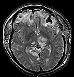
MRI scan showing damage due to brain herniation after TBI

A large percentage of the people killed by brain trauma do not die right away but rather days to weeks after the event; rather than improving after being hospitalized, some 40% of TBI patients deteriorate. Primary brain injury (the damage that occurs at the moment of trauma when tissues and blood vessels are stretched, compressed, and torn) is not adequate to explain this deterioration; rather, it is caused by secondary injury, a complex set of cellular processes and biochemical cascades that occur in the minutes to days following the trauma. These secondary processes can dramatically worsen the damage caused by primary injury and account for the greatest number of TBI deaths occurring in hospitals.

Secondary injury events include damage to the blood–brain barrier, release of factors that cause inflammation, free radical overload, excessive release of the neurotransmitter glutamate (excitotoxicity), influx of calcium and sodium ions into neurons, and dysfunction of mitochondria. Injured axons in the brain's white matter may separate from their cell bodies as a result of secondary injury, potentially killing those neurons. Other factors in secondary injury are changes in the blood flow to the brain; ischemia (insufficient blood flow); cerebral hypoxia (insufficient oxygen in the brain); cerebral edema (swelling of the brain); and raised intracranial pressure (the pressure within the skull). Intracranial pressure may rise due to swelling or a mass effect from a lesion, such as a hemorrhage. As a result, cerebral perfusion pressure (the pressure of blood flow in the brain) is reduced; ischemia results. When the pressure within the skull rises too high, it can cause brain death or brain herniation, in which parts of the brain are squeezed by structures in the skull.

Diagnosis

CT scan showing epidural hematoma (arrow)

Diagnosis is suspected based on lesion circumstances and clinical evidence, most prominently a neurological examination, for example checking whether the pupils constrict normally in response to light and assigning a Glasgow Coma Score. Neuroimaging helps in determining the diagnosis and prognosis and in deciding what treatments to give. DSM-5 can be utilized to diagnose TBI and its psychiatric sequelae.

The preferred radiologic test in the emergency setting is computed tomography (CT): it is quick, accurate, and widely available. Follow-up CT scans may be performed later to determine whether the injury has progressed.

Magnetic resonance imaging (MRI) can show more detail than CT, and can add information about expected outcome in the long term. It is more useful than CT for detecting injury characteristics such as diffuse axonal injury in the longer term; however, MRI is not used in the emergency setting for reasons including its relative inefficacy in detecting bleeds and fractures, its lengthy acquisition of images, the inaccessibility of the patient in the machine, and its incompatibility with metal items used in emergency care. A variant of MRI since 2012 is high-definition fiber tracking (HDFT).

Other techniques may be used to confirm a particular diagnosis. X-rays are still used for head trauma, but evidence suggests they are not useful; head injuries are either so mild that they do not need imaging or severe enough to merit the more accurate CT. Angiography may be used to detect blood vessel pathology when risk factors such as penetrating head trauma are involved. Functional imaging can measure cerebral blood flow or metabolism, inferring neuronal activity in specific regions and potentially helping to predict outcome.

Neuropsychological assessment can be performed to evaluate the long-term cognitive sequelae and to aid in the planning of the rehabilitation. Instruments range from short measures of general mental functioning to complete batteries formed of different domain-specific tests.

Prevention

Demonstration in 1912 of the Warren Safety Helmet, which was designed to protect pilots but has often been wrongly described as a football helmet
Protective sports equipment such as helmets can partially protect athletes from head injury.

Since a major cause of TBI are vehicle accidents, their prevention or the amelioration of their consequences can both reduce the incidence and gravity of TBI. In accidents, damage can be reduced by use of seat belts, child safety seats and motorcycle helmets, and presence of roll bars and airbags. Education programs exist to lower the number of crashes. In addition, changes to public policy and safety laws can be made; these include speed limits, seat belt and helmet laws, and road engineering practices.

Changes to common practices in sports have also been discussed. An increase in use of helmets could reduce the incidence of TBI. Due to the possibility that repeatedly "heading" a ball practicing soccer could cause cumulative brain injury, the idea of introducing protective headgear for players has been proposed. Improved equipment design can enhance safety; softer baseballs reduce head injury risk. Rules against dangerous types of contact, such as "spear tackling" in American football, when one player tackles another head first, may also reduce head injury rates.

Falls can be avoided by installing grab bars in bathrooms and handrails on stairways; removing tripping hazards such as throw rugs; or installing window guards and safety gates at the top and bottom of stairs around young children. Playgrounds with shock-absorbing surfaces such as mulch or sand also prevent head injuries. Child abuse prevention is another tactic; programs exist to prevent shaken baby syndrome by educating about the dangers of shaking children. Gun safety, including keeping guns unloaded and locked, is another preventative measure. Studies on the effect of laws that aim to control access to guns in the United States have been insufficient to determine their effectiveness preventing number of deaths or injuries.

Treatment

It is important to begin emergency treatment within the so-called "golden hour" following the injury. People with moderate to severe injuries are likely to receive treatment in an intensive care unit followed by a neurosurgical ward. Treatment depends on the recovery stage of the patient. In the acute stage, the primary aim is to stabilize the patient and focus on preventing further injury. This is done because the initial damage caused by trauma cannot be reversed. Rehabilitation is the main treatment for the subacute and chronic stages of recovery. International clinical guidelines have been proposed with the aim of guiding decisions in TBI treatment, as defined by an authoritative examination of current evidence.

Acute stage

Tranexamic acid within three hours of a head injury decreases the risk of death. Certain facilities are equipped to handle TBI better than others; initial measures include transporting patients to an appropriate treatment center. Both during transport and in hospital the primary concerns are ensuring proper oxygen supply, maintaining adequate blood flow to the brain, and controlling raised intracranial pressure (ICP), since high ICP deprives the brain of badly needed blood flow and can cause deadly brain herniation. Other methods to prevent damage include management of other injuries and prevention of seizures. Some data supports the use of hyperbaric oxygen therapy to improve outcomes. Further research is required to determine the effectiveness and clinical importance of positioning the head at different angles (degrees of head-of-bed elevation) while the person is being treated in intensive care.

Neuroimaging is helpful but not flawless in detecting raised ICP. A more accurate way to measure ICP is to place a catheter into a ventricle of the brain, which has the added benefit of allowing cerebrospinal fluid to drain, releasing pressure in the skull. Treatment of raised ICP may be as simple as tilting the person's bed and straightening the head to promote blood flow through the veins of the neck. Sedatives, analgesics and paralytic agents are often used. Propofol and midazolam are equally effective as sedatives.

Hypertonic saline can improve ICP by reducing the amount of cerebral water (swelling), though it is used with caution to avoid electrolyte imbalances or heart failure. Mannitol, an osmotic diuretic, appears to be as effective as hypertonic saline at reducing ICP; however, some concerns have been raised regarding some of the studies performed. Hyertonic saline is also suitable in children with severe traumatic brain injury.

Diuretics, drugs that increase urine output to reduce excessive fluid in the system, may be used to treat high intracranial pressures, but may cause hypovolemia (insufficient blood volume). Hyperventilation (larger and/or faster breaths) reduces carbon dioxide levels and causes blood vessels to constrict; this decreases blood flow to the brain and reduces ICP, but it potentially causes ischemia and is, therefore, used only in the short term. Giving corticosteroids is associated with an increased risk of death, and so their routine use is not recommended. There is no strong evidence that the following pharmaceutical interventions should be recommended to routinely treat TBI: magnesium, monoaminergic and dopamine agonists, progesterone, aminosteroids, excitatory amino acid reuptake inhibitors, beta-2 antagonists (bronchodilators), haemostatic and antifibrinolytic drugs.

Endotracheal intubation and mechanical ventilation may be used to ensure proper oxygen supply and provide a secure airway. Hypotension (low blood pressure), which has a devastating outcome in TBI, can be prevented by giving intravenous fluids to maintain a normal blood pressure. Failing to maintain blood pressure can result in inadequate blood flow to the brain. Blood pressure may be kept at an artificially high level under controlled conditions by infusion of norepinephrine or similar drugs; this helps maintain cerebral perfusion. Body temperature is carefully regulated because increased temperature raises the brain's metabolic needs, potentially depriving it of nutrients. Seizures are common. While they can be treated with benzodiazepines, these drugs are used carefully because they can depress breathing and lower blood pressure. Anti-convulsant medications have only been found to be useful for reducing the risk of an early seizure. Phenytoin and Levetiracetam appear to have similar levels of effectiveness for preventing early seizures. People with TBI are more susceptible to side effects and may react adversely to some medications. During treatment monitoring continues for signs of deterioration such as a decreasing level of consciousness.

Traumatic brain injury may cause a range of serious coincidental complications that include cardiac arrhythmias and neurogenic pulmonary edema. These conditions must be adequately treated and stabilised as part of the core care. Surgery can be performed on mass lesions or to eliminate objects that have penetrated the brain. Mass lesions such as contusions or hematomas causing a significant mass effect (shift of intracranial structures) are considered emergencies and are removed surgically. For intracranial hematomas, the collected blood may be removed using suction or forceps or it may be floated off with water. Surgeons look for hemorrhaging blood vessels and seek to control bleeding. In penetrating brain injury, damaged tissue is surgically debrided, and craniotomy may be needed. Craniotomy, in which part of the skull is removed, may be needed to remove pieces of fractured skull or objects embedded in the brain. Decompressive craniectomy (DC) is performed routinely in the very short period following TBI during operations to treat hematomas; part of the skull is removed temporarily (primary DC). DC performed hours or days after TBI in order to control persistently high intracranial pressures (secondary DC), although can reduce intracranial pressure and length of stay in ICU, but have worse Glasgow Coma Scale (GCS) scores, and high chances of death, vegetative state, or severe disability when compared to those receiving standard medical therapies.

Chronic stage
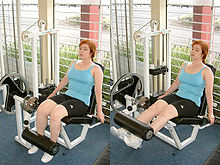
Physical therapy will commonly include muscle strength exercise.

Once medically stable, people may be transferred to a subacute rehabilitation unit of the medical center or to an independent rehabilitation hospital. Rehabilitation aims to improve independent functioning at home and in society, and to help adapt to disabilities. Rehabilitation has demonstrated its general effectiveness when conducted by a team of health professionals who specialize in head trauma. As for any person with neurologic deficits, a multidisciplinary approach is key to optimizing outcome. Physiatrists or neurologists are likely to be the key medical staff involved, but depending on the person, doctors of other medical specialties may also be helpful. Allied health professions such as physiotherapy, speech and language therapy, cognitive rehabilitation therapy, and occupational therapy will be essential to assess function and design the rehabilitation activities for each person. Treatment of neuropsychiatric symptoms such as emotional distress and clinical depression may involve mental health professionals such as therapists, psychologists, and psychiatrists, while neuropsychologists can help to evaluate and manage cognitive deficits. Social workers, rehabilitation support personnel, nutritionists, therapeutic recreationists, and pharmacists are also important members of the TBI rehabilitation team. After discharge from the inpatient rehabilitation treatment unit, care may be given on an outpatient basis. Community-based rehabilitation will be required for a high proportion of people, including vocational rehabilitation; this supportive employment matches job demands to the worker's abilities. People with TBI who cannot live independently or with family may require care in supported living facilities such as group homes. Respite care, including day centers and leisure facilities for disabled people, offers time off for caregivers, and activities for people with TBI.

Pharmacological treatment can help to manage psychiatric or behavioral problems. Medication is also used to control post-traumatic epilepsy; however the preventive use of anti-epileptics is not recommended. In those cases where the person is bedridden due to a reduction of consciousness, has to remain in a wheelchair because of mobility problems, or has any other problem heavily impacting self-caring capacities, caregiving and nursing are critical. The most effective research documented intervention approach is the activation database guided EEG biofeedback approach, which has shown significant improvements in memory abilities of the TBI subject that are far superior than traditional approaches (strategies, computers, medication intervention). Gains of 2.61 standard deviations have been documented. The TBI's auditory memory ability was superior to the control group after the treatment.

There is a promising technology called activation database-guided EEG biofeedback, which has been documented to return a TBI's auditory memory ability to above the control group's performance.

Effect on the gait pattern

The Amsterdam Gait Classification facilitates the assessment of the gait pattern in patients after a traumatic brain injury. It helps to facilitate communication in the interdisciplinary team between those affected, doctors, physiotherapists and orthotists.

In patients who have developed paralysis of the legs in the form of spastic hemiplegia or diplegia as a result of the traumatic brain injury, various gait patterns can be observed, the exact extent of which can only be described with the help of complex gait analysis systems. In order to facilitate interdisciplinary communication in the interdisciplinary team between those affected, doctors, physiotherapists and orthotists, a simple description of the gait pattern is useful. J. Rodda and H. K. Graham already described in 2001 how gait patterns of CP patients can be more easily recognized and defined gait types which they compared in a classification. They also described that gait patterns can vary with age. Building on this, the Amsterdam Gait Classification was developed at the free university in Amsterdam, the VU medisch centrum. A special feature of this classification is that it makes different gait patterns very recognizable and can be used in patients in whom only one leg and both legs are affected. The Amsterdam Gait Classification was developed for viewing patients with cerebral palsy; however, it can be used just as well in patients with traumatic brain injuries. According to the Amsterdam Gait Classification, five gait types are described. To assess the gait pattern, the patient is viewed visually or via a video recording from the side of the leg to be assessed. At the point in time at which the leg to be viewed is in mid stance and the leg not to be viewed is in mid swing, the knee angle and the contact of the foot with the ground are assessed on the one hand.

Classification of the gait pattern according to the Amsterdam Gait Classification: In gait type 1, the knee angle is normal and the foot contact is complete. In gait type 2, the knee angle is hyperextended and the foot contact is complete. In gait type 3, the knee angle is hyperextended and foot contact is incomplete (only on the forefoot). In gait type 4, the knee angle is bent and foot contact is incomplete (only on the forefoot). With gait type 5, which is also known as crouch gait, the knee angle is bent and the foot contact is complete.

Orthotics

Ankle-foot orthosis with dynamic functional elements, whose adjustable spring resistances in plantar and dorsiflexion can be separately adapted to the patient's gait. The orthosis is used to improve safety when standing and walking. (Designation of the orthosis according to the body parts included in the orthosis fitting: ankle and foot, English abbreviation: AFO for ankle-foot orthoses)

To improve the gait pattern, orthotics can be included in the therapy concept. An Orthosis can support physiotherapeutic treatment in setting the right motor impulses in order to create new cerebral connections. The orthosis must meet the requirements of the medical prescription. In addition, the orthosis must be designed by the orthotist in such a way that it achieves the effectiveness of the necessary levers, matching the gait pattern, in order to support the proprioceptive approaches of physiotherapy. The orthotic concepts of the treatment are based on the concepts for the patients with cerebral palsy. The characteristics of the stiffness of the orthosis shells and the adjustable dynamics in the ankle joint are important elements of the orthosis to be considered.

The orthotic concepts of the treatment are based on the concepts for the patients with cerebral palsy. Due to these requirements, the development of orthoses has changed significantly in recent years, especially since around 2010. At about the same time, care concepts were developed that deal intensively with the orthotic treatment of the lower extremities in cerebral palsy. Modern materials and new functional elements enable the rigidity to be specifically adapted to the requirements that fits to the gait pattern of the patient. The adjustment of the stiffness has a decisive influence on the gait pattern and on the energy cost of walking. It is of great advantage if the stiffness of the orthosis can be adjusted separately from one another via resistances of the two functional elements in the two directions of movement, dorsiflexion and plantar flexion.

Prognosis

Prognosis worsens with the severity of injury. Most TBIs are mild and do not cause permanent or long-term disability; however, all severity levels of TBI have the potential to cause significant, long-lasting disability. Permanent disability is thought to occur in 10% of mild injuries, 66% of moderate injuries, and 100% of severe injuries. Most mild TBI is completely resolved within three weeks. Almost all people with mild TBI are able to live independently and return to the jobs they had before the injury, although a small portion have mild cognitive and social impairments. Over 90% of people with moderate TBI are able to live independently, although some require assistance in areas such as physical abilities, employment, and financial managing. Most people with severe closed head injury either die or recover enough to live independently; middle ground is less common. Coma, as it is closely related to severity, is a strong predictor of poor outcome.

Prognosis differs depending on the severity and location of the lesion, and access to immediate, specialised acute management. Subarachnoid hemorrhage approximately doubles mortality. Subdural hematoma is associated with worse outcome and increased mortality, while people with epidural hematoma are expected to have a good outcome if they receive surgery quickly. Diffuse axonal injury may be associated with coma when severe, and poor outcome. Following the acute stage, prognosis is strongly influenced by the patient's involvement in activity that promote recovery, which for most patients requires access to a specialised, intensive rehabilitation service. The Functional Independence Measure is a way to track progress and degree of independence throughout rehabilitation.

Medical complications are associated with a bad prognosis. Examples of such complications include: hypotension (low blood pressure), hypoxia (low blood oxygen saturation), lower cerebral perfusion pressures, and longer times spent with high intracranial pressures. Patient characteristics also influence prognosis. Examples of factors thought to worsen it include: abuse of substances such as illicit drugs and alcohol and age over sixty or under two years (in children, younger age at time of injury may be associated with a slower recovery of some abilities). Other influences that may affect recovery include pre-injury intellectual ability, coping strategies, personality traits, family environment, social support systems and financial circumstances.

Life satisfaction has been known to decrease for individuals with TBI immediately following the trauma, but evidence has shown that life roles, age, and depressive symptoms influence the trajectory of life satisfaction as time passes. Many people with traumatic brain injuries have poor physical fitness following their acute injury and this may result with difficulties in day-to-day activities and increased levels of fatigue.

Complications

The relative risk of post-traumatic seizures (PTS) increases with the severity of traumatic brain injury (TBI).
A CT of the head years after a traumatic brain injury showing an empty space where the damage occurred marked by the arrow

Improvement of neurological function usually occurs for two or more years after the trauma. For many years it was believed that recovery was fastest during the first six months, but there is no evidence to support this. It may be related to services commonly being withdrawn after this period, rather than any physiological limitation to further progress. Children recover better in the immediate time frame and improve for longer periods.

Complications are distinct medical problems that may arise as a result of the TBI. The results of traumatic brain injury vary widely in type and duration; they include physical, cognitive, emotional, and behavioral complications. TBI can cause prolonged or permanent effects on consciousness, such as coma, brain death, persistent vegetative state (in which patients are unable to achieve a state of alertness to interact with their surroundings), and minimally conscious state (in which patients show minimal signs of being aware of self or environment). Lying still for long periods can cause complications including pressure sores, pneumonia or other infections, progressive multiple organ failure, and deep venous thrombosis, which can cause pulmonary embolism. Infections that can follow skull fractures and penetrating injuries include meningitis and abscesses. Complications involving the blood vessels include vasospasm, in which vessels constrict and restrict blood flow, the formation of aneurysms, in which the side of a vessel weakens and balloons out, and stroke.

Movement disorders that may develop after TBI include tremor, ataxia (uncoordinated muscle movements), spasticity (muscle contractions are overactive), myoclonus (shock-like contractions of muscles), and loss of movement range and control (in particular with a loss of movement repertoire). The risk of post-traumatic seizures increases with severity of trauma and is particularly elevated with certain types of brain trauma such as cerebral contusions or hematomas. People with early seizures, those occurring within a week of injury, have an increased risk of post-traumatic epilepsy (recurrent seizures occurring more than a week after the initial trauma). People may lose or experience altered vision, hearing, or smell.

Hormonal disturbances may occur secondary to hypopituitarism, occurring immediately or years after injury in 10 to 15% of TBI patients. Development of diabetes insipidus or an electrolyte abnormality acutely after injury indicate need for endocrinologic work up. Signs and symptoms of hypopituitarism may develop and be screened for in adults with moderate TBI and in mild TBI with imaging abnormalities. Children with moderate to severe head injury may also develop hypopituitarism. Screening should take place 3 to 6 months, and 12 months after injury, but problems may occur more remotely.

Cognitive deficits that can follow TBI include impaired attention; disrupted insight, judgement, and thought; reduced processing speed; distractibility; and deficits in executive functions such as abstract reasoning, planning, problem-solving, and multitasking. Memory loss, the most common cognitive impairment among head-injured people, occurs in 20–79% of people with closed head trauma, depending on severity. People who have had TBI may also have difficulty with understanding or producing spoken or written language, or with more subtle aspects of communication such as body language. Post-concussion syndrome, a set of lasting symptoms experienced after mild TBI, can include physical, cognitive, emotional and behavioral problems such as headaches, dizziness, difficulty concentrating, and depression. Multiple TBIs may have a cumulative effect. A young person who receives a second concussion before symptoms from another one have healed may be at risk for developing a very rare but deadly condition called second-impact syndrome, in which the brain swells catastrophically after even a mild blow, with debilitating or deadly results. About one in five career boxers is affected by chronic traumatic brain injury (CTBI), which causes cognitive, behavioral, and physical impairments. Dementia pugilistica, the severe form of CTBI, affects primarily career boxers years after a boxing career. It commonly manifests as dementia, memory problems, and parkinsonism (tremors and lack of coordination).

TBI may cause emotional, social, or behavioral problems and changes in personality. These may include emotional instability, depression, anxiety, hypomania, mania, apathy, irritability, problems with social judgment, and impaired conversational skills. TBI appears to predispose survivors to psychiatric disorders including obsessive compulsive disorder, substance abuse, dysthymia, clinical depression, bipolar disorder, and anxiety disorders. In patients who have depression after TBI, suicidal ideation is not uncommon; the suicide rate among these persons is increased 2- to 3-fold. Social and behavioral symptoms that can follow TBI include disinhibition, inability to control anger, impulsiveness, lack of initiative, inappropriate sexual activity, asociality and social withdrawal, and changes in personality.

TBI also has a substantial impact on the functioning of family systems Caregiving family members and TBI survivors often significantly alter their familial roles and responsibilities following injury, creating significant change and strain on a family system. Typical challenges identified by families recovering from TBI include: frustration and impatience with one another, loss of former lives and relationships, difficulty setting reasonable goals, inability to effectively solve problems as a family, increased level of stress and household tension, changes in emotional dynamics, and overwhelming desire to return to pre-injury status. In addition, families may exhibit less effective functioning in areas including coping, problem solving and communication. Psychoeducation and counseling models have been demonstrated to be effective in minimizing family disruption.

Epidemiology

Causes of TBI fatalities in the US

TBI is a leading cause of death and disability around the globe and presents a major worldwide social, economic, and health problem. It is the number one cause of coma, it plays the leading role in disability due to trauma, and is the leading cause of brain damage in children and young adults. In Europe it is responsible for more years of disability than any other cause. It also plays a significant role in half of trauma deaths.

Findings on the frequency of each level of severity vary based on the definitions and methods used in studies. A World Health Organization study estimated that between 70 and 90% of head injuries that receive treatment are mild, and a US study found that moderate and severe injuries each account for 10% of TBIs, with the rest mild.

The incidence of TBI varies by age, gender, region and other factors. Findings of incidence and prevalence in epidemiological studies vary based on such factors as which grades of severity are included, whether deaths are included, whether the study is restricted to hospitalized people, and the study's location. The annual incidence of mild TBI is difficult to determine but may be 100–600 people per 100,000.

Mortality

In the US, the case fatality rate is estimated to be 21% by 30 days after TBI. A study on Iraq War soldiers found that severe TBI carries a mortality of 30–50%. Deaths have declined due to improved treatments and systems for managing trauma in societies wealthy enough to provide modern emergency and neurosurgical services. The fraction of those who die after being hospitalized with TBI fell from almost half in the 1970s to about a quarter at the beginning of the 21st century. This decline in mortality has led to a concomitant increase in the number of people living with disabilities that result from TBI.

Biological, clinical, and demographic factors contribute to the likelihood that an injury will be fatal. In addition, outcome depends heavily on the cause of head injury. In the US, patients with fall-related TBIs have an 89% survival rate, while only 9% of patients with firearm-related TBIs survive. In the US, firearms are the most common cause of fatal TBI, followed by vehicle accidents and then falls. Of deaths from firearms, 75% are considered to be suicides.

The incidence of TBI is increasing globally, due largely to an increase in motor vehicle use in low- and middle-income countries. In developing countries, automobile use has increased faster than safety infrastructure could be introduced. In contrast, vehicle safety laws have decreased rates of TBI in high-income countries, which have seen decreases in traffic-related TBI since the 1970s. Each year in the United States, about two million people have a TBI, approximately 675,000 injuries are seen in the emergency department, and about 500,000 patients are hospitalized. The yearly incidence of TBI is estimated at 180–250 per 100,000 people in the US, 281 per 100,000 in France, 361 per 100,000 in South Africa, 322 per 100,000 in Australia, and 430 per 100,000 in England. In the European Union the yearly aggregate incidence of TBI hospitalizations and fatalities is estimated at 235 per 100,000.

Demographics

TBI is present in 85% of traumatically injured children, either alone or with other injuries. The greatest number of TBIs occur in people aged 15–24. Because TBI is more common in young people, its costs to society are high due to the loss of productive years to death and disability. The age groups most at risk for TBI are children ages five to nine and adults over age 80, and the highest rates of death and hospitalization due to TBI are in people over age 65. The incidence of fall-related TBI in First-World countries is increasing as the population ages; thus the median age of people with head injuries has increased.

Regardless of age, TBI rates are higher in males. Men have twice as many TBIs as women do and have a fourfold risk of fatal head injury, and males account for two thirds of childhood and adolescent head trauma; however, when matched for severity of injury, women appear to fare more poorly than men.

Socioeconomic status also appears to affect TBI rates; people with lower levels of education and employment and lower socioeconomic status are at greater risk. Approximately half of those incarcerated in prisons and jails in the United States have had TBIs.

History

The Edwin Smith Papyrus

Head injury is present in ancient myths that may date back before recorded history. Skulls found in battleground graves with holes drilled over fracture lines suggest that trepanation may have been used to treat TBI in ancient times. Ancient Mesopotamians knew of head injury and some of its effects, including seizures, paralysis, and loss of sight, hearing or speech. The Edwin Smith Papyrus, written around 1650–1550 BC, describes various head injuries and symptoms and classifies them based on their presentation and tractability. Ancient Greek physicians including Hippocrates understood the brain to be the center of thought, probably due to their experience observing the effects of head trauma.

Medieval and Renaissance surgeons continued the practice of trepanation for head injury. In the Middle Ages, physicians further described head injury symptoms and the term concussion became more widespread. Concussion symptoms were first described systematically in the 16th century by Berengario da Carpi.

It was first suggested in the 18th century that intracranial pressure rather than skull damage was the cause of pathology after TBI. This hypothesis was confirmed around the end of the 19th century, and opening the skull to relieve pressure was then proposed as a treatment.

In the 19th century it was noted that TBI is related to the development of psychosis. At that time a debate arose around whether post-concussion syndrome was due to a disturbance of the brain tissue or psychological factors. The debate continues today.

Phineas Gage with the tamping iron that entered his left cheek and emerged at the top of his head

Perhaps the first reported case of personality change after brain injury is that of Phineas Gage, who survived an accident in which a large iron rod was driven through his head, destroying one or both of his frontal lobes; numerous cases of personality change after brain injury have been reported since.

The 20th century saw the advancement of technologies that improved treatment and diagnosis such as the development of imaging tools including CT and MRI, and, in the 21st century, diffusion tensor imaging (DTI). The introduction of intracranial pressure monitoring in the 1950s has been credited with beginning the "modern era" of head injury. Until the 20th century, the mortality rate of TBI was high and rehabilitation was uncommon; improvements in care made during World War I reduced the death rate and made rehabilitation possible. Facilities dedicated to TBI rehabilitation were probably first established during World War I. Explosives used in World War I caused many blast injuries; the large number of TBIs that resulted allowed researchers to learn about localization of brain functions. Blast-related injuries are now common problems in returning veterans from Iraq & Afghanistan; research shows that the symptoms of such TBIs are largely the same as those of TBIs involving a physical blow to the head.

In the 1970s, awareness of TBI as a public health problem grew, and a great deal of progress has been made since then in brain trauma research, such as the discovery of primary and secondary brain injury. The 1990s saw the development and dissemination of standardized guidelines for treatment of TBI, with protocols for a range of issues such as drugs and management of intracranial pressure. Research since the early 1990s has improved TBI survival; that decade was known as the "Decade of the Brain" for advances made in brain research.

Research directions

Diagnosis

Quantitative EEG and EEG, which has no specific patterns in TBI is used in research settings to differentiate between mild TBI and no TBI.

Medications

As of 2008, no medication is approved to halt the progression of the initial injury to secondary injury. The variety of pathological events presents opportunities to find treatments that interfere with the damage processes.

Further research is necessary to determine if the vasoconstrictor indomethacin (indometacin) can be used to treat increased pressure in the skull following a TBI. In addition, drugs such as NMDA receptor antagonists to halt neurochemical cascades such as excitotoxicity showed promise in animal trials but failed in clinical trials. These failures could be due to factors including faults in the trial designs or in the insufficiency of a single agent to prevent the array of injury processes involved in secondary injury. Other topics of research have included investigations into mannitol, dexamethasone, barbiturates, magnesium (no strong evidence), and calcium channel blockers.

Procedures

Although neuroprotection methods to decrease secondary injury have been the subject of interest follows TBI, trials to test agents that could halt these cellular mechanisms have met largely with failure as of 2008. For example, interest existed in cooling the injured brain; however, a 2020 Cochrane review did not find enough evidence to see if it was useful or not. Maintaining a normal temperature in the immediate period after a TBI appeared useful. One review found a lower than normal temperature was useful in adults but not children. While two other reviews found it did not appear to be useful. In addition to traditional imaging modalities, there are several devices that help to monitor brain injury and facilitate research. Microdialysis allows ongoing sampling of extracellular fluid for analysis of metabolites that might indicate ischemia or brain metabolism, such as glucose, glycerol, and glutamate. Intraparenchymal brain tissue oxygen monitoring systems (either Licox or Neurovent-PTO) are used routinely in neurointensive care in the US. A non invasive model called CerOx is in development.

Research is also planned to clarify factors correlated to outcome in TBI and to determine in which cases it is best to perform CT scans and surgical procedures.

Hyperbaric oxygen therapy (HBO) has been evaluated as an add on treatment following TBI. The findings of a 2012 Cochrane systematic review does not justify the routine use of hyperbaric oxygen therapy to treat people recovering from a traumatic brain injury. This review also reported that only a small number of randomized controlled trials had been conducted at the time of the review, many of which had methodological problems and poor reporting. HBO for TBI is controversial with further evidence required to determine if it has a role.

Psychological

Further research is required to determine the effectiveness of non-pharmacological treatment approaches for treating depression in children/adolescents and adults with TBI.

As of 2010, the use of predictive visual tracking measurement to identify mild traumatic brain injury was being studied. In visual tracking tests, a head-mounted display unit with eye-tracking capability shows an object moving in a regular pattern. People without brain injury are able to track the moving object with smooth pursuit eye movements and correct trajectory. The test requires both attention and working memory which are difficult functions for people with mild traumatic brain injury. The question being studied, is whether results for people with brain injury will show visual-tracking gaze errors relative to the moving target.

Monitoring pressure

Pressure reactivity index is used to correlate intracranial pressure with arterial blood pressure to give information about the state of cerebral perfusion, thus guiding treatment and prevent excessively high or low blood flow to the brain. However, such method of monitoring intracranial pressure of equal or less than 20 mmHg is no better than imaging and clinical examination that monitor the neurological status of the brain in prolonging the survival, preserving the mental or functional status of the subject.

Sensory processing

In animal models of TBI, sensory processing has been widely studied to show systematic defects arise and are slowly but likely only partially recovered. It is especially characterised by an initial period of decreased activity in upper cortical layers. This period of decreased activity has also been characterised as by specific timing effects in the patterns of cortical activity in these upper layers in response to regular sensory stimuli.

Somatic cell nuclear transfer

From Wikipedia, the free encyclopedia
Somatic cell nuclear transfer can create clones for both reproductive and therapeutic purposes.

In genetics and developmental biology, somatic cell nuclear transfer (SCNT) is a laboratory strategy for creating a viable embryo from a body cell and an egg cell. The technique consists of taking a denucleated oocyte (egg cell) and implanting a donor nucleus from a somatic (body) cell. It is used in both therapeutic and reproductive cloning. In 1996, Dolly the sheep became famous for being the first successful case of the reproductive cloning of a mammal. In January 2018, a team of scientists in Shanghai announced the successful cloning of two female crab-eating macaques (named Zhong Zhong and Hua Hua) from foetal nuclei.

"Therapeutic cloning" refers to the potential use of SCNT in regenerative medicine; this approach has been championed as an answer to the many issues concerning embryonic stem cells (ESCs) and the destruction of viable embryos for medical use, though questions remain on how homologous the two cell types truly are.

Introduction

Somatic cell nuclear transfer is a technique for cloning in which the nucleus of a somatic cell is transferred to the cytoplasm of an enucleated egg. After the somatic cell transfers, the cytoplasmic factors affect the nucleus to become a zygote. The blastocyst stage is developed by the egg to help create embryonic stem cells from the inner cell mass of the blastocyst. The first mammal to be developed by this technique was Dolly the sheep, in 1996.

Early 20th-Century

Although Dolly is generally recognized as the first animal to be cloned using this technique, earlier instances of SCNT exist as early as the 1950s. In particular, the research of Sir John Gurdon in 1958 entailed the cloning of Xenopus laevis utilizing the principles of SCNT. In short, the experiment consisted of inducing a female specimen to ovulate, at which point her eggs were harvested. From here, the egg was enucleated using ultra-violet irradiation to disable the egg's pronucleus. At this point, the prepared egg cell and nucleus from the donor cell were combined, and then incubation and eventual development into a tadpole proceeded. Gurdon's application of SCNT differs from more modern applications and even applications used on other model systems of the time (i.e., Rana pipiens) due to his usage of UV irradiation to enucleate the egg instead of using a pipette to remove the nucleus from the egg.

Process

BTX ECM 2001 electrofusion generator used for SCNT and Cloning applications

The process of somatic cell nuclear transfer involves two different cells. The first being a female gamete, known as the ovum (egg/oocyte). In human SCNT experiments, these eggs are obtained through consenting donors, utilizing ovarian stimulation. The second being a somatic cell, referring to the cells of the human body. Skin cells, fat cells, and liver cells are only a few examples. The genetic material of the donor egg cell is removed and discarded, leaving it 'deprogrammed.' What is left is a somatic cell and an enucleated egg cell. These are then fused by inserting the somatic cell into the 'empty' ovum. After being inserted into the egg, the somatic cell nucleus is reprogrammed by its host egg cell. The ovum, now containing the somatic cell's nucleus, is stimulated with a shock and will begin to divide. The egg is now viable and capable of producing an adult organism containing all necessary genetic information from just one parent. Development will ensue normally and after many mitotic divisions, the single cell forms a blastocyst (an early stage embryo with about 100 cells) with an identical genome to the original organism (i.e. a clone). Stem cells can then be obtained by the destruction of this clone embryo for use in therapeutic cloning or in the case of reproductive cloning the clone embryo is implanted into a host mother for further development and brought to term.

Conventional SCNT requires the use of micromanipulators, which are expensive machines used to accurately manipulate cells. Using the micromanipulator, a scientist makes an opening in the zona pellucida and sucks out the egg cell's original nucleus using a pipette. They then make another opening to a different pipette to inject the donor nucleus. Alternatively, electric energy can be applied to fuse the empty egg cell with a donor cell containing a nucleus.

An alternative technique called "handmade cloning" was described by Indian scientists in 2001. This technique requires no use of a micromanipulator and has been used for the cloning of several livestock species. Removal of the nucleus can be done chemically, by centrifuge, or with the use of a blade. The empty egg is glued to the donor cell with phytohaemagglutinin, then fused using electricity. (If a blade is used, two fusion steps would be required: the first fusion is between the donor and an empty half-egg, the second between the half-size "demi-embryo" and another empty half-egg.)

Applications

Stem cell research

Somatic cell nuclear transplantation has become a focus of study in stem cell research. The aim of carrying out this procedure is to obtain pluripotent cells from a cloned embryo. These cells genetically matched the donor organism from which they came. This gives them the ability to create patient specific pluripotent cells, which could then be used in therapies or disease research.

Embryonic stem cells are undifferentiated cells of an embryo. These cells are deemed to have a pluripotent potential because they have the ability to give rise to all of the tissues found in an adult organism. This ability allows stem cells to create any cell type, which could then be transplanted to replace damaged or destroyed cells. Controversy surrounds human ESC work due to the destruction of viable human embryos, leading scientists to seek alternative methods of obtaining pluripotent stem cells, SCNT is one such method.

A potential use of stem cells genetically matched to a patient would be to create cell lines that have genes linked to a patient's particular disease. By doing so, an in vitro model could be created, would be useful for studying that particular disease, potentially discovering its pathophysiology, and discovering therapies. For example, if a person with Parkinson's disease donated their somatic cells, the stem cells resulting from SCNT would have genes that contribute to Parkinson's disease. The disease specific stem cell lines could then be studied in order to better understand the condition.

Another application of SCNT stem cell research is using the patient specific stem cell lines to generate tissues or even organs for transplant into the specific patient. The resulting cells would be genetically identical to the somatic cell donor, thus avoiding any complications from immune system rejection.

Only a handful of the labs in the world are currently using SCNT techniques in human stem cell research. In the United States, scientists at the Harvard Stem Cell Institute, the University of California San Francisco, the Oregon Health & Science University, Stemagen (La Jolla, CA) and possibly Advanced Cell Technology are currently researching a technique to use somatic cell nuclear transfer to produce embryonic stem cells. In the United Kingdom, the Human Fertilisation and Embryology Authority has granted permission to research groups at the Roslin Institute and the Newcastle Centre for Life. SCNT may also be occurring in China.

Though there has been numerous successes with cloning animals, questions remain concerning the mechanisms of reprogramming in the ovum. Despite many attempts, success in creating human nuclear transfer embryonic stem cells has been limited. There lies a problem in the human cell's ability to form a blastocyst; the cells fail to progress past the eight cell stage of development. This is thought to be a result from the somatic cell nucleus being unable to turn on embryonic genes crucial for proper development. These earlier experiments used procedures developed in non-primate animals with little success.

A research group from the Oregon Health & Science University demonstrated SCNT procedures developed for primates successfully using skin cells. The key to their success was utilizing oocytes in metaphase II (MII) of the cell cycle. Egg cells in MII contain special factors in the cytoplasm that have a special ability in reprogramming implanted somatic cell nuclei into cells with pluripotent states. When the ovum's nucleus is removed, the cell loses its genetic information. This has been blamed for why enucleated eggs are hampered in their reprogramming ability. It is theorized the critical embryonic genes are physically linked to oocyte chromosomes, enucleation negatively affects these factors. Another possibility is removing the egg nucleus or inserting the somatic nucleus causes damage to the cytoplast, affecting reprogramming ability.

Taking this into account the research group applied their new technique in an attempt to produce human SCNT stem cells. In May 2013, the Oregon group reported the successful derivation of human embryonic stem cell lines derived through SCNT, using fetal and infant donor cells. Using MII oocytes from volunteers and their improved SCNT procedure, human clone embryos were successfully produced. These embryos were of poor quality, lacking a substantial inner cell mass and poorly constructed trophectoderm. The imperfect embryos prevented the acquisition of human ESC. The addition of caffeine during the removal of the ovum's nucleus and fusion of the somatic cell and the egg improved blastocyst formation and ESC isolation. The ESC obtain were found to be capable of producing teratomas, expressed pluripotent transcription factors, and expressed a normal 46XX karyotype, indicating these SCNT were in fact ESC-like. This was the first instance of successfully using SCNT to reprogram human somatic cells. This study used fetal and infantile somatic cells to produce their ESC.

In April 2014, an international research team expanded on this break through. There remained the question of whether the same success could be accomplished using adult somatic cells. Epigenetic and age related changes were thought to possibly hinder an adult somatic cells ability to be reprogrammed. Implementing the procedure pioneered by the Oregon research group they indeed were able to grow stem cells generated by SCNT using adult cells from two donors aged 35 and 75, indicating that age does not impede a cell's ability to be reprogrammed.

Late April 2014, the New York Stem Cell Foundation was successful in creating SCNT stem cells derived from adult somatic cells. One of these lines of stem cells was derived from the donor cells of a type 1 diabetic. The group was then able to successfully culture these stem cells and induce differentiation. When injected into mice, cells of all three of the germ layers successfully formed. The most significant of these cells, were those who expressed insulin and were capable of secreting the hormone. These insulin producing cells could be used for replacement therapy in diabetics, demonstrating real SCNT stem cell therapeutic potential.

The impetus for SCNT-based stem cell research has been decreased by the development and improvement of alternative methods of generating stem cells. Methods to reprogram normal body cells into pluripotent stem cells were developed in humans in 2007. The following year, this method achieved a key goal of SCNT-based stem cell research: the derivation of pluripotent stem cell lines that have all genes linked to various diseases. Some scientists working on SCNT-based stem cell research have recently moved to the new methods of induced pluripotent stem cells. Though recent studies have put in question how similar iPS cells are to embryonic stem cells. Epigenetic memory in iPS affects the cell lineage it can differentiate into. For instance, an iPS cell derived from a blood cell using only the yamanaka factors will be more efficient at differentiating into blood cells, while it will be less efficient at creating a neuron. Recent studies indicate however that changes to the epigenetic memory of iPSCs using small molecules can reset them to an almost naive state of pluripotency. Studies have even shown that via tetraploid complementation, an entire viable organism can be created solely from iPSCs. SCNT stem cells have been found to have similar challenges. The cause for low yields in bovine SCNT cloning has, in recent years, been attributed to the previously hidden epigenetic memory of the somatic cells that were being introduced into the oocyte.

Reproductive cloning

This technique is currently the basis for cloning animals (such as the famous Dolly the sheep), and has been proposed as a possible way to clone humans. Using SCNT in reproductive cloning has proven difficult with limited success. High fetal and neonatal death make the process very inefficient. Resulting cloned offspring are also plagued with development and imprinting disorders in non-human species. For these reasons, along with moral and ethical objections, reproductive cloning in humans is proscribed in more than 30 countries. Most researchers believe that in the foreseeable future it will not be possible to use the current cloning technique to produce a human clone that will develop to term. It remains a possibility, though critical adjustments will be required to overcome current limitations during early embryonic development in human SCNT.

There is also the potential for treating diseases associated with mutations in mitochondrial DNA. Recent studies show SCNT of the nucleus of a body cell afflicted with one of these diseases into a healthy oocyte prevents the inheritance of the mitochondrial disease. This treatment does not involve cloning but would produce a child with three genetic parents. A father providing a sperm cell, one mother providing the egg nucleus, and another mother providing the enucleated egg cell.

In 2018, the first successful cloning of primates using somatic cell nuclear transfer, the same method as Dolly the sheep, with the birth of two live female clones (crab-eating macaques named Zhong Zhong and Hua Hua) was reported.

Interspecies nuclear transfer

Interspecies nuclear transfer (iSCNT) is a means of somatic cell nuclear transfer being used to facilitate the rescue of endangered species, or even to restore species after their extinction. The technique is similar to SCNT cloning which typically is between domestic animals and rodents, or where there is a ready supply of oocytes and surrogate animals. However, the cloning of highly endangered or extinct species requires the use of an alternative method of cloning. Interspecies nuclear transfer utilizes a host and a donor of two different organisms that are closely related species and within the same genus. In 2000, Robert Lanza was able to produce a cloned fetus of a gaur, Bos gaurus, combining it successfully with a domestic cow, Bos taurus.

In 2017, the first cloned Bactrian camel was born through iSCNT, using oocytes of dromedary camel and skin fibroblast cells of an adult Bactrian camel as donor nuclei.

Limitations

Somatic cell nuclear transfer (SCNT) can be inefficient due to stresses placed on both the egg cell and the introduced nucleus. This can result in a low percentage of successfully reprogrammed cells. For example, in 1996 Dolly the sheep was born after 277 eggs were used for SCNT, which created 29 viable embryos, giving it a measly 0.3% efficiency. Only three of these embryos survived until birth, and only one survived to adulthood. Millie, the offspring that survived, took 95 attempts to produce. Because the procedure was not automated and had to be performed manually under a microscope, SCNT was very resource intensive. Another reason why there is such high mortality rate with the cloned offspring is due to the fetus being larger than even other large offspring, resulting in death soon after birth. The biochemistry involved in reprogramming the differentiated somatic cell nucleus and activating the recipient egg was also far from understood. Another limitation is trying to use one-cell embryos during the SCNT. When using just one-cell cloned embryos, the experiment has a 65% chance to fail in the process of making morula or blastocyst. The biochemistry also has to be extremely precise, as most late term cloned fetus deaths are the result of inadequate placentation. However, by 2014, researchers were reporting success rates of 70-80% with cloning pigs and in 2016 a Korean company, Sooam Biotech, was reported to be producing 500 cloned embryos a day.

In SCNT, not all of the donor cell's genetic information is transferred, as the donor cell's mitochondria that contain their own mitochondrial DNA are left behind. The resulting hybrid cells retain those mitochondrial structures which originally belonged to the egg. As a consequence, clones such as Dolly that are born from SCNT are not perfect copies of the donor of the nucleus. This fact may also hamper the potential benefits of SCNT-derived tissues and organs for therapy, as there may be an immuno-response to the non-self mtDNA after transplant. Additionally, the genes found in the mitochondria’s genome need to communicate with the cell’s genome and a failure of somatic cell nuclear reprogramming can lead to non communication to the cell’s genome causing SCNT to fail.

Epigenetic factors play an important role in the success or failure of SCNT attempts. The varying gene expression of a previously activated cell and its mRNAs may lead to overexpression, underexpression, or in some cases non functional genes which will affect the developing fetus. One such example of epigenetic limitations to SCNT is regulating histone methylation. Differing regulation of these histone methylation genes can directly affect the transcription of the developing genome, causing failure of the SCNT. Another contributing factor to failure of SCNT includes the X chromosome inactivation in early development of the embryo. A non coding gene called XIST is responsible for inactivating one X chromosome during development, however in SCNT this gene can have abnormal regulation causing mortality to the developing fetus.

Controversy

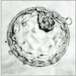
Human blastocyst, showing the inner cell mass (top, right)

Nuclear transfer techniques present a different set of ethical considerations than those associated with the use of other stem cells like embryonic stem cells which are controversial for their requirement to destroy an embryo. These different considerations have led to some individuals and organizations who are not opposed to human embryonic stem cell research to be concerned about, or opposed to, SCNT research.

One concern is that blastula creation in SCNT-based human stem cell research will lead to the reproductive cloning of humans. Both processes use the same first step: the creation of a nuclear transferred embryo, most likely via SCNT. Those who hold this concern often advocate for strong regulation of SCNT to preclude implantation of any derived products for the intention of human reproduction, or its prohibition.

A second important concern is the appropriate source of the eggs that are needed. SCNT requires human egg cells, which can only be obtained from women. The most common source of these eggs today are eggs that are produced and in excess of the clinical need during IVF treatment. This is a minimally invasive procedure, but it does carry some health risks, such as ovarian hyperstimulation syndrome.

One vision for successful stem cell therapies is to create custom stem cell lines for patients. Each custom stem cell line would consist of a collection of identical stem cells each carrying the patient's own DNA, thus reducing or eliminating any problems with rejection when the stem cells were transplanted for treatment. For example, to treat a man with Parkinson's disease, a cell nucleus from one of his cells would be transplanted by SCNT into an egg cell from an egg donor, creating a unique lineage of stem cells almost identical to the patient's own cells. (There would be differences. For example, the mitochondrial DNA would be the same as that of the egg donor. In comparison, his own cells would carry the mitochondrial DNA of his mother.)

Potentially millions of patients could benefit from stem cell therapy, and each patient would require a large number of donated eggs in order to successfully create a single custom therapeutic stem cell line. Such large numbers of donated eggs would exceed the number of eggs currently left over and available from couples trying to have children through assisted reproductive technology. Therefore, healthy young women would need to be induced to sell eggs to be used in the creation of custom stem cell lines that could then be purchased by the medical industry and sold to patients. It is so far unclear where all these eggs would come from.

Stem cell experts consider it unlikely that such large numbers of human egg donations would occur in a developed country because of the unknown long-term public health effects of treating large numbers of healthy young women with heavy doses of hormones in order to induce hyper-ovulation (ovulating several eggs at once). Although such treatments have been performed for several decades now, the long-term effects have not been studied or declared safe to use on a large scale on otherwise healthy women. Longer-term treatments with much lower doses of hormones are known to increase the rate of cancer decades later. Whether hormone treatments to induce hyper-ovulation could have similar effects is unknown. There are also ethical questions surrounding paying for eggs. In general, marketing body parts is considered unethical and is banned in most countries. Human eggs have been a notable exception to this rule for some time.

To address the problem of creating a human egg market, some stem cell researchers are investigating the possibility of creating artificial eggs. If successful, human egg donations would not be needed to create custom stem cell lines. However, this technology may be a long way off.

Policies regarding human SCNT

SCNT involving human cells is currently legal for research purposes in the United Kingdom, having been incorporated into the Human Fertilisation and Embryology Act 1990. Permission must be obtained from the Human Fertilisation and Embryology Authority in order to perform or attempt SCNT.

In the United States, the practice remains legal, as it has not been addressed by federal law. However, in 2002, a moratorium on United States federal funding for SCNT prohibits funding the practice for the purposes of research. Thus, though legal, SCNT cannot be federally funded. American scholars have recently argued that because the product of SCNT is a clone embryo, rather than a human embryo, these policies are morally wrong and should be revised.

In 2003, the United Nations adopted a proposal submitted by Costa Rica, calling on member states to "prohibit all forms of human cloning in as much as they are incompatible with human dignity and the protection of human life." This phrase may include SCNT, depending on interpretation.

The Council of Europe's Convention on Human Rights and Biomedicine and its Additional Protocol to the Convention for the Protection of Human Rights and Dignity of the Human Being with regard to the Application of Biology and Medicine, on the Prohibition of Cloning Human Being appear to ban SCNT of human beings. Of the Council's 45 member states, the Convention has been signed by 31 and ratified by 18. The Additional Protocol has been signed by 29 member nations and ratified by 14.

Stem cell controversy

From Wikipedia, the free encyclopedia
https://en.wikipedia.org/wiki/Stem_cell_controversy
 
The stem cell controversy concerns the ethics of research involving the development and use of human embryos. Most commonly, this controversy focuses on embryonic stem cells. Not all stem cell research involves human embryos. For example, adult stem cells, amniotic stem cells, and induced pluripotent stem cells do not involve creating, using, or destroying human embryos, and thus are minimally, if at all, controversial. Many less controversial sources of acquiring stem cells include using cells from the umbilical cord, breast milk, and bone marrow, which are not pluripotent.

Background

For many decades, stem cells have played an important role in medical research, beginning in 1868 when Ernst Haeckel first used the phrase to describe the fertilized egg which eventually gestates into an organism. The term was later used in 1886 by William Sedgwick to describe the parts of a plant that grow and regenerate. Further work by Alexander Maximow and Leroy Stevens introduced the concept that stem cells are pluripotent. This significant discovery led to the first human bone marrow transplant by E. Donnall Thomas in 1956, which although successful in saving lives, has generated much controversy since. This has included the many complications inherent in stem cell transplantation (almost 200 allogeneic marrow transplants were performed in humans, with no long-term successes before the first successful treatment was made), through to more modern problems, such as how many cells are sufficient for engraftment of various types of hematopoietic stem cell transplants, whether older patients should undergo transplant therapy, and the role of irradiation-based therapies in preparation for transplantation.

The discovery of adult stem cells led scientists to develop an interest in the role of embryonic stem cells, and in separate studies in 1981 Gail Martin and Martin Evans derived pluripotent stem cells from the embryos of mice for the first time. This paved the way for Mario Capecchi, Martin Evans, and Oliver Smithies to create the first knockout mouse, ushering in a whole new era of research on human disease. In 1995 adult stem cell research with human use was patented (US PTO with effect from 1995). In fact, human use was published in World J Surg 1991 & 1999 (B G Matapurkar). Salhan, Sudha (August 2011).

In 1998, James Thomson and Jeffrey Jones derived the first human embryonic stem cells, with even greater potential for drug discovery and therapeutic transplantation. However, the use of the technique on human embryos led to more widespread controversy as criticism of the technique now began from the wider public who debated the moral ethics of questions concerning research involving human embryonic cells.

Potential use in therapy

Since pluripotent stem cells have the ability to differentiate into any type of cell, they are used in the development of medical treatments for a wide range of conditions. Treatments that have been proposed include treatment for physical trauma, degenerative conditions, and genetic diseases (in combination with gene therapy). Yet further treatments using stem cells could potentially be developed due to their ability to repair extensive tissue damage.

Great levels of success and potential have been realized from research using adult stem cells. In early 2009, the FDA approved the first human clinical trials using embryonic stem cells. Only cells from an embryo at the morula stage or earlier are truly totipotent, meaning that they are able to form all cell types including placental cells. Adult stem cells are generally limited to differentiating into different cell types of their tissue of origin. However, some evidence suggests that adult stem cell plasticity may exist, increasing the number of cell types a given adult stem cell can become.

Points of controversy

Destruction of a human embryo is required in order to research new embryonic cell lines. Much of the debate surrounding human embryonic stem cells, therefore, concern ethical and legal quandaries around the destruction of an embryo. Ethical and legal questions such as "At what point does one consider life to begin?" and "Is it just to destroy a human embryo if it has the potential to cure countless numbers of patients and further our understanding of disease?" are central to the controversy. Political leaders debate how to regulate and fund research studies that involve the techniques used to remove the embryo cells. No clear consensus has emerged.

Much of the criticism has been a result of religious beliefs and, in the most high-profile case, US President George W Bush signed an executive order banning the use of federal funding for any stem cell lines other than those already in existence, stating at the time, "My position on these issues is shaped by deeply held beliefs," and "I also believe human life is a sacred gift from our Creator." This ban was in part revoked by his successor Barack Obama, who stated: "As a person of faith, I believe we are called to care for each other and work to ease human suffering. I believe we have been given the capacity and will to pursue this research and the humanity and conscience to do so responsibly."

Alternatives to embryonic stem cells

Some stem cell researchers are working to develop techniques of isolating stem cells with similar potency as embryonic stem cells, but do not require the destruction of a human embryo.

Induced pluripotent stem cells (iPSCs)

Foremost among these was the discovery in August 2006 that human adult somatic cells can be cultured in vitro with the four “Yamanaka factors” (Oct-4, SOX2, c-Myc, KLF4) which effectively returns a cell to the pluripotent state similar to that observed in embryonic stem cells. This major breakthrough won a Nobel Prize for the discoverers, Shinya Yamanaka and John Gurdon. Induced pluripotent stem cells are those derived from adult somatic cells and have the potential to provide an alternative for stem cell research that does not require the destruction of human embryos. Some debate remains about the similarities of these cells to embryonic stem cells as research has shown that the induced pluripotent cells may have a different epigenetic memory or modifications to the genome than embryonic stem cells depending on the tissue of origin and donor the iPSCs come from. While this may be the case, epigenetic manipulation of the cells is possible using small molecules and more importantly, iPSCs from multiple tissues of origin have been shown to give rise to a viable organism similar to the way ESCs can. This allows iPSCs to serve as a powerful tool for tissue generation, drug screening, disease modeling, and personalized medicine that has far fewer ethical considerations than embryonic stem cells that would otherwise serve the same purpose.

Somatic cell nuclear transfer (SCNT)

In an alternative technique, researchers at Harvard University, led by Kevin Eggan and Savitri Marajh, have transferred the nucleus of a somatic cell into an existing embryonic stem cell, thus creating a new stem cell line. This technique known as somatic cell nuclear transfer (SCNT) creates pluripotent cells that are genetically identical to the donor. While the creation of stem cells via SCNT does not destroy an embryo, it requires an oocyte from a donor which opens the door to a whole new set of ethical considerations such as the debate as to whether or not it is appropriate to offer financial incentives to female donors.

Single-Cell blastomere biopsy

Researchers at Advanced Cell Technology, led by Robert Lanza and Travis Wahl, reported the successful derivation of a stem cell line using a process similar to preimplantation genetic diagnosis, in which a single blastomere is extracted from a blastocyst. At the 2007 meeting of the International Society for Stem Cell Research (ISSCR), Lanza announced that his team had succeeded in producing three new stem cell lines without destroying the parent embryos. "These are the first human embryonic cell lines in existence that didn't result from the destruction of an embryo." Lanza is currently in discussions with the National Institutes of Health to determine whether the new technique sidesteps U.S. restrictions on federal funding for ES cell research.

Amniotic fluid stem cells (AFSCs)

Anthony Atala of Wake Forest University says that the fluid surrounding the fetus has been found to contain stem cells that, when used correctly, "can be differentiated towards cell types such as fat, bone, muscle, blood vessel, nerve and liver cells." The extraction of this fluid is not thought to harm the fetus in any way. He hopes "that these cells will provide a valuable resource for tissue repair and for engineered organs, as well." AFSCs have been found to express both embryonic and adult stem cell markers as well as having the ability to be maintained over 250 population doublings.

Umbilical cord blood (UCB)

Similarly, pro-life supporters claim that the use of adult stem cells from sources such as the cord blood has consistently produced more promising results than the use of embryonic stem cells. Research has shown that umbilical cord blood (UCB) is in fact a viable source for stem cells and their progenitors which occur in high frequencies within the fluid. Furthermore, these cells may hold an advantage over induced PSC as they can create large quantities of homogenous cells.

IPSCs and other embryonic stem cell alternatives must still be collected and maintained with the informed consent of the donor as a donor's genetic information is still within the cells and by the definition of pluripotency, each alternative cell type has the potential to give rise to viable organisms. Generation of viable offspring using iPSCs has been shown in mouse models through tetraploid complementation. This potential for the generation of viable organisms and the fact that iPSC cells contain the DNA of donors require that they be handled along the ethical guidelines laid out by the US Food and Drug Administration (FDA), European Medicines Agency (EMA), and International Society for Stem Cell Research (ISSCR).

Viewpoints

Stem cell debates have motivated and reinvigorated the anti-abortion movement, whose members are concerned with the rights and status of the human embryo as an early-aged human life. They believe that embryonic stem cell research profits from and violates the sanctity of life and is tantamount to murder. The fundamental assertion of those who oppose embryonic stem cell research is the belief that human life is inviolable, combined with the belief that human life begins when a sperm cell fertilizes an egg cell to form a single cell. The view of those in favor is that these embryos would otherwise be discarded, and if used as stem cells, they can survive as a part of a living human person.

A portion of stem cell researchers use embryos that were created but not used in in vitro fertility treatments to derive new stem cell lines. Most of these embryos are to be destroyed, or stored for long periods of time, long past their viable storage life. In the United States alone, an estimated at least 400,000 such embryos exist. This has led some opponents of abortion, such as Senator Orrin Hatch, to support human embryonic stem cell research. See also embryo donation.

Medical researchers widely report that stem cell research has the potential to dramatically alter approaches to understanding and treating diseases, and to alleviate suffering. In the future, most medical researchers anticipate being able to use technologies derived from stem cell research to treat a variety of diseases and impairments. Spinal cord injuries and Parkinson's disease are two examples that have been championed by high-profile media personalities (for instance, Christopher Reeve and Michael J. Fox, who have lived with these conditions, respectively). The anticipated medical benefits of stem cell research add urgency to the debates, which has been appealed to by proponents of embryonic stem cell research.

In August 2000, The U.S. National Institutes of Health's Guidelines stated:

... research involving human pluripotent stem cells ... promises new treatments and possible cures for many debilitating diseases and injuries, including Parkinson's disease, diabetes, heart disease, multiple sclerosis, burns and spinal cord injuries. The NIH believes the potential medical benefits of human pluripotent stem cell technology are compelling and worthy of pursuit in accordance with appropriate ethical standards.

In 2006, researchers at Advanced Cell Technology of Worcester, Massachusetts, succeeded in obtaining stem cells from mouse embryos without destroying the embryos. If this technique and its reliability are improved, it would alleviate some of the ethical concerns related to embryonic stem cell research.

Another technique announced in 2007 may also defuse the longstanding debate and controversy. Research teams in the United States and Japan have developed a simple and cost-effective method of reprogramming human skin cells to function much like embryonic stem cells by introducing artificial viruses. While extracting and cloning stem cells is complex and extremely expensive, the newly discovered method of reprogramming cells is much cheaper. However, the technique may disrupt the DNA in the new stem cells, resulting in damaged and cancerous tissue. More research will be required before noncancerous stem cells can be created.

Update of article to include 2009/2010 current stem cell usages in clinical trials: The planned treatment trials will focus on the effects of oral lithium on neurological function in people with chronic spinal cord injury and those who have received umbilical cord blood mononuclear cell transplants to the spinal cord. The interest in these two treatments derives from recent reports indicating that umbilical cord blood stem cells may be beneficial for spinal cord injury and that lithium may promote regeneration and recovery of function after spinal cord injury. Both lithium and umbilical cord blood are widely available therapies that have long been used to treat diseases in humans.

Endorsement

  • Embryonic stem cells have the ability to grow indefinitely in a laboratory environment and can differentiate into almost all types of bodily tissue. This makes embryonic stem cells a prospect for cellular therapies to treat a wide range of diseases.

Human potential and humanity

This argument often goes hand-in-hand with the utilitarian argument, and can be presented in several forms:

  • Embryos are not equivalent to human life while they are still incapable of surviving outside the womb (i.e. they only have the potential for life).
  • More than a third of zygotes do not implant after conception. Thus, far more embryos are lost due to chance than are proposed to be used for embryonic stem cell research or treatments.
  • Blastocysts are a cluster of human cells that have not differentiated into distinct organ tissue, making cells of the inner cell mass no more "human" than a skin cell.
  • Some parties contend that embryos are not human persons, believing that the life of Homo sapiens only begins when the heartbeat develops, which is during the fifth week of pregnancy, or when the brain begins developing activity, which has been detected at 54 days after conception.

Efficiency

  • In vitro fertilization (IVF) generates large numbers of unused embryos (e.g. 70,000 in Australia alone). Many of these thousands of IVF embryos are slated for destruction. Using them for scientific research uses a resource that would otherwise be wasted.
  • While the destruction of human embryos is required to establish a stem cell line, no new embryos have to be destroyed to work with existing stem cell lines. It would be wasteful not to continue to make use of these cell lines as a resource.

Superiority

This is usually presented as a counter-argument to using adult stem cells, as an alternative that does not involve embryonic destruction.

  • Embryonic stem cells make up a significant proportion of a developing embryo, while adult stem cells exist as minor populations within a mature individual (e.g. in every 1,000 cells of the bone marrow, only one will be a usable stem cell). Thus, embryonic stem cells are likely to be easier to isolate and grow ex vivo than adult stem cells.
  • Embryonic stem cells divide more rapidly than adult stem cells, potentially making it easier to generate large numbers of cells for therapeutic means. In contrast, adult stem cell might not divide fast enough to offer immediate treatment.
  • Embryonic stem cells have greater plasticity, potentially allowing them to treat a wider range of diseases.
  • Adult stem cells from the patient's own body might not be effective in treatment of genetic disorders. Allogeneic embryonic stem cell transplantation (i.e. from a healthy donor) may be more practical in these cases than gene therapy of a patient's own cell.
  • DNA abnormalities found in adult stem cells that are caused by toxins and sunlight may make them poorly suited for treatment.
  • Embryonic stem cells have been shown to be effective in treating heart damage in mice.
  • Embryonic stem cells have the potential to cure chronic and degenerative diseases which current medicine has been unable to effectively treat.

Individuality

  • Before the primitive streak is formed when the embryo attaches to the uterus around 14 days after fertilization, two fertilized eggs can combine by fusing together and develop into one person (a tetragametic chimera). Since a fertilized egg has the potential to be two individuals or half of one, some believe it can only be considered a 'potential' person, not an actual one. Those who subscribe to this belief then hold that destroying a blastocyst for embryonic stem cells is ethical.

Viability

  • Viability is another standard under which embryos and fetuses have been regarded as human lives. In the United States, the 1973 Supreme Court case of Roe v. Wade concluded that viability determined the permissibility of abortions performed for reasons other than the protection of the woman's health, defining viability as the point at which a fetus is "potentially able to live outside the mother's womb, albeit with artificial aid." The point of viability was 24 to 28 weeks when the case was decided and has since moved to about 22 weeks due to advancement in medical technology. Embryos used in medical research for stem cells are well below development that would enable viability.

Further uses for stem cells

Adult stem cells have provided many different therapies for illnesses such as Parkinson's disease, leukemia, multiple sclerosis, lupus, sickle-cell anemia, and heart damage (to date, embryonic stem cells have also been used in treatment). Moreover, there have been many advances in adult stem cell research, including a recent study where pluripotent adult stem cells were manufactured from differentiated fibroblast by the addition of specific transcription factors. Newly created stem cells were developed into an embryo and were integrated into newborn mouse tissues, analogous to the properties of embryonic stem cells.

Stated views of groups

Government policy stances

Europe

Austria, Denmark, France, Germany, Portugal and Ireland do not allow the production of embryonic stem cell lines, but the creation of embryonic stem cell lines is permitted in Finland, Greece, the Netherlands, Sweden, and the United Kingdom.

United States

Origins

In 1973, Roe v. Wade legalized abortion in the United States. Five years later, the first successful human in vitro fertilization resulted in the birth of Louise Brown in England. These developments prompted the federal government to create regulations barring the use of federal funds for research that experimented on human embryos. In 1995, the NIH Human Embryo Research Panel advised the administration of President Bill Clinton to permit federal funding for research on embryos left over from in vitro fertility treatments and also recommended federal funding of research on embryos specifically created for experimentation. In response to the panel's recommendations, the Clinton administration, citing moral and ethical concerns, declined to fund research on embryos created solely for research purposes, but did agree to fund research on leftover embryos created by in vitro fertility treatments. At this point, the Congress intervened and passed the 1995 Dickey–Wicker Amendment (the final bill, which included the Dickey-Wicker Amendment, was signed into law by Bill Clinton) which prohibited any federal funding for the Department of Health and Human Services be used for research that resulted in the destruction of an embryo regardless of the source of that embryo.

In 1998, privately funded research led to the breakthrough discovery of human embryonic stem cells (hESC). This prompted the Clinton administration to re-examine guidelines for federal funding of embryonic research. In 1999, the president's National Bioethics Advisory Commission recommended that hESC harvested from embryos discarded after in vitro fertility treatments, but not from embryos created expressly for experimentation, be eligible for federal funding. Though embryo destruction had been inevitable in the process of harvesting hESC in the past (this is no longer the case), the Clinton administration had decided that it would be permissible under the Dickey-Wicker Amendment to fund hESC research as long as such research did not itself directly cause the destruction of an embryo. Therefore, HHS issued its proposed regulation concerning hESC funding in 2001. Enactment of the new guidelines was delayed by the incoming George W. Bush administration which decided to reconsider the issue.

President Bush announced, on August 9, 2001, that federal funds, for the first time, would be made available for hESC research on currently existing embryonic stem cell lines. President Bush authorized research on existing human embryonic stem cell lines, not on human embryos under a specific, unrealistic timeline in which the stem cell lines must have been developed. However, the Bush administration chose not to permit taxpayer funding for research on hESC cell lines not currently in existence, thus limiting federal funding to research in which "the life-and-death decision has already been made." The Bush administration's guidelines differ from the Clinton administration guidelines which did not distinguish between currently existing and not-yet-existing hESC. Both the Bush and Clinton guidelines agree that the federal government should not fund hESC research that directly destroys embryos.

Neither Congress nor any administration has ever prohibited private funding of embryonic research. Public and private funding of research on adult and cord blood stem cells is unrestricted.

U.S. congressional response

In April 2004, 206 members of Congress signed a letter urging President Bush to expand federal funding of embryonic stem cell research beyond what Bush had already supported.

In May 2005, the House of Representatives voted 238–194 to loosen the limitations on federally funded embryonic stem-cell research – by allowing government-funded research on surplus frozen embryos from in vitro fertilization clinics to be used for stem cell research with the permission of donors – despite Bush's promise to veto the bill if passed. On July 29, 2005, Senate Majority Leader William H. Frist (R-TN) announced that he too favored loosening restrictions on federal funding of embryonic stem cell research. On July 18, 2006, the Senate passed three different bills concerning stem cell research. The Senate passed the first bill (the Stem Cell Research Enhancement Act) 63–37, which would have made it legal for the federal government to spend federal money on embryonic stem cell research that uses embryos left over from in vitro fertilization procedures. On July 19, 2006, President Bush vetoed this bill. The second bill makes it illegal to create, grow, and abort fetuses for research purposes. The third bill would encourage research that would isolate pluripotent, i.e., embryonic-like, stem cells without the destruction of human embryos.

In 2005 and 2007, Congressman Ron Paul introduced the Cures Can Be Found Act, with 10 cosponsors. With an income tax credit, the bill favors research upon non-embryonic stem cells obtained from placentas, umbilical cord blood, amniotic fluid, humans after birth, or unborn human offspring who died of natural causes; the bill was referred to committee. Paul argued that hESC research is outside of federal jurisdiction either to ban or to subsidize.

Bush vetoed another bill, the Stem Cell Research Enhancement Act of 2007, which would have amended the Public Health Service Act to provide for human embryonic stem cell research. The bill passed the Senate on April 11 by a vote of 63–34, then passed the House on June 7 by a vote of 247–176. President Bush vetoed the bill on July 19, 2007.

On March 9, 2009, President Obama removed the restriction on federal funding for newer stem cell lines.  Two days after Obama removed the restriction, the president then signed the Omnibus Appropriations Act of 2009, which still contained the long-standing Dickey–Wicker Amendment which bans federal funding of "research in which a human embryo or embryos are destroyed, discarded, or knowingly subjected to risk of injury or death;" the congressional provision effectively prevents federal funding being used to create new stem cell lines by many of the known methods. So, while scientists might not be free to create new lines with federal funding, President Obama's policy allows the potential of applying for such funding into research involving the hundreds of existing stem cell lines as well as any further lines created using private funds or state-level funding. The ability to apply for federal funding for stem cell lines created in the private sector is a significant expansion of options over the limits imposed by President Bush, who restricted funding to the 21 viable stem cell lines that were created before he announced his decision in 2001. The ethical concerns raised during Clinton's time in office continue to restrict hESC research and dozens of stem cell lines have been excluded from funding, now by judgment of an administrative office rather than presidential or legislative discretion.

Funding

In 2005, the NIH funded $607 million worth of stem cell research, of which $39 million was specifically used for hESC. Sigrid Fry-Revere has argued that private organizations, not the federal government, should provide funding for stem-cell research, so that shifts in public opinion and government policy would not bring valuable scientific research to a grinding halt.

In 2005, the State of California took out $3 billion in bond loans to fund embryonic stem cell research in that state.

Asia

China has one of the most permissive human embryonic stem cell policies in the world. In the absence of a public controversy, human embryo stem cell research is supported by policies that allow the use of human embryos and therapeutic cloning.

Religious views

Generally speaking, no group advocates for unrestricted stem cell research, especially in the context of embryonic stem cell research.

Jewish view

According to Rabbi Levi Yitzchak Halperin of the Institute for Science and Jewish Law in Jerusalem, embryonic stem cell research is permitted so long as it has not been implanted in the womb. Not only is it permitted, but research is encouraged, rather than wasting it.

As long as it has not been implanted in the womb and it is still a frozen fertilized egg, it does not have the status of an embryo at all and there is no prohibition to destroy it...

However in order to remove all doubt [as to the permissibility of destroying it], it is preferable not to destroy the pre-embryo unless it will otherwise not be implanted in the woman who gave the eggs (either because there are many fertilized eggs, or because one of the parties refuses to go on with the procedure – the husband or wife – or for any other reason). Certainly it should not be implanted into another woman.... The best and worthiest solution is to use it for life-saving purposes, such as for the treatment of people that suffered trauma to their nervous system, etc.

— Rabbi Levi Yitzchak Halperin, Ma'aseh Choshev vol. 3, 2:6

Similarly, the sole Jewish majority state, Israel, permits research on embryonic stem cells.

Catholicism

The Catholic Church opposes human embryonic stem cell research calling it "an absolutely unacceptable act." The Church supports research that involves stem cells from adult tissues and the umbilical cord, as it "involves no harm to human beings at any state of development." This support has been expressed both politically and financially, with different Catholic groups either raising money indirectly, offering grants, or seeking to pass federal legislation, according to the United States Conference of Catholic Bishops. Specific examples include a grant from the Catholic Archiocese of Sydney which funded research demonstrating the capabilities of adult stem cells, and the U.S. Conference of Catholic Bishops working to pass federal legislation creating a nationwide public bank for umbilical cord blood stem cells.

Baptists

The Southern Baptist Convention opposes human embryonic stem cell research on the grounds that the "Bible teaches that human beings are made in the image and likeness of God (Gen. 1:27; 9:6) and protectable human life begins at fertilization." However, it supports adult stem cell research as it does "not require the destruction of embryos."

Methodism

The United Methodist Church opposes human embryonic stem cell research, saying, "a human embryo, even at its earliest stages, commands our reverence." However, it supports adult stem cell research, stating that there are "few moral questions" raised by this issue.

Pentecostalism

The Assemblies of God opposes human embryonic stem cell research, saying, it "perpetuates the evil of abortion and should be prohibited."

Islam

Islamic scholars generally favor the stance that scientific research and development of stem cells is allowed as long as it benefits society while causing the least amount of harm to the subjects. "Stem cell research is one of the most controversial topics of our time period and has raised many religious and ethical questions regarding the research being done. With there being no true guidelines set forth in the Qur'an against the study of biomedical testing, Muslims have adopted any new studies as long as the studies do not contradict another teaching in the Qur'an. One of the teachings of the Qur'an states that 'Whosoever saves the life of one, it shall be if he saves the life of humankind' (5:32), it is this teaching that makes stem cell research acceptable in the Muslim faith because of its promise of potential medical breakthrough." This statement does not, however, make a distinction between adult, embryonic, or stem-cells. In specific instances, different sources have issued fatwas, or nonbinding but authoritative legal opinions according to Islamic faith, ruling on conduct in stem cell research. The Fatwa of the Islamic Jurisprudence Council of the Islamic World League (December 2003) addressed permissible stem cell sources, as did the Fatwa Khamenei (2002) in Iran. Several different governments in predominantly Muslim countries have also supported stem cell research, notably Iran. but Saudi Arabia religious officials issued a decree that sanctions the use of embryos for therapeutic and research purposes.

The Church of Jesus Christ of Latter-day Saints

The First Presidency of the Church of Jesus Christ of Latter-day Saints "has not taken a position regarding the use of embryonic stem cells for research purposes. The absence of a position should not be interpreted as support for or opposition to any other statement made by Church members, whether they are for or against embryonic stem cell research.”

Globular cluster

From Wikipedia, the free encyclopedia https://en.wikipedia.org/wiki/Globular_cluster     Globular cluster...