Search This Blog

Sunday, April 18, 2021

Nucleus accumbens

From Wikipedia, the free encyclopedia
 
Nucleus accumbens
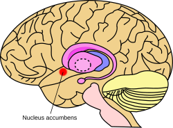
Nucleus accumbens.svg
Approximate location of the nucleus accumbens in the brain
 
Mouse Nucleus Accumbens.pdf
Nucleus accumbens of the mouse brain
 
Details
Part ofMesolimbic pathway
Basal ganglia (Ventral striatum)
PartsNucleus accumbens shell
Nucleus accumbens core
Identifiers
Latinnucleus accumbens septi
Acronym(s)NAc or NAcc
MeSHD009714
NeuroNames277
NeuroLex IDbirnlex_727
TA98A14.1.09.440
TA25558
FMA61889
Anatomical terms of neuroanatomy

The nucleus accumbens (NAc or NAcc; also known as the accumbens nucleus, or formerly as the nucleus accumbens septi, Latin for "nucleus adjacent to the septum") is a region in the basal forebrain rostral to the preoptic area of the hypothalamus. The nucleus accumbens and the olfactory tubercle collectively form the ventral striatum. The ventral striatum and dorsal striatum collectively form the striatum, which is the main component of the basal ganglia. The dopaminergic neurons of the mesolimbic pathway project onto the GABAergic medium spiny neurons of the nucleus accumbens and olfactory tubercle. Each cerebral hemisphere has its own nucleus accumbens, which can be divided into two structures: the nucleus accumbens core and the nucleus accumbens shell. These substructures have different morphology and functions.

Different NAcc subregions (core vs shell) and neuron subpopulations within each region (D1-type vs D2-type medium spiny neurons) are responsible for different cognitive functions. As a whole, the nucleus accumbens has a significant role in the cognitive processing of motivation, aversion, reward (i.e., incentive salience, pleasure, and positive reinforcement), and reinforcement learning (e.g., Pavlovian-instrumental transfer); hence, it has a significant role in addiction. In addition, part of the nucleus accumbens core is centrally involved in the induction of slow-wave sleep. The nucleus accumbens plays a lesser role in processing fear (a form of aversion), impulsivity, and the placebo effect. It is involved in the encoding of new motor programs as well.

Structure

The nucleus accumbens is an aggregate of neurons which is described as having an outer shell and an inner core.

Input

Major glutamatergic inputs to the nucleus accumbens include the prefrontal cortex (particularly the prelimbic cortex and infralimbic cortex), basolateral amygdala, ventral hippocampus, thalamic nuclei (specifically the midline thalamic nuclei and intralaminar nuclei of the thalamus), and glutamatergic projections from the ventral tegmental area (VTA). The nucleus accumbens receives dopaminergic inputs from the ventral tegmental area, which connect via the mesolimbic pathway. The nucleus accumbens is often described as one part of a cortico-basal ganglia-thalamo-cortical loop.

Dopaminergic inputs from the VTA modulate the activity of GABAergic neurons within the nucleus accumbens. These neurons are activated directly or indirectly by euphoriant drugs (e.g., amphetamine, opiates, etc.) and by participating in rewarding experiences (e.g., sex, music, exercise, etc.).

Another major source of input comes from the CA1 and ventral subiculum of the hippocampus to the dorsomedial area of the nucleus accumbens. Slight depolarizations of cells in the nucleus accumbens correlates with positivity of the neurons of the hippocampus, making them more excitable. The correlated cells of these excited states of the medium spiny neurons in the nucleus accumbens are shared equally between the subiculum and CA1. The subiculum neurons are found to hyperpolarize (increase negativity) while the CA1 neurons "ripple" (fire > 50 Hz) in order to accomplish this priming.

The nucleus accumbens is one of the few regions that receive histaminergic projections from the tuberomammillary nucleus (the sole source of histamine neurons in the brain).

Output

The output neurons of the nucleus accumbens send axonal projections to the basal ganglia and the ventral analog of the globus pallidus, known as the ventral pallidum (VP). The VP, in turn, projects to the medial dorsal nucleus of the dorsal thalamus, which projects to the prefrontal cortex as well as the striatum. Other efferents from the nucleus accumbens include connections with the tail of the ventral tegmental area, substantia nigra, and the reticular formation of the pons.

Shell

The nucleus accumbens shell (NAcc shell) is a substructure of the nucleus accumbens. The shell and core together form the entire nucleus accumbens.

Location: The shell is the outer region of the nucleus accumbens, and – unlike the core – is considered to be part of the extended amygdala, located at its rostral pole.

Cell types: Neurons in the nucleus accumbens are mostly medium spiny neurons (MSNs) containing mainly D1-type (i.e., DRD1 and DRD5) or D2-type (i.e., DRD2, DRD3, and DRD4) dopamine receptors. A subpopulation of MSNs contain both D1-type and D2-type receptors, with approximately 40% of striatal MSNs expressing both DRD1 and DRD2 mRNA. These mixed-type NAcc MSNs with both D1-type and D2-type receptors are mostly confined to the NAcc shell. The neurons in the shell, as compared to the core, have a lower density of dendritic spines, less terminal segments, and less branch segments than those in the core. The shell neurons project to the subcommissural part of the ventral pallidum as well as the ventral tegmental area and to extensive areas in the hypothalamus and extended amygdala.

Function: The shell of the nucleus accumbens is involved in the cognitive processing of reward, including subjective "liking" reactions to certain pleasurable stimuli, motivational salience, and positive reinforcement. That NAcc shell has also been shown to mediate specific Pavlovian-instrumental transfer, a phenomenon in which a classically conditioned stimulus modifies operant behavior. A "hedonic hotspot" or pleasure center which is responsible for the pleasurable or "liking" component of some intrinsic rewards is also located in a small compartment within the medial NAcc shell. Addictive drugs have a larger effect on dopamine release in the shell than in the core.

Core

The nucleus accumbens core (NAcc core) is the inner substructure of the nucleus accumbens.

Location: The nucleus accumbens core is part of the ventral striatum, located within the basal ganglia. Cell types: The core of the NAcc is made up mainly of medium spiny neurons containing mainly D1-type or D2-type dopamine receptors. The D1-type medium spiny neurons mediate reward-related cognitive processes, whereas the D2-type medium spiny neurons mediate aversion-related cognition. The neurons in the core, as compared to the neurons in the shell, have an increased density of dendritic spines, branch segments, and terminal segments. From the core, the neurons project to other sub-cortical areas such as the globus pallidus and the substantia nigra. GABA is one of the main neurotransmitters in the NAcc, and GABA receptors are also abundant.

Function: The nucleus accumbens core is involved in the cognitive processing of motor function related to reward and reinforcement and the regulation of slow-wave sleep. Specifically, the core encodes new motor programs which facilitate the acquisition of a given reward in the future. The indirect pathway (i.e., D2-type) neurons in the NAcc core which co-express adenosine A2A receptors activation-dependently promote slow-wave sleep. The NAcc core has also been shown to mediate general Pavlovian-instrumental transfer, a phenomenon in which a classically conditioned stimulus modifies operant behavior.

Cell types

Approximately 95% of neurons in the NAcc are GABAergic medium spiny neurons (MSNs) which primarily express either D1-type or D2-type receptors; about 1–2% of the remaining neuronal types are large aspiny cholinergic interneurons and another 1–2% are GABAergic interneurons. Compared to the GABAergic MSNs in the shell, those in the core have an increased density of dendritic spines, branch segments, and terminal segments. From the core, the neurons project to other sub-cortical areas such as the globus pallidus and the substantia nigra. GABA is one of the main neurotransmitters in the NAcc, and GABA receptors are also abundant. These neurons are also the main projection or output neurons of the nucleus accumbens.

Neurochemistry

Some of the neurotransmitters, neuromodulators, and hormones that signal through receptors within the nucleus accumbens include:

Dopamine: Dopamine is released into the nucleus accumbens following exposure to rewarding stimuli, including recreational drugs like substituted amphetamines, cocaine, nicotine and morphine.

Phenethylamine and tyramine: Phenethylamine and tyramine are trace amines which are synthesized in neurons that express the aromatic amino acid hydroxylase (AADC) enzyme, which includes all dopaminergic neurons. Both compounds function as dopaminergic neuromodulators which regulate the reuptake and release of dopamine into the Nacc via interactions with VMAT2 and TAAR1 in the axon terminal of mesolimbic dopamine neurons.

Glucocorticoids and dopamine: Glucocorticoid receptors are the only corticosteroid receptors in the nucleus accumbens shell. L-DOPA, steroids, and specifically glucocorticoids are currently known to be the only known endogenous compounds that can induce psychotic problems, so understanding the hormonal control over dopaminergic projections with regard to glucocorticoid receptors could lead to new treatments for psychotic symptoms. A recent study demonstrated that suppression of the glucocorticoid receptors led to a decrease in the release of dopamine, which may lead to future research involving anti-glucocorticoid drugs to potentially relieve psychotic symptoms.

GABA: A recent study on rats that used GABA agonists and antagonists indicated that GABAA receptors in the NAcc shell have inhibitory control on turning behavior influenced by dopamine, and GABAB receptors have inhibitory control over turning behavior mediated by acetylcholine.

Glutamate: Studies have shown that local blockade of glutamatergic NMDA receptors in the NAcc core impaired spatial learning. Another study demonstrated that both NMDA and AMPA (both glutamate receptors) play important roles in regulating instrumental learning.

Serotonin (5-HT): Overall, 5-HT synapses are more abundant and have a greater number of synaptic contacts in the NAcc shell than in the core. They are also larger and thicker, and contain more large dense core vesicles than their counterparts in the core.

Function

Reward and reinforcement

The nucleus accumbens, being one part of the reward system, plays an important role in processing rewarding stimuli, reinforcing stimuli (e.g., food and water), and those which are both rewarding and reinforcing (addictive drugs, sex, and exercise). The predominant response of neurons in the nucleus accumbens to the reward sucrose is inhibition; the opposite is true in response to the administration of aversive quinine. Substantial evidence from pharmacological manipulation also suggests that reducing the excitability of neurons in the nucleus accumbens is rewarding, as, for example, would be true in the case of μ-opioid receptor stimulation. The blood oxygen level dependent signal (BOLD) in the nucleus accumbens is selectively increased during the perception of pleasant, emotionally arousing pictures and during mental imagery of pleasant, emotional scenes. However, as BOLD is thought to be an indirect measure of regional net excitation to inhibition, the extent to which BOLD measures valence dependent processing is unknown. Because of the abundance of NAcc inputs from limbic regions and strong NAcc outputs to motor regions, the nucleus accumbens has been described by Gordon Mogensen as the interface between the limbic and motor system.

Tuning of appetitive and defensive reactions in the nucleus accumbens shell. (Above) AMPA blockade requires D1 function in order to produce motivated behaviors, regardless of valence, and D2 function to produce defensive behaviors. GABA agonism, on the other hand, does not require dopamine receptor function.(Below)The expansion of the anatomical regions that produce defensive behaviors under stress, and appetitive behaviors in the home environment produced by AMPA antagonism. This flexibility is less evident with GABA agonism.

The nucleus accumbens is causally related to the experience of pleasure. Microinjections of μ-opioid agonists, δ-opioid agonists or κ-opioid agonists in the rostrodorsal quadrant of the medial shell enhance "liking", while more caudal injections can inhibit disgust reactions, liking reactions, or both. The regions of the nucleus accumbens that can be ascribed a causal role in the production of pleasure are limited both anatomically and chemically, as besides opioid agonists only endocannabinoids can enhance liking. In the nucleus accumbens as a whole, dopamine, GABA receptor agonist or AMPA antagonists solely modify motivation, while the same is true for opioid and endocannabinoids outside of the hotspot in the medial shell. A rostro-caudal gradient exists for the enhancement of appetitive versus fearful responses, the latter of which is traditionally thought to require only D1 receptor function, and the former of which requires both D1 and D2 function. One interpretation of this finding, the disinhibition hypothesis, posits that inhibition of accumbens MSNs (which are GABAergic) disinhibits downstream structures, enabling the expression of appetitive or consummatory behaviors. The motivational effects of AMPA antagonists, and to a lesser extent GABA agonists, is anatomically flexible. Stressful conditions can expand the fear inducing regions, while a familiar environment can reduce the size of the fear inducing region. Furthermore, cortical input from the orbitofrontal cortex (OFC) biases the response towards that of appetitive behavior, and infralimbic input, equivalent to the human subgenual cingulate cortex, suppresses the response regardless of valence.

The nucleus accumbens is neither necessary nor sufficient for instrumental learning, although manipulations can affect performance on instrumental learning tasks. One task where the effect of NAcc lesions is evident is Pavlovian-instrumental transfer (PIT), where a cue paired with a specific or general reward can enhance instrumental responding. Lesions to the core of the NAcc impair performance after devaluation and inhibit the effect of general PIT. On the other hand, lesions to the shell only impair the effect of specific PIT. This distinction is thought to reflect consummatory and appetitive conditioned responses in the NAcc shell and the NAcc core, respectively.

In the dorsal striatum, a dichotomy has been observed between D1-MSNs and D2-MSNs, with the former being reinforcing and enhancing locomotion, and the latter being aversive and reducing locomotion. Such a distinction has been traditionally assumed to apply to the nucleus accumbens as well, but evidence from pharmacological and optogenetics studies is conflicting. Furthermore, a subset of NAcc MSNs express both D1 and D2 MSNs, and pharmacological activation of D1 versus D2 receptors need not necessarily activate the neural populations exactly. While most studies show no effect of selective optogenetic stimulation of D1 or D2 MSNs on locomotor activity, one study has reported a decrease in basal locomotion with D2-MSN stimulation. While two studies have reported reduced reinforcing effects of cocaine with D2-MSN activation, one study has reported no effect. NAcc D2-MSN activation has also been reported to enhance motivation, as assessed by PIT, and D2 receptor activity is necessary for the reinforcing effects of VTA stimulation. A 2018 study reported that D2 MSN activation enhanced motivation via inhibiting the ventral pallidum, thereby disinhibiting the VTA.

Maternal behavior

An fMRI study conducted in 2005 found that when mother rats were in the presence of their pups the regions of the brain involved in reinforcement, including the nucleus accumbens, were highly active. Levels of dopamine increase in the nucleus accumbens during maternal behavior, while lesions in this area upset maternal behavior. When women are presented pictures of unrelated infants, fMRIs show increased brain activity in the nucleus accumbens and adjacent caudate nucleus, proportionate to the degree to which the women find these infants "cute".

Aversion

Activation of D1-type MSNs in the nucleus accumbens is involved in reward, whereas the activation of D2-type MSNs in the nucleus accumbens promotes aversion.

Slow-wave sleep

In late 2017, studies on rodents which utilized optogenetic and chemogenetic methods found that the indirect pathway (i.e., D2-type) medium spiny neurons in the nucleus accumbens core which co-express adenosine A2A receptors and project to the ventral pallidum are involved in the regulation of slow-wave sleep. In particular, optogenetic activation of these indirect pathway NAcc core neurons induces slow-wave sleep and chemogenetic activation of the same neurons increases the number and duration of slow-wave sleep episodes. Chemogenetic inhibition of these NAcc core neurons suppresses sleep. In contrast, the D2-type medium spiny neurons in the NAcc shell which express adenosine A2A receptors have no role in regulating slow-wave sleep.

Clinical significance

Addiction

Current models of addiction from chronic drug use involve alterations in gene expression in the mesocorticolimbic projection. The most important transcription factors that produce these alterations are ΔFosB, cyclic adenosine monophosphate (cAMP) response element binding protein (CREB), and nuclear factor kappa B (NFκB). ΔFosB is the most significant gene transcription factor in addiction since its viral or genetic overexpression in the nucleus accumbens is necessary and sufficient for many of the neural adaptations and behavioral effects (e.g., expression-dependent increases in self-administration and reward sensitization) seen in drug addiction. ΔFosB overexpression has been implicated in addictions to alcohol (ethanol), cannabinoids, cocaine, methylphenidate, nicotine, opioids, phencyclidine, propofol, and substituted amphetamines, among others. Increases in nucleus accumbens ΔJunD expression can reduce or, with a large increase, even block most of the neural alterations seen in chronic drug abuse (i.e., the alterations mediated by ΔFosB).

ΔFosB also plays an important role in regulating behavioral responses to natural rewards, such as palatable food, sex, and exercise. Natural rewards, like drugs of abuse, induce ΔFosB in the nucleus accumbens, and chronic acquisition of these rewards can result in a similar pathological addictive state through ΔFosB overexpression. Consequently, ΔFosB is the key transcription factor involved in addictions to natural rewards as well; in particular, ΔFosB in the nucleus accumbens is critical for the reinforcing effects of sexual reward. Research on the interaction between natural and drug rewards suggests that psychostimulants and sexual behavior act on similar biomolecular mechanisms to induce ΔFosB in the nucleus accumbens and possess cross-sensitization effects that are mediated through ΔFosB.

Similar to drug rewards, non-drug rewards also increase the level of extracellular dopamine in the NAcc shell. Drug-induced dopamine release in the NAcc shell and NAcc core is usually not prone to habituation (i.e., the development of drug tolerance: a decrease in dopamine release from future drug exposure as a result of repeated drug exposure); on the contrary, repeated exposure to drugs that induce dopamine release in the NAcc shell and core typically results in sensitization (i.e., the amount of dopamine that is released in the NAcc from future drug exposure increases as a result of repeated drug exposure). Sensitization of dopamine release in the NAcc shell following repeated drug exposure serves to strengthen stimulus-drug associations (i.e., classical conditioning that occurs when drug use is repeatedly paired with environmental stimuli) and these associations become less prone to extinction (i.e., "unlearning" these classically conditioned associations between drug use and environmental stimuli becomes more difficult). After repeated pairing, these classically conditioned environmental stimuli (e.g., contexts and objects that are frequently paired with drug use) often become drug cues which function as secondary reinforcers of drug use (i.e., once these associations are established, exposure to a paired environmental stimulus triggers a craving or desire to use the drug which they've become associated with).

In contrast to drugs, the release of dopamine in the NAcc shell by many types of rewarding non-drug stimuli typically undergoes habituation following repeated exposure (i.e., the amount of dopamine that is released from future exposure to a rewarding non-drug stimulus normally decreases as a result of repeated exposure to that stimulus).

Depression

In April 2007, two research teams reported on having inserted electrodes into the nucleus accumbens in order to use deep brain stimulation to treat severe depression. In 2010, experiments reported that deep brain stimulation of the nucleus accumbens was successful in decreasing depression symptoms in 50% of patients who did not respond to other treatments such as electroconvulsive therapy. Nucleus accumbens has also been used as a target to treat small groups of patients with therapy-refractory obsessive-compulsive disorder.

Ablation

To treat addiction and in an attempt to treat mental illness radiofrequency ablation of the nucleus accumbens has been performed. The results are inconclusive and controversial.

Placebo effect

Activation of the NAcc has been shown to occur in the anticipation of effectiveness of a drug when a user is given a placebo, indicating a contributing role of the nucleus accumbens in the placebo effect.

 

Caudate nucleus

From Wikipedia, the free encyclopedia
 
Caudate nucleus
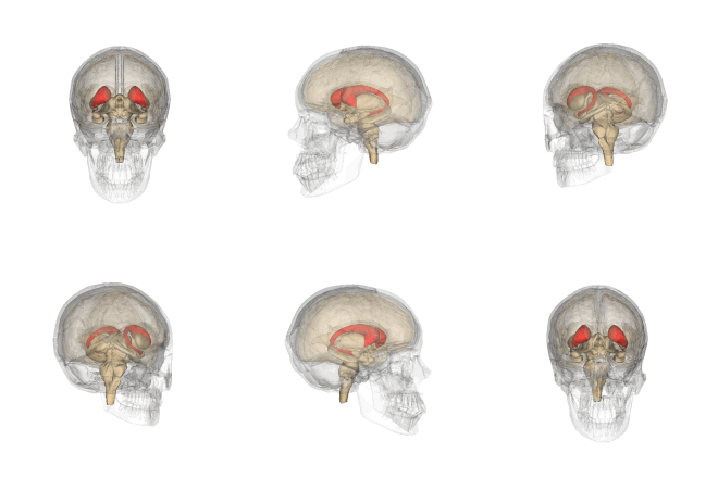
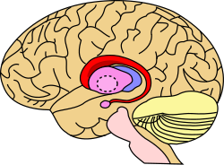
Caudate nucleus.svg
Caudate nucleus (in red) shown within the brain
Telencephalon-Horiconatal.jpg
Transverse Cut of Brain (Horizontal Section), basal ganglia is blue
Details
Part ofdorsal striatum
Identifiers
Latinnucleus caudatus
MeSHD002421
NeuroNames226
NeuroLex IDbirnlex_1373
TA98A14.1.09.502
TA25561
FMA61833
Anatomical terms of neuroanatomy

The caudate nucleus is one of the structures that make up the corpus striatum, which is a component of the basal ganglia. While the caudate nucleus has long been associated with motor processes due to its role in Parkinson's disease, it plays important roles in various other nonmotor functions as well, including procedural learning, associative learning and inhibitory control of action, among other functions. The caudate is also one of the brain structures which compose the reward system and functions as part of the corticobasal gangliathalamic loop.

Structure

Caudate nucleus within the skull

Together with the putamen, the caudate forms the dorsal striatum, which is considered a single functional structure; anatomically, it is separated by a large white matter tract, the internal capsule, so it is sometimes also referred to as two structures: the medial dorsal striatum (the caudate) and the lateral dorsal striatum (the putamen). In this vein, the two are functionally distinct not as a result of structural differences, but merely due to the topographical distribution of function.

The caudate nuclei are located near the center of the brain, sitting astride the thalamus. There is a caudate nucleus within each hemisphere of the brain. Individually, they resemble a C-shape structure with a wider "head" (caput in Latin) at the front, tapering to a "body" (corpus) and a "tail" (cauda). Sometimes a part of the caudate nucleus is referred to as the "knee" (genu). The caudate head receives its blood supply from the lenticulostriate artery while the tail of the caudate receives its blood supply from the anterior choroidal artery.

This is a transverse view of the caudate nucleus from a structural MR image.
Transverse view of the caudate nucleus from a structural MR image

The head and body of the caudate nucleus form part of the floor of the anterior horn of the lateral ventricle. After the body travels briefly towards the back of the head, the tail curves back toward the anterior, forming the roof of the inferior horn of the lateral ventricle. This means that a coronal (on a plane parallel to the face) section that cuts through the tail will also cross the body and head of the caudate nucleus.

Neurochemistry

The caudate is highly innervated by dopaminergic neurons that originate from the substantia nigra pars compacta (SNc). The SNc is located in the midbrain and contains cell projections to the caudate and putamen, utilizing the neurotransmitter dopamine. There are also additional inputs from various association cortices.

Motor functions

Spatial mnemonic processing

The caudate nucleus integrates spatial information with motor behavior formulation. Selective impairment of spatial working memory in subjects with Parkinson's disease and the knowledge of the disease's impact on the amount of dopamine supplied to the striatum have linked the caudate nucleus to spatial and nonspatial mnemonic processing. Spatially dependent motor preparation has been linked to the caudate nucleus through event-related fMRI analysis techniques. Activity in the caudate nucleus was demonstrated to be greater during tasks featuring spatial and motoric memory demands than those that involved nonspatial tasks. Specifically, spatial working memory activity has been observed, via fMRI studies of delayed recognition, to be greater in the caudate nucleus when the activity immediately preceded a motor response. These results indicate that the caudate nucleus could be involved in coding a motor response. With this in mind, the caudate nucleus could be involved in the recruitment of the motor system to support working memory performance by the mediation of sensory-motor transformations.

Directed movements

The caudate nucleus contributes importantly to body and limbs posture and the speed and accuracy of directed movements. Deficits in posture and accuracy during paw usage tasks were observed following the removal of caudate nuclei in felines. A delay in initiating performance and the need to constantly shift body position were both observed in cats following partial removal of the nuclei.

Following the application of cocaine to the caudate nucleus and the resulting lesions produced, a "leaping or forward movement" was observed in monkeys. Due to its association with damage to the caudate, this movement demonstrates the inhibitory nature of the caudate nucleus. The "motor release" observed as a result of this procedure indicates that the caudate nucleus inhibits the tendency for an animal to move forward without resistance.

Cognitive functions

Goal-directed action

A review of neuroimaging studies, anatomical studies of caudate connectivity, and behavioral studies reveals a role for the caudate in executive functioning. A study of Parkinson's patients (see below) may also contribute to a growing body of evidence.

A two-pronged approach of neuroimaging (including PET and fMRI) and anatomical studies expose a strong relationship between the caudate and cortical areas associated with executive functioning: "non-invasive measures of anatomical and functional connectivity in humans demonstrate a clear link between the caudate and executive frontal areas."

Meanwhile, behavioral studies provide another layer to the argument: recent studies suggest that the caudate is fundamental to goal-directed action, that is, "the selection of behavior based on the changing values of goals and a knowledge of which actions lead to what outcomes." One such study presented rats with levers that triggered the release of a cinnamon flavored solution. After the rats learned to press the lever, the researchers changed the value of the outcome (the rats were taught to dislike the flavor either by being given too much of the flavor, or by making the rats ill after drinking the solution) and the effects were observed. Normal rats pressed the lever less frequently, while rats with lesions in the caudate did not suppress the behavior as effectively. In this way, the study demonstrates the link between the caudate and goal-directed behavior; rats with damaged caudate nuclei had difficulty assessing the changing value of the outcome. In a 2003-human behavioral study, a similar process was repeated, but the decision this time was whether or not to trust another person when money was at stake. While here the choice was far more complex––the subjects were not simply asked to press a lever, but had to weigh a host of different factors––at the crux of the study was still behavioral selection based on changing values of outcomes.

In short, neuroimagery and anatomical studies support the assertion that the caudate plays a role in executive functioning, while behavioral studies deepen our understanding of the ways in which the caudate guides some of our decision-making processes.

Memory

The dorsal-prefrontal cortex subcortical loop involving the caudate nucleus has been linked to deficits in working memory, specifically in schizophrenic patients. Functional imaging has shown activation of this subcortical loop during working memory tasks in primates and healthy human subjects. The caudate may be affiliated with deficits involving working memory from before illness onset as well. Caudate nucleus volume has been found to be inversely associated with perseverative errors on spatial working memory tasks.

The amygdala sends direct projections to the caudate nucleus. Both the amygdala and the caudate nucleus have direct and indirect projections to the hippocampus. The influence of the amygdala on memory processing in the caudate nucleus has been demonstrated with the finding that lesions involving the connections between these two structures "block the memory-enhancing effects of oxotremorine infused into the caudate nucleus". In a study involving rats given water-maze training, the caudate nucleus was discovered to enhance memory of visually cued training after amphetamine was infused post-training into the caudate.

Learning

In a 2005 study, subjects were asked to learn to categorize visual stimuli by classifying images and receiving feedback on their responses. Activity associated with successful classification learning (correct categorization) was concentrated to the body and tail of the caudate, while activity associated with feedback processing (the result of incorrect categorization) was concentrated to the head of the caudate.

Sleep

Bilateral lesions in the head of the caudate nucleus in cats were correlated with a decrease in the duration of deep slow wave sleep during the sleep-wakefulness cycle. With a decrease in total volume of deep slow wave sleep, the transition of short-term memory to long-term memory may also be affected negatively. However, the effects of caudate nuclei removal on the sleep-wakefulness pattern of cats have not been permanent. Normalization has been discovered after a period of three months following caudate nuclei ablation. This discovery could be due to the inter-related nature of the roles of the caudate nucleus and the frontal cortex in controlling levels of central nervous system activation. The cats with caudate removal, although permanently hyperactive, had a significant decrease in rapid eye movement sleep (REMS) time that only lasted for about two months. However, afrontal cats had a permanent decrease in REMS time and only a temporary period of hyperactivity.

Contrasting with associations between "deep", REM sleep and the caudate nucleus, a study involving EEG and fMRI measures during human sleep cycles has indicated that the caudate nucleus demonstrates reduced activity during non-REM sleep across all sleep stages. Additionally, studies of human caudate nuclei volume in congenital central hypoventilation syndrome (CCHS) subjects established a correlation between CCHS and a significant reduction in left and right caudate volume. CCHS is a genetic disorder that affects the sleep cycle due to a reduced drive to breathe. Therefore, the caudate nucleus has been suggested to play a role in human sleep cycles.

Emotion

The caudate nucleus has been implicated in responses to visual beauty, and has been suggested as one of the "neural correlates of romantic love".

Approach-attachment behavior and affect are also controlled by the caudate nucleus. Cats with bilateral removal of the caudate nuclei persistently approached and followed objects, attempting to contact the target, while exhibiting a friendly disposition by the elicitation of treading of the forelimbs and purring. The magnitude of the behavioral responses was correlated to the extent of the removal of the nuclei. Reports of human patients with selective damage to the caudate nucleus show unilateral caudate damage resulting in loss of drive, obsessive-compulsive disorder, stimulus-bound perseverative behavior, and hyperactivity. Most of these deficits can be classified as relating to approach-attachment behaviors, from approaching a target to romantic love.

Language

Neuroimaging studies reveal that people who can communicate in multiple languages activate exactly the same brain regions regardless of the language. A 2006 publication studies this phenomenon and identifies the caudate as a center for language control. In perhaps the most illustrative case, a trilingual subject with a lesion to the caudate was observed. The patient maintained language comprehension in her three languages, but when asked to produce language, she involuntarily switched between the three languages. In short, "these and other findings with bilingual patients suggest that the left caudate is required to monitor and control lexical and language alternatives in production tasks."

Local shape deformations of the medial surface of the caudate have been correlated with verbal learning capacity for females and the number of perseverance errors on spatial and verbal fluency working memory tasks for males. Specifically, a larger caudate nucleus volume has been linked with better verbal fluency performance.

A neurological study of glossolalia showed a significant reduction in activity in the left caudate nucleus during glossolalia compared to singing in English.

Threshold control

The brain contains large collections of neurons reciprocally connected by excitatory synapses, thus forming large network of elements with positive feedback. It is difficult to see how such a system can operate without some mechanism to prevent explosive activation. There is some indirect evidence that the caudate may perform this regulatory role by measuring the general activity of cerebral cortex and controlling the threshold potential.

Clinical significance

Alzheimer's disease

A 2013 study has suggested a link between Alzheimer's patients and the caudate nucleus. MRI images were used to estimate the volume of caudate nuclei in patients with Alzheimer's and normal volunteers. The study found a "significant reduction in the caudate volume" in Alzheimer's patients when compared to the normal volunteers. While the correlation does not indicate causation, the finding may have implications for early diagnosis.

Parkinson's disease

Parkinson's disease is likely the most studied basal ganglia disorder. Patients with this progressive neurodegenerative disorder often first experience movement related symptoms (the three most common being tremors at rest, muscular rigidity, and akathisia) which are later combined with various cognitive deficiencies, including dementia. Parkinson's disease depletes dopaminergic neurons in the nigrostriatal tract, a dopamine pathway that is connected to the head of the caudate. As such, many studies have correlated the loss of dopaminergic neurons that send axons to the caudate nucleus and the degree of dementia in Parkinson's patients. And while a relationship has been drawn between the caudate and Parkinson's motor deficiencies, the caudate has also been associated with Parkinson's concomitant cognitive impairments. One review contrasts the performance of patients with Parkinson's and patients that strictly suffered from frontal-lobe damage in the Tower of London test. The differences in performance between the two types of patients (in a test that, in short, requires subjects to select appropriate intermediate goals with a larger goal in mind) draws a link between the caudate and goal-directed action. However, the studies are not conclusive. While the caudate has been associated with executive function (see "Goal-Directed Action"), it remains "entirely unclear whether executive deficits in [Parkinson's patients] reflect pre-dominantly their cortical or subcortical damage."

Huntington's disease

In Huntington's disease, a genetic mutation occurs in the HTT gene which encodes for Htt protein. The Htt protein interacts with over 100 other proteins, and appears to have multiple biological functions. The behavior of this mutated protein is not completely understood, but it is toxic to certain cell types, particularly in the brain. Early damage is most evident in the striatum, but as the disease progresses, other areas of the brain are also more conspicuously affected. Early symptoms are attributable to functions of the striatum and its cortical connections—namely control over movement, mood and higher cognitive function.

Attention-deficit hyperactivity disorder

A 2002 study draws a relationship between caudate asymmetry and symptoms related to ADHD. The authors used MR images to compare the relative volumes of the caudate nuclei (as the caudate is a bilateral structure), and drew a connection between any asymmetries and symptoms of ADHD: "The degree of caudate asymmetry significantly predicted cumulative severity ratings of inattentive behaviors." This correlation is congruent with previous associations of the caudate with attentional functioning. A more recent 2018 study replicated these findings, and demonstrated that the caudate asymmetries related to ADHD were more pronounced in the dorsal medial regions of the caudate.

Schizophrenia

The volume of white matter in the caudate nucleus has been linked with patients diagnosed with Schizophrenia. A 2004 study uses magnetic resonance imaging to compare the relative volume of white matter in the caudate among Schizophrenia patients. Those patients who suffer from the disorder have "smaller absolute and relative volumes of white matter in the caudate nucleus than healthy subjects."

Bipolar type I

A 2014 study found Type I Bipolar patients had relatively higher volume of gray and white matter in the caudate nucleus and other areas associated with reward processing and decision making, compared to controls and Bipolar II subjects. Overall the amount of gray and white matter in Bipolar patients was lower than controls.

Obsessive-compulsive disorder

It has been theorized that the caudate nucleus may be dysfunctional in persons with obsessive compulsive disorder (OCD), in that it may perhaps be unable to properly regulate the transmission of information regarding worrying events or ideas between the thalamus and the orbitofrontal cortex.

A neuroimaging study with positron emission tomography found that the right caudate nucleus had the largest change in glucose metabolism after patients had been treated with paroxetine. Recent SDM meta-analyses of voxel-based morphometry studies comparing people with OCD and healthy controls have found people with OCD to have increased grey matter volumes in bilateral lenticular nuclei, extending to the caudate nuclei, while decreased grey matter volumes in bilateral dorsal medial frontal/anterior cingulate gyri. These findings contrast with those in people with other anxiety disorders, who evince decreased (rather than increased) grey matter volumes in bilateral lenticular / caudate nuclei, while also decreased grey matter volumes in bilateral dorsal medial frontal/anterior cingulate gyri.


Curiosity

From Wikipedia, the free encyclopedia
Curious children gather around photographer Toni Frissell, looking at her camera (circa 1945)

Curiosity (from Latin cūriōsitās, from cūriōsus "careful, diligent, curious", akin to cura "care") is a quality related to inquisitive thinking such as exploration, investigation, and learning, evident by observation in humans and other animals. Curiosity is heavily associated with all aspects of human development, in which derives the process of learning and desire to acquire knowledge and skill.

The term curiosity can also be used to denote the behavior or emotion of being curious, in regard to the desire to gain knowledge or information. Curiosity as a behavior and emotion is attributed over millennia as the driving force behind not only human development, but developments in science, language, and industry.

Causes

Children peer over shoulders to see what their friends are reading.

Curiosity can be seen as an innate quality of many different species. It is common to human beings at all ages from infancy through adulthood, and is easy to observe in many other animal species; these include apes, cats, and rodents. Early definitions cite curiosity as a motivated desire for information. This motivational desire has been said to stem from a passion or an appetite for knowledge, information, and understanding.

These traditional ideas of curiosity have recently expanded to look at the difference between curiosity as the innate exploratory behavior that is present in all animals and curiosity as the desire for knowledge that is specifically attributed to humans.

Daniel Berlyne recognized three major variables playing a role in evoking curiosity; namely, psychophysical variables, ecological variables, and collative variables. Psychophysical variables correspond to physical intensity, while ecological variables to motivational significance and task relevance. Collative variables are called “collative” because they involve a comparison between different stimuli or features, which may be actually perceived or which may be recalled from memory. Berlyne mentioned four collative variables; namely, novelty, complexity, uncertainty, and conflict. At the same time, he suggested that all collative variables probably involve conflict. Additionally, he considered three variables supplementary to novelty, namely change, surprisingness, and incongruity. Finally, curiosity may not only be aroused by the perception of some stimulus associated with the aforementioned variables ("specific exploration"), but also by a lack of stimulation, out of “boredom” ("diversive exploration").

Curiosity-driven behavior

Curiosity-driven behavior is often defined as behavior through which knowledge is gained, and should therefore encompass all behaviors that provide access to or increase sensory information. Berlyne divided curiosity-driven behavior into three categories; namely, orienting responses, locomotor exploration, and investigatory responses, or, investigatory manipulation. Previously, Berlyne had already suggested that curiosity also includes verbal activities, such as asking questions, and symbolic activities, consisting of internally fueled mental processes such as thinking ("epistemic exploration").

Theories

Like other desires and need states that take on an appetitive quality (e.g. food), curiosity is linked with exploratory behavior and experiences of reward. Curiosity can be described as positive emotions and acquiring knowledge; when one's curiosity has been aroused it is considered inherently rewarding and pleasurable. Discovering new information may also be rewarding because it can help reduce undesirable states of uncertainty rather than stimulating interest. Theories have arisen in attempts to further understand this need to rectify states of uncertainty and the desire to participate in pleasurable experiences of exploratory behaviors.

Curiosity-drive theory

Curiosity-drive theory relates to the undesirable experiences of "uncertainty". The reduction of these unpleasant feelings, in turn, is rewarding. This theory suggests that people desire coherence and understanding in their thought processes. When this coherence is disrupted by something that is unfamiliar, uncertain, or ambiguous, it is curiosity-drive that attempts to gather information and knowledge of the unfamiliar to restore coherent thought processes. Through this theory, the general concept dictates that curiosity is developed strictly out of the desire to make sense of unfamiliar aspects of one's environment through interaction of exploratory behaviors. Once understanding of the unfamiliar has been achieved and coherence has been restored, these behaviors and desires will subside.

Subsets of curiosity-drive theory differ on whether curiosity is a primary or secondary drive and if this curiosity-drive is originated due to one's need to make sense of and regulate their environment or if it is caused by an external stimulus. Causes can range from basic needs that need to be satisfied (e.g. hunger, thirst) to needs in fear induced situations. Each of these subset theories state that whether the need is primary or secondary curiosity is developed from experiences that create a sensation of uncertainty or perceived unpleasantness. Curiosity then acts as a means in which to dispel this uncertainty. By exhibiting curious and exploratory behavior, one is able to gain knowledge of the unfamiliar and thus reduce the state of uncertainty or unpleasantness. This theory, however, does not address the idea that curiosity can often be displayed even in the absence of new or unfamiliar situations. This type of exploratory behavior is common in many species. Take the example of a human toddler who, if bored in his current situation devoid of arousing stimuli, will walk about until something interesting is found. The observation of curiosity even in the absence of novel stimuli pinpoints one of the major shortcomings in the curiosity-drive model.

Optimal-arousal theory

Optimal-arousal theory developed out of the need to explain the desire for some to seek out opportunities to engage in exploratory behaviors without the presence of uncertain or ambiguous situations. Optimal-arousal theory attempts to explain this aspect of curiosity by suggesting that one can be motivated to maintain a pleasurable sense of arousal through these exploratory behaviors.

The concept of optimal-arousal of curiosity suggests that there is a tendency to maintain an optimal level of arousal. When a stimulus is encountered that is associated with complexity, uncertainty, conflict, or novelty, this will increase arousal, and exploratory behavior is employed to learn about that stimulus and thereby reduce arousal again. In contrast, if the environment is boring and lacks excitement, arousal is reduced and exploratory behavior will be engaged in order to increase information input and stimulation, and thereby increasing arousal again. This theory addresses both curiosity elicited by uncertain or unfamiliar situations and curiosity elicited in the absence of such situations.

Cognitive-consistency theory

Cognitive-consistency theories assume that "when two or more simultaneously active cognitive structures are logically inconsistent, arousal is increased, which activates processes with the expected consequence of increasing consistency and decreasing arousal." Similar to optimal-arousal theory, cognitive-consistency theory suggests that there is a tendency to maintain arousal at a preferred, or expected, level, but it also explicitly links the amount of arousal to the amount of experienced inconsistency between an expected situation and the actually perceived situation. When this inconsistency is small, exploratory behavior triggered by curiosity is employed to gather information with which expectancy can be updated through learning to match perception, thereby reducing inconsistency. This approach puts curiosity in a broader perspective, also involving aggression and fear. That is, if the inconsistency is larger, fear or aggressive behavior may be employed to alter the perception in order to make it match expectancy, depending on the size of the inconsistency as well as the specific context. Aggressive behavior is assumed to alter perception by forcefully manipulating it into matching the expected situation, while uninhibited fear results in fleeing, thereby removing the inconsistent stimulus from the perceptual field and resolving the inconsistency.

Integration of the reward pathway into theory

Taking into account the shortcomings of both curiosity-drive and optimal-arousal theories, attempts have been made to integrate neurobiological aspects of reward, wanting, and pleasure into a more comprehensive theory for curiosity. Research suggests that the act of wanting and desiring new information directly involves mesolimbic pathways of the brain that directly account for dopamine activation. The use of these pathways and dopamine activation may account for the assigning of value to new information and then interpreting as reward. This aspect of neurobiology can accompany curiosity-drive theory in motivating exploratory behavior.

Role of neurological aspects and structures

Although the phenomenon of curiosity is widely regarded, its root causes are relatively unknown beyond theory. However, recent studies have provided some insight into the neurological mechanisms that make up what is known as the reward pathway which may impact characteristics associated with curiosity, such as learning, memory, and motivation. Due to the complex nature of curiosity, research that focuses on specific neural processes with these characteristics can help create a better understanding the phenomenon of curiosity as a whole. The following are characteristics of curiosity and their links to neural aspects that can be thought of as essential in creating exploratory behaviors.

Motivation and reward

Dopamine Pathway in the Brain

The drive to learn new information or perform some action is often initiated by the anticipation of reward. In this way, the concepts of motivation and reward are naturally tied to the notion of curiosity.

This idea of reward is defined as the positive reinforcement of an action that encourages a particular behavior by using the emotional sensations of relief, pleasure, and satisfaction that correlate with happiness. Many areas in the brain are used to process reward and come together to form what is called the reward pathway. In this pathway many neurotransmitters play a role in the activation of the reward sensation, including dopamine, serotonin and opioid chemicals.

Dopamine is linked to the process of curiosity, as it is responsible for assigning and retaining reward values of information gained. Research suggests higher amounts of dopamine is released when the reward is unknown and the stimulus is unfamiliar, compared to activation of dopamine when stimulus is familiar.

Nucleus accumbens

The nucleus accumbens is a formation of neurons and is important in reward pathway activation. As previously mentioned, the reward pathway is an integral part in the induction of curiosity. The release of dopamine in investigating response to novel or exciting stimuli. The fast dopamine release observed during childhood and adolescence is important in development, as curiosity and exploratory behavior are the largest facilitators of learning during early years.

In addition, the sensation pleasure of "liking" can occur when opioids are released by nucleus accumbens. This helps someone evaluate the unfamiliar situation or environment and attach value to the novel object. These processes of both wanting and liking play a role in activating the reward system of the brain, and perhaps in the stimulation of curious or information-seeking tendencies as well.

Caudate nucleus

The caudate nucleus, is a region of the brain that is highly responsive to dopamine. The caudate nucleus is another component of the reward pathway. Research has suggested the role of the caudate nucleus anticipates the possibility of and is in anticipation of reward of exploratory behavior and gathered information, thus contributing to factors of curiosity.

Anterior cortices

Regions of the anterior cortices correspond to both conflict and arousal and, as such, seem to reinforce certain exploratory models of curiosity.

Cortisol

Cortisol is a chemical known for its role in stress regulation. However, cortisol may also be associated with curious or exploratory behavior. Findings in recent studies suggesting the role of cortisol with curiosity support the idea of optimal arousal theory. It is suggested the release of a small amount cortisol causing stress encourages curious behavior, while too much stress can initiate a "back away" response.

Attention

Attention is important to the understanding of curiosity because it directly correlates with one's abilities to selectively focus and concentrate on particular stimuli in the surrounding environment. As there are limited cognitive and sensory resources to understand and evaluate various stimuli, attention allows the brain to better focus on what it perceives to be the most important or relevant of these stimuli. Individuals tend to focus their energies on stimuli that are particularly stimulating or engaging. Indicating that the more attention a stimulus garners, the more frequent one's energy and focus will be directed towards that stimulus. This idea suggests an individual will focus their attention on new or unfamiliar stimuli in an effort to better understand or make sense of the unknown over the more familiar or repetitive stimuli, creating the idea that curiosity demands attention.

Striatum

The striatum is a part of the brain which coordinates motivation with body movement. It would seem natural that the striatum plays a role in attention and reward anticipation, both of which are important in the provocation of curiosity.

Precuneus

The precuneus is a region of the brain that is involved in attention, episodic memory, and visuospatial processing. There has been a correlation found between the amount of grey matter in the precuneus and levels of curious and exploratory behaviors; suggesting that the precuneus density has an influence on levels of curiosity.

Memory and learning

Memory plays an important role in the understanding of curiosity. If curiosity is the desire to seek out and understand unfamiliar or novel stimuli, one's memory is important in determining if the stimuli is indeed unfamiliar.

Memory is the process by which the brain can store and access information. In order to determine if the stimulus is novel, an individual must remember if the stimulus has been encountered before. Thus, memory plays an integral role in dictating the level of novelty or unfamiliarity, and the level of need for curiosity.

It can also be suggested that curiosity can affect memory. As previously mentioned, stimuli that are novel tend to capture more of our attention. Additionally, novel stimuli usually have a reward value associated with them, the anticipated reward of what learning that new information may bring. With stronger associations and more attention devoted to a stimulus, it is probable that the memory formed from that stimulus will be longer lasting and easier to recall, both of which facilitate better learning.

Hippocampus and the parahippocampal gyrus

The hippocampus is important in memory formation and recall and therefore instrumental in determining the novelty of various stimuli. Research suggests the hippocampus is involved in generating the underlying motivation to explore for the purpose of learning.

The parahippocampal gyrus (PHG), the area of grey matter surrounding the hippocampus, has recently been implicated in the process of curiosity. This finding suggests that the PHG may be involved in the amplification of curiosity more so than the primary induction of curiosity.

Amygdala

The amygdala often is associated with emotional processing, particularly for the emotion of fear, as well as memory. It is suggested the amygdala is important in processing emotional reactions towards novel or unexpected stimuli and the induction of exploratory behavior. This implies a potential connection between curiosity levels and the amygdala. However, more research is needed on direct correlation.

Early development

Jean Piaget is considered to be the most influential child researcher. He argued that babies and children are constantly trying to make sense of their reality and that it contributed to their intellectual development. According to Piaget, children develop hypotheses, conduct experiments and then reassess their hypotheses depending on what they observe. Piaget was the first to closely document children's actions and interpret them as consistent, calculated effort to test and learn about their environment.

There is no universally accepted definition for curiosity in children. Most research on curiosity has been focused on adults and which typically used self-report measures are inappropriate and inapplicable for studying children. Curiosity is mostly thought of as attributable to a mature person and is characterized in young children as a fledgling feature of their outlook on the world.

Exploratory behaviour is commonly observed in children and is associated with their curiosity development. Several studies look at children's curiosity by simply observing their interaction with novel and familiar toys.

There has been evidence found of a relationship between the anxiety children might feel and their curiosity. One study found that object curiosity in 11-year-olds was negatively related to psychological maladjusted so children who exhibit more anxiety in classroom settings engaged in less curious behaviour. It has also been suggested that certain aspects of classroom learning is dependent on curiosity which can be affected by students' anxiety.

Other measures of childhood curiosity have used exploratory behaviour as a basis but differing on how which parts of this behaviour is best to focus on. Some studies have examined children's preference for complexity/the unknown as a basis for their curiosity measure; others have relied on novelty preference as their basis.

Researchers have also looked at the relationship between a child's reaction to surprise and curiosity. It has been suggested that children are further motivated to learn when dealing with uncertainty. It is argued that their reactions to not having their expectations met would fuel their curiosity more than the introduction of a novel or complex object would.

Ethicality

There is a widely held belief that children's curiosity becomes discouraged throughout the process of formal education: "Children are born scientists. From the first ball they send flying to the ant they watch carry a crumb, children use science's tools—enthusiasm, hypotheses, tests, conclusions—to uncover the world's mysteries. But somehow students seem to lose what once came naturally." 

Sir Ken Robinson discusses a similar phenomenon in his TED Talk titled "Do schools kill creativity?" When curiosity in young people leads to knowledge-gathering it is widely seen as a positive.

Impact from disease

Left: normal brain. Right: AD afflicted brain. Severe degeneration of areas implicated in curiosity

Different neurodegenerative diseases or other psychological disorders can affect various characteristics of curiosity, for instance Alzheimer's disease's effects on memory or depression on motivation and reward. Alzheimer's is a neurodegenerative disease that directly affects the capability and capacity for memory. Depression is a mood disorder that is characterized by a lack of interest in one's environment and feelings of sadness or hopelessness. A lack of curiosity for novel stimuli might also be used as a potential predictor for these and other illnesses.

Morbid curiosity

A crowd mills around the site of a car accident in Czechoslovakia in 1980.

Morbid curiosity exemplifies an aspect of curiosity that can be seen as focused on objects of death, violence, or any other event that may cause harm physically or emotionally.

The idea of morbid curiosity typically is described as having an addictive quality. This addictive aspect of the need to understand or make sense of topics that surround harm, violence or death can be attributed to the idea of one's need to relate unusual and often difficult circumstances to a primary emotion or experience of their own, described as meta-emotions.

Understanding these difficult circumstances dates back to Aristotle in his Poetics, stating, "We enjoy and admire paintings of objects that in themselves would annoy or disgust us."

State and trait curiosity

There are two distinct classifications of types of curiosity: state and trait curiosity. Both types determine whether curiosity comes from within or outside of a person. State curiosity is external such as wondering why things happen for the sake of just curiousness, for example, wondering why most stores open at 8 a.m. This type of curiosity tends to be the most relatable for people on a day-to-day basis since state curiosity relates to high levels of reward. On the other hand, trait curiosity relates to people who are interested in learning. Generally, it could be trying out a new sport or food, or traveling to a new unknown place. One can look at curiosity as the urge that draws people out of their comfort zones and fears as the agent that keeps them within those zones.

Curiosity in artificial intelligence

AI agents are able to display curiosity, and curiosity in AI can be useful for improving the success of an AI agent at various tasks. In artificial intelligence, curiosity is typically defined quantitatively, as the uncertainty the agent has in predicting its own actions given its current state.

In 2019, a study trained AI agent to play video games, but they were rewarded only for curiosity. The agents reliably learned advantageous game behaviors based solely on the curiosity reward.

Operator (computer programming)

From Wikipedia, the free encyclopedia https://en.wikipedia.org/wiki/Operator_(computer_programmin...