Search This Blog

Sunday, April 29, 2018

The Greenhouse Effect and Thermodynamics

Posted by Chris Colose on Wednesday, 15 June, 2011
Original page:  https://www.skepticalscience.com/print.php?n=803
 
When we think about problems in planetary climate-- whether it be the greenhouse effect of Venus, Snowball Earth, extreme orbits, the range of habitability around others stars, or what exotic atmospheres one might encounter on other planets-- we must be prepared to think well outside the "climate box" in terms of scenarios and possibilities.  Whatever alien situation we can think of however, we are necessarily constrained by the laws of physics to create a self-consistent picture that distinguishes reality from science fiction.  Among these laws of physics are the many well-established rules governing the behavior of radiant energy and its interaction with air, and also the statistical behavior of gases in local thermodynamic equilibrium.  Just as an incredibly trivial equation of state emerges in the thermodynamic limit from very complex molecular dynamics (which ultimately describes a relationship between fundamental variables in our atmosphere), we can make many general remarks concerning the energy balance and temperature structure of planetary atmospheres, even with exceedingly complex behavior at the interface of fluid dynamics, chemical interactions, and energy/momentum transfer.

The nearby rocky planets (e.g. Mercury, Venus, Earth, Mars) gain and lose energy radiatively, and come into thermal equilibrium when the magnitude of the absorbed solar radiation equals the outgoing emission by the planet (which is in the far-infrared part of the electromagnetic spectrum for all planets in our solar system, but could just as well be primarily in the visible for very hot planets orbiting close to their host star). This is not always the case: on the gaseous planets, observations show that the outgoing thermal radiation exceeds the incoming solar energy by significant amounts (this excess is nearly a factor of three for Neptune).  This is because the giant planets have an internal heat source.  On Earth or Venus, internal heating takes the form of radioactive decay, although it is negligible for energy budget purposes, since the energy flux is many orders of magnitude smaller than the incoming solar energy flux.  Radioactive decay is not responsible for the infrared excess on gas planets either; instead, the interior heat source takes the form of Kelvin-Helmholtz contraction—a way of converting potential energy into kinetic energy as the whole atmosphere contracts into the center (i.e., becoming more centrally condensed), heating the gas interiors.  This is a critical component of giant gas planet evolution, and the process is also what makes young stars hot enough in the center to eventually fuse hydrogen, although Jupiter is not nearly massive enough to reach this point.

Introducing an infrared absorbing atmosphere into the picture complicates things, since now radiation is lost to space less efficiently than with no atmosphere (for a given temperature). In essence, the surface temperature acts as a slave to the way energy flows operate between our sun , the planet, and the overlying air and eventually adjusts to maintain equilibrium at the top and bottom of the atmosphere.  The critical ingredient for the greenhouse effect (aside from IR absorbers, obviously) is that the temperature structure of the atmosphere is one that declines with height.  This is because in order to make the planet lose radiant heat less efficiently, you need to replace the “radiating surface” near the ground with a weaker “radiating surface” in the upper, colder atmosphere (Fig 1)


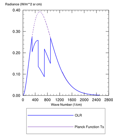
Figure 1: Spectrum (Radiance vs. wavenumber) for a Planck Body at 300 K (purple dashed) and the OLR with an IR absorbing greenhouse gas

Figure 1 is plotted as a somewhat “contrived” greenhouse substance that works like this: Our ground has a temperature Ts, with a colder temperature above the surface (e.g. the stratosphere).  Plotted are the Planck function for the surface temperature (purple dashed) and actual outgoing radiation (OLR, curve).  The Planck function gives the distribution of energy intensity vs. wavenumber (or wavelength, or frequency, depending on your favorite characterization of an electromagnetic wave) for a blackbody at some specified temperature.

The blue curve titled “OLR” is the actual spectrum of this hypothetical planet with a hypothetical greenhouse gas in the atmosphere.  The difference between that blue spectrum and the Planck (purple) spectrum for the ground temperature arises because our greenhouse gas happens to be blocking radiation from exiting directly to space at 600 cm-1 and the surrounding regions.  Even toward the “wings” at 400 or 800 cm-1 it is making the atmosphere “partially opaque.” This is fairly standard qualitative behavior for a greenhouse gas, especially CO2, although there are exceptions.

This plot is computed for a fixed temperature, so the end result of adding the greenhouse gas is to reduce the total outgoing radiation (the specific amount is whatever chunk is taken out of the Planck curve).  This creates a situation where the planet temporarily takes in more energy than it loses, and as a consequence the ground temperature must rise to increase emission and restore equilibrium.

To think about this another way, emission at wavenumbers where the atmosphere is strongly absorbing will always be closer to a "sensor" that is recording the emission than wavenumbers where the atmosphere is transparent.  If the sensor is a satellite looking down from space, it will see warm, surface emission in transparent ("window") wavenumbers, but for opaque wavenumbers, emission emanates from the high atmosphere.

Similarly, for a surface sensor looking up, emission from opaque regions is seen to come from very near the surface, whereas for transparent wavenumbers the sensor is recording the  ~3 K temperature of microwave background radiation in space. In this post, we're thinking about the sensor looking down.  

Brief Technical aside: Let’s define a “mean radiating pressure" of the planet, which we’ll call pr, where the atmosphere becomes optically thin enough to lose its radiation to space directly rather than being absorbed in a higher layer. Since pressure decreases with height, the radiating pressure will decrease as the optical thickness of the atmosphere increases (i.e., more radiation is preferentially leaking out higher in the atmosphere where it is colder when you add greenhouse gases).  Conversely, the radiating pressure is at the surface (pr=ps) with no greenhouse effect. It is easy to show that for an atmosphere whose temperature profile is dry adiabatic, that the radiating pressure is given by:
 
where the ratio cp/R is approximately 7/2 for Earth air; the numerator in the brackets is the absorbed solar radiation, σ is the Stefan-Boltzmann constant, and Tis the surface temperature.  For Earth, the mean radiating pressure would thus be at ~650 millibars, rather than at sea level (1000 mb) with no atmosphere (in reality, it would be smaller than this, since the real lapse rate is less steep than the dry adiabat).  See also Figure 2, to show how decreasing pr increases the surface temperature.
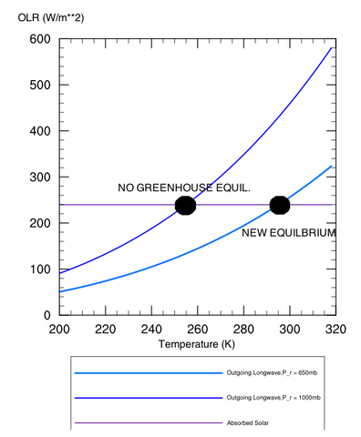
Figure 2: Depiction of how increasing the radiating height of a planet increases the surface temperature.  Equilibrium is reached when the outgoing long-wave energy curves intersect the absorbed solar radiation curve.

Does this all violate Thermodynamics?

The reason greenhouse warming does not violate thermodynamics is because the planet is not an energetically closed system, and receives a constant influx of energy from the sun.  The reduction in outgoing energy flow by the atmosphere can therefore heat the planet toward a value slightly closer to the solar temperature.  If the sun turned off, the greenhouse effect would be irrelevant (even assuming you could keep your atmosphere in the air at all without everything condensing out).  Some people on the blogs have claimed that because a colder atmosphere radiates toward a warmer surface, there is some thermodynamic inconsistency with the second law.  First, note that I have not said a word about back-radiation to the surface, primarily because it doesn’t give proper insight into the way energy balance is adjusted and determined.  But to the point, cold objects still radiate energy and a photon doesn’t care whether it’s traveling toward a warm object.  So yes, colder objects can and do radiate toward (and heat!) warmer objects.  Standard measurements (from Grant Petty's Radiation book) of back-radiation should be simple proof that this occurs.  Keep in mind that the net two-way energy flow is always from warm to cold.

Let’s now compare the theoretical Fig. 1 spectrum with a real Venus spectrum (Fig 3).

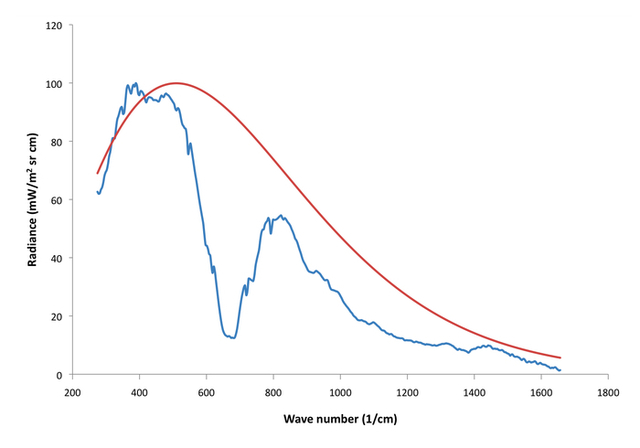
Figure 3:260 K blackbody spectrum (red) with observed Venus spectrum from The Venera 15 orbiter (blue). 

Here, the red curve is a 260 K blackbody Planck spectrum and the blue is a typical Venus spectrum I plotted which was obtained from the Soviet Venera 15 orbiter.  Keep in mind that the Venusian surface radiates at ~735 K, so the fact that the whole spectrum is seen to radiate at Earth or Mars like temperatures is a good indication that the atmosphere is highly opaque in the infrared spectrum.  Most of this is CO2, but other constituents like water vapor, SO2, and sulfur-water clouds are very important too, along with some other minor species.

Some Remarks about Pressure

It has been argued on some blogs that high pressures can cause high temperatures, and the argument has taken a variety of forms.  One is that p= ρRT (the ideal gas law) implies that a high p means a high T.  Of course, the pressure is 90x higher on Venus but the temperature is only 2-3 times higher than Earth, so such a straightforward proportion obviously doesn’t work.  The temperature must satisfy energy balance considerations, so a better way to think about the problem is to fix T (with other information, namely radiation) and solve for the density, which is of course much higher on Venus.  You can't get all the information from the equation of state alone.  The other argument is that some “insulative” property of gases could keep Venus hot at high pressure, even if the whole atmosphere were transparent to outgoing light.  One way to heat Venus would be to compress its atmosphere, but this would be temporary and eventually the temperature must relax back to its equilibrium value determined by energy conservation considerations.  The way things work is that heat is sluggishly migrated upward by radiation or convection until it finally reaches a point where the air is optically thin enough to let radiation leak out to space.  This doesn’t happen in a transparent atmosphere.

So does pressure matter for the greenhouse effect? The answer is yes, and the prime reason it matters is that collisions between molecules act to “smooth out” absorption and fill in the window regions where air is transparent.  Unlike the quantum nature of absorption and emission, the kinetic energy of moving molecules is not quantized, so it is possible for colliding molecules to impart kinetic energy on the absorber and make up the energy deficit required to make a quantum leap from one energy level to another.  There are some other broadening mechanisms too, but this is by far most important in the lower atmosphere.

Aside from the fact that a 90 bar atmosphere can hold much more greenhouse gas, pressure broadening is huge on Venus, but you can only smooth things out and fill in the windows so much.  Where pressure broadening would really make a difference is to put in a 1 bar atmosphere (even N2) on a very low dense atmosphere like Mars.  The reason why Mars does not currently generate a strong greenhouse effect, even at over 90% CO2, is that the spectral lines are too narrow to have a sizable effect.  Even with almost two orders of magnitude more CO2 per square meter than Earth, the equivalent width is less on Mars.  The equivalent width is a measure of the area of absorption taken out by a molecule (see the wiki article for further explanation on its definition).  The following diagrams illustrate the OLR change in a 250 ppm CO2 atmosphere at Earthlike pressure (Fig. 4a) and 100x Earth pressure (Fig. 4b) (note that the same mixing ratio in the 100 bar atmosphere implies more greenhouse gas overall).  
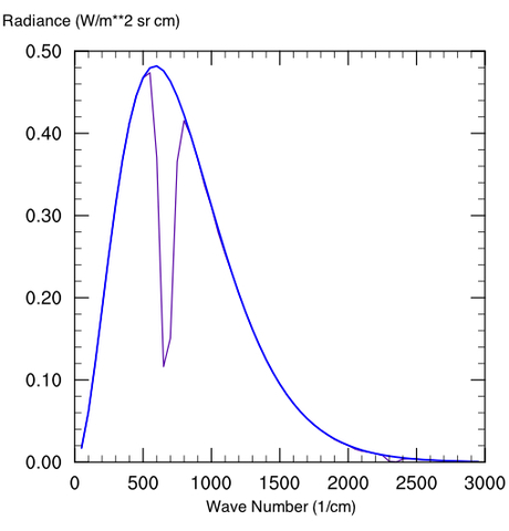
Figure 5: 250 ppm CO2 mixing ratio for an atmosphere at a) Earthlike pressure and b) 100x Earth pressure

Note that at very high CO2 concentrations, a lot of new absorption features come into play that are irrelevant on modern Earth.  The water vapor and sulfur-bearing compounds on Venus also help to fill in some window regions considerably.   Also unlike Earth, Venus has a non-negligible scattering greenhouse component too (by inhibiting cooling through IR scattering rather than absorption and emission).  These make direct planetary comparisons useless, except that Venus is a case in point of how much a greenhouse effect can matter in planetary climate discussions.

Note also that very dense atmospheres also raise the albedo through Rayleigh scattering; this is the same process that make our skies blue.  A pure Venusian CO2atmosphere raises the albedo to a moderately high ~40%, somewhat short of its current albedo (~77%, because of clouds), but still higher than Earth.  This remark is primarily true for planets orbiting sun-like stars, but for lower temperature stars (like M-dwarfs) the Rayleigh scattering is much less important, since the spectrum of the starlight itself is red-shifted, and Rayleigh scattering favors shorter (bluer) wavelengths.

Could a purely diatomic molecule atmosphere generate a greenhouse effect?

The answer, again, is yes.  This may be surprising because something like H2 or N2 doesn’t have the molecular symmetry (to make a dipole moment) that we commonly attribute as a defining characteristic of greenhouse gases.  Similarly, Pressure broadening doesn’t broaden anything that isn’t there to begin with.  But for very dense atmosphere, frequent enough collisions between diatomic molecules can temporarily make a ”four-atom” molecule that behaves like a greenhouse gas.  This effect is much more pronounced at colder temperatures, since the time of collision is longer at low velocities.  Collision induced (as opposed to broadened) absorption has been best studied on Titan, but it’s important on the gaseous planets, as well as some theoretical atmosphere with several tens of bars of H2 or He that are relatively dense and cold.  It’s unimportant on Earth, since the temperatures are high enough and density low enough.

Lapse Rates and Tropopause Height

Several other bloggers have been under the impression that the lapse rate “causes” high surface temperatures on a place like Venus, the idea being that the tropopause is very high and so one can extrapolate down the adiabat very far to reach a high temperature.  As should be obvious from the preceding section, the entire reason why you’re allowed to extrapolate such a far distance is because of the greenhouse effect, which increases the altitude where emission in the opaque regions of the spectrum take place.  In fact, on Venus the high tropopause is a a consequence of the high optical thickness. 

In radiative-convective equilibrium, the atmosphere transports sufficient heat vertically (by convection) to prevent the lapse rate from exceeding some critical value, so that a stratosphere can exist in radiative equilibrium (with a thermal balance between ozone heating and CO2 cooling) atop a troposphere where both radiative and dynamical fluxes are important.   The lapse rate just describes the manner in which temperature changes vertically; it isn’t some supply of energy and you need to specify the temperature at the surface by some other means.  The reason an adiabatic lapse rate might develop and the height to which it extends is most certainly not independent of radiation, which provides a basis for global energy flows.

An adiabatic lapse rate only needs to develop by convection where air parcels at the surface become buoyant with respect to the air above it.  In an infrared transparent atmosphere with no sources and sinks of energy, convection would eventually give out and the tropopause would migrate to the surface, developing a deep isothermal region.

In conclusion, the "greenhouse effect" is a very real physical phenomenon and has no inconsistencies with thermodynamics or any other field of inquiry (and in fact,emerges from these disciplines).  It can be just as important in determining the global temperature as the distance to the sun, and is especially important on Venus.

Acknowledgments: I would like to thank Ludmila Zasova for the Venus Venera spectral data used in Figure 3 (which was provided by David Crisp).  I also made use of Dr. Ray Pierrehumbert's online Python code that supplements his new textbook for image production.

Further Recommended Reading: Pierrehumbert RT 2011: Infrared radiation and planetary temperature. Physics Today 64, 33-38, online here [PDF]

ScienceofDoom - no specific link, as he has a large number of articles on Energy Balance and radiative transfer...great multi-series introduction if you wade through the pages

Comment On "Falsification Of The Atmospheric Co2 Greenhouse Effects Within The Frame Of Physics", by Joshua B. Halpern, Christopher M. Colose, Chris Ho-Stuart, Joel D. Shore, Arthur P. Smith And Jörg Zimmermann, in IJMP(B), Vol 24, Iss 10, Apr 20, 2010, pp 1309-1332
Several part series on Venus, by Brian Angliss, starting with this post

Brainstem

From Wikipedia, the free encyclopedia 
 
Brainstem
1311 Brain Stem.jpg
The three distinct parts of the brainstem are colored in this sagittal section of a human brain.
 
Details
Part of Brain
Parts Medulla, Pons, Midbrain
Identifiers
Latin truncus encephali
MeSH D001933
NeuroNames 2052, 236
NeuroLex ID birnlex_1565
TA A14.1.03.009
FMA 79876

The brainstem (or brain stem) is the posterior part of the brain, adjoining and structurally continuous with the spinal cord. In the human brain the brainstem includes the midbrain, the pons, and the medulla oblongata. Sometimes the diencephalon, the caudal part of the forebrain, is included.[1]

The brainstem provides the main motor and sensory innervation to the face and neck via the cranial nerves. Of the twelve pairs of cranial nerves, ten pairs come from the brainstem. Though small, this is an extremely important part of the brain as the nerve connections of the motor and sensory systems from the main part of the brain to the rest of the body pass through the brainstem. This includes the corticospinal tract (motor), the posterior column-medial lemniscus pathway (fine touch, vibration sensation, and proprioception), and the spinothalamic tract (pain, temperature, itch, and crude touch).

The brainstem also plays an important role in the regulation of cardiac and respiratory function. It also regulates the central nervous system, and is pivotal in maintaining consciousness and regulating the sleep cycle. The brainstem has many basic functions including heart rate, breathing, sleeping, and eating.

Structure

Midbrain

 
Structures of the brainstem
Cross-section of the midbrain at the level of the superior colliculus.
 
Cross-section of the midbrain at the level of the inferior colliculus.
 
The midbrain is divided into three parts: tectum, tegmentum, and the ventral tegmentum. The tectum (Latin:roof), which forms the ceiling. The tectum comprises the paired structure of the superior and inferior colliculi and is the dorsal covering of the cerebral aqueduct. The inferior colliculus, is the principal midbrain nucleus of the auditory pathway and receives input from several peripheral brainstem nuclei, as well as inputs from the auditory cortex. Its inferior brachium (arm-like process) reaches to the medial geniculate nucleus of the diencephalon. Superior to the inferior colliculus, the superior colliculus marks the rostral midbrain. It is involved in the special sense of vision and sends its superior brachium to the lateral geniculate body of the diencephalon. The tegmentum which forms the floor of the midbrain, and is ventral to the cerebral aqueduct. Several nuclei, tracts, and the reticular formation are contained here. The ventral tegmentum is composed of paired cerebral peduncles. These transmit axons of upper motor neurons.
The midbrain consists of:

Pons

The pons lies between the medulla oblongata and the midbrain. It contains tracts that carry signals from the cerebrum to the medulla and to the cerebellum and also tracts that carry sensory signals to the thalamus. The pons is connected to the cerebellum by the cerebellar peduncles. The pons houses the respiratory pneumotaxic center and apneustic center that make up the pontine respiratory group in the respiratory center. The pons co-ordinates activities of the cerebellar hemispheres.[citation needed]

Medulla oblongata

The medulla oblongata often just referred to as the medulla, is the lower half of the brainstem continuous with the spinal cord. Its upper part is continuous with the pons.[2] The medulla contains the cardiac, respiratory, vomiting and vasomotor centres dealing with heart rate, breathing and blood pressure.

Ventral view of medulla and pons

Cross-section of the middle pons (at the level of cranial nerve V).
 
Cross-section of the inferior pons (at the level of the facial genu).
 
Cross-section of the rostral (superior) medulla.
 
Cross-section of the middle medulla.
 
Cross-section of the inferior medulla.

In the medial part of the medulla is the anterior median fissure. Moving laterally on each side are the medullary pyramids. The pyramids contain the fibers of the corticospinal tract (also called the pyramidal tract), or the upper motor neuronal axons as they head inferiorly to synapse on lower motor neuronal cell bodies within the anterior grey column of the spinal cord.

The anterolateral sulcus is lateral to the pyramids. Emerging from the anterolateral sulci are the CN XII (hypoglossal nerve) rootlets. Lateral to these rootlets and the anterolateral sulci are the olives. The olives are swellings in the medulla containing underlying inferior nucleary nuclei (containing various nuclei and afferent fibers). Lateral (and dorsal) to the olives are the rootlets for CN IX (glossopharyngeal), CN X (vagus) and CN XI (accessory nerve). The pyramids end at the pontine medulla junction, noted most obviously by the large basal pons. From this junction, CN VI (abducens nerve), CN VII (facial nerve) and CN VIII (vestibulocochlear nerve) emerge. At the level of the midpons, CN V (the trigeminal nerve) emerges. Cranial nerve III (the oculomotor nerve) emerges ventrally from the midbrain, while the CN IV (the trochlear nerve) emerges out from the dorsal aspect of midbrain.

Between the two pyramids can be seen a decussation of fibres which marks the transition from the medulla to the spinal cord. The medulla is above the decussation and the spinal cord below.

Dorsal view of medulla and pons

The most medial part of the medulla is the posterior median sulcus. Moving laterally on each side is the fasciculus gracilis, and lateral to that is the fasciculus cuneatus. Superior to each of these, and directly inferior to the obex, are the gracile and cuneate tubercles, respectively. Underlying these are their respective nuclei. The obex marks the end of the fourth ventricle and the beginning of the central canal. The posterior intermediate sulcus separates the fasciculus gracilis from the fasciculus cuneatus. Lateral to the fasciculus cuneatus is the lateral funiculus.

Superior to the obex is the floor of the fourth ventricle. In the floor of the fourth ventricle, various nuclei can be visualized by the small bumps that they make in the overlying tissue. In the midline and directly superior to the obex is the vagal trigone and superior to that it the hypoglossal trigone. Underlying each of these are motor nuclei for the respective cranial nerves. Superior to these trigones are fibers running laterally in both directions. These fibers are known collectively as the striae medullares. Continuing in a rostral direction, the large bumps are called the facial colliculi. Each facial colliculus, contrary to their names, do not contain the facial nerve nuclei. Instead, they have facial nerve axons traversing superficial to underlying abducens (CN VI) nuclei. Lateral to all these bumps previously discussed is an indented line, or sulcus that runs rostrally, and is known as the sulcus limitans. This separates the medial motor neurons from the lateral sensory neurons. Lateral to the sulcus limitans is the area of the vestibular system, which is involved in special sensation. Moving rostrally, the inferior, middle, and superior cerebellar peduncles are found connecting the midbrain to the cerebellum. Directly rostral to the superior cerebellar peduncle, there is the superior medullary velum and then the two trochlear nerves. This marks the end of the pons as the inferior colliculus is directly rostral and marks the caudal midbrain.

Development

The adult human brainstem emerges from two of the three primary vesicles formed of the neural tube. The mesencephalon is the second of the three primary vesicles, and does not further differentiate into a secondary vesicle. This will become the midbrain. The third primary vesicle, the rhombencephalon (hindbrain) will further differentiate into two secondary vesicles, the metencephalon and the myelencephalon. The metencephalon will become the cerebellum and the pons. The more caudal myelencephalon will become the medulla.

Blood supply

The main supply of blood to the brainstem is provided by the basilar arteries and the vertebral arteries.[3]

Cranial nerves

Ten of the twelve pairs of cranial nerves either target or are sourced from the brainstem.[4] The nuclei of the oculomotor nerve (III) and trochlear nerve (IV) are located in the midbrain. The nuclei of the trigeminal nerve (V), abducens nerve (VI), facial nerve (VII) and vestibulocochlear nerve (VIII) are located in the pons. The nuclei of the glossopharyngeal nerve (IX), vagus nerve (X), accessory nerve (XI) and hypoglossal nerve (XII) are located in the medulla. The fibers of these cranial nerves exit the brainstem from these nuclei.[5]

Function

There are three main functions of the brainstem:
  1. The brainstem plays a role in conduction. That is, all information relayed from the body to the cerebrum and cerebellum and vice versa must traverse the brainstem. The ascending pathways coming from the body to the brain are the sensory pathways and include the spinothalamic tract for pain and temperature sensation and the dorsal column, fasciculus gracilis, and cuneatus for touch, proprioception, and pressure sensation (both of the body). (The facial sensations have similar pathways, and will travel in the spinothalamic tract and the medial lemniscus also.) Descending tracts are upper motor neurons destined to synapse on lower motor neurons in the ventral horn and posterior horn. In addition, there are upper motor neurons that originate in the brainstem's vestibular, red, tectal, and reticular nuclei, which also descend and synapse in the spinal cord.
  2. The cranial nerves III-XII emerge from the brainstem.[6] These cranial nerves supply the face, head, and viscera. (The first two pairs of cranial nerves arise from the cerebrum).
  3. The brainstem has integrative functions being involved in cardiovascular system control, respiratory control, pain sensitivity control, alertness, awareness, and consciousness. Thus, brainstem damage is a very serious and often life-threatening problem.

Clinical significance

Diseases of the brainstem can result in abnormalities in the function of cranial nerves that may lead to visual disturbances, pupil abnormalities, changes in sensation, muscle weakness, hearing problems, vertigo, swallowing and speech difficulty, voice change, and co-ordination problems. Localizing neurological lesions in the brainstem may be very precise, although it relies on a clear understanding on the functions of brainstem anatomical structures and how to test them.

Brainstem stroke syndrome can cause a range of impairments including locked-in syndrome.

Duret haemorrhages are areas of bleeding in the midbrain and upper pons due to a downward traumatic displacement of the brainstem.[7]

Cysts known as syrinxes can affect the brainstem, in a condition called syringobulbia. These fluid-filed cavities can be congenital, acquired or the result of a tumor.

Criteria for claiming brainstem death in the UK have developed in order to make the decision of when to stop ventilation of somebody who could not otherwise sustain life. These determining factors are that the patient is irreversibly unconscious and incapable of breathing unaided. All other possible causes must be ruled out that might otherwise indicate a temporary condition. The state of irreversible brain damage has to be unequivocal. There are brainstem reflexes that are checked for by two senior doctors so that imaging technology is unnecessary. The absence of the cough and gag reflexes, of the corneal reflex and the vestibulo-ocular reflex need to be established; the pupils of the eyes must be fixed and dilated; there must be an absence of motor response to stimulation and an absence of breathing marked by concentrations of carbon dioxide in the arterial blood. All of these tests must be repeated after a certain time before death can be declared.[8]

Additional images

  • The midbrain, pons, and medulla oblongata are labelled on this coronal section of the human brain.
  • Brainstem. Anterior face.Deep dissection
  • Brainstem. Posterior face.Deep dissec
  • Neuroscience of sex differences

    From Wikipedia, the free encyclopedia

    Neuroscience of sex differences is the study of the characteristics of the brain that separate the male brain and the female brain. Psychological sex differences are thought by some to reflect the interaction of genes, hormones and social learning on brain development throughout the lifespan.

    Some evidence from brain morphology and function studies indicates that male and female brains cannot always be assumed to be identical from either a structural or functional perspective, and some brain structures are sexually dimorphic.[1][2]

    Experts note that neural sexual dimorphisms in humans exist only as averages, with overlapping variabilities,[3] and that it is unknown to what extent each is influenced by genetics or environment, even in adulthood.[4][5]

    History

    Ideas of differences in the male and female brain circulated during the time of ancient Greek philosophers around 850 B.C. Aristotle claimed that males did not "receive their soul" until 40 days post-gestation and females did not until 80 days. In 1854, Emil Huschke discovered that "the frontal lobe in the male is all of 1% larger than that of the female."[6] As the 19th century progressed, scientists began researching sexual dimorphisms in the brain significantly more.[7] Until around 21 years ago, scientists knew of several structural sexual dimorphisms of the brain, but they did not think that gender had any impact on how the human brain performs daily tasks. Through molecular, animal, and neuroimaging studies, a great deal of information regarding the differences between male and female brains and how much they differ in regards to both structure and function has been uncovered.[8]

    Evolutionary explanations

    Sexual selection

    It is thought that male and female differences in learning ability have contributed to sexual selection and mate preference throughout evolution. The hippocampus has even been found to exhibit seasonal activity in some mammals where it is active during breeding periods but inactive during hibernation; this is because spatial learning is more present during the breeding season.[9]

    Females show enhanced information recall compared to males. This may be due to the fact that females have a more intricate evaluation of risk-scenario contemplation, based on a prefrontal cortical control of the amygdala. For example, the ability to recall information better than males most likely originated from sexual selective pressures on females during competition with other females in mate selection. Recognition of social cues was an advantageous characteristic because it ultimately maximized offspring and was therefore selected for during evolution.[1]

    Oxytocin is a hormone that induces contraction of the uterus and lactation in mammals. It is also a characteristic hormone of nursing mothers. Studies have found that oxytocin improves spatial memory. Through activation of the MAP kinase pathway, oxytocin plays a role in the enhancement of long-term synaptic plasticity, which is a change in strength between two neurons over a synapse that lasts for minutes or longer, and long-term memory. This hormone may have helped mothers remember the location of distant food sources so they could better nurture their offspring.[1]

    Male vs. female brain anatomy

    Hemisphere differences

    A popular theory regarding language functions suggests that women use both hemispheres more equally, whereas men are more strongly lateralized to the left hemisphere.[10] This theory found initial support in a high-profile study of 19 men and 19 women, which found stronger lateralization in men during one of the three language tasks assessed.[11] In 2008, some researchers concluded that further studies have failed to replicate this finding, and a meta-analysis of 29 studies comparing language lateralization in males and females found no overall difference.[12] However, in 2013, researchers at the Perelman School of Medicine at the University of Pennsylvania mapped notable differences in male and female neural wiring. The study used diffusion tensor imaging of 949 individuals aged 8–22 years, and concluded that in all supratentorial regions of the brain inter-hemispheric connectivity was greater in women's and girls' brains, whereas intra-hemispheric connectivity was greater in the brains of men and boys. The effect was reversed in cerebellar connections.[13] The detected differences in neural connectivity were negligible up to the age of 13, but became much more prominent in the 14 to 17-year-olds.[13] In terms of the potential effect on behaviour, the authors concluded, "Overall, the results suggest that male brains are structured to facilitate connectivity between perception and coordinated action, whereas female brains are designed to facilitate communication between analytical and intuitive processing modes".[13]

    Amygdala

    image of Amygdala
    The amygdala (red) in a human brain.

    According to some researchers, "the research on sex differences in the amygdala has produced conflicting results".[14] After correcting for the overall difference in brain volume between men and women, a 2016 meta-analysis indicated that the amygdala is not significantly larger in either sex.[15] Some studies, however, report increased amygdala activity during the processing of affective scenes in men relative to women (Schienle et al., 2005; Goldstein et al., 2010), and meta-analysis supports this view, showing larger effect sizes in studies of affective processing including only men compared with those including only women (Sergerie et al., 2008). However several studies using similar stimuli have reported a larger amygdala response in women (Klein et al., 2003; McClure et al., 2004; Hofer et al., 2006; Domes et al., 2010), and others have reported no sex difference at all (Wrase et al., 2003; Caseras et al., 2007; Aleman and Swart, 2008). A possible explanation for these inconsistent results is that sex differences in amygdala response are valence-dependent. Furthermore, according to other researchers,[16] "Correlation analyses revealed that gray matter thickness in left ventromedial PFC was inversely correlated with task-related activation in the amygdala. These data add support to a general role of the ventromedial PFC in regulating activity of the amygdala."

    Research has been done on post-traumatic stress disorder (PTSD), an anxiety disorder found in both sexes, which is particularly common in war veterans, assault victims and women who have experienced abuse. Emotional memory encoding varies in the amygdala on the right and left and occurs equally for both genders: the right triggers unpleasant and fear-related memories, both declarative (conscious) and episodic (nonconcious).[17]

    Amygdala volume correlates positively with fearfulness in girls but not in boys.[18]

    Hippocampus

    Several studies have indicated that the hippocampi of men and women differ anatomically, neurochemically, and also in degree of long-term potentiation.[19] Such evidence indicates that sex may influence the role of the hippocampus in learning. By contrast, a 2016 meta-analysis indicated that the hippocampus does not differ in volume between men and women.[20] One experiment examined the effects of stress on Pavlovian conditioning performance in both sexes and found that males' performance under stress was enhanced while female performance was impaired. Activation of the hippocampus is more dominant on the left side of hippocampus in females, while it is more dominant on the right side in males. This in turn influences cognitive reasoning; women use more verbal strategies than men when performing a task that requires cognitive thinking.[21] The hippocampus's relationship with other structures in the brain influences learning and has been found to be sexually dimorphic as well.[1]

    Estradiol has been found to influence hippocampal development. Studies have shown endogenous neurogenesis, or the internally-driven formation of new neurons, to be higher in the male hippocampus than in that of the female. However, following exogenous estradiol injections, the number of new neurons in the hippocampi of females reached levels equivalent to those found in the hippocampi of males. Conversely, injecting estradiol in males did not increase neurogenesis in males. However, antagonizing endogenous estradiol in males reduced neurogenesis, but did not change the number of new neurons in females. Thus, hippocampal sex differences appear to be mediated in part through endogenous estradiol levels.[22]

    Frontal lobe

    The ventromedial prefrontal cortex (VMPC) plays a key role in social emotional processing. In accordance with the sexual dimorphism of the amygdala, the right VMPC is more dominant in an active limbic system for males while the left is more dominant in females. These differences carry out to a behavioral level. For example, Koscik et al. wrote:
    "A man with a unilateral right VMPC lesion, who was well educated and had worked successfully as a minister, was entirely unable to return to any form of gainful employment after his brain damage. He requires supervision for daily tasks and demonstrates severe disturbances in behavior and emotional regulation, including impulsivity and poor judgment. By contrast, a man with a unilateral left VMPC lesion was able to return to his job at a grain elevator and remains successfully employed there. He is remarkably free of disturbances to his social life and emotional functioning."[23]

    Orbital prefrontal cortex

    Positron emission tomography studies have shown that men and women ranging from the ages of 19 to 32 years old metabolize glucose at significantly different rates in the orbital prefrontal cortex. Infant males who exhibited lesions on their orbital prefrontal cortex struggled with object reversal experiments, but females exhibiting such lesions did not have impaired performance in object reversal.[24]

    Other regions and not region-specific

    There are sex differences in locus coeruleus dendritic structure that allow for an increased reception and processing of limbic information in females compared to males.[18]

    Aggressive and defiant behavior is also associated with decreased right anterior cingulate cortex (ACC) volume in boys.[18]

    According to the neuroscience journal review series Progress in Brain Research, it has been found that males have larger and longer planum temporale and Sylvian fissure while females have significantly larger proportionate volumes to total brain volume in the superior temporal cortex, Broca's area, the hippocampus and the caudate.[25] The midsagittal and fiber numbers in the anterior commissure that connect the temporal poles and mass intermedia that connects the thalami is also larger in females.[25]

    The journal review also found that the male brain volume was slightly larger than the female brain volume, a difference is often attributed to the larger average body and skull sizes of males. In female brains, however, greater cortical thickness, cortical complexity and cortical surface area are observed after adjusting for brain volume differences.[25] Given that cortical complexity and cortical features are positively correlated with intelligence, researchers postulated that these differences in cortical complexity may have been an evolutionary adaptation to the difference in brain volume of males and females.[25]

    White/grey matter

    Global and regional grey matter (GM) differs in men and women. Women have larger left orbitofrontal GM volumes and overall cortical thickness than men.[26] Behavioral implications of the greater volume have not yet been discovered. Women have a higher percentage of GM, whereas men have a higher percentage of white matter (WM) and of CSF (cerebrospinal fluid). In men the percentage of GM was higher in the left hemisphere, the percentage of WM was symmetric, and the percentage of CSF was higher in the right. Women showed no asymmetries. Both GM and WM volumes correlated moderately with global, verbal, and spatial performance across groups. However, the regression of cognitive performance and WM volume was significantly steeper in women.[27]

    In a 2013 meta-analysis, researchers found on average males had larger grey matter volume in bilateral amygdalae, hippocampi, anterior parahippocampal gyri, posterior cingulate gyri, precuneus, putamen and temporal poles, areas in the left posterior and anterior cingulate gyri, and areas in the cerebellum bilateral VIIb, VIIIa and Crus I lobes, left VI and right Crus II lobes.[2] On the other hand, females on average had larger grey matter volume at the right frontal pole, inferior and middle frontal gyri, pars triangularis, planum temporale/parietal operculum, anterior cingulate gyrus, insular cortex, and Heschl's gyrus; bilateral thalami and precuneus; the left parahippocampal gyrus and lateral occipital cortex (superior division).[2] The meta-analysis found larger volumes in females were most pronounced in areas in the right hemisphere related to language in addition to several limbic structures such as the right insular cortex and anterior cingulate gyrus.[2]

    Amber Ruigrok's 2013 meta-analysis also found greater grey matter density in the average male left amygdala, hippocampus, insula, pallidum, putamen, claustrum and right cerebellum.[2] The meta-analysis also found greater grey matter density in the average female left frontal pole[2]

    Brain networks

    A 2014 meta-analysis by researcher Ashley C.Hill found that although men and women commonly used the same brain networks for working memory, specific regions were gender specific.[28] For example, both men and women's active working memory networks composed of bilateral middle frontal gyri, left cingulate gyrus, right precuneus, left inferior and superior parietal lobes, right claustrum, and left middle temporal gyrus but women also tended have consistent activity in the limbic regions such as the anterior cingulate, bilateral amygdala and right hippocampus while men tended to have a distributed networks spread out among the cerebellum, portions of the superior parietal lobe, the left insula and bilateral thalamus.[28] In the work of[29] the authors have computed structural connectomes of 96 subjects of the Human Connectome Project, and they have proven that in numerous graph-theoretical parameters, the structural connectome of women are significantly better connected than the connectome of men. For example, women's connectome has more edges, higher minimum bipartition width, larger eigengap, greater minimum vertex cover than that of men. The minimum bipartition width (or the minimum balanced cut (graph theory)) is well-known measure of quality of computer multistage interconnection networks, it describes the possible bottlenecks in network communication: The higher this value is, the better is the network. The larger eigengap shows that the female connectome is better expander graph than the connectome of males. The better expanding property, the higher minimum bipartition width and the greater minimum vertex cover show deep advantages in network connectivity in the case of female braingraph.

    Brain differences between homo- and heterosexuals

    Brain wiring comparisons of homosexuals and persons of the opposite sex show that homosexuals may be born with a predisposition to be homosexual. Research at the Stockholm Brain Institute in Sweden found that homosexual men and heterosexual women have similar brain characteristics. Specifically, these similarities are in the overall size of the brain and the activity of the amygdala. The same is for heterosexual men and homosexual women. Molecular biologist at the National Institutes of Health, Dean Hamer, says, "this is from a series of observations showing there's a biological reason for sexual orientation".[30]

    Ivanka Savic – Berglund conducted a study in which MRIs were used to measure the volume and shapes of the brain. She also used PET scans to view blood flow to the amygdala. Savic – Berglund found that in homosexual men and heterosexual women, the blood flowed to areas involved in fear and anxiety, whereas in heterosexual men and homosexual women, it tended to flow to pockets linked to aggression. When looking at hemisphere differences, the right hemisphere was found to be slightly larger than the left in heterosexual men and homosexual women, whereas those of homosexual men and heterosexual women were more symmetrical.[31]

    Research has indicated that the corpus callosum is larger in homosexual men than in heterosexual men. This is significant because the corpus callosum is a structure that is developed early. In the Journal Science Simon LeVay showed that the third interstitial nucleus of the hypothalamus has neurons that are packed more together in homosexual men than in heterosexual men.[32] Connections from the amygdala to other parts of the brain are similar between homosexuals and persons of the opposite gender as shown through PET and MRI scans. For example, in homosexual men and heterosexual women, there were more connections from the left amygdala. In homosexual women and heterosexual men, there were more connections from the right amygdala. LeVay's results were not replicated in other studies. A 2001 study that attempted to replicate the findings concluded that "Although there was a trend for INAH3 to occupy a smaller volume in homosexual men than in heterosexual men, there was no difference in the number of neurons within the nucleus based on sexual orientation."[33]

    Neurochemical differences

    Hormones

    Steroid hormones have several effects on brain development as well as maintenance of homeostasis throughout adulthood. One effect they exhibit is on the hypothalamus, where they increase synapse formation.[34] Estrogen receptors have been found in the hypothalamus, pituitary gland, hippocampus, and frontal cortex, indicating the estrogen plays a role in brain development. Gonadal hormone receptors have also been found in the basal forebrain nuclei.[35]

    Estrogen and the female brain

    Estradiol influences cognitive function, specifically by enhancing learning and memory in a dose-sensitive manner. Too much estrogen can have negative effects by weakening performance of learned tasks as well as hindering performance of memory tasks; this can result in females exhibiting poorer performance of such tasks when compared to males.[36]

    It has been suggested that during development, estrogen can exhibit both feminizing and defeminizing effects on the human brain; high levels of estrogen induce male neural traits to develop while moderate levels induce female traits. In females, defeminizing effects are resisted because of the presence of α-fetoprotein (AFP), a carrier protein proposed to transport estrogen into brain cells, allowing the female brain to properly develop. The role of AFP is significant at crucial stages of development, however. Prenatally, AFP blocks estrogen. Postnatally, AFP decreases to ineffective levels; therefore, it is probable that estrogen exhibits its effects on female brain development postnatally.[37]

    Ovariectomies, surgeries inducing menopause, or natural menopause cause fluctuating and decreased estrogen levels in women. This in turn can "attenuate the effects" of endogenous opioid peptides.  Opioid peptides are known to play a role in emotion and motivation. β-endorphin (β-EP), an endogenous opioid peptide, content has been found to decrease (in varying amounts/brain region), post ovariectomy, in female rats within the hypothalamus, hippocampus, and pituitary gland. Such a change in β-EP levels could be the cause of mood swings, behavioral disturbances, and hot flashes in post menopausal women.[35]

    Testosterone and the male brain

    Testosterone has been found to play a big role during development but may have independent effects on sexually dimorphic brain regions in adulthood. Studies have shown that the medial amygdala of male hamsters exhibits lateralization and sexual dimorphism prior to puberty. Furthermore, organization of this structure during development is influenced by the presence of androgens and testosterone. This is evident when comparing medial amygdala volume of male and female rats, adult male brains have a medial amygdala of greater volume than do adult female brains which is partially due to androgen circulation.[38] It also heavily influences male development; a study found that perinatal females introduced to elevated testosterone levels exhibited male behavior patterns. In the absence of testosterone, female behavior is retained.[34] Testosterone's influence on the brain is caused by organizational developmental effects. It has been shown to influence proaptotic proteins so that they increase neuronal cell death in certain brain regions. Another way testosterone affects brain development is by aiding in the construction of the "limbic hypothalamic neural networks".[34]

    Similar to how estrogen enhances memory and learning in women, testosterone has been found to enhance memory recall in men. In a study testing a correlation between memory recall and testosterone levels in men, "fMRI analysis revealed that higher testosterone levels were related to increased brain activation in the amygdala during encoding of neutral pictures".[39]

    Oxytocin and Vasopressin

    Oxytocin is positively correlated with maternal behaviours, social recognition, social contact, sexual behaviour and pair bonding. Oxytocin appears at higher levels in women than in men.[40] Vasopressin on the other hand is more present in men and mediates sexual behavior, aggression and other social functions.[40][41]

    Neurotransmitters

    Whole level 5-HT serotonin levels are higher in women versus men while men synthesize serotonin significantly faster than women. Healthy women also have higher 5-HT transport availability in the diencephalon and brainstem areas of the brain.[42] Dopamine function is also increased in women especially dopamine transporter which regulates the availability of receptors. Women before the onset of menopause synthesize higher levels of striatal presynaptic dopamine than age-matched men.[42] Other neurotransmitters like μ-opioids show significantly higher binding potential in the cerebellum, amygdala and the thalamus for women than it does so for men.[43] Women are also more dependent on norepinephrine in the formation of long term emotional memories than men are.[43]

    Male vs. female brain functionality

    Neural masculinization is a developmental process where different sex hormones assist in the expression of male behavior.[44]

    Stress

    image of stress regions in brain
    Regions of the brain associated with stress and fear.

    Stress has been found to induce an increase in serotonin, norepinephrine, and dopamine levels within the basolateral amygdala of male rats, but not within that of female rats. Furthermore, object recognition is impaired in males as a result of short term stress exposure. Neurochemical levels in the brain can change under the influence of stress exposure, particularly in regions associated with spatial and non-spatial memory, such as the prefrontal cortex and the hippocampus. Dopamine metabolite levels decrease post stress in male rats' brains, specifically within the CA1 region of the hippocampus.[45]

    In female rats, both short term (1 hour) and long term (21 days) stress has been found to actually enhance spatial memory. Under stress, male rats exhibit deleterious effects on spatial memory, however female rats show a degree of resistance to this phenomenon. Stressed female rats' norepinephrine (NE) levels go up by about 50% in their prefrontal cortex while that of male rats goes down 50%.[45]

    Cognitive tasks

    It was once thought that sex differences in cognitive task and problem solving did not occur until puberty. However, new evidence now suggests that cognitive and skill differences are present earlier in development. For example, researchers have found that three- and four-year-old boys were better at targeting and at mentally rotating figures within a clock face than girls of the same age were. Prepubescent girls, however, excelled at recalling lists of words. These sex differences in cognition correspond to patterns of ability rather than overall intelligence. Laboratory settings are used to systematically study the sexual dimorphism in problem solving task performed by adults.[46]

    On average, males excel relative to females at certain spatial tasks. Specifically, males have an advantage in tests that require the mental rotation or manipulation of an object.[47] In a computer simulation of a maze task, males completed the task faster and with fewer errors than their female counterparts. Additionally, males have displayed higher accuracy in tests of targeted motor skills, such as guiding projectiles.[46] Males are also faster on reaction time and finger tapping tests.[48]

    On average, females excel relative to males on tests that measure recollection. They have an advantage on processing speed involving letters, digits and rapid naming tasks.[48] Females tend to have better object location memory and verbal memory.[49] They also perform better at verbal learning.[50] Females have better performance at matching items and precision tasks, such as placing pegs into designated holes. In maze and path completion tasks, males learn the goal route in fewer trials than females, but females remember more of the landmarks presented. This shows that females use landmarks in everyday situations to orient themselves more than males. Females are better at remembering whether objects had switched places or not.[46]

    Studies using the Iowa gambling task, or Iowa Card Task, have examined cognitive reasoning and decision-making in males and females. A study in which participants of various age groups who were asked to perform the Iowa Card Task produced data showing that males and females differ in their decision making processes on the neurological level. The study suggests that decision-making in females may be guided by avoidance of negativity while decision making in males is mainly guided by assessing the long term outcome of a situation. They also found that males outperformed females in the Iowa Card Task, but there was a negative correlation between elevated testosterone levels and performance in the card task which indicates gonadal hormones influence decision-making.[24]

    Volcanic gas

    From Wikipedia, the free encyclopedia Not to be confused with Pyroclastic flow . Volcanic gases entering th...