Search This Blog

Saturday, June 23, 2018

Dorsolateral prefrontal cortex

From Wikipedia, the free encyclopedia
 
Dorsolateral Prefrontal Cortex
Prefrontal1.png
An illustration of brain's prefrontal region
Details
Identifiers
Latin Cortex praefrontalis dorsolateralis
FMA 276189

The dorsolateral prefrontal cortex (DLPFC or DL-PFC) is an area in the prefrontal cortex of the brain of humans and non-human primates. It is one of the most recently derived parts of the human brain. It undergoes a prolonged period of maturation which lasts until adulthood.[1] The DLPFC is not an anatomical structure, but rather a functional one. It lies in the middle frontal gyrus of humans (i.e., lateral part of Brodmann's area (BA) 9 and 46[2]). In macaque monkeys, it is around the principal sulcus (i.e., in Brodmann's area 46[3][4][5]). Other sources consider that DLPFC is attributed anatomically to BA 9 and 46[6] and BA 8, 9 and 10.[1]

The DLPFC has connections with the orbitofrontal cortex, as well as the thalamus, parts of the basal ganglia (specifically, the dorsal caudate nucleus), the hippocampus, and primary and secondary association areas of neocortex (including posterior temporal, parietal, and occipital areas).[7] The DLPFC is also the end point for the dorsal pathway (stream), which is concerned with how to interact with stimuli.

An important function of the DLPFC is the executive functions, such as working memory, cognitive flexibility,[8] planning, inhibition, and abstract reasoning.[9] However, the DLPFC is not exclusively responsible for the executive functions. All complex mental activity requires the additional cortical and subcortical circuits with which the DLPFC is connected.[10] The DLPFC is also the highest cortical area that is involved in motor planning, organization and regulation.[10]

Structure

As the DLPFC is composed of spatial selective neurons, it has a neural circuitry that encompasses the entire range of sub-functions necessary to carry out an integrated response, such as: sensory input, retention in short-term memory, and motor signaling.[11] Historically, the DLPFC was defined by its connection to: the superior temporal cortex, the posterior parietal cortex, the anterior and posterior cingulate, the premotor cortex, the retrosplenial cortex, and the neocerebellum.[1] These connections allow the DLPFC to regulate the activity of those regions, as well as to receive information from and be regulated by those regions.[1]

Function

Primary functions

The DLPFC is known for its involvement in the executive functions, which is an umbrella term for the management of cognitive processes,[12] including working memory, cognitive flexibility,[13] and planning.[14] A couple of tasks have been very prominent in the research on the DLPFC, such as the A-not-B task, the delayed response task and object retrieval tasks.[1] The behavioral task that is most strongly linked to DLPFC is the combined A-not-B/delayed response task, in which the subject has to find a hidden object after a certain delay. This task requires holding information in mind (working memory), which is believed to be one of the functions of DLPFC.[1] The importance of DLPFC for working memory was strengthened by studies with adult macaques. Lesions that destroyed DLPFC disrupted the macaques’ performance of the A-not-B/delayed response task, whereas lesions to other brain parts did not impair their performance on this task.[1]

DLPFC is not required for the memory of a single item. Thus, damage to the dorsolateral prefrontal cortex does not impair recognition memory.[15] Nevertheless, if two items must be compared from memory, the involvement of DLPFC is required. People with damaged DLPFC are not able to identify a picture they had seen, after some time, when given the opportunity to choose from two pictures.[15] Moreover, these subjects also failed in Wisconsin Card-Sorting Test as they lose track of the currently correct rule and persistently organize their cards in the previously correct rule.[16] In addition, as DLPFC deals with waking thought and reality testing, it is not active when one is asleep.[16] Likewise, DLPFC is most frequently related to the dysfunction of drive, attention and motivation.[17] Patients with minor DLPFC damage display disinterest in their surroundings and are deprived of spontaneity in language as well as behavior.[17] Patients may also be less alert than normal to people and events they know.[17] Damage to this region in a person also leads to the lack of motivation to do things for themselves and/or for others.[17]

Decision making

The DLPFC is involved in both risky and moral decision making; when individuals have to make moral decisions like how to distribute limited resources, the DLPFC is activated.[18] This region is also active when costs and benefits of alternative choices are of interest.[19] Similarly, when options for choosing alternatives are present, the DLPFC evokes a preference towards the most equitable option and suppresses the temptation to maximize personal gain.[20]

Working memory

Working memory is the system that actively holds multiple pieces of transitory information in the mind, where they can be manipulated. The DLPFC is important for working memory;[21] reduced activity in this area correlates to poor performance on working memory tasks.[22] However, other areas of the brain are involved in working memory as well.[23]

There is an ongoing discussion if the DLPFC is specialized in a certain type of working memory, namely computational mechanisms for monitoring and manipulating items, or if it has a certain content, namely visuospatial information, which makes it possible to mentally represent coordinates within the spatial domain.[21]

There have also been some suggestions that the function of the DLPFC in verbal and spatial working memory is lateralised into the left and right hemisphere, respectively. Smith, Jonides and Koeppe (1996)[24] observed a lateralisation of DLPFC activations during verbal and visual working memory. Verbal working memory tasks mainly activated the left DLPFC and visual working memory tasks mainly activated the right DLPFC. Murphy et al. (1998)[25] also found that verbal working memory tasks activated the right and left DLPFC, whereas spatial working memory tasks predominantly activated the left DLPFC. Reuter-Lorenz et al. (2000)[26] found that activations of the DLPFC showed prominent lateralisation of verbal and spatial working memory in young adults, whereas in older adults this lateralisation was less noticeable. It was proposed that this reduction in lateralisation could be due to recruitment of neurons from the opposite hemisphere to compensate for neuronal decline with ageing.

Secondary functions

The DLPFC may also be involved in the act of deception and lying,[27] which is thought to inhibit normal tendency to truth telling. Research also suggests that using TMS on the DLPFC can impede a person's ability to lie or to tell the truth.[28]

Additionally, supporting evidence suggests that the DLPFC may also play a role in conflict-induced behavioral adjustment, for instance when an individual decides what to do when faced with conflicting rules.[29] One way in which this has been tested is through the Stroop test,[30] in which subjects are shown a name of a color printed in colored ink and then are asked to name the color of the ink as fast as possible. Conflict arises when the color of the ink does not match the name of the printed color. During this experiment, tracking of the subjects’ brain activity showed a noticeable activity within the DLPFC.[30] The activation of the DLPFC correlated with the behavioral performance, which suggests that this region maintains the high demands of the task to resolve conflict, and thus in theory plays a role in taking control.[30]

DLPFC may also be associated with human intelligence. However, even when correlations are found between the DLPFC and human intelligence, that does not mean that all human intelligence is a function of the DLPFC. In other words, this region may be attributed to general intelligence on a broader scale as well as very specific roles, but not all roles. For example, using imaging studies like PET and fMRI indicate DLPFC involvement in deductive, syllogistic reasoning.[31] Specifically, when involved in activities that require syllogistic reasoning, left DLPFC areas are especially and consistently active.[31]

The DLPFC may also be involved in threat-induced anxiety.[32] In one experiment, participants were asked to rate themselves as behaviorally inhibited or not. Those who rated themselves as behaviorally inhibited, moreover, showed greater tonic (resting) activity in the right-posterior DLPFC.[32] Such activity is able to be seen through Electroencephalogram (EEG) recordings. Individuals who are behaviorally inhibited are more likely to experience feelings of stress and anxiety when faced with a particularly threatening situation.[32] In one theory, anxiety susceptibility may increase as a result of present vigilance. Evidence for this theory includes neuroimaging studies that demonstrate DLPFC activity when an individual experiences vigilance.[32] More specifically, it is theorized that threat-induced anxiety may also be connected to deficits in resolving problems, which leads to uncertainty.[32] When an individual experiences uncertainty, there is increased activity in the DLPFC. In other words, such activity can be traced back to threat-induced anxiety.

Social cognition

Among the prefrontal lobes, the DLPFC seems to be the one that has the least direct influence on social behavior, yet it does seem to give clarity and organization to social cognition.[9] The DLPFC seems to contribute to social functions through the operation of its main speciality the executive functions, for instance when handling complex social situations.[9] Social areas in which the role of the DLPFC is investigated are, amongst others, social perspective taking[7] and inferring the intentions of other people,[7] or theory of mind;[9] the suppression of selfish behavior,[7][33] and commitment in a relationship.[34]

Relation to neurotransmitters

As the DLPFC undergoes long maturational changes, one change that has been attributed to the DLPFC for making early cognitive advances is the increasing level of the neurotransmitter dopamine in the DLPFC.[1] In studies where adult macaques' dopamine receptors were blocked, it was seen that the adult macaques had deficits in the A-not-B task, as if the DFPLC was taken out altogether. A similar situation was seen when the macaques were injected with MPTP, which reduces the level of dopamine in the DLPFC.[1] Even though there have been no physiological studies about involvement of cholinergic actions in sub-cortical areas, behavioral studies indicate that the neurotransmitter acetylcholine is essential for working memory function of the DLPFC.[35]

Clinical significance

Schizophrenia

Schizophrenia may be partially attributed to a lack in activity in the frontal lobe.[16] The dorsolateral prefrontal cortex is especially underactive when a person suffers from chronic schizophrenia. Schizophrenia is also related to lack of dopamine neurotransmitter in the frontal lobe.[16] The DLPFC dysfunctions are unique among the schizophrenia patients as those that are diagnosed with depression do not tend to have the same abnormal activation in the DLPFC during working memory-related tasks.[22] Working memory is dependent upon the DLPFC’s stability and functionality, thus reduced activation of the DLPFC causes schizophrenic patients to perform poorly on tasks involving working memory. The poor performance contributes to the added capacity limitations in working memory that is greater than the limits on normal patients.[36] The cognitive processes that deal heavily with the DLPFC, such as memory, attention, and higher order processing, are the functions that once distorted contribute to the illness.[22]

Depression

Along with regions of the brains such as the limbic system, the dorsolateral prefrontal cortex deals heavily with major depressive disorder (MDD). The DLPFC may contribute to depression due to being involved with the disorder on an emotional level during the suppression stage.[37] While working memory tasks seem to activate the DLPFC normally,[38] its decreased grey matter volume correlates to its decreased activity. The DLPFC may also have ties to the ventromedial prefrontal cortex in their functions with depression.[37] This can be attributed to how the DLPFC’s cognitive functions can also involve emotions, and the VMPFC’s emotional effects can also involve self-awareness or self-reflection. Damage or lesion to the DLPFC can also lead to increased expression of depression symptoms.

Stress

Exposure to severe stress may also be linked to damage in the DLPFC.[39] More specifically, acute stress has a negative impact on the higher cognitive function known as working memory (WM), which is also traced to be a function of the DLPFC.[39] In an experiment, researchers used functional magnetic resonance imaging (fMRI) to record the neural activity in healthy individuals who participated in tasks while in a stressful environment.[39] When stress successfully impacted the subjects, their neural activity showed reduced working memory related activity in the DLPFC.[39] These findings not only demonstrate the importance of the DLPFC region in relation to stress, but they also suggest that the DLPFC may play a role in other psychiatric disorders. In patients with post-traumatic stress disorder (PTSD), for example, daily sessions of right dorsolateral prefrontal repetitive transcranial magnetic stimulation (rTMS) at a frequency of 10 Hz resulted in more effective therapeutic stimulation.[40]

Substance abuse

Substance abuse of drugs, or substance use disorder (SUD), may correlate with dorsolateral prefrontal cortex dysfunction.[41] Those who abuse drugs have been shown to engage in increased risky behavior, possibly correlating with a dysfunction of the DLPFC. The executive controlling functions of the DLPFC in individuals who display drug abuse may have a connection that is lessen from risk factoring areas such as the anterior cingulate cortex and insula.[41] This weakened connection is even shown in healthy subjects, such as a patient who continued to make risky decisions with a disconnect between their DLPFC and insula. Lesions of the DLPFC may result in irresponsibility and freedom from inhibitions,[42] and the abuse of drugs can invoke the same response of willingness or inspiration to engage in daring activity.

Alcohol

Alcohol can create an effect on the functionality of the prefrontal cortex and can contribute to the regulation of alcoholism.[43] As the ACC works to inhibit any inappropriate behaviors through processing information to the executive network of the DLPFC,[43] as noted before this disruption in communication can lead to these actions being made. In a task known as Cambridge risk task, SUD participants have been shown to have a lower activation of their DLPFC. Specifically in a test related to alcoholism, a task called the Wheel of Fortune (WOF) had adolescents with a family history of alcoholism present lower DLPFC activation.[41] Adolescents that have had no family members with a history of alcoholism did not exhibit the same decrease of activity.[citation needed]

Transcranial magnetic stimulation

From Wikipedia, the free encyclopedia
Transcranial magnetic stimulation
Transcranial magnetic stimulation.jpg
Transcranial magnetic stimulation (schematic diagram)
MeSH D050781

Transcranial magnetic stimulation (TMS) is a method in which a changing magnetic field is used to cause electric current to flow in a small region of the brain via electromagnetic induction. During a TMS procedure, a magnetic field generator, or "coil", is placed near the head of the person receiving the treatment.[1]:3 The coil is connected to a pulse generator, or stimulator, that delivers a changing electric current to the coil.[2]

TMS is used diagnostically to measure the connection between the central nervous system and skeletal muscle to evaluate damage in a wide variety of disease states, including stroke, multiple sclerosis, amyotrophic lateral sclerosis, movement disorders, and motor neuron diseases.[3]

Evidence suggests it is useful for neuropathic pain[4] and treatment-resistant major depressive disorder.[4][5] A 2015 Cochrane review found that there was not enough evidence to determine its effectiveness in treating schizophrenia.[6] For negative symptoms another review found possible efficacy.[4] As of 2014, all other investigated uses of repetitive TMS have only possible or no clinical efficacy.[4]

Matching the discomfort of TMS to distinguish true effects from placebo is an important and challenging issue that influences the results of clinical trials.[4][7][8][9] Adverse effects of TMS are uncommon, and include fainting and rarely seizure.[7] Other adverse effects of TMS include discomfort or pain, hypomania, cognitive changes, hearing loss, and inadvertent current induction in implanted devices such as pacemakers or defibrillators.[7]

Medical uses

The use of TMS can be divided into diagnostic and therapeutic uses.

Diagnosis

TMS can be used clinically to measure activity and function of specific brain circuits in humans.[3] The most robust and widely accepted use is in measuring the connection between the primary motor cortex and a muscle to evaluate damage from stroke, multiple sclerosis, amyotrophic lateral sclerosis, movement disorders, motor neuron disease and injuries and other disorders affecting the facial and other cranial nerves and the spinal cord.[3][10][11][12] TMS has been suggested as a means of assessing short-interval intracortical inhibition (SICI) which measures the internal pathways of the motor cortex but this use has not yet been validated.[13]

Treatment

For neuropathic pain, for which there is little effective treatment, high-frequency (HF) repetitive TMS (rTMS) appears effective.[4] For treatment-resistant major depressive disorder, HF-rTMS of the left dorsolateral prefrontal cortex (DLPFC) appears effective and low-frequency (LF) rTMS of the right DLPFC has probable efficacy.[4][5] The Royal Australia and New Zealand College of Psychiatrists has endorsed rTMS for treatment resistant MDD.[14] As of October 2008, the US Food and Drug Administration authorized the use of rTMS as an effective treatment for clinical depression.[15]

Adverse effects

Although TMS is generally regarded as safe, risks increase for therapeutic rTMS compared to single or paired TMS for diagnostic purposes.[16] In the field of therapeutic TMS, risks increase with higher frequencies.[7]

The greatest immediate risk is the rare occurrence of syncope (fainting) and even less commonly, induced seizures.[7][17]

Other adverse short-term effects of TMS include discomfort or pain, transient induction of hypomania, transient cognitive changes, transient hearing loss, transient impairment of working memory, and induced currents in electrical circuits in implanted devices.[7]

Devices and procedure

During a transcranial magnetic stimulation (TMS) procedure, a magnetic field generator, or "coil" is placed near the head of the person receiving the treatment.[1]:3 The coil produces small electric currents in the region of the brain just under the coil via electromagnetic induction. The coil is positioned by finding anatomical landmarks on the skull including, but not limited to, the inion or the nasion.[18] The coil is connected to a pulse generator, or stimulator, that delivers electric current to the coil.[2]

Most devices provide a shallow magnetic field that affects neurons mostly on the surface of the brain, delivered with coil shaped like the number eight. Some devices can provide magnetic fields that can penetrate deeper, are used for "deep TMS", and have different types of coils including the H-coil the C-core coil, and the circular crown coil; as of 2013 the H coil used in devices made by Brainsway were the most developed.[19]

Theta-burst stimulation

Theta-burst stimulation (TBS) is a popular protocol, as opposed to stimulation patterns based on other neural oscillation patterns (e.g. alpha-burst) used in transcranial magnetic stimulation. It was originally described by Huang in 2005.[20] The protocol has been used clinical for multiple types of disorders. A specific example, for major depressive disorder with stimulation of both right and left dorsolateral prefrontal cortex (DLPFC) is as follows: The left is stimulated intermediately (iTBS) while the right is inhibited via continuous stimulation (cTBS). In the theta-burst stimulation pattern, 3 pulses are administered at 50 Hz, every 200 ms. In the intermittent theta burst stimulation pattern (iTBS), a 2-second train of TBS is repeated every 10 s for a total of 190 s (600 pulses). In the continuous theta burst stimulation paradigm (cTBS), a 40 s train of uninterrupted TBS is given (600 pulses).

In a March 2015 publication, Bakker[21] demonstrated with 185 patients evenly divided between the standard 10 Hz protocol (30 min) and the theta-burst stimulation, that the outcome (reduction of Ham-D and BDI scores) was the same.

Society and culture

Regulatory approvals

Neurosurgery planning

Nexstim obtained 510(k) FDA clearance for the assessment of the primary motor cortex for pre-procedural planning in December 2009[22] and for neurosurgical planning in June 2011.[23]

Depression

A number of deep TMS have received FDA 510k clearance to market for use in adults with treatment resistant major depressive disorders.[24][25][26][27][28]

Migraine

The use of single-pulse TMS was approved by the FDA for treatment of migraines in December 2013.[29] It is approved as a Class II medical device under the "de novo pathway".[30][31]

Others

In the European Economic Area, various versions of Deep TMS H-coils has CE marking for Alzheimer's disease,[32] autism,[32] bipolar disorder,[33] epilepsy [34] chronic pain[33] major depressive disorder[33] Parkinson's disease,[33][35] posttraumatic stress disorder (PTSD),[33] schizophrenia (negative symptoms)[33] and to aid smoking cessation.[32] One review found tentative benefit for cognitive enhancement in healthy people.[36]

Health insurance

United States

Commercial health insurance
In 2013, several commercial health insurance plans in the United States, including Anthem, Health Net, and Blue Cross Blue Shield of Nebraska and of Rhode Island, covered TMS for the treatment of depression for the first time.[37] In contrast, UnitedHealthcare issued a medical policy for TMS in 2013 that stated there is insufficient evidence that the procedure is beneficial for health outcomes in patients with depression. UnitedHealthcare noted that methodological concerns raised about the scientific evidence studying TMS for depression include small sample size, lack of a validated sham comparison in randomized controlled studies, and variable uses of outcome measures.[38] Other commercial insurance plans whose 2013 medical coverage policies stated that the role of TMS in the treatment of depression and other disorders had not been clearly established or remained investigational included Aetna, Cigna and Regence.[39]
Medicare
Policies for Medicare coverage vary among local jurisdictions within the Medicare system,[40] and Medicare coverage for TMS has varied among jurisdictions and with time. For example:
  • In early 2012 in New England, Medicare covered TMS for the first time in the United States.[41] However, that jurisdiction later decided to end coverage after October, 2013.[42]
  • In August 2012, the jurisdiction covering Arkansas, Louisiana, Mississippi, Colorado, Texas, Oklahoma, and New Mexico determined that there was insufficient evidence to cover the treatment,[43] but the same jurisdiction subsequently determined that Medicare would cover TMS for the treatment of depression after December 2013.[44]

United Kingdom's National Health Service

The United Kingdom's National Institute for Health and Care Excellence (NICE) issues guidance to the National Health Service (NHS) in England, Wales, Scotland and Northern Ireland. NICE guidance does not cover whether or not the NHS should fund a procedure. Local NHS bodies (primary care trusts and hospital trusts) make decisions about funding after considering the clinical effectiveness of the procedure and whether the procedure represents value for money for the NHS.[45]

NICE evaluated TMS for severe depression (IPG 242) in 2007, and subsequently considered TMS for reassessment in January 2011 but did not change its evaluation.[46] The Institute found that TMS is safe, but there is insufficient evidence for its efficacy.[46]

In January 2014, NICE reported the results of an evaluation of TMS for treating and preventing migraine (IPG 477). NICE found that short-term TMS is safe but there is insufficient evidence to evaluate safety for long-term and frequent uses. It found that evidence on the efficacy of TMS for the treatment of migraine is limited in quantity, that evidence for the prevention of migraine is limited in both quality and quantity.[47]

Technical information

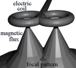
TMS focal field.png

TMS – Butterfly Coils

TMS uses electromagnetic induction to generate an electric current across the scalp and skull without physical contact.[48] A plastic-enclosed coil of wire is held next to the skull and when activated, produces a magnetic field oriented orthogonally to the plane of the coil. The magnetic field passes unimpeded through the skin and skull, inducing an oppositely directed current in the brain that activates nearby nerve cells in much the same way as currents applied directly to the cortical surface.[49]

The path of this current is difficult to model because the brain is irregularly shaped and electricity and magnetism are not conducted uniformly throughout its tissues. The magnetic field is about the same strength as an MRI, and the pulse generally reaches no more than 5 centimeters into the brain unless using the deep transcranial magnetic stimulation variant of TMS.[50] Deep TMS can reach up to 6 cm into the brain to stimulate deeper layers of the motor cortex, such as that which controls leg motion.[51]

Mechanism of action

 \mathbf B = \frac{\mu_0}{4\pi} I \int_C \frac{d\mathbf l \times \mathbf{\hat r}}{r^2}
it has been shown that a current through a wire generates a magnetic field around that wire. Transcranial magnetic stimulation is achieved by quickly discharging current from a large capacitor into a coil to produce pulsed magnetic fields between 2 and 3 T.[52] By directing the magnetic field pulse at a targeted area of the brain, one can either depolarize or hyperpolarize neurons in the brain. The magnetic flux density pulse generated by the current pulse through the coil causes an electric field as explained by the Maxwell-Faraday equation,

\nabla \times \mathbf {E} =-{\frac {\partial \mathbf {B} }{\partial t}} .
This electric field causes a change in the transmembrane current of the neuron, which leads to the depolarization or hyperpolarization of the neuron and the firing of an action potential.[52]

The exact details of how TMS functions are still being explored. The effects of TMS can be divided into two types depending on the mode of stimulation:
  • Single or paired pulse TMS causes neurons in the neocortex under the site of stimulation to depolarize and discharge an action potential. If used in the primary motor cortex, it produces muscle activity referred to as a motor evoked potential (MEP) which can be recorded on electromyography. If used on the occipital cortex, 'phosphenes' (flashes of light) might be perceived by the subject. In most other areas of the cortex, the participant does not consciously experience any effect, but his or her behaviour may be slightly altered (e.g., slower reaction time on a cognitive task), or changes in brain activity may be detected using sensing equipment.[53]
  • Repetitive TMS produces longer-lasting effects which persist past the initial period of stimulation. rTMS can increase or decrease the excitability of the corticospinal tract depending on the intensity of stimulation, coil orientation, and frequency. The mechanism of these effects is not clear, though it is widely believed to reflect changes in synaptic efficacy akin to long-term potentiation (LTP) and long-term depression (LTD).[54]
MRI images, recorded during TMS of the motor cortex of the brain, have been found to match very closely with PET produced by voluntary movements of the hand muscles innervated by TMS, to 5–22 mm of accuracy.[55] The localisation of motor areas with TMS has also been seen to correlate closely to MEG[56] and also fMRI.[57]

Coil types

The design of transcranial magnetic stimulation coils used in either treatment or diagnostic/experimental studies may differ in a variety of ways. These differences should be considered in the interpretation of any study result, and the type of coil used should be specified in the study methods for any published reports.

The most important considerations include:
  • the type of material used to construct the core of the coil
  • the geometry of the coil configuration
  • the biophysical characteristics of the pulse produced by the coil.
With regard to coil composition, the core material may be either a magnetically inert substrate (i.e., the so-called 'air-core' coil design), or possess a solid, ferromagnetically active material (i.e., the so-called 'solid-core' design). Solid core coil design result in a more efficient transfer of electrical energy into a magnetic field, with a substantially reduced amount of energy dissipated as heat, and so can be operated under more aggressive duty cycles often mandated in therapeutic protocols, without treatment interruption due to heat accumulation, or the use of an accessory method of cooling the coil during operation. Varying the geometric shape of the coil itself may also result in variations in the focality, shape, and depth of cortical penetration of the magnetic field. Differences in the coil substance as well as the electronic operation of the power supply to the coil may also result in variations in the biophysical characteristics of the resulting magnetic pulse (e.g., width or duration of the magnetic field pulse). All of these features should be considered when comparing results obtained from different studies, with respect to both safety and efficacy.[58]

A number of different types of coils exist, each of which produce different magnetic field patterns. Some examples:
  • round coil: the original type of TMS coil
  • figure-eight coil (i.e., butterfly coil): results in a more focal pattern of activation
  • double-cone coil: conforms to shape of head, useful for deeper stimulation
  • four-leaf coil: for focal stimulation of peripheral nerves[59]
  • H-coil: for deep transcranial magnetic stimulation
Design variations in the shape of the TMS coils allow much deeper penetration of the brain than the standard depth of 1.5–2.5 cm. Circular crown coils, Hesed (or H-core) coils, double cone coils, and other experimental variations can induce excitation or inhibition of neurons deeper in the brain including activation of motor neurons for the cerebellum, legs and pelvic floor. Though able to penetrate deeper in the brain, they are less able to produce a focused, localized response and are relatively non-focal.[7]

History

Luigi Galvani did pioneering research on the effects of electricity on the body in the late 1700s, and laid the foundations for the field of electrophysiology.[60] In the 1800s Michael Faraday discovered that an electrical current had a corresponding magnetic field, and that changing one, could change the other.[61] Work to directly stimulate the human brain with electricity started in the late 1800s, and by the 1930s electroconvulsive therapy has been developed by Italian physicians Cerletti and Bini.[60] ECT became widely used to treat mental illness and became overused as it began to be seen as a "psychiatric panacea", and a backlash against it grew in the 1970s.[60] Around that time Anthony T. Barker began exploring use of magnetic fields to alter electrical signalling in the brain, and the first stable TMS devices were developed around 1985.[60][61] They were originally intended as diagnostic and research devices, and only later were therapeutic uses explored.[60][61] The first TMS devices were approved by the FDA in October 2008.[60]

Research

TMS research in animal studies is limited due to early FDA approval of TMS treatment of drug-resistant depression. Because of this, there has been no specific coils for animal models. Hence, there are limited number of TMS coils that can be used for animal studies.[62] There are some attempts in the literature showing new coil designs for mice with an improved stimulation profile.[63]

Areas of research include:

Study blinding

It is difficult to establish a convincing form of "sham" TMS to test for placebo effects during controlled trials in conscious individuals, due to the neck pain, headache and twitching in the scalp or upper face associated with the intervention.[4][7] "Sham" TMS manipulations can affect cerebral glucose metabolism and MEPs, which may confound results.[76] This problem is exacerbated when using subjective measures of improvement.[7] Placebo responses in trials of rTMS in major depression are negatively associated with refractoriness to treatment, vary among studies and can influence results.[77]

A 2011 review found that only 13.5% of 96 randomized control studies of rTMS to the dorsolateral prefrontal cortex had reported blinding success and that, in those studies, people in real rTMS groups were significantly more likely to think that they had received real TMS, compared with those in sham rTMS groups.[78] Depending on the research question asked and the experimental design, matching the discomfort of rTMS to distinguish true effects from placebo can be an important and challenging issue.

Psychophysics

From Wikipedia, the free encyclopedia
 
Psychophysics quantitatively investigates the relationship between physical stimuli and the sensations and perceptions they produce. Psychophysics has been described as "the scientific study of the relation between stimulus and sensation"[1] or, more completely, as "the analysis of perceptual processes by studying the effect on a subject's experience or behaviour of systematically varying the properties of a stimulus along one or more physical dimensions".[2]

Psychophysics also refers to a general class of methods that can be applied to study a perceptual system. Modern applications rely heavily on threshold measurement,[3] ideal observer analysis, and signal detection theory.[4]

Psychophysics has widespread and important practical applications. For example, in the study of digital signal processing, psychophysics has informed the development of models and methods of lossy compression. These models explain why humans perceive very little loss of signal quality when audio and video signals are formatted using lossy compression.

History

Many of the classical techniques and theories of psychophysics were formulated in 1860 when Gustav Theodor Fechner in Leipzig published Elemente der Psychophysik (Elements of Psychophysics).[5] He coined the term "psychophysics", describing research intended to relate physical stimuli to the contents of consciousness such as sensations (Empfindungen). As a physicist and philosopher, Fechner aimed at developing a method that relates matter to the mind, connecting the publicly observable world and a person's privately experienced impression of it. His ideas were inspired by experimental results on the sense of touch and light obtained in the early 1830s by the German physiologist Ernst Heinrich Weber in Leipzig,[6][7] most notably those on the minimum discernible difference in intensity of stimuli of moderate strength (just noticeable difference; jnd) which Weber had shown to be a constant fraction of the reference intensity, and which Fechner referred to as Weber's law. From this, Fechner derived his well-known logarithmic scale, now known as Fechner scale. Weber's and Fechner's work formed one of the bases of psychology as a science, with Wilhelm Wundt founding the first laboratory for psychological research in Leipzig (Institut für experimentelle Psychologie). Fechner's work systematised the introspectionist approach (psychology as the science of consciousness), that had to contend with the Behaviorist approach in which even verbal responses are as physical as the stimuli. During the 1930s, when psychological research in Nazi Germany essentially came to a halt, both approaches eventually began to be replaced by use of stimulus-response relationships as evidence for conscious or unconscious processing in the mind.[8] Fechner's work was studied and extended by Charles S. Peirce, who was aided by his student Joseph Jastrow, who soon became a distinguished experimental psychologist in his own right. Peirce and Jastrow largely confirmed Fechner's empirical findings, but not all. In particular, a classic experiment of Peirce and Jastrow rejected Fechner's estimation of a threshold of perception of weights, as being far too high. In their experiment, Peirce and Jastrow in fact invented randomized experiments: They randomly assigned volunteers to a blinded, repeated-measures design to evaluate their ability to discriminate weights.[9][10][11][12] Peirce's experiment inspired other researchers in psychology and education, which developed a research tradition of randomized experiments in laboratories and specialized textbooks in the 1900s.[9][10][11][12] The Peirce–Jastrow experiments were conducted as part of Peirce's application of his pragmaticism program to human perception; other studies considered the perception of light, etc.[13] Jastrow wrote the following summary: "Mr. Peirce’s courses in logic gave me my first real experience of intellectual muscle. Though I promptly took to the laboratory of psychology when that was established by Stanley Hall, it was Peirce who gave me my first training in the handling of a psychological problem, and at the same time stimulated my self-esteem by entrusting me, then fairly innocent of any laboratory habits, with a real bit of research. He borrowed the apparatus for me, which I took to my room, installed at my window, and with which, when conditions of illumination were right, I took the observations. The results were published over our joint names in the Proceedings of the National Academy of Sciences. The demonstration that traces of sensory effect too slight to make any registry in consciousness could none the less influence judgment, may itself have been a persistent motive that induced me years later to undertake a book on The Subconscious." This work clearly distinguishes observable cognitive performance from the expression of consciousness.

Modern approaches to sensory perception, such as research on vision, hearing, or touch, measure what the perceiver's judgment extracts from the stimulus, often putting aside the question what sensations are being experienced. One leading method is based on signal detection theory, developed for cases of very weak stimuli. However, the subjectivist approach persists among those in the tradition of Stanley Smith Stevens (1906–1973). Stevens revived the idea of a power law suggested by 19th century researchers, in contrast with Fechner's log-linear function (cf. Stevens' power law). He also advocated the assignment of numbers in ratio to the strengths of stimuli, called magnitude estimation. Stevens added techniques such as magnitude production and cross-modality matching. He opposed the assignment of stimulus strengths to points on a line that are labeled in order of strength. Nevertheless, that sort of response has remained popular in applied psychophysics. Such multiple-category layouts are often misnamed Likert scaling after the question items used by Likert to create multi-item psychometric scales, e.g., seven phrases from "strongly agree" through "strongly disagree".

Omar Khaleefa[14] has argued that the medieval scientist Alhazen should be considered the founder of psychophysics. Although al-Haytham made many subjective reports regarding vision, there is no evidence that he used quantitative psychophysical techniques and such claims have been rebuffed.[15]

Thresholds

Psychophysicists usually employ experimental stimuli that can be objectively measured, such as pure tones varying in intensity, or lights varying in luminance. All the senses have been studied: vision, hearing, touch (including skin and enteric perception), taste, smell and the sense of time. Regardless of the sensory domain, there are three main areas of investigation: absolute thresholds, discrimination thresholds and scaling.

A threshold (or limen) is the point of intensity at which the participant can just detect the presence of a stimulus (absolute threshold[16]) or the presence of a difference between two stimuli (difference threshold[7]). Stimuli with intensities below the threshold are considered not detectable (hence: sub-liminal). Stimuli at values close enough to a threshold will often be detectable some proportion of occasions; therefore, a threshold is considered to be the point at which a stimulus, or change in a stimulus, is detected some proportion p of occasions.

Detection

An absolute threshold is the level of intensity of a stimulus at which the subject is able to detect the presence of the stimulus some proportion of the time (a p level of 50% is often used).[17] An example of an absolute threshold is the number of hairs on the back of one's hand that must be touched before it can be felt – a participant may be unable to feel a single hair being touched, but may be able to feel two or three as this exceeds the threshold. Absolute threshold is also often referred to as detection threshold. Several different methods are used for measuring absolute thresholds (as with discrimination thresholds; see below).

Discrimination

A difference threshold (or just-noticeable difference, JND) is the magnitude of the smallest difference between two stimuli of differing intensities that the participant is able to detect some proportion of the time (the percentage depending on the kind of task). To test this threshold, several different methods are used. The subject may be asked to adjust one stimulus until it is perceived as the same as the other (method of adjustment), may be asked to describe the direction and magnitude of the difference between two stimuli, or may be asked to decide whether intensities in a pair of stimuli are the same or not (forced choice). The just-noticeable difference (JND) is not a fixed quantity; rather, it depends on how intense the stimuli being measured are and the particular sense being measured.[18]  Weber's Law states that the just-noticeable difference of a stimulus is a constant proportion despite variation in intensity.[19]

In discrimination experiments, the experimenter seeks to determine at what point the difference between two stimuli, such as two weights or two sounds, is detectable. The subject is presented with one stimulus, for example a weight, and is asked to say whether another weight is heavier or lighter (in some experiments, the subject may also say the two weights are the same). At the point of subjective equality (PSE), the subject perceives the two weights to be the same. The just-noticeable difference,[20] or difference limen (DL), is the magnitude of the difference in stimuli that the subject notices some proportion p of the time (50% is usually used for p in the comparison task). In addition, a two-alternative forced choice (2-afc) paradigm can be used to assess the point at which performance reduces to chance on a discrimination between two alternatives (p will then typically be 75% since p=50% corresponds to chance in the 2-afc task).

Absolute and difference thresholds are sometimes considered similar in principle because there is always background noise interfering with our ability to detect stimuli.[6][21]

Experimentation

In psychophysics, experiments seek to determine whether the subject can detect a stimulus, identify it, differentiate between it and another stimulus, or describe the magnitude or nature of this difference.[6][7] Software for psychophysical experimentation is overviewed by Strasburger.[22]

Classical psychophysical methods

Psychophysical experiments have traditionally used three methods for testing subjects' perception in stimulus detection and difference detection experiments: the method of limits, the method of constant stimuli and the method of adjustment.[23]

Method of limits

In the ascending method of limits, some property of the stimulus starts out at a level so low that the stimulus could not be detected, then this level is gradually increased until the participant reports that they are aware of it. For example, if the experiment is testing the minimum amplitude of sound that can be detected, the sound begins too quietly to be perceived, and is made gradually louder. In the descending method of limits, this is reversed. In each case, the threshold is considered to be the level of the stimulus property at which the stimuli are just detected.[23]

In experiments, the ascending and descending methods are used alternately and the thresholds are averaged. A possible disadvantage of these methods is that the subject may become accustomed to reporting that they perceive a stimulus and may continue reporting the same way even beyond the threshold (the error of habituation). Conversely, the subject may also anticipate that the stimulus is about to become detectable or undetectable and may make a premature judgment (the error of anticipation).

To avoid these potential pitfalls, Georg von Békésy introduced the staircase procedure in 1960 in his study of auditory perception. In this method, the sound starts out audible and gets quieter after each of the subject's responses, until the subject does not report hearing it. At that point, the sound is made louder at each step, until the subject reports hearing it, at which point it is made quieter in steps again. This way the experimenter is able to "zero in" on the threshold.[23]

Method of constant stimuli

Instead of being presented in ascending or descending order, in the method of constant stimuli the levels of a certain property of the stimulus are not related from one trial to the next, but presented randomly. This prevents the subject from being able to predict the level of the next stimulus, and therefore reduces errors of habituation and expectation. For 'absolute thresholds' again the subject reports whether he or she is able to detect the stimulus.[23] For 'difference thresholds' there has to be a constant comparison stimulus with each of the varied levels. Friedrich Hegelmaier described the method of constant stimuli in an 1852 paper.[24] This method allows for full sampling of the psychometric function, but can result in a lot of trials when several conditions are interleaved.

Method of adjustment

The method of adjustment asks the subject to control the level of the stimulus, instructs them to alter it until it is just barely detectable against the background noise, or is the same as the level of another stimulus. This is repeated many times. This is also called the method of average error.[23] In this method the observer himself controls the magnitude of the variable stimulus beginning with a variable that is distinctly greater or lesser than a standard one and he varies it until he is satisfied by the subjectivity of two. The difference between the variable stimuli and the standard one is recorded after each adjustment and the error is tabulated for a considerable series. At the end mean is calculated giving the average error which can be taken as the measure of sensitivity.

Adaptive psychophysical methods

The classic methods of experimentation are often argued to be inefficient. This is because, in advance of testing, the psychometric threshold is usually unknown and much data is collected at points on the psychometric function that provide little information about the parameter of interest, usually the threshold. Adaptive staircase procedures (or the classical method of adjustment) can be used such that the points sampled are clustered around the psychometric threshold. However, the cost of this efficiency is that there is less information regarding the psychometric function's shape. Adaptive methods can be optimized for estimating the threshold only, or threshold and slope. Adaptive methods are classified into staircase procedures (see below) and Bayesian or maximum-likelihood methods. Staircase methods rely on the previous response only and are easier to implement. Bayesian methods take the whole set of previous stimulus-response pairs into account and are believed to be more robust against lapses in attention.[25]

Staircase procedures

Diagram showing a specific staircase procedure: Transformed Up/Down Method (1 up/ 2 down rule). Until the first reversal (which is neglected) the simple up/down rule and a larger step size is used.

Staircases usually begin with a high intensity stimulus, which is easy to detect. The intensity is then reduced until the observer makes a mistake, at which point the staircase 'reverses' and intensity is increased until the observer responds correctly, triggering another reversal. The values for the last of these 'reversals' are then averaged. There are many different types of staircase procedures, using different decision and termination rules. Step-size, up/down rules and the spread of the underlying psychometric function dictate where on the psychometric function they converge.[25] Threshold values obtained from staircases can fluctuate wildly, so care must be taken in their design. Many different staircase algorithms have been modeled and some practical recommendations suggested by Garcia-Perez.[26]

One of the more common staircase designs (with fixed-step sizes) is the 1-up-N-down staircase. If the participant makes the correct response N times in a row, the stimulus intensity is reduced by one step size. If the participant makes an incorrect response the stimulus intensity is increased by the one size. A threshold is estimated from the mean midpoint of all runs. This estimate approaches, asymptotically, the correct threshold.

Bayesian and maximum-likelihood procedures

Bayesian and maximum-likelihood adaptive procedures behave, from the observer's perspective, similar to the staircase procedures. The choice of the next intensity level works differently, however: After each observer response, from the set of this and all previous stimulus/response pairs the likelihood is calculated of where the threshold lies. The point of maximum likelihood is then chosen as the best estimate for the threshold, and the next stimulus is presented at that level (since a decision at that level will add the most information). In a Bayesian procedure, a prior likelihood is further included in the calculation.[25] Compared to staircase procedures, Bayesian and ML procedures are more time-consuming to implement but are considered to be more robust. Well-known procedures of this kind are Quest,[27] ML-PEST,[28] and Kontsevich & Tyler’s method.[29]

Magnitude estimation

In the prototypical case, people are asked to assign numbers in proportion to the magnitude of the stimulus. This psychometric function of the geometric means of their numbers is often a power law with stable, replicable exponent. Although contexts can change the law and exponent, that change too is stable and replicable. Instead of numbers, other sensory or cognitive dimensions can be used to match a stimulus and the method then becomes "magnitude production" or "cross-modality matching". The exponents of those dimensions found in numerical magnitude estimation predict the exponents found in magnitude production. Magnitude estimation generally finds lower exponents for the psychophysical function than multiple-category responses, because of the restricted range of the categorical anchors, such as those used by Likert as items in attitude scales.
Political psychology

From Wikipedia, the free encyclopedia ...