Search This Blog

Friday, July 13, 2018

Polydendrocytes

From Wikipedia, the free encyclopedia
Polydendrocytes (also known as NG2 cells, NG2 glia, or oligodendrocyte progenitor cells) are process-bearing glial cells (neuroglia) in the mammalian central nervous system (CNS) that are identified by the expression of the NG2 chondroitin sulfate proteoglycan (CSPG4)  and the alpha receptor for platelet-derived growth factor (PDGFRA). They are distinct from other cell populations such as neurons, astrocytes, oligodendrocytes, microglia, and neural stem cells and are recognized as the fourth major glial cell type in the mammalian CNS.[citation needed] Studies have implicated polydendrocytes in many cellular and physiological processes. Polydendrocytes in the postnatal mouse CNS and those grown in culture generate oligodendrocytes, and thus they are often equated with oligodendrocyte progenitor cells (OPCs). Under some culture conditions, polydendrocytes give rise to astrocytes. A subpopulation of polydendrocytes in the gray matter of the embryonic CNS also generates protoplasmic astrocytes. In addition, polydendrocytes express receptors for various neurotransmitters and undergo membrane depolarization when they receive synaptic inputs from neurons.

History

It had been known since the early 1900s that astrocytes, oligodendrocytes, and microglia make up the major glial cell populations in the mammalian CNS. The presence of another glial cell population had escaped recognition because of the lack of a suitable marker to identify them in tissue sections. The notion that there exists a population of glial progenitor cells in the developing and mature CNS began to be entertained in the late 1980s by several independent groups. In one series of studies on the development and origin of oligodendrocytes in the rodent CNS, a population of immature cells that appeared to be precursors to oligodendrocytes was identified by the expression of the GD3 ganglioside.[3][4]

In a separate series of studies, cells from perinatal rat optic nerves that expressed the A2B5 ganglioside were shown to differentiate into oligodendrocytes in culture.[5] Subsequently, A2B5+ cells from other CNS regions and from adult CNS were also shown to generate oligodendrocytes. Based on the observation that these cells require PDGF for their proliferation and expansion, the expression of the alpha receptor for platelet-derived growth factor (Pdgfra) was used to search for the in vivo correlates of the A2B5+ cells, which led to the discovery of a unique population of Pdgfra+ cells in the CNS whose appearance and distribution were consistent with those of developing oligodendrocytes.[6]

Independently, Stallcup and colleagues generated an antiserum that recognized a group of rat brain tumor cell lines, which exhibited properties that were intermediate between those of typical neurons and glial cells. Biochemical studies showed that the antiserum recognized a chondroitin sulfate proteoglycan with a core glycoprotein of 300 kDa,[7] and the antigen was named NG2 (nerve/glial antigen 2).[8][9] NG2 was found to be expressed on A2B5+ oligodendrocyte precursor cells isolated from the perinatal rat CNS tissues and on process-bearing cells in the CNS in vivo.[7][10] Comparison of NG2 and Pdgfra expression revealed that NG2 and Pdgfra are expressed on the same population of cells in the CNS.[11] These cells represent 2-9% of all the cells and remain proliferative in the mature CNS.[12]

Origin and fate

Origin

In the embryonic spinal cord, a major source of polydendrocytes is the ventral ventricular zone of the pMN domain marked by the expression of the transcription factors Olig1 and Olig2 and the p3 domain that expresses Nkx2.2, which are induced by the morphogen Shh (sonic hedgehog). Some polydendrocytes also arise from the dorsal ventricular zone. In the forebrain, three regionally distinct sources have been shown to generate polydendrocytes sequentially: an early ventral source from the medial ganglionic eminence marked by Nkx2.1, followed by progenitor cells in the lateral ganglionic eminence marked by Gsh2, and finally the dorsal neocortical germinal zone marked by Emx1.[13] After the committed progenitor cells exit the germinal zones, they begin to express NG2 and Pdgfra and expand by local proliferation and migration and eventually occupy the entire CNS parenchyma. Polydendrocytes continue to exist in the adult CNS and retain their proliferative ability throughout life.

Fate

The fate of polydendrocytes has been highly debated.[14] Using Cre-Lox recombination-mediated genetic fate mapping, several labs have reported the fate of polydendrocytes using different Cre driver and reporter mouse lines;[15][16][17][18] reviewed in reference.[19] The consensus of these studies is that polydendrocytes generate predominantly oligodendrocytes in both gray and white matter. The rate at which they generate oligodendrocytes declines with age and is greater in white matter than in gray matter. These studies revealed that up to 30% of the oligodendrocytes that exist in the adult corpus callosum are generated de novo from polydendrocytes over a period of 2 months. It is not known whether all polydendrocytes eventually generate oligodendrocytes while self-renewing its population or whether some remain as polydendrocytes throughout the life of the animal and never differentiate into oligodendrocytes.

Using NG2cre mice, it was shown that polydendrocytes in the prenatal and perinatal gray matter of the ventral forebrain and spinal cord generate protoplasmic astrocytes in addition to oligodendrocytes. However, contrary to the prediction from optic nerve cultures, polydendrocytes in white matter do not generate astrocytes. When the oligodendrocyte transcription factor Olig2 is deleted specifically in polydendrocytes, there is a region- and age-dependent switch in the fate of polydendrocytes from oligodendrocytes to astrocytes.[20]

Although controversy still continues about the neuronal fate of polydendrocytes, the consensus from a number of recent genetic fate mapping studies described above seems to be that polydendrocytes do not generate a significant number of neurons under normal conditions, and that they are distinct from neural stem cells that reside in the subventricular zone.[21]

Functions

In remyelination

Polydendrocytes retain the ability to proliferate in adulthood and comprise 70-90% of the proliferating cell population in the mature CNS.[12][22] Under conditions in the developing and mature CNS where a reduction in the normal number of oligodendrocytes or myelin occurs, polydendrocytes react promptly by undergoing increased proliferation. In acute or chronic demyelinated lesions created in the rodent CNS by chemical agents such as lysolecithin or cuprizone, polydendrocytes proliferate in response to demyelination, and the proliferated cells differentiate into remyelinating oligodendrocytes.[23][24] Similarly, polydendrocyte proliferation occurs in other types of injury that are accompanied by loss of myelin, such as spinal cord injury.[25]

If polydendrocytes were capable of giving rise to myelinating oligodendrocytes, one would expect complete remyelination of pathologically demyelinated lesions such as those seen in multiple sclerosis (MS). However, complete myelin regeneration is usually not observed clinically or in chronic experimental models. Possible explanations for remyelination failure include depletion of polydendrocytes over time, failure to recruit polydendrocytes to the demyelinated lesion, and failure of recruited polydendrocytes to differentiate into mature oligodendrocytes.[25]

Numerous factors have been shown to regulate polydendrocyte proliferation, migration, and differentiation [25][25] (reviewed in [26][27][28]). In fresh MS lesions, clusters of HNK-1+ oligodendrocytes have been observed,[29] which suggests that under favorable conditions polydendrocytes expand around demyelinated lesions and generate new oligodendrocytes. In chronic MS lesions where remyelination is incomplete, there is evidence that there are oligodendrocytes with processes extending toward demyelinated axons, but they do not seem to be able to generate new myelin.[30] The mechanisms that regulate differentiation of polydendrocytes into myelinating oligodendrocytes are an actively investigated area of research.

Another unanswered question is whether the polydendrocyte pool eventually becomes depleted after they are used to generate remyelinating cells. Clonal analysis of isolated polydendrocytes in the normal mouse forebrain suggests that in the adult, most clones originating from single polydendrocytes consist of either a heterogeneous population containing both oligodendrocytes and polydendrocytes or consist exclusively of polydendrocytes, suggesting that polydendrocytes in the adult CNS are able to self-renew and are not depleted under normal conditions.[31] However, it is not known whether this dynamic is altered in response to demyelinating lesions.

Neuron-polydendrocyte interactions

There is substantial evidence that indicates a functional interaction between polydendrocytes and neurons.

Node of Ranvier

Polydendrocytes extend their processes to the nodes of Ranvier [32] and together with astrocyte processes make up the nodal glial complex. Since the nodes of Ranvier contain a high density of voltage-dependent sodium channels and allow regenerative action potentials to be generated, it is speculated that this location allows polydendrocytes to sense and possibly respond to neuronal activity

Neuron-polydendrocyte synapse

Studies have shown that neurons form synapses with polydendrocytes in both gray matter [33] and white matter.[32][34] Polydendrocytes express the AMPA type glutamate receptors and GABAA receptors and undergo small membrane depolarizations when stimulated by glutamate or GABA that is vesicularly released from presynaptic terminals. Electron microscopy revealed polydendrocyte membranes apposed to neuronal presynaptic terminals filled with synaptic vesicles. Polydendrocytes lose their ability to respond to synaptic inputs from neurons as they differentiate into mature oligodendrocytes.[35][36]

Polydendrocytes can undergo cell division while maintaining synaptic inputs from neurons.[37] These observations suggest that cells that receive neuronal synaptic inputs and those that differentiate into oligodendrocytes are not mutually exclusive cell populations but that the same population of polydendrocytes can receive synaptic inputs and generate myelinating oligodendrocytes. The functional significance of the neuron-polydendrocyte synapses remains to be elucidated.

Neuroglia

From Wikipedia, the free encyclopedia

Neuroglia
Glial Cell Types.png
Illustration of the four different types of glial cells found in the central nervous system: ependymal cells (light pink), astrocytes (green), microglial cells (dark red), and oligodendrocytes (light blue).
Details
PrecursorNeuroectoderm for macroglia, and hematopoietic stem cells for microglia
SystemNervous system
Identifiers
MeSHD009457
TAA14.0.00.005
THH2.00.06.2.00001
FMA54541
Anatomical terms of microanatomy
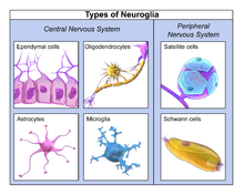
Neuroglia, also called glial cells or simply glia, are non-neuronal cells in the central nervous system (brain and spinal cord) and the peripheral nervous system. They maintain homeostasis, form myelin, and provide support and protection for neurons. In the central nervous system, glial cells include oligodendrocytes, astrocytes, ependymal cells, and microglia, and in the peripheral nervous system glial cells include Schwann cells and satellite cells. They have four main functions: (1) To surround neurons and hold them in place (2) To supply nutrients and oxygen to neurons (3) To insulate one neuron from another (4) To destroy pathogens and remove dead neurons. They also play a role in neurotransmission and synaptic connections, and in physiological processes like breathing.

Neuroglia, also called glial cells or simply glia, are non-neuronal cells in the central nervous system (brain and spinal cord) and the peripheral nervous system. They maintain homeostasis, form myelin, and provide support and protection for neurons. In the central nervous system, glial cells include oligodendrocytes, astrocytes, ependymal cells, and microglia, and in the peripheral nervous system glial cells include Schwann cells and satellite cells. They have four main functions: (1) To surround neurons and hold them in place (2) To supply nutrients and oxygen to neurons (3) To insulate one neuron from another (4) To destroy pathogens and remove dead neurons. They also play a role in neurotransmission and synaptic connections, and in physiological processes like breathing.

Glia were discovered in 1856, by the pathologist Rudolf Virchow in his search for a "connective tissue" in the brain. The term derives from Greek γλία and γλοία "glue"(/ˈɡlə/ or /ˈɡlə/), and suggests the original impression that they were the glue of the nervous system.

Types

Neuroglia of the brain shown by Golgi's method
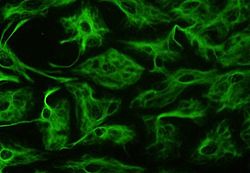
Astrocytes can be identified in culture because, unlike other mature glia, they express glial fibrillary acidic protein (GFAP)
Glial cells in a rat brain stained with an antibody against GFAP
Different types of neuroglia

Macroglia

Derived from ectodermal tissue.

Location Name Description
CNS Astrocytes The most abundant type of macroglial cell in the CNS,[6] astrocytes (also called astroglia) have numerous projections that link neurons to their blood supply while forming the blood-brain barrier. They regulate the external chemical environment of neurons by removing excess potassium ions, and recycling neurotransmitters released during synaptic transmission. Astrocytes may regulate vasoconstriction and vasodilation by producing substances such as arachidonic acid, whose metabolites are vasoactive.
Astrocytes signal each other using ATP. The gap junctions (also known as electrical synapses) between astrocytes allow the messenger molecule IP3 to diffuse from one astrocyte to another. IP3 activates calcium channels on cellular organelles, releasing calcium into the cytoplasm. This calcium may stimulate the production of more IP3 and cause release of ATP through channels in the membrane made of pannexins. The net effect is a calcium wave that propagates from cell to cell. Extracellular release of ATP, and consequent activation of purinergic receptors on other astrocytes, may also mediate calcium waves in some cases.
In general, there are two types of astrocytes, protoplasmic and fibrous, similar in function but distinct in morphology and distribution. Protoplasmic astrocytes have short, thick, highly branched processes and are typically found in gray matter. Fibrous astrocytes have long, thin, less branched processes and are more commonly found in white matter.
It has recently been shown that astrocyte activity is linked to blood flow in the brain, and that this is what is actually being measured in fMRI.[7] They also have been involved in neuronal circuits playing an inhibitory role after sensing changes in extracellular calcium.[8]
CNS Oligodendrocytes Oligodendrocytes are cells that coat axons in the central nervous system (CNS) with their cell membrane, forming a specialized membrane differentiation called myelin, producing the myelin sheath. The myelin sheath provides insulation to the axon that allows electrical signals to propagate more efficiently.[9]
CNS Ependymal cells Ependymal cells, also named ependymocytes, line the spinal cord and the ventricular system of the brain. These cells are involved in the creation and secretion of cerebrospinal fluid (CSF) and beat their cilia to help circulate the CSF and make up the blood-CSF barrier. They are also thought to act as neural stem cells.[10]
CNS Radial glia Radial glia cells arise from neuroepithelial cells after the onset of neurogenesis. Their differentiation abilities are more restricted than those of neuroepithelial cells. In the developing nervous system, radial glia function both as neuronal progenitors and as a scaffold upon which newborn neurons migrate. In the mature brain, the cerebellum and retina retain characteristic radial glial cells. In the cerebellum, these are Bergmann glia, which regulate synaptic plasticity. In the retina, the radial Müller cell is the glial cell that spans the thickness of the retina and, in addition to astroglial cells,[11] participates in a bidirectional communication with neurons.[12]
PNS Schwann cells Similar in function to oligodendrocytes, Schwann cells provide myelination to axons in the peripheral nervous system (PNS). They also have phagocytotic activity and clear cellular debris that allows for regrowth of PNS neurons.[13]
PNS Satellite cells Satellite glial cells are small cells that surround neurons in sensory, sympathetic, and parasympathetic ganglia.[14] These cells help regulate the external chemical environment. Like astrocytes, they are interconnected by gap junctions and respond to ATP by elevating intracellular concentration of calcium ions. They are highly sensitive to injury and inflammation, and appear to contribute to pathological states, such as chronic pain.[15]
PNS Enteric glial cells Are found in the intrinsic ganglia of the digestive system. They are thought to have many roles in the enteric system, some related to homeostasis and muscular digestive processes.[16]

Microglia

Microglia are specialized macrophages capable of phagocytosis that protect neurons of the central nervous system.[17] They are derived from the earliest wave of mononuclear cells that originate in yolk sac blood islands early in development, and colonize the brain shortly after the neural precursors begin to differentiate.[18]

These cells are found in all regions of the brain and spinal cord. Microglial cells are small relative to macroglial cells, with changing shapes and oblong nuclei. They are mobile within the brain and multiply when the brain is damaged. In the healthy central nervous system, microglia processes constantly sample all aspects of their environment (neurons, macroglia and blood vessels). In a healthy brain, microglia direct the immune response to brain damage and play an important role in the inflammation that accompanies the damage. Many diseases and disorders are associated with deficient microglia, such as Alzheimer's disease, Parkinson's disease, and ALS.

Other

Pituicytes from the posterior pituitary are glia cells with characteristics in common to astrocytes.[19] Tanycytes in the median eminence of the hypothalamus are a type of ependymal cell that descend from radial glia and line the base of the third ventricle.[20]

Total number

In general, neuroglial cells are smaller than neurons; there are about 86 billion neurons and 85 billion "non-neuronal" (glia) cells in the human male brain. Glial cells make up about half the total volume of the brain and spinal cord.[21][22]^ The ratio varies from one part of the brain to another. The glia/neuron ratio in the cerebral cortex is 3.72 (60.84 billion glia (72%); 16.34 billion neurons), while that of the cerebellum is only 0.23 (16.04 billion glia; 69.03 billion neurons). The ratio in the cerebral cortex gray matter is 1.48, with 3.76 for the gray and white matter combined.[21] The ratio of the basal ganglia, diencephalon and brainstem combined is 11.35.[21]

Most cerebral cortex glia are oligodendrocytes (75.6%); astrocytes account for 17.3% and microglia for 6.5%.[23]

Development

23-week fetal brain culture astrocyte

Most glia are derived from ectodermal tissue of the developing embryo, in particular the neural tube and crest. The exception is microglia, which are derived from hemopoietic stem cells. In the adult, microglia are largely a self-renewing population and are distinct from macrophages and monocytes, which infiltrate the injured and diseased CNS.

In the central nervous system, glia develop from the ventricular zone of the neural tube. These glia include the oligodendrocytes, ependymal cells, and astrocytes. In the peripheral nervous system, glia derive from the neural crest. These PNS glia include Schwann cells in nerves and satellite glial cells in ganglia.

Current research involving glial cells in the human cochlea proposes that these cells are the common precursor to both mature Schwann cells and satellite glial cells. Additionally, the peripheral glial cells located along the peripheral processes expressed NGFR, indicating a phenotype distinct from the peripheral glial cells located along the central processes.[24]

Capacity to divide

Glia retain the ability to undergo cell division in adulthood, whereas most neurons cannot. The view is based on the general deficiency of the mature nervous system in replacing neurons after an injury, such as a stroke or trauma, while very often there is a profound proliferation of glia, or gliosis near or at the site of damage. However, detailed studies found no evidence that 'mature' glia, such as astrocytes or oligodendrocytes, retain the ability of mitosis. Only the resident oligodendrocyte precursor cells seem to keep this ability after the nervous system matures. On the other hand, there are a few regions in the mature nervous system, such as the dentate gyrus of the hippocampus and the subventricular zone, where generation of new neurons can be observed.[25]

Glial cells are known to be capable of mitosis. By contrast, scientific understanding of whether neurons are permanently post-mitotic,[26] or capable of mitosis,[27][28][29] is still developing. In the past, glia had been considered[by whom?] to lack certain features of neurons. For example, glial cells were not believed to have chemical synapses or to release transmitters. They were considered to be the passive bystanders of neural transmission. However, recent studies have shown this to be untrue.[30]

Functions

Some glial cells function primarily as the physical support for neurons. Others regulate the internal environment of the brain, especially the fluid surrounding neurons and their synapses, and nutrify neurons. During early embryogenesis, glial cells direct the migration of neurons and produce molecules that modify the growth of axons and dendrites.

Neuron repair and development

Glia are also crucial in the development of the nervous system and in processes such as synaptic plasticity and synaptogenesis. Glia have a role in the regulation of repair of neurons after injury. In the central nervous system (CNS), glia suppress repair. Glial cells known as astrocytes enlarge and proliferate to form a scar and produce inhibitory molecules that inhibit regrowth of a damaged or severed axon. In the peripheral nervous system (PNS), glial cells known as Schwann cells promote repair. After axonal injury, Schwann cells regress to an earlier developmental state to encourage regrowth of the axon. This difference between the CNS and the PNS, raises hopes for the regeneration of nervous tissue in the CNS. For example, a spinal cord may be able to be repaired following injury or severance. Schwann cells are also known as neuri-lemmocytes. These cells envelop nerve fibers of the PNS by winding repeatedly around a nerve fiber with the nucleus inside of it. This process creates a myelin sheath, which not only aids in conductivity but also assists in the regeneration of damaged fibers.

Myelin sheath creation

Oligodendrocytes are another type of glial cell of the CNS. These dendrocytes resemble an octopus bulbous body and contain up to fifteen arm-like processes. Each “arm” reaches out to a nerve fiber and spirals around it, creating a myelin sheath. This myelin sheath insulates the nerve fiber from the extracellular fluid as well as speeds up the signal conduction in the nerve fiber.[31]

Neurotransmission

Recent research indicates that glial cells of the hippocampus and cerebellum participate in synaptic transmission, regulate the clearance of neurotransmitters from the synaptic cleft, and release gliotransmitters such as ATP, which modulate synaptic function.[32]

Astrocytes are crucial in clearance of neurotransmitters from within the synaptic cleft, which provides distinction between arrival of action potentials and prevents toxic build-up of certain neurotransmitters such as glutamate (excitotoxicity). It is also thought that glia play a role in many neurological diseases, including Alzheimer's disease.[33] Furthermore, at least in vitro, astrocytes can release gliotransmitter glutamate in response to certain stimulation. Another unique type of glial cell, the oligodendrocyte precursor cells or OPCs, have very well-defined and functional synapses from at least two major groups of neurons.[34] The only notable differences between neurons and glial cells are neurons' possession of axons and dendrites, and capacity to generate action potentials.

Clinical significance

Neoplastic glial cells stained with an antibody against GFAP (brown), from a brain biopsy

While glial cells in the PNS frequently assist in regeneration of lost neural functioning, loss of neurons in the CNS does not result in a similar reaction from neuroglia.[13] In the CNS, regrowth will only happen if the trauma was mild, and not severe.[35] When severe trauma presents itself, the survival of the remaining neurons becomes the optimal solution. However, some studies investigating the role of glial cells in Alzheimer's Disease are beginning to contradict the usefulness of this feature, and even claim it can "exacerbate" the disease.[36] In addition to impacting the potential repair of neurons in Alzheimer's Disease, scarring and inflammation from glial cells have been further implicated in the degeneration of neurons caused by Amyotrophic lateral sclerosis.[37]

In addition to neurodegenerative diseases, a wide range of harmful exposure, such as hypoxia, or physical trauma, can lead to the end result of physical damage to the CNS.[35] Generally, when damage occurs to the CNS, glial cells cause Apoptosis among the surrounding cellular bodies.[35] Then, there is a large amount of microglial activity, which results in inflammation, and finally, there is a heavy release of growth inhibiting molecules.[35]

History

Glia were first described in 1856 by the pathologist Rudolf Virchow in a comment to his 1846 publication on connective tissue. A more detailed description of glial cells was provided in the 1858 book Cellular Pathology by the same author.[38]

When markers for different types of cells were analyzed, Einstein's brain was discovered to contain significantly more glia than normal brains in the left angular gyrus, an area thought to be responsible for mathematical processing and language.[39]

The ratio of glia to neurons increases with our definition of intelligence. Not only does the ratio of glia to neurons increase through evolution, but so does the size of the glia. Astroglial cells in the human have a volume 27 times greater than the same cells in the mouse's brain.[40]

These important scientific findings may begin to shift the neuron-specific perspective into a more holistic view of the brain which encompasses the glial cells as well. The glia's importance is becoming ever more clear as time goes on and new research is conducted. For most of the last century, scientists had written off glial cells as being nothing more than the structure and foundations that hold the neurons in place. But now, there is direct evidence that correlates the number of glial cells in the brain with the amount of intelligence that any given species possesses.[41] Future research will begin to shed light on the mysterious, yet increasingly crucial, role of glial cells.

The Coming Merging of Mind and Machine

February 21, 2001 by Ray Kurzweil
Original link:  http://www.kurzweilai.net/the-coming-merging-of-mind-and-machine

Ray Kurzweil predicts a future with direct brain-to-computer access and conscious machines. From Scientific American.

Originally published September 1, 1999 in Scientific American. Published on KurzweilAI.net February 22, 2001.

Sometime early in the next century, the intelligence of machines will exceed that of humans. Within several decades, machines will exhibit the full range of human intellect, emotions and skills, ranging from musical and other creative aptitudes to physical movement. They will claim to have feelings and, unlike today’s virtual personalities, will be very convincing when they tell us so. By 2019 a $1,000 computer will at least match the processing power of the human brain. By 2029 the software for intelligence will have been largely mastered, and the average personal computer will be equivalent to 1,000 brains.

Within three decades, the author maintains, neural implants will be available that interface directly to our brain cells. The implants would enhance sensory experiences and improve our memory and thinking.

Once computers achieve a level of intelligence comparable to that of humans, they will necessarily soar past it. For example, if I learn French, I can’t readily download that learning to you. The reason is that for us, learning involves successions of stunningly complex patterns of interconnections among brain cells (neurons) and among the concentrations of biochemicals, known as neurotransmitters, that enable impulses to travel from neuron to neuron. We have no way of quickly downloading these patterns. But quick downloading will allow our nonbiological creations to share immediately what they learn with billions of other machines. Ultimately, nonbiological entities will master not only the sum total of their own knowledge but all of ours as well.

As this happens, there will no longer be a clear distinction between human and machine. We are already putting computers–neural implants–directly into people’s brains to counteract Parkinson’s disease and tremors from multiple sclerosis. We have cochlear implants that restore hearing. A retinal implant is being developed in the U.S. that is intended to provide at least some visual perception for some blind individuals, basically by replacing certain visual-processing circuits of the brain. Recently scientists from Emory University implanted a chip in the brain of a paralyzed stroke victim that allows him to use his brainpower to move a cursor across a computer screen.

In the 2020s neural implants will improve our sensory experiences, memory and thinking. By 2030, instead of just phoning a friend, you will be able to meet in, say, a virtual Mozambican game preserve that will seem compellingly real. You will be able to have any type of experience–business, social, sexual–with anyone, real or simulated, regardless of physical proximity.

How Life and Technology Evolve

To gain insight into the kinds of forecasts I have just made, it is important to recognize that technology is advancing exponentially. An exponential process starts slowly, but eventually its pace increases extremely rapidly. (A fuller documentation of my argument is contained in my new book, The Age of Spiritual Machines.)

The evolution of biological life and the evolution of technology have both followed the same pattern: they take a long time to get going, but advances build on one another and progress erupts at an increasingly furious pace. We are entering that explosive part of the technological evolution curve right now.

Consider: It took billions of years for Earth to form. It took two billion more for life to begin and almost as long for molecules to organize into the first multicellular plants and animals about 700 million years ago. The pace of evolution quickened as mammals inherited Earth some 65 million years ago. With the emergence of primates, evolutionary progress was measured in mere millions of years, leading to Homo sapiens perhaps 500,000 years ago.

The evolution of technology has been a continuation of the evolutionary process that gave rise to us-the technology-creating species-in the first place. It took tens of thousands of years for our ancestors to figure out that sharpening both sides of a stone created useful tools. Then, earlier in this millennium, the time required for a major paradigm shift in technology had shrunk to hundreds of years.

The pace continued to accelerate during the 19th century, during which technological progress was equal to that of the 10 centuries that came before it. Advancement in the first two decades of the 20th century matched that of the entire 19th century. Today significant technological transformations take just a few years; for example, the World Wide Web, already a ubiquitous form of communication and commerce, did not exist just nine years ago.

Computing technology is experiencing the same exponential growth. Over the past several decades, a key factor in this expansion has been described by Moore’s Law. Gordon Moore, a co-founder of Intel, noted in the mid-1960s that technologists had been doubling the density of transistors on integrated circuits every 12 months. This meant computers were periodically doubling both in capacity and in speed per unit cost. In the mid-1970s Moore revised his observation of the doubling time to a more accurate estimate of about 24 months, and that trend has persisted through the 1990s.

After decades of devoted service, Moore’s Law will have run its course around 2019. By that time, transistor features will be just a few atoms in width. But new computer architectures will continue the exponential growth of computing. For example, computing cubes are already being designed that will provide thousands of layers of circuits, not just one as in today’s computer chips. Other technologies that promise orders-of-magnitude increases in computing density include nanotube circuits built from carbon atoms, optical computing, crystalline computing and molecular computing.

We can readily see the march of computing by plotting the speed (in instructions per second) per $1,000 (in constant dollars) of 49 famous calculating machines spanning the 20th century [see graph below]. The graph is a study in exponential growth: computer speed per unit cost doubled every three years between 1910 and 1950 and every two years between 1950 and 1966 and is now doubling every year. It took 90 years to achieve the first $1,000 computer capable of executing one million instructions per second (MIPS). Now we add an additional MIPS to a $1,000 computer every day.

Why Returns Accelerate

Why do we see exponential progress occurring in biological life, technology and computing? It is the result of a fundamental attribute of any evolutionary process, a phenomenon I call the Law of Accelerating Returns. As order exponentially increases (which reflects the essence of evolution), the time between salient events grows shorter. Advancement speeds up. The returns–the valuable products of the process–accelerate at a nonlinear rate. The escalating growth in the price performance of computing is one important example of such accelerating returns.

A frequent criticism of predictions is that they rely on an unjustified extrapolation of current trends, without considering the forces that may alter those trends. But an evolutionary process accelerates because it builds on past achievements, including improvements in its own means for further evolution. The resources it needs to continue exponential growth are its own increasing order and the chaos in the environment in which the evolutionary process takes place, which provides the options for further diversity. These two resources are essentially without limit.

The Law of Accelerating Returns shows that by 2019 a $1,000 personal computer will have the processing power of the human brain–20 million billion calculations per second. Neuroscientists came up with this figure by taking an estimation of the number of neurons in the brain, 100 billion, and multiplying it by 1,000 connections per neuron and 200 calculations per second per connection. By 2055, $1,000 worth of computing will equal the processing power of all human brains on Earth (of course, I may be off by a year or two).

The accelerating rate of progress in computing is demonstrated by this graph, which shows the amount of computing speed that $1,000 (in constant dollars) would buy, plotted as a function of time. Computer power per unit cost is now doubling every year.

Programming Intelligence

That’s the prediction for processing power, which is a necessary but not sufficient condition for achieving human-level intelligence in machines. Of greater importance is the software of intelligence.

One approach to creating this software is to painstakingly program the rules of complex processes. We are getting good at this task in certain cases; the CYC (as in “encyclopedia”) system designed by Douglas B. Lenat of Cycorp has more than one million rules that describe the intricacies of human common sense, and it is being applied to Internet search engines so that they return smarter answers to our queries.

Another approach is “complexity theory” (also known as chaos theory) computing, in which self-organizing algorithms gradually learn patterns of information in a manner analogous to human learning. One such method, neural nets, is based on simplified mathematical models of mammalian neurons. Another method, called genetic (or evolutionary) algorithms, is based on allowing intelligent solutions to develop gradually in a simulated process of evolution.

Ultimately, however, we will learn to program intelligence by copying the best intelligent entity we can get our hands on: the human brain itself. We will reverse-engineer the human brain, and fortunately for us it’s not even copyrighted!

The most immediate way to reach this goal is by destructive scanning: take a brain frozen just before it was about to expire and examine one very thin slice at a time to reveal every neuron, interneuronal connection and concentration of neurotransmitters across each gap between neurons (these gaps are called synapses). One condemned killer has already allowed his brain and body to be scanned, and all 15 billion bytes of him can be accessed on the National Library of Medicine’s Web site. The resolution of these scans is not nearly high enough for our purposes, but the data at least enable us to start thinking about these issues.

We also have noninvasive scanning techniques, including high-resolution magnetic resonance imaging (MRI) and others. Their increasing resolution and speed will eventually enable us to resolve the connections between neurons. The rapid improvement is again a result of the Law of Accelerating Returns, because massive computation is the main element in higher-resolution imaging.

Another approach would be to send microscopic robots (or “nanobots”) into the bloodstream and program them to explore every capillary, monitoring the brain’s connections and neurotransmitter concentrations.

Fantastic Voyage

Although sophisticated robots that small are still several decades away at least, their utility for probing the innermost recesses of our bodies would be far-reaching. They would communicate wirelessly with one another and report their findings to other computers. The result would be a noninvasive scan of the brain taken from within.

Most of the technologies required for this scenario already exist, though not in the microscopic size required. Miniaturizing them to the tiny sizes needed, however, would reflect the essence of the Law of Accelerating Returns. For example, the translators on an integrated circuit have been shrinking by a factor of approximately 5.6 in each linear dimension every 10 years.

The capabilities of these embedded nanobots would not be limited to passive roles such as monitoring. Eventually they could be built to communicate directly with the neuronal circuits in our brains, enhancing or extending our mental capabilities. We already have electronic devices that can communicate with neurons by detecting their activity and either triggering nearby neurons to fire or suppressing them from firing. The embedded nanobots will be capable of reprogramming neural connections to provide virtual-reality experiences and to enhance our pattern recognition and other cognitive faculties.

To decode and understand the brain’s information-processing methods (which, incidentally, combine both digital and analog methods), it is not necessary to see every connection, because there is a great deal of redundancy within each region. We are already applying insights from early stages of this reverse-engineering process. For example, in speech recognition, we have already decoded and copied the brain’s early stages of sound processing.

Perhaps more interesting than this scanning-the-brain-to-understand-it approach would be scanning the brain for the purpose of downloading it. We would map the locations, interconnections, and contents of all the neurons, synapses and neurotransmitter concentrations. The entire organization, including the brain’s memory, would then be re-created on a digital-analog computer.

To do this, we would need to understand local brain processes, and progress is already under way. Theodore W. Berger and his co-workers at the University of Southern California have built integrated circuits that precisely match the processing characteristics of substantial clusters of neurons. Carver A. Mead and his colleagues at the California Institute of Technology have built a variety of integrated circuits that emulate the digital-analog characteristics of mammalian neural circuits.

Developing complete maps of the human brain is not as daunting as it may sound. The Human Genome Project seemed impractical when it was first proposed. At the rate at which it was possible to scan genetic codes 12 years ago, it would have taken thousands of years to complete the genome. But in accordance with the Law of Accelerating Returns, the ability to sequence DNA has been accelerating. The latest estimates are that the entire human genome will be completed in just a few years.

By the third decade of the 21st century, we will be in a position to create complete, detailed maps of the computationally relevant features of the human brain and to re-create these designs in advanced neural computers. We will provide a variety of bodies for our machines, too, from virtual bodies in virtual reality to bodies comprising swarms of nanobots. In fact, humanoid robots that ambulate and have lifelike facial expressions are already being developed at several laboratories in Tokyo.

Will It Be Conscious?

Such possibilities prompt a host of intriguing issues and questions. Suppose we scan someone’s brain and reinstate the resulting “mind file” into a suitable computing medium. Will the entity that emerges from such an operation be conscious? This being would appear to others to have very much the same personality, history and memory. For some, that is enough to define consciousness. For others, such as physicist and author James Trefil, no logical reconstruction can attain human consciousness, although Trefil concedes that computers may become conscious in some new way.

At what point do we consider an entity to be conscious, to be self-aware, to have free will? How do we distinguish a process that is conscious from one that just acts as if it is conscious? If the entity is very convincing when it says, “I’m lonely, please keep me company,” does that settle the issue?

If you ask the “person” in the machine, it will strenuously claim to be the original person. If we scan, let’s say, me and reinstate that information into a neural computer, the person who emerges will think he is (and has been) me (or at least he will act that way). He will say, “I grew up in Queens, New York, went to college at M.I.T., stayed in the Boston area, walked into a scanner there and woke up in the machine here. Hey, this technology really works.” But wait, is this really me? For one thing, old Ray (that’s me) still exists in my carbon-cell-based brain.

Will the new entity be capable of spiritual experiences? Because its brain processes are effectively identical, its behavior will be comparable to that of the person it is based on. So it will certainly claim to have the full range of emotional and spiritual experiences that a person claims to have.

No objective test can absolutely determine consciousness. We cannot objectively measure subjective experience (this has to do with the very nature of the concepts “objective” and “subjective”). We can measure only correlates of it, such as behavior. The new entities will appear to be conscious, and whether or not they actually are will not affect their behavior. Just as we debate today the consciousness of nonhuman entities such as animals, we will surely debate the potential consciousness of nonbiological intelligent entities. From a practical perspective, we will accept their claims. They’ll get mad if we don’t.

Before the next century is over, the Law of Accelerating Returns tells us, Earth’s technology-creating species-us-will merge with our own technology. And when that happens, we might ask: What is the difference between a human brain enhanced a millionfold by neural implants and a nonbiological intelligence based on the reverse-engineering of the human brain that is subsequently enhanced and expanded?

The engine of evolution used its innovation from one period (humans) to create the next (intelligent machines). The subsequent milestone will be for the machines to create their own next generation without human intervention.

An evolutionary process accelerates because it builds on its own means for further evolution. Humans have beaten evolution. We are creating intelligent entities in considerably less time than it took the evolutionary process that created us. Human intelligence–a product of evolution–has transcended it. So, too, the intelligence that we are now creating in computers will soon exceed the intelligence of its creators.

The Coming Merger of Mind and Machine reproduced with permission. Copyright (C) Scientific American, Inc. All rights reserved.

Original article at Sciam.com

Nucleus accumbens

From Wikipedia, the free encyclopedia
 
Nucleus accumbens
Gray727-Brodman.png
Medial surface, person facing to the left. Nucleus accumbens is very roughly in Brodmann area 34
 
Mouse Nucleus Accumbens.pdf
Nucleus accumbens of the mouse brain
Details
Part of Mesolimbic pathway
Basal ganglia (Ventral striatum)
Parts Nucleus accumbens shell
Nucleus accumbens core
Identifiers
Latin nucleus accumbens septi
Acronym(s) NAc or NAcc
MeSH D009714
NeuroNames 277
NeuroLex ID birnlex_727
TA A14.1.09.440
FMA 61889
Anatomical terms of neuroanatomy

The nucleus accumbens (NAc or NAcc), also known as the accumbens nucleus, or formerly as the nucleus accumbens septi (Latin for nucleus adjacent to the septum) is a region in the basal forebrain rostral to the preoptic area of the hypothalamus. The nucleus accumbens and the olfactory tubercle collectively form the ventral striatum. The ventral striatum and dorsal striatum collectively form the striatum, which is the main component of the basal ganglia. The dopaminergic neurons of the mesolimbic pathway project onto the GABAergic medium spiny neurons of the nucleus accumbens and olfactory tubercle. Each cerebral hemisphere has its own nucleus accumbens, which can be divided into two structures: the nucleus accumbens core and the nucleus accumbens shell. These substructures have different morphology and functions.

Different NAcc subregions (core vs shell) and neuron subpopulations within each region (D1-type vs. D2-type medium spiny neurons) are responsible for different cognitive functions.[5][6] As a whole, the nucleus accumbens has a significant role in the cognitive processing of motivation, aversion, reward (i.e., incentive salience, pleasure, and positive reinforcement), and reinforcement learning (e.g., Pavlovian-instrumental transfer);[4][7][8][9][10] hence, it has a significant role in addiction.[4][8] In addition, part of the nucleus accumbens core is centrally involved in the induction of slow-wave sleep.[11][12][13][14] The nucleus accumbens plays a lesser role in processing fear (a form of aversion), impulsivity, and the placebo effect.[15][16][17] It is involved in the encoding of new motor programs as well.[4]

Structure

The nucleus accumbens is an aggregate of neurons which is described as having an outer shell and an inner core.[4]

Input

Major glutamatergic inputs to the nucleus accumbens include the prefrontal cortex (particularly the prelimbic cortex and infralimbic cortex), basolateral amygdala, ventral hippocampus, thalamic nuclei (specifically the midline thalamic nuclei and intralaminar nuclei of the thalamus), and glutamatergic projections from the ventral tegmental area.[18] The nucleus accumbens receives dopaminergic inputs from the ventral tegmental area (VTA), which connect via the mesolimbic pathway. The nucleus accumbens is often described as one part of a cortico-basal ganglia-thalamo-cortical loop.[19]

Dopaminergic inputs from the VTA modulate the activity of GABAergic neurons within the nucleus accumbens. These neurons are activated directly or indirectly by euphoriant drugs (e.g., amphetamine, opiates, etc.) and by participating in rewarding experiences (e.g., sex, music, exercise, etc.).[20][21]

Another major source of input comes from the CA1 and ventral subiculum of the hippocampus to the dorsomedial area of the nucleus accumbens. Slight depolarizations of cells in the nucleus accumbens correlates with positivity of the neurons of the hippocampus, making them more excitable. The correlated cells of these excited states of the medium spiny neurons in the nucleus accumbens are shared equally between the subiculum and CA1. The subiculum neurons are found to hyperpolarize (increase negativity) while the CA1 neurons "ripple" (fire > 50 Hz) in order to accomplish this priming.[22]

The nucleus accumbens is one of the few regions that receive histaminergic projections from the tuberomammillary nucleus (the sole source of histamine neurons in the brain).[23]

Output

The output neurons of the nucleus accumbens send axonal projections to the basal ganglia and the ventral analog of the globus pallidus, known as the ventral pallidum (VP). The VP, in turn, projects to the medial dorsal nucleus of the dorsal thalamus, which projects to the prefrontal cortex as well as the striatum. Other efferents from the nucleus accumbens include connections with the tail of the ventral tegmental area,[24] substantia nigra, and the reticular formation of the pons.[1]

Shell

The nucleus accumbens shell (NAcc shell) is a substructure of the nucleus accumbens. The shell and core together form the entire nucleus accumbens.

Location: The shell is the outer region of the nucleus accumbens, and – unlike the core – is considered to be part of the extended amygdala, located at its rostral pole.

Cell types: Neurons in the nucleus accumbens are mostly medium spiny neurons (MSNs) containing mainly D1-type (i.e., DRD1 and DRD5) or D2-type (i.e., DRD2, DRD3, and DRD4) dopamine receptors. A subpopulation of MSNs contain both D1-type and D2-type receptors, with approximately 40% of striatal MSNs expressing both DRD1 and DRD2 mRNA.[19][25][26] These mixed-type NAcc MSNs with both D1-type and D2-type receptors are mostly confined to the NAcc shell.[19] The neurons in the shell, as compared to the core, have a lower density of dendritic spines, less terminal segments, and less branch segments than those in the core. The shell neurons project to the subcommissural part of the ventral pallidum as well as the ventral tegmental area and to extensive areas in the hypothalamus and extended amygdala.[27][28][29]

Function: The shell of the nucleus accumbens is involved in the cognitive processing of reward, including subjective "liking" reactions to certain pleasurable stimuli, motivational salience, and positive reinforcement.[4][5][30][31] That NAcc shell has also been shown to mediate specific Pavlovian-instrumental transfer, a phenomenon in which a classically conditioned stimulus modifies operant behavior.[32][9][10] A "hedonic hotspot" or pleasure center which is responsible for the pleasurable or "liking" component of some intrinsic rewards is also located in a small compartment within the medial NAcc shell.[30][33][34] The D1-type medium spiny neurons in the Nacc shell mediate reward-related cognitive processes,[5][35][36] whereas the D2-type medium spiny neurons in the NAcc shell mediate aversion-related cognition.[6] Addictive drugs have a larger effect on dopamine release in the shell than in the core.[4]

Core

The nucleus accumbens core (NAcc core) is the inner substructure of the nucleus accumbens.

Location: The nucleus accumbens core is part of the ventral striatum, located within the basal ganglia.

Cell types: The core of the NAcc is made up mainly of medium spiny neurons containing mainly D1-type or D2-type dopamine receptors. The neurons in the core, as compared to the neurons in the shell, have an increased density of dendritic spines, branch segments, and terminal segments. From the core, the neurons project to other sub-cortical areas such as the globus pallidus and the substantia nigra. GABA is one of the main neurotransmitters in the NAcc, and GABA receptors are also abundant.[27][29]

Function: The nucleus accumbens core is involved in the cognitive processing of motor function related to reward and reinforcement and the regulation of slow-wave sleep.[4][11][12][13] Specifically, the core encodes new motor programs which facilitate the acquisition of a given reward in the future.[4] The indirect pathway (i.e., D2-type) neurons in the NAcc core which co-express adenosine A2A receptors activation-dependently promote slow-wave sleep.[11][12][13] The NAcc core has also been shown to mediate general Pavlovian-instrumental transfer, a phenomenon in which a classically conditioned stimulus modifies operant behavior.[32][9][10]

Cell types

Approximately 95% of neurons in the NAcc are GABAergic medium spiny neurons (MSNs) which primarily express either D1-type or D2-type receptors;[20] about 1–2% of the remaining neuronal types are large aspiny cholinergic interneurons and another 1–2% are GABAergic interneurons.[20] Compared to the GABAergic MSNs in the shell, those in the core have an increased density of dendritic spines, branch segments, and terminal segments. From the core, the neurons project to other sub-cortical areas such as the globus pallidus and the substantia nigra. GABA is one of the main neurotransmitters in the NAcc, and GABA receptors are also abundant.[27][29] These neurons are also the main projection or output neurons of the nucleus accumbens.

Neurochemistry

Some of the neurotransmitters, neuromodulators, and hormones that signal through receptors within the nucleus accumbens include:

Dopamine: Dopamine is released into the nucleus accumbens following exposure to rewarding stimuli, including recreational drugs like substituted amphetamines, cocaine, and morphine.[37][38]

Phenethylamine and tyramine: Phenethylamine and tyramine are trace amines which are synthesized in neurons that express the aromatic amino acid hydroxylase (AADC) enzyme, which includes all dopaminergic neurons.[39] Both compounds function as dopaminergic neuromodulators which regulate the reuptake and release of dopamine into the Nacc via interactions with VMAT2 and TAAR1 in the axon terminal of mesolimbic dopamine neurons.

Glucocorticoids and dopamine: Glucocorticoid receptors are the only corticosteroid receptors in the nucleus accumbens shell. L-DOPA, steroids, and specifically glucocorticoids are currently known to be the only known endogenous compounds that can induce psychotic problems, so understanding the hormonal control over dopaminergic projections with regards to glucocorticoid receptors could lead to new treatments for psychotic symptoms. A recent study demonstrated that suppression of the glucocorticoid receptors led to a decrease in the release of dopamine, which may lead to future research involving anti-glucocorticoid drugs to potentially relieve psychotic symptoms.[40]

GABA: A recent study on rats that used GABA agonists and antagonists indicated that GABAA receptors in the NAc shell have inhibitory control on turning behavior influenced by dopamine, and GABAB receptors have inhibitory control over turning behavior mediated by acetylcholine.[27][41]

Glutamate: Studies have shown that local blockade of glutamatergic NMDA receptors in the NAcc core impaired spatial learning.[42] Another study demonstrated that both NMDA and AMPA (both glutamate receptors) play important roles in regulating instrumental learning.[43]

Serotonin (5-HT): Overall, 5-HT synapses are more abundant and have a greater number of synaptic contacts in the NAc shell than in the core. They are also larger and thicker, and contain more large dense core vesicles than their counterparts in the core.

Function

Reward and reinforcement

The nucleus accumbens, being one part of the reward system, plays an important role in processing rewarding stimuli, reinforcing stimuli (e.g., food and water), and those which are both rewarding and reinforcing (addictive drugs, sex, and exercise).[4][44] The predominant response of neurons in the nucleus accumbens to the reward sucrose is inhibition; the opposite is true in response to the administration of aversive quinine.[45] Substantial evidence from pharmacological manipulation also suggests that reducing the excitability of neurons in the nucleus accumbens is rewarding, as, for example, would be true in the case of μ-opioid receptor stimulation.[46] The blood oxygen level dependent signal (BOLD) in the nucleus accumbens is selectively increased during the perception of pleasant, emotionally arousing pictures and during mental imagery of pleasant, emotional scenes. However, as BOLD is thought to be an indirect measure of regional net excitation to inhibition, the extent to which BOLD measures valence dependent processing is unknown.[47][48] Because of the abundance of NAcc inputs from limbic regions and strong NAcc outputs to motor regions, the nucleus accumbens has been described by Gordon Mogensen as the interface between the limbic and motor system.[49][50]
 
Tuning of appetitive and defensive reactions in the nucleus accumbens shell. (Above) AMPA blockade requires D1 function in order to produce motivated behaviors, regardless of valence, and D2 function to produce defensive behaviors. GABA agonism, on the other hand, does not requires dopamine receptor function.(Below)The expansion of the anatomical regions that produce defensive behaviors under stress, and appetitive behaviors in the home environment produced by AMPA antagonism. This flexibility is less evident with GABA agonism.[51]

The nucleus accumbens is causally related to the experience of pleasure. Microinjections of μ-opioid agonists, δ-opioid agonists or κ-opioid agonists in the rostrodorsal quadrant of the medial shell enhance "liking", while more caudal injections can inhibit disgust reactions, liking reactions, or both.[30] The regions of the nucleus accumbens that can be ascribed a causal role in the production of pleasure are limited both anatomically and chemically, as besides opioid agonists only endocannabinoids can enhance liking. In the nucleus accumbens as a whole, dopamine, GABA receptor agonist or AMPA antagonists solely modify motivation, while the same is true for opioid and endocannabinoids outside of the hotspot in the medial shell. A rostro-caudal gradient exists for the enhancement of appetitive versus fearful responses, the later of which is traditionally thought to require only D1 receptor function, and the former of which requires both D1 and D2 function. One interpretation of this finding, the disinhibition hypothesis, posits that inhibition of accumbens MSNs(which are GABAergic) disinhibits downstream structures, enabling the expression of appetitive or consummatory behaviors.[52] The motivational effects of AMPA antagonists, and to a lesser extent GABA agonists, is anatomically flexible. Stressful conditions can expand the fear inducing regions, while a familiar environment can reduce the size of the fear inducing region. Furthermore, cortical input from the orbitofrontal cortex (OFC) biases the response towards that of appetitive behavior, and infralimbic input, equivalent to the human subgenual cingulate cortex, suppresses the response regardless of valence.[30]

The nucleus accumbens is neither necessary nor sufficient for instrumental learning, although manipulations can affect performance on instrumental learning tasks. One task where the effect of NAc lesions is evident is Pavlovian-instrumental transfer (PIT), where a cue paired with a specific or general reward can enhance instrumental responding. Lesions to the core of the NAc impair performance after devaluation and inhibit the effect of general PIT. On the other hand, lesions to the shell only impair the effect of specific PIT. This distinction is thought to reflect consummatory and appetitive conditioned responses in the NAc shell and the NAc core, respectively.[53]

In the dorsal striatum, a dichotomy has been observed between D1-MSNs and D2-MSNs, with the former being reinforcing and enhancing locomotion, and the latter being aversive and reducing locomotion. Such a distinction has been traditionally assumed to apply to the nucleus accumbens as well, but evidence from pharmacological and optogenetics studies is conflicting. Furthermore, a subset of NAc MSNs express both D1 and D2 MSNs, and pharmacological activation of D1 versus D2 receptors need not necessarily activate the neural populations exactly. While most studies show no effect of selective optogenetic stimulation of D1 or D2 MSNs on locomotor activity, one study has reported a decrease in basal locomotion with D2-MSN stimulation. While two studies have reported reduced reinforcing effects of cocaine with D2-MSN activation, one study has reported no effect. NAc D2-MSN activation has also been reported to enhance motivation, as assessed by PIT, and D2 receptor activity is necessary for the reinforcing effects of VTA stimulation.[54] A 2018 study reported that D2 MSN activation enhanced motivation via inhibiting the ventral pallidum, thereby disinhibiting the VTA.[55]

Maternal behavior

An fMRI study conducted in 2005 found that when mother rats were in the presence of their pups the regions of the brain involved in reinforcement, including the nucleus accumbens, were highly active.[56] Levels of dopamine increase in the nucleus accumbens during maternal behavior, while lesions in this area upset maternal behavior.[57] When women are presented pictures of unrelated infants, fMRIs show increased brain activity in the nucleus accumbens and adjacent caudate nucleus, proportionate to the degree to which the women find these infants "cute".[58]

Aversion

Activation of D1-type MSNs in the nucleus accumbens is involved in reward, whereas the activation of D2-type MSNs in the nucleus accumbens promotes aversion.[6]

Slow-wave sleep

In late 2017, studies on rodents which utilized optogenetic and chemogenetic methods found that the indirect pathway (i.e., D2-type) medium spiny neurons in the nucleus accumbens core which co-express adenosine A2A receptors and project to the ventral pallidum are involved in the regulation of slow-wave sleep.[11][12][13][14] In particular, optogenetic activation of these indirect pathway NAcc core neurons induces slow-wave sleep and chemogenetic activation of the same neurons increases the number and duration of slow-wave sleep episodes.[12][13][14] Chemogenetic inhibition of these NAcc core neurons suppresses sleep.[12][13] In contrast, the D2-type medium spiny neurons in the NAcc shell which express adenosine A2A receptors have no role in regulating slow-wave sleep.[12][13]

Clinical significance

Addiction

Current models of addiction from chronic drug use involve alterations in gene expression in the mesocorticolimbic projection.[20][59][60] The most important transcription factors that produce these alterations are ΔFosB, cyclic adenosine monophosphate (cAMP) response element binding protein (CREB), and nuclear factor kappa B (NFκB).[20] ΔFosB is the most significant gene transcription factor in addiction since its viral or genetic overexpression in the nucleus accumbens is necessary and sufficient for many of the neural adaptations and behavioral effects (e.g., expression-dependent increases in self-administration and reward sensitization) seen in drug addiction.[20][35][61] ΔFosB overexpression has been implicated in addictions to alcohol (ethanol), cannabinoids, cocaine, methylphenidate, nicotine, opioids, phencyclidine, propofol, and substituted amphetamines, among others.[20][59][61][62][63] Increases in nucleus accumbens ΔJunD expression can reduce or, with a large increase, even block most of the neural alterations seen in chronic drug abuse (i.e., the alterations mediated by ΔFosB).[20]

ΔFosB also plays an important role in regulating behavioral responses to natural rewards, such as palatable food, sex, and exercise.[20][21] Natural rewards, like drugs of abuse, induce ΔFosB in the nucleus accumbens, and chronic acquisition of these rewards can result in a similar pathological addictive state through ΔFosB overexpression.[20][21][44] Consequently, ΔFosB is the key transcription factor involved in addictions to natural rewards as well;[20][21][44] in particular, ΔFosB in the nucleus accumbens is critical for the reinforcing effects of sexual reward.[21] Research on the interaction between natural and drug rewards suggests that psychostimulants and sexual behavior act on similar biomolecular mechanisms to induce ΔFosB in the nucleus accumbens and possess cross-sensitization effects that are mediated through ΔFosB.[44][64]

Similar to drug rewards, non-drug rewards also increase the level of extracellular dopamine in the NAcc shell. Drug-induced dopamine release in the NAcc shell and NAcc core is usually not prone to habituation (i.e., the development of drug tolerance: a decrease in dopamine release from future drug exposure as a result of repeated drug exposure); on the contrary, repeated exposure to drugs that induce dopamine release in the NAcc shell and core typically results in sensitization (i.e., the amount of dopamine that is released in the NAcc from future drug exposure increases as a result of repeated drug exposure). Sensitization of dopamine release in the NAcc shell following repeated drug exposure serves to strengthen stimulus-drug associations (i.e., classical conditioning that occurs when drug use is repeatedly paired with environmental stimuli) and these associations become less prone to extinction (i.e., "unlearning" these classically conditioned associations between drug use and environmental stimuli becomes more difficult). After repeated pairing, these classically conditioned environmental stimuli (e.g., contexts and objects that are frequently paired with drug use) often become drug cues which function as secondary reinforcers of drug use (i.e., once these associations are established, exposure to a paired environmental stimulus triggers a craving or desire to use the drug which they've become associated with).[27][38]

In contrast to drugs, the release of dopamine in the NAcc shell by many types of rewarding non-drug stimuli typically undergoes habituation following repeated exposure (i.e., the amount of dopamine that is released from future exposure to a rewarding non-drug stimulus normally decreases as a result of repeated exposure to that stimulus).[27][38]
 
Summary of addiction-related plasticity
Form of neuroplasticity
or behavioral plasticity
Type of reinforcer Sources
Opiates Psychostimulants High fat or sugar food Sexual intercourse Physical exercise
(aerobic)
Environmental
enrichment
ΔFosB expression in
nucleus accumbens D1-type MSNs
[44]
Behavioral plasticity
Escalation of intake Yes Yes Yes


[44]
Psychostimulant
cross-sensitization
Yes Not applicable Yes Yes Attenuated Attenuated [44]
Psychostimulant
self-administration

[44]
Psychostimulant
conditioned place preference
[44]
Reinstatement of drug-seeking behavior

[44]
Neurochemical plasticity
CREB phosphorylation
in the nucleus accumbens

[44]
Sensitized dopamine response
in the nucleus accumbens
No Yes No Yes

[44]
Altered striatal dopamine signaling DRD2, ↑DRD3 DRD1, ↓DRD2, ↑DRD3 DRD1, ↓DRD2, ↑DRD3
DRD2 DRD2 [44]
Altered striatal opioid signaling No change or
μ-opioid receptors
μ-opioid receptors
κ-opioid receptors
μ-opioid receptors μ-opioid receptors No change No change [44]
Changes in striatal opioid peptides dynorphin
No change: enkephalin
dynorphin enkephalin
dynorphin dynorphin [44]
Mesocorticolimbic synaptic plasticity
Number of dendrites in the nucleus accumbens


[44]
Dendritic spine density in
the nucleus accumbens


[44]

Depression

In April 2007, two research teams reported on having inserted electrodes into the nucleus accumbens in order to use deep brain stimulation to treat severe depression.[65] In 2010, experiments reported that deep brain stimulation of the nucleus accumbens was successful in decreasing depression symptoms in 50% of patients who did not respond to other treatments such as electroconvulsive therapy.[66] Nucleus accumbens has also been used as a target to treat small groups of patients with therapy-refractory obsessive-compulsive disorder.[67]

Ablation

To treat addiction and in an attempt to treat mental illness radiofrequency ablation of the nucleus accumbens has been performed. The results are inconclusive and controversial.[68][69]

Placebo effect

Activation of the NAcc has been shown to occur in the anticipation of effectiveness of a drug when a user is given a placebo, indicating a contributing role of the nucleus accumbens in the placebo effect.[16][70]

Additional images

Green development

From Wikipedia, the free encyclopedia https://en.wikipedia.org/w...