Search This Blog

Saturday, September 22, 2018

Embryogenesis

From Wikipedia, the free encyclopedia

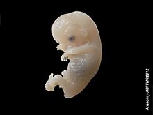
Embryogenesis is the process by which the embryo forms and develops. In mammals, the term refers chiefly to early stages of prenatal development, whereas the terms fetus and fetal development describe later stages.

Embryogenesis starts with the fertilization of the egg cell (ovum) by a sperm cell, (spermatozoon). Once fertilized, the ovum is referred to as a zygote, a single diploid cell. The zygote undergoes mitotic divisions with no significant growth (a process known as cleavage) and cellular differentiation, leading to development of a multicellular embryo.

Although embryogenesis occurs in both animal and plant development, this article addresses the common features among different animals, with some emphasis on the embryonic development of vertebrates and mammals.

Fertilization and the zygote

The egg cell is generally asymmetric, having an "animal pole" (future ectoderm and mesoderm) and a "vegetal pole" (future endoderm). It is covered with protective envelopes, with different layers. The first envelope – the one in contact with the membrane of the egg – is made of glycoproteins and is known as the vitelline membrane (zona pellucida in mammals). Different taxa show different cellular and acellular envelopes englobing the vitelline membrane.

Fertilization (also known as 'conception', 'fecundation' and 'syngamy') is the fusion of gametes to produce a new organism. In animals, the process involves a sperm fusing with an ovum, which eventually leads to the development of an embryo. Depending on the animal species, the process can occur within the body of the female in internal fertilisation, or outside in the case of external fertilisation. The fertilized egg cell is known as the zygote.

To prevent more than one sperm fertilizing the egg, polyspermy, fast block and slow block to polyspermy are used. Fast block, the membrane potential rapidly depolarizing and then returning to normal, happens immediately after an egg is fertilized by a single sperm. Slow block begins the first few seconds after fertilization and is when the release of calcium causes the cortical reaction, various enzymes releasing from cortical granules in the eggs plasma membrane, to expand and harden the outside membrane, preventing more sperm from entering.

Cleavage and morula

Cell divisions (cleavage)

Cell division with no significant growth, producing a cluster of cells that is the same size as the original zygote, is called cleavage. At least four initial cell divisions occur, resulting in a dense ball of at least sixteen cells called the morula. The different cells derived from cleavage, up to the blastula stage, are called blastomeres. Depending mostly on the amount of yolk in the egg, the cleavage can be holoblastic (total) or meroblastic (partial).

Holoblastic cleavage occurs in animals with little yolk in their eggs, such as humans and other mammals who receive nourishment as embryos from the mother, via the placenta or milk, such as might be secreted from a marsupium. On the other hand, meroblastic cleavage occurs in animals whose eggs have more yolk (i.e. birds and reptiles). Because cleavage is impeded in the vegetal pole, there is an uneven distribution and size of cells, being more numerous and smaller at the animal pole of the zygote.

In holoblastic eggs the first cleavage always occurs along the vegetal-animal axis of the egg, and the second cleavage is perpendicular to the first. From here the spatial arrangement of blastomeres can follow various patterns, due to different planes of cleavage, in various organisms:

Cleavage patterns followed by holoblastic and meroblastic eggs
Holoblastic Meroblastic


The end of cleavage is known as midblastula transition and coincides with the onset of zygotic transcription.

In amniotes, the cells of the morula are at first closely aggregated, but soon they become arranged into an outer or peripheral layer, the trophoblast, which does not contribute to the formation of the embryo proper, and an inner cell mass, from which the embryo is developed. Fluid collects between the trophoblast and the greater part of the inner cell-mass, and thus the morula is converted into a vesicle, called the blastodermic vesicle. The inner cell mass remains in contact, however, with the trophoblast at one pole of the ovum; this is named the embryonic pole, since it indicates the location where the future embryo will develop.

Formation of the blastula

After the 7th cleavage has produced 128 cells, the embryo is called a blastula. The blastula is usually a spherical layer of cells (the blastoderm) surrounding a fluid-filled or yolk-filled cavity (the blastocoel)

Mammals at this stage form a structure called the blastocyst, characterized by an inner cell mass that is distinct from the surrounding blastula. The blastocyst must not be confused with the blastula; even though they are similar in structure, their cells have different fates.

Before gastrulation, the cells of the trophoblast become differentiated into two strata: The outer stratum forms a syncytium (i.e., a layer of protoplasm studded with nuclei, but showing no evidence of subdivision into cells), termed the syncytiotrophoblast, while the inner layer, the cytotrophoblast or "Layer of Langhans", consists of well-defined cells. As already stated, the cells of the trophoblast do not contribute to the formation of the embryo proper; they form the ectoderm of the chorion and play an important part in the development of the placenta. On the deep surface of the inner cell mass, a layer of flattened cells, called the endoderm, is differentiated and quickly assumes the form of a small sac, called the yolk sac. Spaces appear between the remaining cells of the mass and, by the enlargement and coalescence of these spaces, a cavity called the amniotic cavity is gradually developed. The floor of this cavity is formed by the embryonic disk, which is composed of a layer of prismatic cells, the embryonic ectoderm, derived from the inner cell mass and lying in apposition with the endoderm.

Formation of the germ layers

The embryonic disk becomes oval and then pear-shaped, the wider end being directed forward. Near the narrow, posterior end, an opaque streak, called the primitive streak, makes its appearance and extends along the middle of the disk for about one-half of its length; at the anterior end of the streak there is a knob-like thickening termed the primitive node or knot, (known as Hensen's knot in birds). A shallow groove, the primitive groove, appears on the surface of the streak, and the anterior end of this groove communicates by means of an aperture, the blastopore, with the yolk sac. The primitive streak is produced by a thickening of the axial part of the ectoderm, the cells of which multiply, grow downward, and blend with those of the subjacent endoderm. From the sides of the primitive streak a third layer of cells, the mesoderm, extends laterally between the ectoderm and endoderm; the caudal end of the primitive streak forms the cloacal membrane. The blastoderm now consists of three layers, named from without inward: ectoderm, mesoderm, and endoderm; each has distinctive characteristics and gives rise to certain tissues of the body. For many mammals, it is sometime during formation of the germ layers that implantation of the embryo in the uterus of the mother occurs.

Formation of the gastrula

During gastrulation cells migrate to the interior of the blastula, subsequently forming two (in diploblastic animals) or three (triploblastic) germ layers. The embryo during this process is called a gastrula. The germ layers are referred to as the ectoderm, mesoderm and endoderm. In diploblastic animals only the ectoderm and the endoderm are present.
  • Among different animals, different combinations of the following processes occur to place the cells in the interior of the embryo:
    • Epiboly – expansion of one cell sheet over other cells
    • Ingression – migration of individual cells into the embryo (cells move with pseudopods)
    • Invagination – infolding of cell sheet into embryo, forming the mouth, anus, and archenteron.
    • Delamination – splitting or migration of one sheet into two sheets
    • Involution – inturning of cell sheet over the basal surface of an outer layer
    • Polar proliferation – Cells at the polar ends of the blastula/gastrula proliferate, mostly at the animal pole.
  • Other major changes during gastrulation:
In most animals, a blastopore is formed at the point where cells are entering the embryo. Two major groups of animals can be distinguished according to the blastopore's fate. In deuterostomes the anus forms from the blastopore, while in protostomes it develops into the mouth.

Formation of the early nervous system – neural groove, tube and notochord

In front of the primitive streak, two longitudinal ridges, caused by a folding up of the ectoderm, make their appearance, one on either side of the middle line formed by the streak. These are named the neural folds; they commence some little distance behind the anterior end of the embryonic disk, where they are continuous with each other, and from there gradually extend backward, one on either side of the anterior end of the primitive streak. Between these folds is a shallow median groove, the neural groove. The groove gradually deepens as the neural folds become elevated, and ultimately the folds meet and coalesce in the middle line and convert the groove into a closed tube, the neural tube or canal, the ectodermal wall of which forms the rudiment of the nervous system. After the coalescence of the neural folds over the anterior end of the primitive streak, the blastopore no longer opens on the surface but into the closed canal of the neural tube, and thus a transitory communication, the neurenteric canal, is established between the neural tube and the primitive digestive tube. The coalescence of the neural folds occurs first in the region of the hind brain, and from there extends forward and backward; toward the end of the third week, the front opening (anterior neuropore) of the tube finally closes at the anterior end of the future brain, and forms a recess that is in contact, for a time, with the overlying ectoderm; the hinder part of the neural groove presents for a time a rhomboidal shape, and to this expanded portion the term sinus rhomboidalis has been applied. Before the neural groove is closed, a ridge of ectodermal cells appears along the prominent margin of each neural fold; this is termed the neural crest or ganglion ridge, and from it the spinal and cranial nerve ganglia and the ganglia of the sympathetic nervous system are developed. By the upward growth of the mesoderm, the neural tube is ultimately separated from the overlying ectoderm.

Dissection of human embryo

The cephalic end of the neural groove exhibits several dilatations that, when the tube is shut, assume the form of three vesicles; these constitute the three primary cerebral vesicles, and correspond, respectively, to the future 'fore-brain' (prosencephalon), 'midbrain' (mesencephalon), and 'hind-brain' (rhombencephalon) (Fig. 18). The walls of the vesicles are developed into the nervous tissue and neuroglia of the brain, and their cavities are modified to form its ventricles. The remainder of the tube forms the spinal cord (medulla spinalis); from its ectodermal wall the nervous and neuroglial elements of the spinal cord are developed, while the cavity persists as the central canal.

Formation of the early septum

The extension of the mesoderm takes place throughout the whole of the embryonic and extra-embryonic areas of the ovum, except in certain regions. One of these is seen immediately in front of the neural tube. Here the mesoderm extends forward in the form of two crescentic masses, which meet in the middle line so as to enclose behind them an area that is devoid of mesoderm. Over this area, the ectoderm and endoderm come into direct contact with each other and constitute a thin membrane, the buccopharyngeal membrane, which forms a septum between the primitive mouth and pharynx.

Early formation of the heart and other primitive structures

In front of the buccopharyngeal area, where the lateral crescents of mesoderm fuse in the middle line, the pericardium is afterward developed, and this region is therefore designated the pericardial area. A second region where the mesoderm is absent, at least for a time, is that immediately in front of the pericardial area. This is termed the proamniotic area, and is the region where the proamnion is developed; in humans, however, it appears that a proamnion is never formed. A third region is at the hind end of the embryo, where the ectoderm and endoderm come into apposition and form the cloacal membrane.

Somitogenesis

Somitogenesis is the process by which somites (primitive segments) are produced. These segmented tissue blocks differentiate into skeletal muscle, vertebrae, and dermis of all vertebrates.

Somitogenesis begins with the formation of somitomeres (whorls of concentric mesoderm) marking the future somites in the presomitic mesoderm (unsegmented paraxial). The presomitic mesoderm gives rise to successive pairs of somites, identical in appearance that differentiate into the same cell types but the structures formed by the cells vary depending upon the anteroposterior (e.g., the thoracic vertebrae have ribs, the lumbar vertebrae do not). Somites have unique positional values along this axis and it is thought that these are specified by the Hox homeotic genes.

Toward the end of the second week after fertilization, transverse segmentation of the paraxial mesoderm begins, and it is converted into a series of well-defined, more or less cubical masses, also known as the somites, which occupy the entire length of the trunk on either side of the middle line from the occipital region of the head. Each segment contains a central cavity (known as a myocoel), which, however, is soon filled with angular and spindle-shape cells. The somites lie immediately under the ectoderm on the lateral aspect of the neural tube and notochord, and are connected to the lateral mesoderm by the intermediate cell mass. Those of the trunk may be arranged in the following groups, viz.: cervical 8, thoracic 12, lumbar 5, sacral 5, and coccygeal from 5 to 8. Those of the occipital region of the head are usually described as being four in number. In mammals, somites of the head can be recognized only in the occipital region, but a study of the lower vertebrates leads to the belief that they are present also in the anterior part of the head and that, altogether, nine segments are represented in the cephalic region.

Organogenesis

Human embryo, 8-9 weeks, 38 mm

At some point after the different germ layers are defined, organogenesis begins. The first stage in vertebrates is called neurulation, where the neural plate folds forming the neural tube (see above). Other common organs or structures that arise at this time include the heart and somites, but from now on embryogenesis follows no common pattern among the different taxa of the animal kingdom.

In most animals organogenesis, along with morphogenesis, results in a larva. The hatching of the larva, which must then undergo metamorphosis, marks the end of embryonic development.

Multicellular organism

From Wikipedia, the free encyclopedia
 
Multicellular organism
Temporal range: Mesoproterozoic–present
C elegans stained.jpg
In this image, a wild-type Caenorhabditis elegans is stained to highlight the nuclei of its cells.

Multicellular organisms are organisms that consist of more than one cell, in contrast to unicellular organisms.

All species of animals, land plants and most fungi are multicellular, as are many algae, whereas a few organisms are partially uni- and partially multicellular, like slime molds and social amoebae such as the genus Dictyostelium.

Multicellular organisms arise in various ways, for example by cell division or by aggregation of many single cells. Colonial organisms are the result of many identical individuals joining together to form a colony. However, it can often be hard to separate colonial protists from true multicellular organisms, because the two concepts are not distinct; colonial protists have been dubbed "pluricellular" rather than "multicellular".

Evolutionary history

Occurrence

Multicellularity has evolved independently at least 46 times in eukaryotes, and also in some prokaryotes, like cyanobacteria, myxobacteria, actinomycetes, Magnetoglobus multicellularis or Methanosarcina. However, complex multicellular organisms evolved only in six eukaryotic groups: animals, fungi, brown algae, red algae, green algae, and land plants. It evolved repeatedly for Chloroplastida (green algae and land plants), once or twice for animals, once for brown algae, three times in the fungi (chytrids, ascomycetes and basidiomycetes) and perhaps several times for slime molds and red algae. The first evidence of multicellularity is from cyanobacteria-like organisms that lived 3–3.5 billion years ago. To reproduce, true multicellular organisms must solve the problem of regenerating a whole organism from germ cells (i.e. sperm and egg cells), an issue that is studied in evolutionary developmental biology. Animals have evolved a considerable diversity of cell types in a multicellular body (100–150 different cell types), compared with 10–20 in plants and fungi.

Loss of multicellularity

Loss of multicellularity occurred in some groups. Fungi are predominantly multicellular, though early diverging lineages are largely unicellular (e.g. Microsporidia) and there have been numerous reversions to unicellularity across fungi (e.g. Saccharomycotina, Cryptococcus, and other yeasts). It may also have occurred in some red algae (e.g. Porphyridium), but it is possible that they are primitively unicellular. Loss of multicellularity is also considered probable in some green algae (e.g. Chlorella vulgaris and some Ulvophyceae). In other groups, generally parasites, a reduction of multicellularity occurred, in number or types of cells (e.g. the myxozoans, multicellular organisms, earlier thought to be unicellular, are probably extremely reduced cnidarians).

Cancer

Multicellular organisms, especially long-living animals, face the challenge of cancer, which occurs when cells fail to regulate their growth within the normal program of development. Changes in tissue morphology can be observed during this process. Cancer in animals (metazoans) has often been described as a loss of multicellularity. There is a discussion about the possibility of existence of cancer in other multicellular organisms or even in protozoa. For example, plant galls have been characterized as tumors but some authors argue that plants do not develop cancer.

Separation of somatic and germ cells

In some multicellular groups, which are called Weismannists, a separation between a sterile somatic cell line and a germ cell line evolved. However, Weismannist development is relatively rare (e.g. vertebrates, arthropods, Volvox), as great part of species have the capacity for somatic embryogenesis (e.g. land plants, most algae, many invertebrates).

Hypotheses for origin

Tetrabaena socialis consists of four cells.

One hypothesis for the origin of multicellularity is that a group of function-specific cells aggregated into a slug-like mass called a grex, which moved as a multicellular unit. This is essentially what slime molds do. Another hypothesis is that a primitive cell underwent nucleus division, thereby becoming a coenocyte. A membrane would then form around each nucleus (and the cellular space and organelles occupied in the space), thereby resulting in a group of connected cells in one organism (this mechanism is observable in Drosophila). A third hypothesis is that as a unicellular organism divided, the daughter cells failed to separate, resulting in a conglomeration of identical cells in one organism, which could later develop specialized tissues. This is what plant and animal embryos do as well as colonial choanoflagellates.

Because the first multicellular organisms were simple, soft organisms lacking bone, shell or other hard body parts, they are not well preserved in the fossil record. One exception may be the demosponge, which may have left a chemical signature in ancient rocks. The earliest fossils of multicellular organisms include the contested Grypania spiralis and the fossils of the black shales of the Palaeoproterozoic Francevillian Group Fossil B Formation in Gabon (Gabonionta). The Doushantuo Formation has yielded 600 million year old microfossils with evidence of multicellular traits.

Until recently, phylogenetic reconstruction has been through anatomical (particularly embryological) similarities. This is inexact, as living multicellular organisms such as animals and plants are more than 500 million years removed from their single-cell ancestors. Such a passage of time allows both divergent and convergent evolution time to mimic similarities and accumulate differences between groups of modern and extinct ancestral species. Modern phylogenetics uses sophisticated techniques such as alloenzymes, satellite DNA and other molecular markers to describe traits that are shared between distantly related lineages.

The evolution of multicellularity could have occurred in a number of different ways, some of which are described below:

The symbiotic theory

This theory suggests that the first multicellular organisms occurred from symbiosis (cooperation) of different species of single-cell organisms, each with different roles. Over time these organisms would become so dependent on each other they would not be able to survive independently, eventually leading to the incorporation of their genomes into one multicellular organism. Each respective organism would become a separate lineage of differentiated cells within the newly created species.
This kind of severely co-dependent symbiosis can be seen frequently, such as in the relationship between clown fish and Riterri sea anemones. In these cases, it is extremely doubtful whether either species would survive very long if the other became extinct. However, the problem with this theory is that it is still not known how each organism's DNA could be incorporated into one single genome to constitute them as a single species. Although such symbiosis is theorized to have occurred (e.g. mitochondria and chloroplasts in animal and plant cells—endosymbiosis), it has happened only extremely rarely and, even then, the genomes of the endosymbionts have retained an element of distinction, separately replicating their DNA during mitosis of the host species. For instance, the two or three symbiotic organisms forming the composite lichen, although dependent on each other for survival, have to separately reproduce and then re-form to create one individual organism once more.

The cellularization (syncytial) theory

This theory states that a single unicellular organism, with multiple nuclei, could have developed internal membrane partitions around each of its nuclei. Many protists such as the ciliates or slime molds can have several nuclei, lending support to this hypothesis. However, the simple presence of multiple nuclei is not enough to support the theory. Multiple nuclei of ciliates are dissimilar and have clear differentiated functions. The macronucleus serves the organism's needs, whereas the micronucleus is used for sexual reproduction with exchange of genetic material. Slime molds syncitia form from individual amoeboid cells, like syncitial tissues of some multicellular organisms, not the other way round. To be deemed valid, this theory needs a demonstrable example and mechanism of generation of a multicellular organism from a pre-existing syncytium.

The colonial theory

The Colonial Theory of Haeckel, 1874, proposes that the symbiosis of many organisms of the same species (unlike the symbiotic theory, which suggests the symbiosis of different species) led to a multicellular organism. At least some, it is presumed land-evolved, multicellularity occurs by cells separating and then rejoining (e.g. cellular slime molds) whereas for the majority of multicellular types (those that evolved within aquatic environments), multicellularity occurs as a consequence of cells failing to separate following division. The mechanism of this latter colony formation can be as simple as incomplete cytokinesis, though multicellularity is also typically considered to involve cellular differentiation.

ColonialFlagellateHypothesis.png

The advantage of the Colonial Theory hypothesis is that it has been seen to occur independently in 16 different protoctistan phyla. For instance, during food shortages the amoeba Dictyostelium groups together in a colony that moves as one to a new location. Some of these amoeba then slightly differentiate from each other. Other examples of colonial organisation in protista are Volvocaceae, such as Eudorina and Volvox, the latter of which consists of up to 500–50,000 cells (depending on the species), only a fraction of which reproduce. For example, in one species 25–35 cells reproduce, 8 asexually and around 15–25 sexually. However, it can often be hard to separate colonial protists from true multicellular organisms, as the two concepts are not distinct; colonial protists have been dubbed "pluricellular" rather than "multicellular".

The Synzoospore theory

Some authors suggest that the origin of multicellularity, at least in Metazoa, occurred due to a transition from temporal to spatial cell differentiation, rather than through a gradual evolution of cell differentiation, as affirmed in Haeckel’s Gastraea theory.

GK-PID

About 800 million years ago, a minor genetic change in a single molecule called guanylate kinase protein-interaction domain (GK-PID) may have allowed organisms to go from a single cell organism to one of many cells.

The role of viruses

Genes borrowed from viruses have recently been identified as playing a crucial role in the differentiation of multicellular tissues and organs and even in sexual reproduction, in the fusion of egg cell and sperm. Such fused cells are also involved in metazoan membranes such as those that prevent chemicals crossing the placenta and the brain body separation. Two viral components have been identified. The first is syncytin, which came from a virus. The second identified in 2007 is called EFF1, which helps form the skin of Caenorhabditis elegans, part of a whole family of FF proteins. Felix Rey, of the Pasteur Institute in Paris has constructed the 3D structure of the EFF1 protein and shown it does the work of linking one cell to another, in viral infections. The fact that all known cell fusion molecules are viral in origin suggests that they have been vitally important to the inter-cellular communication systems that enabled multicellularity. Without the ability of cellular fusion, colonies could have formed, but anything even as complex as a sponge would not have been possible.

Advantages

Multicellularity allows an organism to exceed the size limits normally imposed by diffusion: single cells with increased size have a decreased surface-to-volume ratio and have difficulty absorbing sufficient nutrients and transporting them throughout the cell. Multicellular organisms thus have the competitive advantages of an increase in size without its limitations. They can have longer lifespans as they can continue living when individual cells die. Multicellularity also permits increasing complexity by allowing differentiation of cell types within one organism.

Evolution of cells

From Wikipedia, the free encyclopedia

Evolution of cells refers to the evolutionary origin and subsequent evolutionary development of cells. Cells first emerged at least 3.8 billion years ago.

The first cells


The origin of cells was the most important step in the evolutionary theory of life on Earth. The birth of the cell marked the passage from pre-biotic chemistry to partitioned units resembling modern cells. The final transition to living entities that fulfill all the definitions of modern cells depended on the ability to evolve effectively by natural selection. This transition has been called the Darwinian transition.

If life is viewed from the point of view of replicator molecules, cells satisfy two fundamental conditions: protection from the outside environment and confinement of biochemical activity. The former condition is needed to keep complex molecules stable in a varying and sometimes aggressive environment; the latter is fundamental for the evolution of biocomplexity. If the freely floating molecules that code for enzymes are not enclosed in cells, the enzymes will automatically benefit the neighbouring replicator molecules. The consequences of diffusion in non-partitioned life forms might be viewed as "parasitism by default." Therefore, the selection pressure on replicator molecules will be lower, as the 'lucky' molecule that produces the better enzyme has no definitive advantage over its close neighbors. If the molecule is enclosed in a cell membrane, then the enzymes coded will be available only to the replicator molecule itself. That molecule will uniquely benefit from the enzymes it codes for, giving it a better chance to multiply.

Partitioning may have begun from cell-like spheroids formed by proteinoids, which are observed by heating amino acids with phosphoric acid as a catalyst. They bear much of the basic features provided by cell membranes. Proteinoid-based protocells enclosing RNA molecules could have been the first cellular life forms on Earth.

Another possibility is that the shores of the ancient coastal waters may have served as a mammoth laboratory, aiding in the countless experiments necessary to bring about the first cell. Waves breaking on the shore create a delicate foam composed of bubbles. Shallow coastal waters also tend to be warmer, further concentrating the molecules through evaporation. While bubbles made mostly of water tend to burst quickly, oily bubbles are much more stable, lending more time to the particular bubble to perform these crucial experiments. The phospholipid is a good example of a common oily compound prevalent in the prebiotic seas.

Phospholipids are composed of a hydrophilic head on one end, and a hydrophobic tail on the other. They possess an important characteristic for the construction of cell membranes; they can come together to form a bilayer membrane. A lipid monolayer bubble can only contain oil, and is not conducive to harbouring water-soluble organic molecules, but a lipid bilayer bubble  can contain water, and was a likely precursor to the modern cell membrane. If a protein came along that increased the integrity of its parent bubble, then that bubble had an advantage. Primitive reproduction may have occurred when the bubbles burst, releasing the results of the experiment into the surrounding medium. Once enough of the right compounds were released into the medium, the development of the first prokaryotes, eukaryotes, and multi-cellular organisms could be achieved.

Community metabolism

The common ancestor of the now existing cellular lineages (eukaryotes, bacteria, and archaea) may have been a community of organisms that readily exchanged components and genes. It would have contained:
  • Autotrophs that produced organic compounds from CO2, either photosynthetically or by inorganic chemical reactions;
  • Heterotrophs that obtained organics by leakage from other organisms
  • Saprotrophs that absorbed nutrients from decaying organisms
  • Phagotrophs that were sufficiently complex to envelop and digest particulate nutrients, including other organisms.
The eukaryotic cell seems to have evolved from a symbiotic community of prokaryotic cells. DNA-bearing organelles like mitochondria and chloroplasts are remnants of ancient symbiotic oxygen-breathing bacteria and cyanobacteria, respectively, where at least part of the rest of the cell may have been derived from an ancestral archaean prokaryote cell. This concept is often termed the endosymbiotic theory. There is still debate about whether organelles like the hydrogenosome predated the origin of mitochondria.

How the current lineages of microbes evolved from this postulated community is currently unsolved but subject to intense research by biologists, stimulated by the great flow of new discoveries in genome science.

Genetic code and the RNA world

Modern evidence suggests that early cellular evolution occurred in a biological realm radically distinct from modern biology. It is thought that in this ancient realm, the current genetic role of DNA was largely filled by RNA, and catalysis also was largely mediated by RNA (that is, by ribozyme counterparts of enzymes). This concept is known as the RNA world hypothesis.

According to this hypothesis, the ancient RNA world transitioned into the modern cellular world via the evolution of protein synthesis, followed by replacement of many cellular ribozyme catalysts by protein-based enzymes. Proteins are much more flexible in catalysis than RNA due to the existence of diverse amino acid side chains with distinct chemical characteristics. The RNA record in existing cells appears to preserve some 'molecular fossils' from this RNA world. These RNA fossils include the ribosome itself (in which RNA catalyses peptide-bond formation), the modern ribozyme catalyst RNase P, and tRNAs.

The nearly universal genetic code preserves some evidence for the RNA world. For instance, recent studies of transfer RNAs, the enzymes that charge them with amino acids (the first step in protein synthesis) and the way these components recognise and exploit the genetic code, have been used to suggest that the universal genetic code emerged before the evolution of the modern amino acid activation method for protein synthesis.

Canonical patterns

Although the evolutionary origins of the major lineages of modern cells are disputed, the primary distinctions between the three major lineages of cellular life (called domains) are firmly established.
In each of these three domains, DNA replication, transcription, and translation all display distinctive features. There are three versions of ribosomal RNAs, and generally three versions of each ribosomal protein, one for each domain of life. These three versions of the protein synthesis apparatus are called the canonical patterns, and the existence of these canonical patterns provides the basis for a definition of the three domains - Bacteria, Archaea, and Eukarya (or Eukaryota) - of currently existing cells.

Using genomics to infer early lines of evolution

Instead of relying a single gene such as the small-subunit ribosomal RNA (SSU rRNA) gene to reconstruct early evolution, or a few genes, scientific effort has shifted to analyzing complete genome sequences.

Evolutionary trees based only on SSU rRNA alone do not capture the events of early eukaryote evolution accurately, and the progenitors of the first nucleated cells are still uncertain. For instance, analysis of the complete genome of the eukaryote yeast shows that many of its genes are more closely related to bacterial genes than they are to archaea, and it is now clear that archaea were not the simple progenitors of the eukaryotes, in contradiction to earlier findings based on SSU rRNA and limited samples of other genes.

One hypothesis is that the first nucleated cell arose from two distinctly different ancient prokaryotic (non-nucleated) species that had formed a symbiotic relationship with one another to carry out different aspects of metabolism. One partner of this symbiosis is proposed to be a bacterial cell, and the other an archaeal cell. It is postulated that this symbiotic partnership progressed via the cellular fusion of the partners to generate a chimeric or hybrid cell with a membrane bound internal structure that was the forerunner of the nucleus. The next stage in this scheme was transfer of both partner genomes into the nucleus and their fusion with one another. Several variations of this hypothesis for the origin of nucleated cells have been suggested. Other biologists dispute this conception and emphasize the community metabolism theme, the idea that early living communities would comprise many different entities to extant cells, and would have shared their genetic material more extensively than current microbes.

Quotes

"The First Cell arose in the previously pre-biotic world with the coming together of several entities that gave a single vesicle the unique chance to carry out three essential and quite different life processes. These were: (a) to copy informational macromolecules, (b) to carry out specific catalytic functions, and (c) to couple energy from the environment into usable chemical forms. These would foster subsequent cellular evolution and metabolism. Each of these three essential processes probably originated and was lost many times prior to The First Cell, but only when these three occurred together was life jump-started and Darwinian evolution of organisms began." (Koch and Silver, 2005)
"The evolution of modern cells is arguably the most challenging and important problem the field of Biology has ever faced. In Darwin's day the problem could hardly be imagined. For much of the 20th century it was intractable. In any case, the problem lay buried in the catch-all rubric "origin of life"---where, because it is a biological not a (bio)chemical problem, it was effectively ignored. Scientific interest in cellular evolution started to pick up once the universal phylogenetic tree, the framework within which the problem had to be addressed, was determined . But it was not until microbial genomics arrived on the scene that biologists could actually do much about the problem of cellular evolution." (Carl Woese, 2002) 

Platinum group

From Wikipedia, the free encyclopedia ...