Search This Blog

Monday, July 20, 2020

Sensory nervous system

From Wikipedia, the free encyclopedia
 
Sensory nervous system
Gray722.svg
Typical sensory system: the visual system, illustrated by the classic Gray's FIG. 722– This scheme shows the flow of information from the eyes to the central connections of the optic nerves and optic tracts, to the visual cortex. Area V1 is the region of the brain which is engaged in vision.
Details
Identifiers
Latinorgana sensuum
MeSHD012679
TAA15.0.00.000
FMA75259
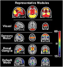
The visual system and the somatosensory system are active even during resting state fMRI
 
 
Activation and response in the sensory nervous system
 
The sensory nervous system is a part of the nervous system responsible for processing sensory information. A sensory system consists of sensory neurons (including the sensory receptor cells), neural pathways, and parts of the brain involved in sensory perception. Commonly recognized sensory systems are those for vision, hearing, touch, taste, smell, and balance. In short, senses are transducers from the physical world to the realm of the mind where we interpret the information, creating our perception of the world around us.
Organisms need information to solve at least three kinds of problems: (a) to maintain an appropriate environment, i.e., homeostasis; (b) to time activities (e.g., seasonal changes in behavior) or synchronize activities with those of conspecifics; and (c) to locate and respond to resources or threats (e.g., by moving towards resources or evading or attacking threats). Organisms also need to transmit information in order to influence another's behavior: to identify themselves, warn conspecifics of danger, coordinate activities, or deceive.
The receptive field is the area of the body or environment to which a receptor organ and receptor cells respond. For instance, the part of the world an eye can see, is its receptive field; the light that each rod or cone can see, is its receptive field. Receptive fields have been identified for the visual system, auditory system and somatosensory system.

Stimulus

Sensory systems code for four aspects of a stimulus; type (modality), intensity, location, and duration. Arrival time of a sound pulse and phase differences of continuous sound are used for sound localization. Certain receptors are sensitive to certain types of stimuli (for example, different mechanoreceptors respond best to different kinds of touch stimuli, like sharp or blunt objects). Receptors send impulses in certain patterns to send information about the intensity of a stimulus (for example, how loud a sound is). The location of the receptor that is stimulated gives the brain information about the location of the stimulus (for example, stimulating a mechanoreceptor in a finger will send information to the brain about that finger). The duration of the stimulus (how long it lasts) is conveyed by firing patterns of receptors. These impulses are transmitted to the brain through afferent neurons.

Senses and receptors

While debate exists among neurologists as to the specific number of senses due to differing definitions of what constitutes a sense, Gautama Buddha and Aristotle classified five ‘traditional’ human senses which have become universally accepted: touch, taste, smell, sight, and hearing. Other senses that have been well-accepted in most mammals, including humans, include nociception, equilibrioception, kinaesthesia, and thermoception. Furthermore, some nonhuman animals have been shown to possess alternate senses, including magnetoception and electroreception.

Receptors

The initialization of sensation stems from the response of a specific receptor to a physical stimulus. The receptors which react to the stimulus and initiate the process of sensation are commonly characterized in four distinct categories: chemoreceptors, photoreceptors, mechanoreceptors, and thermoreceptors. All receptors receive distinct physical stimuli and transduce the signal into an electrical action potential. This action potential then travels along afferent neurons to specific brain regions where it is processed and interpreted.

Chemoreceptors

Chemoreceptors, or chemosensors, detect certain chemical stimuli and transduce that signal into an electrical action potential. The two primary types of chemoreceptors are:

Photoreceptors

Photoreceptors are capable of phototransduction, a process which converts light (electromagnetic radiation) into, among other types of energy, a membrane potential. The three primary types of photoreceptors are: Cones are photoreceptors which respond significantly to color. In humans the three different types of cones correspond with a primary response to short wavelength (blue), medium wavelength (green), and long wavelength (yellow/red). Rods are photoreceptors which are very sensitive to the intensity of light, allowing for vision in dim lighting. The concentrations and ratio of rods to cones is strongly correlated with whether an animal is diurnal or nocturnal. In humans rods outnumber cones by approximately 20:1, while in nocturnal animals, such as the tawny owl, the ratio is closer to 1000:1. Ganglion Cells reside in the adrenal medulla and retina where they are involved in the sympathetic response. Of the ~1.3 million ganglion cells present in the retina, 1-2% are believed to be photosensitive ganglia. These photosensitive ganglia play a role in conscious vision for some animals, and are believed to do the same in humans.

Mechanoreceptors

Mechanoreceptors are sensory receptors which respond to mechanical forces, such as pressure or distortion. While mechanoreceptors are present in hair cells and play an integral role in the vestibular and auditory systems, the majority of mechanoreceptors are cutaneous and are grouped into four categories:
  • Slowly adapting type 1 receptors have small receptive fields and respond to static stimulation. These receptors are primarily used in the sensations of form and roughness.
  • Slowly adapting type 2 receptors have large receptive fields and respond to stretch. Similarly to type 1, they produce sustained responses to a continued stimuli.
  • Rapidly adapting receptors have small receptive fields and underlie the perception of slip.
  • Pacinian receptors have large receptive fields and are the predominant receptors for high-frequency vibration.

Thermoreceptors

Thermoreceptors are sensory receptors which respond to varying temperatures. While the mechanisms through which these receptors operate is unclear, recent discoveries have shown that mammals have at least two distinct types of thermoreceptors:
TRPV1 is a heat-activated channel that acts as a small heat detecting thermometer in the membrane which begins the polarization of the neural fiber when exposed to changes in temperature. Ultimately, this allows us to detect ambient temperature in the warm/hot range. Similarly, the molecular cousin to TRPV1, TRPM8, is a cold-activated ion channel that responds to cold. Both cold and hot receptors are segregated by distinct subpopulations of sensory nerve fibers, which shows us that the information coming into the spinal cord is originally separate. Each sensory receptor has its own “labeled line” to convey a simple sensation experienced by the recipient. Ultimately, TRP channels act as thermosensors, channels that help us to detect changes in ambient temperatures.

Nociceptors

Nociceptors respond to potentially damaging stimuli by sending signals to the spinal cord and brain. This process, called nociception, usually causes the perception of pain. They are found in internal organs, as well as on the surface of the body. Nociceptors detect different kinds of damaging stimuli or actual damage. Those that only respond when tissues are damaged are known as "sleeping" or "silent" nociceptors.
  • Thermal nociceptors are activated by noxious heat or cold at various temperatures.
  • Mechanical nociceptors respond to excess pressure or mechanical deformation.
  • Chemical nociceptors respond to a wide variety of chemicals, some of which are signs of tissue damage. They are involved in the detection of some spices in food.

Sensory cortex

All stimuli received by the receptors listed above are transduced to an action potential, which is carried along one or more afferent neurons towards a specific area of the brain. While the term sensory cortex is often used informally to refer to the somatosensory cortex, the term more accurately refers to the multiple areas of the brain at which senses are received to be processed. For the five traditional senses in humans, this includes the primary and secondary cortices of the different senses: the somatosensory cortex, the visual cortex, the auditory cortex, the primary olfactory cortex, and the gustatory cortex. Other modalities have corresponding sensory cortex areas as well, including the vestibular cortex for the sense of balance.

Somatosensory cortex

Located in the parietal lobe, the primary somatosensory cortex is the primary receptive area for the sense of touch and proprioception in the somatosensory system. This cortex is further divided into Brodmann areas 1, 2, and 3. Brodmann area 3 is considered the primary processing center of the somatosensory cortex as it receives significantly more input from the thalamus, has neurons highly responsive to somatosensory stimuli, and can evoke somatic sensations through electrical stimulation. Areas 1 and 2 receive most of their input from area 3. There are also pathways for proprioception (via the cerebellum), and motor control (via Brodmann area 4). See also: S2 Secondary somatosensory cortex.

The human eye is the first element of a sensory system: in this case, vision, for the visual system.

Visual cortex

The visual cortex refers to the primary visual cortex, labeled V1 or Brodmann area 17, as well as the extrastriate visual cortical areas V2-V5. Located in the occipital lobe, V1 acts as the primary relay station for visual input, transmitting information to two primary pathways labeled the dorsal and ventral streams. The dorsal stream includes areas V2 and V5, and is used in interpreting visual ‘where’ and ‘how.’ The ventral stream includes areas V2 and V4, and is used in interpreting ‘what.’ Increases in Task-negative activity are observed in the ventral attention network, after abrupt changes in sensory stimuli, at the onset and offset of task blocks, and at the end of a completed trial.

Auditory cortex

Located in the temporal lobe, the auditory cortex is the primary receptive area for sound information. The auditory cortex is composed of Brodmann areas 41 and 42, also known as the anterior transverse temporal area 41 and the posterior transverse temporal area 42, respectively. Both areas act similarly and are integral in receiving and processing the signals transmitted from auditory receptors

Primary olfactory cortex

Located in the temporal lobe, the primary olfactory cortex is the primary receptive area for olfaction, or smell. Unique to the olfactory and gustatory systems, at least in mammals, is the implementation of both peripheral and central mechanisms of action. The peripheral mechanisms involve olfactory receptor neurons which transduce a chemical signal along the olfactory nerve, which terminates in the olfactory bulb. The chemoreceptors in the receptor neurons that start the signal cascade are G protein-coupled receptors. The central mechanisms include the convergence of olfactory nerve axons into glomeruli in the olfactory bulb, where the signal is then transmitted to the anterior olfactory nucleus, the piriform cortex, the medial amygdala, and the entorhinal cortex, all of which make up the primary olfactory cortex. 

In contrast to vision and hearing, the olfactory bulbs are not cross-hemispheric; the right bulb connects to the right hemisphere and the left bulb connects to the left hemisphere. 

Gustatory cortex

The gustatory cortex is the primary receptive area for taste. The word taste is used in a technical sense to refer specifically to sensations coming from taste buds on the tongue. The five qualities of taste detected by the tongue include sourness, bitterness, sweetness, saltiness, and the protein taste quality, called umami. In contrast, the term flavor refers to the experience generated through integration of taste with smell and tactile information. The gustatory cortex consists of two primary structures: the anterior insula, located on the insular lobe, and the frontal operculum, located on the frontal lobe. Similarly to the olfactory cortex, the gustatory pathway operates through both peripheral and central mechanisms. Peripheral taste receptors, located on the tongue, soft palate, pharynx, and esophagus, transmit the received signal to primary sensory axons, where the signal is projected to the nucleus of the solitary tract in the medulla, or the gustatory nucleus of the solitary tract complex. The signal is then transmitted to the thalamus, which in turn projects the signal to several regions of the neocortex, including the gustatory cortex.

The neural processing of taste is affected at nearly every stage of processing by concurrent somatosensory information from the tongue, that is, mouthfeel. Scent, in contrast, is not combined with taste to create flavor until higher cortical processing regions, such as the insula and orbitofrontal cortex.

Human sensory system

The human sensory system consists of the following subsystems:

Diseases

Disability-adjusted life year for sense organ diseases per 100,000 inhabitants in 2002.
  no data
  less than 200
  200-400
  400-600
  600-800
  800-1000
  1000-1200
  1200-1400
  1400-1600
  1600-1800
  1800-2000
  2000-2300
  more than 2300
Biological system

From Wikipedia, the free encyclopedia
 
 
A biological system is a complex network of biologically relevant entities. Biological organization spans several scales and are determined based different structures depending on what the system is. Examples of biological systems at the macro scale are populations of organisms. On the organ and tissue scale in mammals and other animals, examples include the circulatory system, the respiratory system, and the nervous system. On the micro to the nanoscopic scale, examples of biological systems are cells, organelles, macromolecular complexes and regulatory pathways. A biological system is not to be confused with a living system, such as a living organism.

Organ and tissue systems

An example of a system: The brain, the cerebellum, the spinal cord, and the nerves are the four basic components of the nervous system.
 
These specific systems are widely studied in human anatomy and are also present in many other animals.

History

The notion of system (or apparatus) relies upon the concept of vital or organic function: a system is a set of organs with a definite function. This idea was already present in Antiquity (Galen, Aristotle), but the application of the term "system" is more recent. For example, the nervous system was named by Monro (1783), but Rufus of Ephesus (c. 90-120), clearly viewed for the first time the brain, spinal cord, and craniospinal nerves as an anatomical unit, although he wrote little about its function, nor gave a name to this unit.

The enumeration of the principal functions - and consequently of the systems - remained almost the same since Antiquity, but the classification of them has been very various, e.g., compare Aristotle, Bichat, Cuvier.

The notion of physiological division of labor, introduced in the 1820s by the French physiologist Henri Milne-Edwards, allowed to "compare and study living things as if they were machines created by the industry of man." Inspired in the work of Adam Smith, Milne-Edwards wrote that the "body of all living beings, whether animal or plant, resembles a factory ... where the organs, comparable to workers, work incessantly to produce the phenomena that constitute the life of the individual." In more differentiated organisms, the functional labor could be apportioned between different instruments or systems (called by him as appareils).

Cellular Organelle Systems

The exact components of a cell are determined by whether the cell is a eukaryote or prokaryote
  • Nucleus: storage of genetic material; control center of the cell.
  • Cytosol: component of the cytoplasm consisting of jelly-like fluid in which organelles are suspended within
  • Cell membrane (plasma membrane):
  • Endoplasmic reticulum: outer part of the nuclear envelope forming a continuous channel used for transportation; consists of the rough endoplasmic reticulum and the smooth endoplasmic reticulum
    • Rough endoplasmic reticulum (RER): considered "rough" due to the ribosomes attached to the channeling; made up of cisternae that allow for protein production
    • Smooth endoplasmic reticulum (SER): storage and synthesis of lipids and steroid hormones as well as detoxification
  • Ribosome: site of biological protein synthesis essential for internal activity and cannot be reproduced in other organs
  • Mitochondrion (mitochondria): powerhouse of the cell; site of cellular respiration producing ATP (adenosine triphosphate)
  • Lysosome: center of breakdown for unwanted/unneeded material within the cell
  • Peroxisome: breaks down toxic materials from the contained digestive enzymes such as H2O2(hydrogen peroxide)
  • Golgi apparatus (eukaryotic only): folded network involved in modification, transport, and secretion
  • Chloroplast (prokaryotic only): site of photosynthesis; storage of chlorophyll

Epitope

From Wikipedia, the free encyclopedia
https://en.wikipedia.org/wiki/Epitope 
An epitope, also known as antigenic determinant, is the part of an antigen that is recognized by the immune system, specifically by antibodies, B cells, or T cells. For example, the epitope is the specific piece of the antigen to which an antibody binds. The part of an antibody that binds to the epitope is called a paratope. Although epitopes are usually non-self proteins, sequences derived from the host that can be recognized (as in the case of autoimmune diseases) are also epitopes.

The epitopes of protein antigens are divided into two categories, conformational epitopes and linear epitopes, based on their structure and interaction with the paratope. Conformational and linear epitopes interact with the paratope based on the 3-D conformation adopted by the epitope, which is determined by the surface features of the involved epitope residues and the shape or tertiary structure of other segments of the antigen. A conformational epitope is formed by the 3-D conformation adopted by the interaction of discontiguous amino acid residues. In contrast, a linear epitope is formed by the 3-D conformation adopted by the interaction of contiguous amino acid residues. A linear epitope is not determined solely by the primary structure of the involved amino acids. Residues that flank such amino acid residues, as well as more distant amino acid residues of the antigen affect the ability of the primary structure residues to adopt the epitope's 3-D conformation. The proportion of epitopes that are conformational is unknown.

Function

T cell epitopes

T cell epitopes are presented on the surface of an antigen-presenting cell, where they are bound to MHC molecules. In humans, professional antigen-presenting cells are specialized to present MHC class II peptides, whereas most nucleated somatic cells present MHC class I peptides. T cell epitopes presented by MHC class I molecules are typically peptides between 8 and 11 amino acids in length, whereas MHC class II molecules present longer peptides, 13-17 amino acids in length, and non-classical MHC molecules also present non-peptidic epitopes such as glycolipids.

Cross-activity

Epitopes are sometimes cross-reactive. This property is exploited by the immune system in regulation by anti-idiotypic antibodies (originally proposed by Nobel laureate Niels Kaj Jerne). If an antibody binds to an antigen's epitope, the paratope could become the epitope for another antibody that will then bind to it. If this second antibody is of IgM class, its binding can upregulate the immune response; if the second antibody is of IgG class, its binding can downregulate the immune response.

Epitope mapping

Epitopes can be mapped using protein microarrays, and with the ELISpot or ELISA techniques. Another technique involves high-throughput mutagenesis, an epitope mapping strategy developed to improve rapid mapping of conformational epitopes on structurally complex proteins.

MHC class I and II epitopes can be reliably predicted by computational means alone, although not all in-silico T cell epitope prediction algorithms are equivalent in their accuracy.

Epitope tags

Epitopes are often used in proteomics and the study of other gene products. Using recombinant DNA techniques genetic sequences coding for epitopes that are recognized by common antibodies can be fused to the gene. Following synthesis, the resulting epitope tag allows the antibody to find the protein or other gene product enabling lab techniques for localisation, purification, and further molecular characterization. Common epitopes used for this purpose are Myc-tag, HA-tag, FLAG-tag, GST-tag, 6xHis, V5-tag and OLLAS. Peptides can also be bound by proteins that form covalent bonds to the peptide, allowing irreversible immobilisation These strategies have also been successfully applied to the develohttps://en.wikipedia.org/wiki/Epitopepment of "epitope-focused" vaccine design.

Neoantigenic determinant

A neoantigenic determinant is an epitope on a neoantigen, which is a newly formed antigen that has not been previously recognized by the immune system. Neoantigens are often associated with tumor antigens and are found in oncogenic cells. Neoantigens and, by extension, neoantigenic determinants can be formed when a protein undergoes further modification within a biochemical pathway such as glycosylation, phosphorylation or proteolysis. This, by altering the structure of the protein, can produce new epitopes that are called neoantigenic determinants as they give rise to new antigenic determinants. Recognition requires separate, specific antibodies.

Antigen

From Wikipedia, the free encyclopedia
 
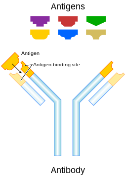
An illustration that shows how antigens induce the immune system response by interacting with an antibody that matches the antigen's molecular structure.
 
In immunology, an antigen (Ag) is a molecule or molecular structure, such as may be present at the outside of a pathogen, that can be bound to by an antigen-specific antibody (Ab) or B cell antigen receptor (BCR). The presence of antigens in the body normally triggers an immune response. The term "antigen" originally described a structural molecule that binds specifically to an antibody only in the form of native antigen. It was expanded later to refer to any molecule or a linear molecular fragment after processing the native antigen that can be recognized by T-cell receptor (TCR). BCR and TCR are both highly variable antigen receptors diversified by somatic V(D)J recombination. Both T cells and B cells are cellular components of adaptive immunity. [1] The Ag abbreviation stands for an antibody generator.

Antigens are "targeted" by antibodies. Each antibody is specifically produced by the immune system to match an antigen after cells in the immune system come into contact with it; this allows a precise identification or matching of the antigen and the initiation of a tailored response. The antibody is said to "match" the antigen in the sense that it can bind to it due to an adaptation in a region of the antibody; because of this, many different antibodies are produced, each able to bind a different antigen while sharing the same basic structure. In most cases, an adapted antibody can only react to and bind one specific antigen; in some instances, however, antibodies may cross-react and bind more than one antigen. 

Also, an antigen is a molecule that binds to Ag-specific receptors, but cannot necessarily induce an immune response in the body by itself. Antigens are usually proteins, peptides (amino acid chains) and polysaccharides (chains of monosaccharides/simple sugars) but lipids and nucleic acids become antigens only when combined with proteins and polysaccharides. In general, saccharides and lipids (as opposed to peptides) qualify as antigens but not as immunogens since they cannot elicit an immune response on their own. Furthermore, for a peptide to induce an immune response (activation of T-cells by antigen-presenting cells) it must be a large enough size, since peptides too small will also not elicit an immune response. 

The antigen may originate from within the body ("self-antigen") or from the external environment ("non-self"). The immune system is supposed to identify and attack "non-self" invaders from the outside world or modified/harmful substances present in the body and usually does not react to self-antigens under normal homeostatic conditions due to negative selection of T cells in the thymus.

Vaccines are examples of antigens in an immunogenic form, which are intentionally administered to a recipient to induce the memory function of adaptive immune system toward the antigens of the pathogen invading that recipient.

Etymology

Paul Ehrlich coined the term antibody (in German Antikörper) in his side-chain theory at the end of the 19th century.[6] In 1899, Ladislas Deutsch (Laszlo Detre) (1874–1939) named the hypothetical substances halfway between bacterial constituents and antibodies "substances immunogenes ou antigenes" (antigenic or immunogenic substances). He originally believed those substances to be precursors of antibodies, just as zymogen is a precursor of an enzyme. But, by 1903, he understood that an antigen induces the production of immune bodies (antibodies) and wrote that the word antigen is a contraction of antisomatogen (Immunkörperbildner). The Oxford English Dictionary indicates that the logical construction should be "anti(body)-gen".

Terminology

  • Epitope – The distinct surface features of an antigen, its antigenic determinant. Antigenic molecules, normally "large" biological polymers, usually present surface features that can act as points of interaction for specific antibodies. Any such feature constitutes an epitope. Most antigens have the potential to be bound by multiple antibodies, each of which is specific to one of the antigen's epitopes. Using the "lock and key" metaphor, the antigen can be seen as a string of keys (epitopes) each of which matches a different lock (antibody). Different antibody idiotypes, each have distinctly formed complementarity-determining regions.
  • Allergen – A substance capable of causing an allergic reaction. The (detrimental) reaction may result after exposure via ingestion, inhalation, injection, or contact with skin.
  • Superantigen – A class of antigens that cause non-specific activation of T-cells, resulting in polyclonal T-cell activation and massive cytokine release.
  • Tolerogen – A substance that invokes a specific immune non-responsiveness due to its molecular form. If its molecular form is changed, a tolerogen can become an immunogen.
  • Immunoglobulin-binding protein – Proteins such as protein A, protein G, and protein L that are capable of binding to antibodies at positions outside of the antigen-binding site. While antigens are the "target" of antibodies, immunoglobulin-binding proteins "attack" antibodies.
  • T-dependent antigen – Antigens that require the assistance of T cells to induce the formation of specific antibodies.
  • T-independent antigen – Antigens that stimulate B cells directly.
  • Immunodominant antigens – Antigens that dominate (over all others from a pathogen) in their ability to produce an immune response. T cell responses typically are directed against a relatively few immunodominant epitopes, although in some cases (e.g., infection with the malaria pathogen Plasmodium spp.) it is dispersed over a relatively large number of parasite antigens.
Antigen-presenting cells present antigens in the form of peptides on histocompatibility molecules. The T cell selectively recognize the antigens; depending on the antigen and the type of the histocompatibility molecule, different types of T cells will be activated. For T Cell Receptor (TCR) recognition, the peptide must be processed into small fragments inside the cell and presented by a major histocompatibility complex (MHC). The antigen cannot elicit the immune response without the help of an immunologic adjuvant. Similarly, the adjuvant component of vaccines plays an essential role in the activation of the innate immune system.

An immunogen is an antigen substance (or adduct) that is able to trigger a humoral (innate) or cell-mediated immune response. It first initiates an innate immune response, which then causes the activation of the adaptive immune response. An antigen binds the highly variable immunoreceptor products (B cell receptor or T cell receptor) once these have been generated. Immunogens are those antigens, termed immunogenic, capable of inducing an immune response.

At the molecular level, an antigen can be characterized by its ability to bind to an antibody's variable Fab region. Different antibodies have the potential to discriminate among specific epitopes present on the antigen surface. A hapten is a small molecule that changes the structure of an antigenic epitope. In order to induce an immune response, it needs to be attached to a large carrier molecule such as a protein (a complex of peptides). Antigens are usually carried by proteins and polysaccharides, and less frequently, lipids. This includes parts (coats, capsules, cell walls, flagella, fimbriae, and toxins) of bacteria, viruses, and other microorganisms. Lipids and nucleic acids are antigenic only when combined with proteins and polysaccharides. Non-microbial non-self antigens can include pollen, egg white, and proteins from transplanted tissues and organs or on the surface of transfused blood cells.

Sources

Antigens can be classified according to their source.

Exogenous antigens

Exogenous antigens are antigens that have entered the body from the outside, for example, by inhalation, ingestion or injection. The immune system's response to exogenous antigens is often subclinical. By endocytosis or phagocytosis, exogenous antigens are taken into the antigen-presenting cells (APCs) and processed into fragments. APCs then present the fragments to T helper cells (CD4+) by the use of class II histocompatibility molecules on their surface. Some T cells are specific for the peptide:MHC complex. They become activated and start to secrete cytokines, substances that activate cytotoxic T lymphocytes (CTL), antibody-secreting B cells, macrophages and other particles. 

Some antigens start out as exogenous and later become endogenous (for example, intracellular viruses). Intracellular antigens can be returned to circulation upon the destruction of the infected cell.

Endogenous antigens

Endogenous antigens are generated within normal cells as a result of normal cell metabolism, or because of viral or intracellular bacterial infection. The fragments are then presented on the cell surface in the complex with MHC class I molecules. If activated cytotoxic CD8+ T cells recognize them, the T cells secrete various toxins that cause the lysis or apoptosis of the infected cell. In order to keep the cytotoxic cells from killing cells just for presenting self-proteins, the cytotoxic cells (self-reactive T cells) are deleted as a result of tolerance (negative selection). Endogenous antigens include xenogenic (heterologous), autologous and idiotypic or allogenic (homologous) antigens. Sometimes antigens are part of the host itself in an autoimmune disease.

Autoantigens

An autoantigen is usually a normal protein or protein complex (and sometimes DNA or RNA) that is recognized by the immune system of patients suffering from a specific autoimmune disease. Under normal conditions, these antigens should not be the target of the immune system, but in autoimmune diseases, their associated T cells are not deleted and instead attack.

Neoantigens

Neoantigens are those that are entirely absent from the normal human genome. As compared with nonmutated self-antigens, neoantigens are of relevance to tumor control, as the quality of the T cell pool that is available for these antigens is not affected by central T cell tolerance. Technology to systematically analyze T cell reactivity against neoantigens became available only recently.[15] Neoantigens can be directly detected and quantified through a method called MANA-SRM developed by a molecular diagnostics company, Complete Omics Inc., through collaborating with a team in Johns Hopkins University School of Medicine. 

Viral antigens

For virus-associated tumors, such as cervical cancer and a subset of head and neck cancers, epitopes derived from viral open reading frames contribute to the pool of neoantigens.

Tumor antigens

Tumor antigens are those antigens that are presented by MHC class I or MHC class II molecules on the surface of tumor cells. Antigens found only on such cells are called tumor-specific antigens (TSAs) and generally result from a tumor-specific mutation. More common are antigens that are presented by tumor cells and normal cells, called tumor-associated antigens (TAAs). Cytotoxic T lymphocytes that recognize these antigens may be able to destroy tumor cells.

Tumor antigens can appear on the surface of the tumor in the form of, for example, a mutated receptor, in which case they are recognized by B cells.

For human tumors without a viral etiology, novel peptides (neo-epitopes) are created by tumor-specific DNA alterations.
Process
A large fraction of human tumor mutations is effectively patient-specific. Therefore, neoantigens may also be based on individual tumor genomes. Deep-sequencing technologies can identify mutations within the protein-coding part of the genome (the exome) and predict potential neoantigens. In mice models, for all novel protein sequences, potential MHC-binding peptides were predicted. The resulting set of potential neoantigens was used to assess T cell reactivity. Exome–based analyses were exploited in a clinical setting, to assess reactivity in patients treated by either tumor-infiltrating lymphocyte (TIL) cell therapy or checkpoint blockade. Neoantigen identification was successful for multiple experimental model systems and human malignancies.

The false-negative rate of cancer exome sequencing is low—i.e.: the majority of neoantigens occur within exonic sequence with sufficient coverage. However, the vast majority of mutations within expressed genes do not produce neoantigens that are recognized by autologous T cells.

As of 2015 mass spectrometry resolution is insufficient to exclude many false positives from the pool of peptides that may be presented by MHC molecules. Instead, algorithms are used to identify the most likely candidates. These algorithms consider factors such as the likelihood of proteasomal processing, transport into the endoplasmic reticulum, affinity for the relevant MHC class I alleles and gene expression or protein translation levels.

The majority of human neoantigens identified in unbiased screens display a high predicted MHC binding affinity. Minor histocompatibility antigens, a conceptually similar antigen class are also correctly identified by MHC binding algorithms. Another potential filter examines whether the mutation is expected to improve MHC binding. The nature of the central TCR-exposed residues of MHC-bound peptides is associated with peptide immunogenicity.

Nativity

A native antigen is an antigen that is not yet processed by an APC to smaller parts. T cells cannot bind native antigens, but require that they be processed by APCs, whereas B cells can be activated by native ones.

Antigenic specificity

Antigenic specificity is the ability of the host cells to recognize an antigen specifically as a unique molecular entity and distinguish it from another with exquisite precision. Antigen specificity is due primarily to the side-chain conformations of the antigen. It is measurable and need not be linear or of a rate-limited step or equation.

B cell

From Wikipedia, the free encyclopedia
 
B lymphocyte cell
Human B Lymphocyte (28942386960).jpg
Transmission electron micrograph of a human B cell
Details
SystemImmune system
Identifiers
Latinlymphocytus B
MeSHD001402
FMA62869
Basic B cell function: bind to an antigen, receive help from a cognate helper T cell, and differentiate into a plasma cell that secretes large amounts of antibodies
3D rendering of a B cell

B cells, also known as B lymphocytes, are a type of white blood cell of the lymphocyte subtype. They function in the humoral immunity component of the adaptive immune system by secreting antibodies. Additionally, B cells present antigens (they are also classified as professional antigen-presenting cells (APCs)) and secrete cytokines. In mammals, B cells mature in the bone marrow, which is at the core of most bones. In birds, B cells mature in the bursa of Fabricius, a lymphoid organ where they were first discovered by Chang and Glick, (B for bursa) and not from bone marrow as commonly believed. 

B cells, unlike the other two classes of lymphocytes, T cells and natural killer cells, express B cell receptors (BCRs) on their cell membrane. BCRs allow the B cell to bind to a specific antigen, against which it will initiate an antibody response.

Development

B cells develop from hematopoietic stem cells (HSCs) that originate from bone marrow. HSCs first differentiate into multipotent progenitor (MPP) cells, then common lymphoid progenitor (CLP) cells. From here, their development into B cells occurs in several stages (shown in image to the right), each marked by various gene expression patterns and immunoglobulin H chain and L chain gene loci arrangements, the latter due to B cells undergoing V(D)J recombination as they develop.

Early B cell development: from stem cell to immature B cell

B cells undergo two types of selection while developing in the bone marrow to ensure proper development, both involving B cell receptors (BCR) on the surface of the cell. Positive selection occurs through antigen-independent signaling involving both the pre-BCR and the BCR. If these receptors do not bind to their ligand, B cells do not receive the proper signals and cease to develop. Negative selection occurs through the binding of self-antigen with the BCR; If the BCR can bind strongly to self-antigen, then the B cell undergoes one of four fates: clonal deletion, receptor editing, anergy, or ignorance (B cell ignores signal and continues development). This negative selection process leads to a state of central tolerance, in which the mature B cells don't bind with self antigens present in the bone marrow.

To complete development, immature B cells migrate from the bone marrow into the spleen as transitional B cells, passing through two transitional stages: T1 and T2. Throughout their migration to the spleen and after spleen entry, they are considered T1 B cells. Within the spleen, T1 B cells transition to T2 B cells. T2 B cells differentiate into either follicular (FO) B cells or marginal zone (MZ) B cells depending on signals received through the BCR and other receptors. Once differentiated, they are now considered mature B cells, or naive B cells.

Transitional B cell development: from immature B cell to MZ B cell or mature (FO) B cell

Activation

B cell activation: from immature B cell to plasma cell or memory B cell

B cell activation occurs in the secondary lymphoid organs (SLOs), such as the spleen and lymph nodes. After B cells mature in the bone marrow, they migrate through the blood to SLOs, which receive a constant supply of antigen through circulating lymph. At the SLO, B cell activation begins when the B cell binds to an antigen via its BCR. Although the events taking place immediately after activation have yet to be completely determined, it is believed that B cells are activated in accordance with the kinetic segregation model, initially determined in T lymphocytes. This model denotes that before antigen stimulation, receptors diffuse through the membrane coming into contact with Lck and CD45 in equal frequency, rendering a net equilibrium of phosphorylation and non-phosphorylation. It is only when the cell comes in contact with an antigen presenting cell that the larger CD45 is displaced due to the close distance between the two membranes. This allows for net phosphorylation of the BCR and the initiation of the signal transduction pathway. Of the three B cell subsets, FO B cells preferentially undergo T cell-dependent activation while MZ B cells and B1 B cells preferentially undergo T cell-independent activation.

B cell activation is enhanced through the activity of CD21, a surface receptor in complex with surface proteins CD19 and CD81 (all three are collectively known as the B cell coreceptor complex). When a BCR binds an antigen tagged with a fragment of the C3 complement protein, CD21 binds the C3 fragment, co-ligates with the bound BCR, and signals are transduced through CD19 and CD81 to lower the activation threshold of the cell.

T cell-dependent activation

Antigens that activate B cells with the help of T-cell are known as T cell-dependent (TD) antigens and include foreign proteins. They are named as such because they are unable to induce a humoral response in organisms that lack T cells. B cell responses to these antigens takes multiple days, though antibodies generated have a higher affinity and are more functionally versatile than those generated from T cell-independent activation.

Once a BCR binds a TD antigen, the antigen is taken up into the B cell through receptor-mediated endocytosis, degraded, and presented to T cells as peptide pieces in complex with MHC-II molecules on the cell membrane. T helper (TH) cells, typically follicular T helper (TFH) cells recognize and bind these MHC-II-peptide complexes through their T cell receptor (TCR). Following TCR-MHC-II-peptide binding, T cells express the surface protein CD40L as well as cytokines such as IL-4 and IL-21. CD40L serves as a necessary co-stimulatory factor for B cell activation by binding the B cell surface receptor CD40, which promotes B cell proliferation, immunoglobulin class switching, and somatic hypermutation as well as sustains T cell growth and differentiation. T cell-derived cytokines bound by B cell cytokine receptors also promote B cell proliferation, immunoglobulin class switching, and somatic hypermutation as well as guide differentiation. After B cells receive these signals, they are considered activated.

T-dependent B cell activation

Once activated, B cells participate in a two-step differentiation process that yields both short-lived plasmablasts for immediate protection and long-lived plasma cells and memory B cells for persistent protection. The first step, known as the extrafollicular response, occurs outside lymphoid follicles but still in the SLO. During this step activated B cells proliferate, may undergo immunoglobulin class switching, and differentiate into plasmablasts that produce early, weak antibodies mostly of class IgM. The second step consists of activated B cells entering a lymphoid follicle and forming a germinal center (GC), which is a specialized microenvironment where B cells undergo extensive proliferation, immunoglobulin class switching, and affinity maturation directed by somatic hypermutation. These processes are facilitated by TFH cells within the GC and generate both high-affinity memory B cells and long-lived plasma cells. Resultant plasma cells secrete large amounts of antibody and either stay within the SLO or, more preferentially, migrate to bone marrow.

T cell-independent activation

Antigens that activate B cells without T cell help are known as T cell-independent (TI) antigens and include foreign polysaccharides and unmethylated CpG DNA. They are named as such because they are able to induce a humoral response in organisms that lack T cells. B cell response to these antigens is rapid, though antibodies generated tend to have lower affinity and are less functionally versatile than those generated from T cell-dependent activation.

As with TD antigens, B cells activated by TI antigens need additional signals to complete activation, but instead of receiving them from T cells, they are provided either by recognition and binding of a common microbial constituent to toll-like receptors (TLRs) or by extensive crosslinking of BCRs to repeated epitopes on a bacterial cell. B cells activated by TI antigens go on to proliferate outside lymphoid follicles but still in SLOs (GCs do not form), possibly undergo immunoglobulin class switching, and differentiate into short-lived plasmablasts that produce early, weak antibodies mostly of class IgM, but also some populations of long-lived plasma cells.

Memory B cell activation

Memory B cell activation begins with the detection and binding of their target antigen, which is shared by their parent B cell. Some memory B cells can be activated without T cell help, such as certain virus-specific memory B cells, but others need T cell help. Upon antigen binding, the memory B cell takes up the antigen through receptor-mediated endocytosis, degrades it, and presents it to T cells as peptide pieces in complex with MHC-II molecules on the cell membrane. Memory T helper (TH) cells, typically memory follicular T helper (TFH) cells, that were derived from T cells activated with the same antigen recognize and bind these MHC-II-peptide complexes through their TCR. Following TCR-MHC-II-peptide binding and the relay of other signals from the memory TFH cell, the memory B cell is activated and differentiates either into plasmablasts and plasma cells via an extrafollicular response or enter a germinal center reaction where they generate plasma cells and more memory B cells. It is unclear whether the memory B cells undergo further affinity maturation within these secondary GCs.

B cell types

  • Plasmablast - A short-lived, proliferating antibody-secreting cell arising from B cell differentiation. Plasmablasts are generated early in an infection and their antibodies tend to have a weaker affinity towards their target antigen compared to plasma cell. Plasmablasts can result from T cell-independent activation of B cells or the extrafollicular response from T cell-dependent activation of B cells.
  • Plasma cell - A long-lived, non-proliferating antibody-secreting cell arising from B cell differentiation. There is evidence that B cells first differentiate into a plasmablast-like cell, then differentiate into a plasma cell. Plasma cells are generated later in an infection and, compared to plasmablasts, have antibodies with a higher affinity towards their target antigen due to affinity maturation in the germinal center (GC) and produce more antibodies. Plasma cells typically result from the germinal center reaction from T cell-dependent activation of B cells, however they can also result from T cell-independent activation of B cells.
  • Lymphoplasmacytoid cell - A cell with a mixture of B lymphocyte and plasma cell morphological features that is thought to be closely related to or a subtype of plasma cells. This cell type is found in pre-malignant and malignant plasma cell dyscrasias that are associated with the secretion of IgM monoclonal proteins; these dyscrasias include IgM monoclonal gammopathy of undetermined significance and Waldenström's macroglobulinemia.
  • Memory B cell - Dormant B cell arising from B cell differentiation. Their function is to circulate through the body and initiate a stronger, more rapid antibody response (known as the anamnestic secondary antibody response) if they detect the antigen that had activated their parent B cell (memory B cells and their parent B cells share the same BCR, thus they detect the same antigen). Memory B cells can be generated from T cell-dependent activation through both the extrafollicular response and the germinal center reaction as well as from T cell-independent activation of B1 cells.
  • B-2 cell - FO B cells and MZ B cells.
    • Follicular (FO) B Cell (also known as a B-2 cell) - Most common type of B cell and, when not circulating through the blood, is found mainly in the lymphoid follicles of secondary lymphoid organs (SLOs). They are responsible for generating the majority of high-affinity antibodies during an infection.
    • Marginal zone (MZ) B cell - Found mainly in the marginal zone of the spleen and serves as a first line of defense against blood-borne pathogens, as the marginal zone receives large amounts of blood from the general circulation. They can undergo both T cell-independent and T cell-dependent activation, but preferentially undergo T cell-independent activation.
  • B-1 cell - Arises from a developmental pathway different from FO B cells and MZ B cells. In mice, they predominantly populate the peritoneal cavity and pleural cavity, generate natural antibodies (antibodies produced without infection), defend against mucosal pathogens, and primarily exhibit T cell-independent activation. A true homologue of mouse B-1 cells has not been discovered in humans, though various cell populations similar to B-1 cells have been described.
  • Regulatory B (Breg) cell - An immunosuppressive B cell type that stops the expansion of pathogenic, pro-inflammatory lymphocytes through the secretion of IL-10, IL-35, and TGF-β. Also, it promotes the generation of regulatory T (Treg) cells by directly interacting with T cells to skew their differentiation towards Tregs. No common Breg cell identity has been described and many Breg cell subsets sharing regulatory functions have been found in both mice and humans. It is currently unknown if Breg cell subsets are developmentally linked and how exactly differentiation into a Breg cell occurs. There is evidence showing that nearly all B cell types can differentiate into a Breg cell through mechanisms involving inflammatory signals and BCR recognition.

B cell-related pathology

Autoimmune disease can result from abnormal B cell recognition of self-antigens followed by the production of autoantibodies. Autoimmune diseases where disease activity is correlated with B cell activity include scleroderma, multiple sclerosis, systemic lupus erythematosus, type 1 diabetes, post-infectious IBS, and rheumatoid arthritis.

Epigenetic

A study that investigated the methylome of B cells along their differentiation cycle, using whole-genome bisulfite sequencing (WGBS), showed that there is a hypomethylation from the earliest stages to the most differentiated stages. The largest methylation difference is between the stages of germinal center B cells and memory B cells. Furthermore, this study showed that there is a similarity between B cell tumors and long-lived B cells in their DNA methylation signatures.

Butane

From Wikipedia, the free encyclopedia ...