Search This Blog

Monday, October 5, 2020

Luminosity

From Wikipedia, the free encyclopedia
 
The Sun has an intrinsic luminosity of 3.83×1026 Watts. In astronomy, this amount is equal to one solar luminosity, represented by the symbol L. A star with four times the radiative power of the sun has a luminosity of L.

Luminosity is an absolute measure of radiated electromagnetic power (light), the radiant power emitted by a light-emitting object.

In astronomy, luminosity is the total amount of electromagnetic energy emitted per unit of time by a star, galaxy, or other astronomical object.

In SI units, luminosity is measured in joules per second, or watts. In astronomy, values for luminosity are often given in the terms of the luminosity of the Sun, L. Luminosity can also be given in terms of the astronomical magnitude system: the absolute bolometric magnitude (Mbol) of an object is a logarithmic measure of its total energy emission rate, while absolute magnitude is a logarithmic measure of the luminosity within some specific wavelength range or filter band.

In contrast, the term brightness in astronomy is generally used to refer to an object's apparent brightness: that is, how bright an object appears to an observer. Apparent brightness depends on both the luminosity of the object and the distance between the object and observer, and also on any absorption of light along the path from object to observer. Apparent magnitude is a logarithmic measure of apparent brightness. The distance determined by luminosity measures can be somewhat ambiguous, and is thus sometimes called the luminosity distance.

Measurement

When not qualified, the term "luminosity" means bolometric luminosity, which is measured either in the SI units, watts, or in terms of solar luminosities (L). A bolometer is the instrument used to measure radiant energy over a wide band by absorption and measurement of heating. A star also radiates neutrinos, which carry off some energy (about 2% in the case of our Sun), contributing to the star's total luminosity. The IAU has defined a nominal solar luminosity of 3.828×1026 W to promote publication of consistent and comparable values in units of the solar luminosity.

While bolometers do exist, they cannot be used to measure even the apparent brightness of a star because they are insufficiently sensitive across the electromagnetic spectrum and because most wavelengths do not reach the surface of the Earth. In practice bolometric magnitudes are measured by taking measurements at certain wavelengths and constructing a model of the total spectrum that is most likely to match those measurements. In some cases, the process of estimation is extreme, with luminosities being calculated when less than 1% of the energy output is observed, for example with a hot Wolf-Rayet star observed only in the infrared. Bolometric luminosities can also be calculated using a bolometric correction to a luminosity in a particular passband.

The term luminosity is also used in relation to particular passbands such as a visual luminosity of K-band luminosity. These are not generally luminosities in the strict sense of an absolute measure of radiated power, but absolute magnitudes defined for a given filter in a photometric system. Several different photometric systems exist. Some such as the UBV or Johnson system are defined against photometric standard stars, while others such as the AB system are defined in terms of a spectral flux density.

Stellar luminosity

A star's luminosity can be determined from two stellar characteristics: size and effective temperature. The former is typically represented in terms of solar radii, R, while the latter is represented in kelvins, but in most cases neither can be measured directly. To determine a star's radius, two other metrics are needed: the star's angular diameter and its distance from Earth. Both can be measured with great accuracy in certain cases, with cool supergiants often having large angular diameters, and some cool evolved stars having masers in their atmospheres that can be used to measure the parallax using VLBI. However, for most stars the angular diameter or parallax, or both, are far below our ability to measure with any certainty. Since the effective temperature is merely a number that represents the temperature of a black body that would reproduce the luminosity, it obviously cannot be measured directly, but it can be estimated from the spectrum.

An alternative way to measure stellar luminosity is to measure the star's apparent brightness and distance. A third component needed to derive the luminosity is the degree of interstellar extinction that is present, a condition that usually arises because of gas and dust present in the interstellar medium (ISM), the Earth's atmosphere, and circumstellar matter. Consequently, one of astronomy's central challenges in determining a star's luminosity is to derive accurate measurements for each of these components, without which an accurate luminosity figure remains elusive. Extinction can only be measured directly if the actual and observed luminosities are both known, but it can be estimated from the observed colour of a star, using models of the expected level of reddening from the interstellar medium.

In the current system of stellar classification, stars are grouped according to temperature, with the massive, very young and energetic Class O stars boasting temperatures in excess of 30,000 K while the less massive, typically older Class M stars exhibit temperatures less than 3,500 K. Because luminosity is proportional to temperature to the fourth power, the large variation in stellar temperatures produces an even vaster variation in stellar luminosity. Because the luminosity depends on a high power of the stellar mass, high mass luminous stars have much shorter lifetimes. The most luminous stars are always young stars, no more than a few million years for the most extreme. In the Hertzsprung–Russell diagram, the x-axis represents temperature or spectral type while the y-axis represents luminosity or magnitude. The vast majority of stars are found along the main sequence with blue Class O stars found at the top left of the chart while red Class M stars fall to the bottom right. Certain stars like Deneb and Betelgeuse are found above and to the right of the main sequence, more luminous or cooler than their equivalents on the main sequence. Increased luminosity at the same temperature, or alternatively cooler temperature at the same luminosity, indicates that these stars are larger than those on the main sequence and they are called giants or supergiants.

Blue and white supergiants are high luminosity stars somewhat cooler than the most luminous main sequence stars. A star like Deneb, for example, has a luminosity around 200,000 L, a spectral type of A2, and an effective temperature around 8,500 K, meaning it has a radius around 203 R (1.41×10¹¹ m). For comparison, the red supergiant Betelgeuse has a luminosity around 100,000 L, a spectral type of M2, and a temperature around 3,500 K, meaning its radius is about 1,000 R (7.0×10¹¹ m). Red supergiants are the largest type of star, but the most luminous are much smaller and hotter, with temperatures up to 50,000 K and more and luminosities of several million L, meaning their radii are just a few tens of R. For example, R136a1 has a temperature over 50,000 K and a luminosity of more than 8,000,000 L (mostly in the UV), it is only 35 R (2.4×10¹⁰ m).

Radio luminosity

The luminosity of a radio source is measured in W Hz−1, to avoid having to specify a bandwidth over which it is measured. The observed strength, or flux density, of a radio source is measured in Jansky where 1 Jy = 10−26 W m−2 Hz−1.

For example, consider a 10W transmitter at a distance of 1 million metres, radiating over a bandwidth of 1 MHz. By the time that power has reached the observer, the power is spread over the surface of a sphere with area 4πr2 or about 1.26×10¹³ m2, so its flux density is 10 / 106 / 1.26×10¹³ W m−2 Hz−1 = 108 Jy.

More generally, for sources at cosmological distances, a k-correction must be made for the spectral index α of the source, and a relativistic correction must be made for the fact that the frequency scale in the emitted rest frame is different from that in the observer's rest frame. So the full expression for radio luminosity, assuming isotropic emission, is

where Lν is the luminosity in W Hz−1, Sobs is the observed flux density in W m−2 Hz−1, DL is the luminosity distance in metres, z is the redshift, α is the spectral index (in the sense , and in radio astronomy, assuming thermal emission the spectral index is typically equal to 2.)

For example, consider a 1 Jy signal from a radio source at a redshift of 1, at a frequency of 1.4 GHz. Ned Wright's cosmology calculator calculates a luminosity distance for a redshift of 1 to be 6701 Mpc = 2×1026 m giving a radio luminosity of 10−26 × 4π(2×1026)2 / (1+1)(1+2) = 6×1026 W Hz−1.

To calculate the total radio power, this luminosity must be integrated over the bandwidth of the emission. A common assumption is to set the bandwidth to the observing frequency, which effectively assumes the power radiated has uniform intensity from zero frequency up to the observing frequency. In the case above, the total power is 4×1027 × 1.4×109 = 5.7×1036 W. This is sometimes expressed in terms of the total (i.e. integrated over all wavelengths) luminosity of the Sun which is 3.86×1026 W, giving a radio power of 1.5×10¹⁰ L.

Luminosity formulae

Point source S is radiating light equally in all directions. The amount passing through an area A varies with the distance of the surface from the light.

The Stefan–Boltzmann equation applied to a black body gives the value for luminosity for a black body, an idealized object which is perfectly opaque and non-reflecting:

,

where A is the surface area, T is the temperature (in Kelvins) and σ is the Stefan–Boltzmann constant, with a value of 5.670374419...×10−8 W⋅m−2⋅K−4.

Imagine a point source of light of luminosity that radiates equally in all directions. A hollow sphere centered on the point would have its entire interior surface illuminated. As the radius increases, the surface area will also increase, and the constant luminosity has more surface area to illuminate, leading to a decrease in observed brightness.

,

where

is the area of the illuminated surface.
is the flux density of the illuminated surface.

The surface area of a sphere with radius r is , so for stars and other point sources of light:

,

where is the distance from the observer to the light source.

For stars on the main sequence, luminosity is also related to mass approximately as below:

.

If we define as the mass of the star in terms of solar masses, the above relationship can be simplified as follows:

.

Relationship to magnitude

Luminosity is an intrinsic measurable property of a star independent of distance. The concept of magnitude, on the other hand, incorporates distance. The apparent magnitude is a measure of the diminishing flux of light as a result of distance according to the inverse-square law. The Pogson logarithmic scale is used to measure both apparent and absolute magnitudes, the latter corresponding to the brightness of a star or other celestial body as seen if it would be located at an interstellar distance of 10 parsecs (3.1×10¹⁷ metres). In addition to this brightness decrease from increased distance, there is an extra decrease of brightness due to extinction from intervening interstellar dust.

By measuring the width of certain absorption lines in the stellar spectrum, it is often possible to assign a certain luminosity class to a star without knowing its distance. Thus a fair measure of its absolute magnitude can be determined without knowing its distance nor the interstellar extinction.

In measuring star brightnesses, absolute magnitude, apparent magnitude, and distance are interrelated parameters—if two are known, the third can be determined. Since the Sun's luminosity is the standard, comparing these parameters with the Sun's apparent magnitude and distance is the easiest way to remember how to convert between them, although officially, zero point values are defined by the IAU.

The magnitude of a star, a unitless measure, is a logarithmic scale of observed visible brightness. The apparent magnitude is the observed visible brightness from Earth which depends on the distance of the object. The absolute magnitude is the apparent magnitude at a distance of 10 pc (3.1×10¹⁷ m), therefore the bolometric absolute magnitude is a logarithmic measure of the bolometric luminosity.

The difference in bolometric magnitude between two objects is related to their luminosity ratio according to:

where:

is the bolometric magnitude of the first object
is the bolometric magnitude of the second object.
is the first object's bolometric luminosity
is the second object's bolometric luminosity

The zero point of the absolute magnitude scale is actually defined as a fixed luminosity of 3.0128×1028 W. Therefore, the absolute magnitude can be calculated from a luminosity in watts:

where L0 is the zero point luminosity 3.0128×1028 W

and the luminosity in watts can be calculated from an absolute magnitude (although absolute magnitudes are often not measured relative to an absolute flux):


Divine light

From Wikipedia, the free encyclopedia

In theology, divine light (also called divine radiance or divine refulgence) is an aspect of divine presence, specifically an unknown and mysterious ability of angels or human beings to express themselves communicatively through spiritual means, rather than through physical capacities.

Spirituality

The term light has been used in spirituality (vision, enlightenment, darshan, Tabor Light). Bible commentators such as John W. Ritenbaugh see the presence of light as a metaphor of truth, good and evil, knowledge and ignorance. In the first Chapter of the Bible, Elohim is described as creating light by fiat and seeing the light to be good. In Hinduism, Diwali — the festival of lights — is a celebration of the victory of light over darkness. A mantra in Bṛhadāraṇyaka Upaniṣad (1.3.28) urges God to 'from darkness, lead us unto Light'. The Rig Veda includes nearly two dozen hymns to the dawn and its goddess, Ushas. And Buddhist scripture speaks of numerous buddhas of light, including a Buddha of Boundless Light, a Buddha of Unimpeded Light, and Buddhas of Unopposed Light, of Pure Light, of Incomparable Light, and of Unceasing Light.

Various local religious concepts exist:

Zoroastrianism

Light is the core concept in Iranian mysticism. The main roots of this thought is in the Zoroastrian beliefs, which defines The Supreme God Ahura Mazda as the source of light. This very essential attribute is manifested in various schools of thought in Persian mysticism and philosophy. Later this notion has been dispensed into the whole Middle East, having a great effect of shaping the paradigms of different religions and philosophies emerging one after another in the region. After the Arab invasion, this concept has been incorporated into the Islamic teachings by Iranian thinkers, most famous of them Shahab al-Din Suhrawardi, who is the founder of the illumination philosophy.

Although this school had stemmed from the Iranian culture and beliefs, it has spread far into Europe and can be seen and traced in the teachings of the Enlightenment era, Renaissance movement, and even the secret cults as early Illuminati.

Manichaeism

Manichaeism, the most widespread Western religion prior to Christianity, was based on the belief that god was, literally, light. From about 250-350 CE devout Manichees followed the teachings of self-proclaimed prophet Mani. Mani's faithful, who could be found from Greece to China, believed in warring kingdoms of Light and Darkness, in "beings of light," and in a Father of Light who would conquer the demons of darkness and remake the earth through shards of light found in human souls. Manichaeism also co-opted other religions, including Buddhist teachings in its scripture and worshipping a Jesus the Luminous who was crucified on a cross of pure light. Among the many followers of Manicheaism was the young Augustine, who later wrote, "I thought that you, Lord God and Truth, were like a luminous body of immense size, and myself a bit of that body." When he converted to Christianity in 386 CE, Augustine denounced Manicheaism. But by then, the faith had been supplanted by ascendant Christianity. Manichaeism's legacy is the word Manichaean -- relating to a dualistic view of the world, dividing things into either good or evil, light or dark, black or white.

Sant Mat

In the terminology of Sant Mat, Light and Sound are the two main and expressions of God and from them all the creation comes into existence. Inner Light (and Inner Sound) can be experienced with and after an initiation by a competent Guru during meditation, and are considered the better way to reach Enlightenment.

Eastern Orthodox Church

In the Eastern Orthodox tradition, the Divine Light illuminates the intellect of man through "theoria" or contemplation. In the Gospel of John, the opening verses describe God as Light: "In Him was life and the life was the light of men. And the light shines in the darkness and the darkness did not comprehend it." (John 1:5)

In John 8:12, Christ proclaims "I am the light of the world", bringing the Divine Light to mankind. The Tabor Light, also called the Uncreated Light, was revealed to the three apostles present at the Transfiguration.

Blindsight

From Wikipedia, the free encyclopedia

Blindsight is the ability of people who are cortically blind due to lesions in their striate cortex, also known as the primary visual cortex or V1, to respond to visual stimuli that they do not consciously see. The majority of studies on blindsight are conducted on patients who have the conscious blindness on only one side of their visual field. Following the destruction of the striate cortex, patients are asked to detect, localize, and discriminate amongst visual stimuli that are presented to their blind side, often in a forced-response or guessing situation, even though they do not consciously recognize the visual stimulus. Research shows that blind patients achieve a higher accuracy than would be expected from chance alone. Type 1 blindsight is the term given to this ability to guess—at levels significantly above chance—aspects of a visual stimulus (such as location or type of movement) without any conscious awareness of any stimuli. Type 2 blindsight occurs when patients claim to have a feeling that there has been a change within their blind area—e.g. movement—but that it was not a visual percept. Blindsight challenges the common belief that perceptions must enter consciousness to affect our behavior; showing that our behavior can be guided by sensory information of which we have no conscious awareness. It may be thought of as a converse of the form of anosognosia known as Anton–Babinski syndrome, in which there is full cortical blindness along with the confabulation of visual experience.

History

We owe much of our current understanding of blindsight to early experiments on monkeys. One monkey in particular, Helen, could be considered the "star monkey in visual research" because she was the original blindsight subject. Helen was a macaque monkey that had been decorticated; specifically, her primary visual cortex (V1) was completely removed, blinding her. Nevertheless, under certain specific situations, Helen exhibited sighted behavior. Her pupils would dilate and she would blink at stimuli that threatened her eyes. Furthermore, under certain experimental conditions, she could detect a variety of visual stimuli, such as the presence and location of objects, as well as shape, pattern, orientation, motion, and color. In many cases, she was able to navigate her environment and interact with objects as if she were sighted.

A similar phenomenon was also discovered in humans. Subjects who had suffered damage to their visual cortices due to accidents or strokes reported partial or total blindness. In spite of this, when they were prompted they could "guess" with above-average accuracy about the presence and details of objects, much like the animal subjects, and they could even catch objects that were tossed at them. The subjects never developed any kind of confidence in their abilities. Even when told of their successes, they would not begin to spontaneously make "guesses" about objects, but instead still required prompting. Furthermore, blindsight subjects rarely express the amazement about their abilities that sighted people would expect them to express.

Describing blindsight

Patients with blindsight have damage to the system that produces visual perception (the visual cortex of the brain and some of the nerve fibers that bring information to it from the eyes) rather than to the underlying brain system controlling eye movements. The phenomenon shows how, after the more complex perception system is damaged, people can use the underlying control system to guide hand movements towards an object even though they cannot see what they are reaching for. Hence, visual information can control behavior without producing a conscious sensation. This ability of those with blindsight to act as if able to see objects that they are unconscious of suggests that consciousness is not a general property of all parts of the brain, but is produced by specialised parts of it.

Blindsight patients show awareness of single visual features, such as edges and motion, but cannot gain a holistic visual percept. This suggests that perceptual awareness is modular and that—in sighted individuals—there is a "binding process that unifies all information into a whole percept", which is interrupted in patients with such conditions as blindsight and visual agnosia. Therefore, object identification and object recognition are thought to be separate processes and occur in different areas of the brain, working independently from one another. The modular theory of object perception and integration would account for the "hidden perception" experienced in blindsight patients. Research has shown that visual stimuli with the single visual features of sharp borders, sharp onset/offset times, motion, and low spatial frequency contribute to, but are not strictly necessary for, an object's salience in blindsight.

Cause

There are three theories for the explanation of blindsight. The first states that after damage to area V1, other branches of the optic nerve deliver visual information to the superior colliculus and several other areas, including parts of the cerebral cortex. In turn, these areas might then control the blindsight responses.

Another explanation for the phenomenon of blindsight is that even though the majority of a person's visual cortex may be damaged, tiny islands of functioning tissue remain. These islands aren't large enough to provide conscious perception, but nevertheless enough for some unconscious visual perception.

A third theory is that the information required to determine the distance to and velocity of an object in object space is determined by the lateral geniculate nucleus before the information is projected to the visual cortex. In a normal subject, these signals are used to merge the information from the eyes into a three-dimensional representation (which includes the position and velocity of individual objects relative to the organism), extract a vergence signal to benefit the precision (previously auxiliary) optical system, and extract a focus control signal for the lenses of the eyes. The stereoscopic information is attached to the object information passed to the visual cortex.

Evidence of blindsight can be indirectly observed in children as young as two months, although there is difficulty in determining the type in a patient who is not old enough to answer questions.

Evidence in animals

In a 1995 experiment, researchers attempted to show that monkeys with lesions in or even wholly removed striate cortexes also experienced blindsight. To study this, they had the monkeys complete tasks similar to those commonly used on human subjects. The monkeys were placed in front of a monitor and taught to indicate whether a stationary object or nothing was present in their visual field when a tone was played. Then the monkeys performed the same task except the stationary objects were presented outside of their visual field. The monkeys performed very similar to human participants and were unable to perceive the presence of stationary objects outside of their visual field.

Another 1995 study by the same group sought to prove that monkeys could also be conscious of movement in their deficit visual field despite not being consciously aware of the presence of an object there. To do this, researchers used another standard test for humans which was similar to the previous study except moving objects were presented in the deficit visual field. Starting from the center of the deficit visual field, the object would either move up, down, or to the right. The monkeys performed identically to humans on the test, getting them right almost every time. This showed that the monkey's ability to detect movement is separate from their ability to consciously detect an object in their deficit visual field, and gave further evidence for the claim that damage to the striate cortex plays a large role in causing the disorder.

Several years later, another study compared and contrasted the data collected from monkeys and that of a specific human patient with blindsight, GY. GY's striate cortical region was damaged through trauma at the age of eight, though for the most part he retained full functionality, GY was not consciously aware of anything in his right visual field. In the monkeys, the striate cortex of the left hemisphere was surgically removed. By comparing the test results of both GY and the monkeys, the researchers concluded that similar patterns of responses to stimuli in the "blind" visual field can be found in both species.

Research

Lawrence Weiskrantz and colleagues showed in the early 1970s that if forced to guess about whether a stimulus is present in their blind field, some observers do better than chance. This ability to detect stimuli that the observer is not conscious of can extend to discrimination of the type of stimulus (for example, whether an 'X' or 'O' has been presented in the blind field).

Electrophysiological evidence from the late 1970s has shown that there is no direct retinal input from S-cones to the superior colliculus, implying that the perception of color information should be impaired. However, more recent evidence point to a pathway from S-cones to the superior colliculus, opposing previous research and supporting the idea that some chromatic processing mechanisms are intact in blindsight.

Patients shown images on their blind side of people expressing emotions correctly guessed the emotion most of the time. The movement of facial muscles used in smiling and frowning were measured and reacted in ways that matched the kind of emotion in the unseen image. Therefore, the emotions were recognized without involving conscious sight.

A 2011 study found that a young woman with a unilateral lesion of area V1 could scale her grasping movement as she reached out to pick up objects of different sizes placed in her blind field, even though she could not report the sizes of the objects. Similarly, another patient with unilateral lesion of area V1 could avoid obstacles placed in his blind field when he reached toward a target that was visible in his intact visual field. Even though he avoided the obstacles, he never reported seeing them.

A study reported in 2008 asked patient GY to misstate where in his visual field a distinctive stimulus was presented. If the stimulus was in the upper part of his visual field, he was to say it was in the lower part, and vice versa. He was able to misstate, as requested, in his left visual field (with normal conscious vision); but he tended to fail in the task—to state the location correctly—when the stimulus was in his blindsight (right) visual field. This failure rate worsened when the stimulus was clearer, indicating that failure was not simply due to unreliability of blindsight.

Case studies

Researchers applied the same type of tests that were used to study blindsight in animals to a patient referred to as DB. The normal techniques that were used to assess visual acuity in humans involved asking them to verbally describe some visually recognizable aspect of an object or objects. DB was given forced-choice tasks to complete instead. The results of DB's guesses showed that DB was able to determine shape and detect movement at some unconscious level, despite not being visually aware of this. DB themselves chalked up the accuracy of their guesses to be merely coincidental.

The discovery of the condition known as blindsight raised questions about how different types of visual information, even unconscious information, may be affected and sometimes even unaffected by damage to different areas of the visual cortex. Previous studies had already demonstrated that even without conscious awareness of visual stimuli humans could still determine certain visual features such as presence in the visual field, shape, orientation and movement. But, in a newer study evidence showed that if the damage to the visual cortex occurs in areas above the primary visual cortex the conscious awareness of visual stimuli itself is not damaged. Blindsight is a phenomenon that shows that even when the primary visual cortex is damaged or removed a person can still perform actions guided by unconscious visual information. So even when damage occurs in the area necessary for conscious awareness of visual information, other functions of the processing of these visual percepts are still available to the individual. The same also goes for damage to other areas of the visual cortex. If an area of the cortex that is responsible for a certain function is damaged, it will only result in the loss of that particular function or aspect, functions that other parts of the visual cortex are responsible for remain intact.

Alexander and Cowey investigated how contrasting brightness of stimuli affects blindsight patients' ability to discern movement. Prior studies have already shown that blindsight patients are able to detect motion even though they claim they do not see any visual percepts in their blind fields. The subjects of the study were two patients who suffered from hemianopsia—blindness in more than half of their visual field. Both of the subjects had displayed the ability to accurately determine the presence of visual stimuli in their blind hemifields without acknowledging an actual visual percept previously.

To test the effect of brightness on the subject's ability to determine motion they used a white background with a series of colored dots. They would alter the contrast of the brightness of the dots compared to the white background in each different trial to see if the participants performed better or worse when there was a larger discrepancy in brightness or not. Their procedure was to have the participants face the display for a period of time and ask them to tell the researchers when the dots were moving. The subjects focused on the display through two equal length time intervals. They would tell the researchers whether they thought the dots were moving during the first or the second time interval.

When the contrast in brightness between the background and the dots was higher, both of the subjects could discern motion more accurately than they would have statistically by just guessing. However one of the subjects was not able to accurately determine whether or not blue dots were moving regardless of the brightness contrast, but he/she was able to do so with every other color dot. When the contrast was highest the subjects were able to tell whether or not the dots were moving with very high rates of accuracy. Even when the dots were white, but still of a different brightness from the background, the subjects could still determine if they were moving or not. But, regardless of the dots' color the subjects could not tell when they were in motion or not when the white background and the dots were of similar brightness.

Kentridge, Heywood, and Weiskrantz used the phenomenon of blindsight to investigate the connection between visual attention and visual awareness. They wanted to see if their subject—who exhibited blindsight in other studies—could react more quickly when his/her attention was cued without the ability to be visually aware of it. The researchers wanted to show that being conscious of a stimulus and paying attention to it was not the same thing.

To test the relationship between attention and awareness, they had the participant try to determine where a target was and whether it was oriented horizontally or vertically on a computer screen. The target line would appear at one of two different locations and would be oriented in one of two directions. Before the target would appear an arrow would become visible on the screen and sometimes it would point to the correct position of the target line and less frequently it would not, this arrow was the cue for the subject. The participant would press a key to indicate whether the line was horizontal or vertical, and could then also indicate to an observer whether or not he/she actually had a feeling that any object was there or not—even if they couldn't see anything. The participant was able to accurately determine the orientation of the line when the target was cued by an arrow before the appearance of the target, even though these visual stimuli did not equal awareness in the subject who had no vision in that area of his/her visual field. The study showed that even without the ability to be visually aware of a stimulus the participant could still focus his/her attention on this object.

In 2003, a patient known as TN lost use of his primary visual cortex, area V1. He had two successive strokes, which knocked out the region in both his left and right hemispheres. After his strokes, ordinary tests of TN's sight turned up nothing. He could not even detect large objects moving right in front of his eyes. Researchers eventually began to notice that TN exhibited signs of blindsight and in 2008 decided to test their theory. They took TN into a hallway and asked him to walk through it without using the cane he always carried after having the strokes. TN was not aware at the time, but the researchers had placed various obstacles in the hallway to test if he could avoid them without conscious use of his sight. To the researchers' delight, he moved around every obstacle with ease, at one point even pressing himself up against the wall to squeeze past a trashcan placed in his way. After navigating through the hallway, TN reported that he was just walking the way he wanted to, not because he knew anything was there.

In another case study, a girl had brought her grandfather in to see a neuropsychologist. The girl's grandfather, Mr. J., had had a stroke which had left him completely blind apart from a tiny spot in the middle of his visual field. The neuropsychologist, Dr. M., performed an exercise with him. The doctor helped Mr. J. to a chair, had him sit down, and then asked to borrow his cane. The doctor then asked, "Mr. J., please look straight ahead. Keep looking that way, and don't move your eyes or turn your head. I know that you can see a little bit straight ahead of you, and I don't want you to use that piece of vision for what I'm going to ask you to do. Fine. Now, I'd like you to reach out with your right hand [and] point to what I'm holding." Mr. J. then replied, "But I don't see anything—I'm blind!" The doctor then said, "I know, but please try, anyway." Mr. J then shrugged and pointed, and was surprised when his finger encountered the end of the cane which the doctor was pointing toward him. After this, Mr. J. said that "it was just luck". The doctor then turned the cane around so that the handle side was pointing towards Mr. J. He then asked for Mr. J. to grab hold of the cane. Mr. J. reached out with an open hand and grabbed hold of the cane. After this, the doctor said, "Good. Now put your hand down, please." The doctor then rotated the cane 90 degrees, so that the handle was oriented vertically. The doctor then asked Mr. J. to reach for the cane again. Mr. J. did this, and he turned his wrist so that his hand matched the orientation of the handle. This case study shows that—although (on a conscious level) Mr. J. was completely unaware of any visual abilities that he may have had—he was able to orient his grabbing motions as if he had no visual impairments.

Brain regions involved

Visual processing in the brain goes through a series of stages. Destruction of the primary visual cortex leads to blindness in the part of the visual field that corresponds to the damaged cortical representation. The area of blindness – known as a scotoma – is in the visual field opposite the damaged hemisphere and can vary from a small area up to the entire hemifield. Visual processing occurs in the brain in a hierarchical series of stages (with much crosstalk and feedback between areas). The route from the retina through V1 is not the only visual pathway into the cortex, though it is by far the largest; it is commonly thought that the residual performance of people exhibiting blindsight is due to preserved pathways into the extrastriate cortex that bypass V1. What is surprising is that activity in these extrastriate areas is apparently insufficient to support visual awareness in the absence of V1.

To put it in a more complex way, recent physiological findings suggest that visual processing takes place along several independent, parallel pathways. One system processes information about shape, one about color, and one about movement, location and spatial organization. This information moves through an area of the brain called the lateral geniculate nucleus, located in the thalamus, and on to be processed in the primary visual cortex, area V1 (also known as the striate cortex because of its striped appearance). People with damage to V1 report no conscious vision, no visual imagery, and no visual images in their dreams. However, some of these people still experience the blindsight phenomenon, though this too is controversial, with some studies showing a limited amount of consciousness without V1 or projections relating to it.

The superior colliculus and prefrontal cortex also have a major role in awareness of a visual stimulus.

Lateral geniculate nucleus

Mosby's Dictionary of Medicine, Nursing & Health Professions defines the LGN as "one of two elevations of the lateral posterior thalamus receiving visual impulses from the retina via the optic nerves and tracts and relaying the impulses to the calcarine (visual) cortex".

What is seen in the left and right visual field is taken in by each eye and brought back to the optic disc via the nerve fibres of the retina. From the optic disc, visual information travels through the optic nerve and into the optic chiasm. Visual information then enters the optic tract and travels to four different areas of the brain including the superior colliculus, pretectum of the mid brain, the suprachiasmatic nucleus of the hypothalamus, and the lateral geniculate nucleus (LGN). Most axons from the LGN will then travel to the primary visual cortex.

Injury to the primary visual cortex, including lesions and other trauma, leads to the loss of visual experience. However, the residual vision that is left cannot be attributed to V1. According to Schmid et al., "thalamic lateral geniculate nucleus has a causal role in V1-independent processing of visual information". This information was found through experiments using fMRI during activation and inactivation of the LGN and the contribution the LGN has on visual experience in monkeys with a V1 lesion. These researchers concluded that the magnocellular system of the LGN is less affected by the removal of V1, which suggests that it is because of this system in the LGN that blindsight occurs.

Furthermore, once the LGN was inactivated, virtually all of the extrastriate areas of the brain no longer showed a response on the fMRI. The information leads to a qualitative assessment that included "scotoma stimulation, with the LGN intact had fMRI activation of ~20% of that under normal conditions". This finding agrees with the information obtained from, and fMRI images of, patients with blindsight. The same study also supported the conclusion that the LGN plays a substantial role in blindsight. Specifically, while injury to V1 does create a loss of vision, the LGN is less affected and may result in the residual vision that remains, causing the "sight" in blindsight. 

Functional magnetic resonance imaging was has also been employed to conduct brain scans in normal, healthy human volunteers to attempt to demonstrate that visual motion can bypass V1, through a connection from the LGN to the human middle temporal complex. Their findings concluded that there was an indeed a connection of visual motion information that went directly from the LGN to the hMT+ bypassing V1 completely. Evidence also suggests that, following a traumatic injury to V1, there is still a direct pathway from the retina through the LGN to the extrastriate visual areas. The extrastriate visual areas include parts of the occipital lobe that surround V1. In non-human primates, these often include V2, V3, and V4.

In a study conducted in primates, after partial ablation of area V1, areas V2 and V3 were still excited by visual stimulus. Other evidence suggests that "the LGN projections that survive V1 removal are relatively sparse in density, but are nevertheless widespread and probably encompass all extrastriate visual areas," including V2, V4, V5 and the inferotemporal cortex region.

Hemispatial neglect

From Wikipedia, the free encyclopedia
 
Hemispatial neglect
Other namesHemiagnosia, hemineglect, unilateral neglect, spatial neglect, contralateral neglect, unilateral visual inattention, hemi-inattention, neglect syndrome, one-side neglect, or contralateral hemispatialagnosia
Gray1197.png
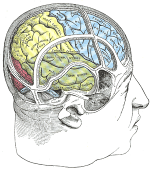
Hemispatial neglect is most frequently associated with a lesion of the right parietal lobe (in yellow, at top).
SpecialtyPsychiatry, Neurology

Hemispatial neglect is a neuropsychological condition in which, after damage to one hemisphere of the brain is sustained, a deficit in attention to and awareness of one side of the field of vision is observed. It is defined by the inability of a person to process and perceive stimuli on one side of the body or environment, where that inability is not due to a lack of sensation. Hemispatial neglect is very commonly contralateral to the damaged hemisphere, but instances of ipsilesional neglect (on the same side as the lesion) have been reported.

Presentation

Hemispatial neglect results most commonly from strokes and brain unilateral injury to the right cerebral hemisphere, with rates in the critical stage of up to 80% causing visual neglect of the left-hand side of space. Neglect is often produced by massive strokes in the middle cerebral artery region and is variegated, so that most sufferers do not exhibit all of the syndrome's traits. Right-sided spatial neglect is rare because there is redundant processing of the right space by both the left and right cerebral hemispheres, whereas in most left-dominant brains the left space is only processed by the right cerebral hemisphere. Although it most strikingly affects visual perception ('visual neglect'), neglect in other forms of perception can also be found, either alone or in combination with visual neglect.

For example, a stroke affecting the right parietal lobe of the brain can lead to neglect for the left side of the visual field, causing a patient with neglect to behave as if the left side of sensory space is nonexistent (although they can still turn left). In an extreme case, a patient with neglect might fail to eat the food on the left half of their plate, even though they complain of being hungry. If someone with neglect is asked to draw a clock, their drawing might show only the numbers 12 to 6, or all 12 numbers might be on one half of the clock face with the other half distorted or blank. Neglect patients may also ignore the contralesional side of their body; for instance, they might only shave, or apply make-up to, the non-neglected side. These patients may frequently collide with objects or structures such as door frames on the side being neglected.

Neglect may also present as a delusional form, where the patient denies ownership of a limb or an entire side of the body. Since this delusion often occurs alone, without the accompaniment of other delusions, it is often labeled as a monothematic delusion.

Neglect not only affects present sensation but memory and recall perception as well. A patient suffering from neglect may also, when asked to recall a memory of a certain object and then draw said object, draw only half of the object. It is unclear, however, if this is due to a perceptive deficit of the memory (to the patient having lost pieces of spatial information of the memory) or whether the information within the memory is whole and intact but simply being ignored, the same way portions of a physical object in the patient's presence would be ignored.

Some forms of neglect may also be very mild—for example, in a condition called extinction where competition from the ipsilesional stimulus impedes perception of the contralesional stimulus. These patients, when asked to fixate on the examiner's nose, can detect fingers being wiggled on the affected side. If the examiner were to wiggle his or her fingers on both the affected and unaffected sides of the patient, the patient will report seeing movement only on the ipsilesional side.

Effects

Though it is frequently underappreciated, unilateral neglect can have dramatic consequences. It has more negative effect on functional ability, as measured by the Barthel ADL index, than age, sex, power, side of stroke, balance, proprioception, cognition, and premorbid ADL status. Its presence within the first 10 days of a stroke is a stronger predictor of poor functional recovery after one year than several other variables, including hemiparesis, hemianopsia, age, visual memory, verbal memory, and visuoconstructional ability. Neglect is probably among the reasons patients with right hemisphere damage are twice as likely to fall as those with left-side brain damage. Patients with neglect take longer to rehabilitate and make less daily progress than other patients with similar functional status. Patients with neglect are also less likely to live independently than patients who have both severe aphasia and right hemiparesis.

Causes

Brain areas in the parietal and frontal lobes are associated with the deployment of attention (internally, or through eye movements, head turns or limb reaches) into contralateral space. Neglect is most closely related to damage to the temporo-parietal junction and posterior parietal cortex. The lack of attention to the left side of space can manifest in the visual, auditory, proprioceptive, and olfactory domains. Although hemispatial neglect often manifests as a sensory deficit (and is frequently co-morbid with sensory deficit), it is essentially a failure to pay sufficient attention to sensory input.

Although hemispatial neglect has been identified following left hemisphere damage (resulting in the neglect of the right side of space), it is most common after damage to the right hemisphere.  This disparity is thought to reflect the fact that the right hemisphere of the brain is specialized for spatial perception and memory, whereas the left hemisphere is specialized for language - there is redundant processing of the right visual fields by both hemispheres. Hence the right hemisphere is able to compensate for the loss of left hemisphere function, but not vice versa. Neglect is not to be confused with hemianopsia. Hemianopsia arises from damage to the primary visual pathways cutting off the input to the cerebral hemispheres from the retinas. Neglect is damage to the processing areas. The cerebral hemispheres receive the input, but there is an error in the processing that is not well understood.

Theories of mechanism

Researchers have argued whether neglect is a disorder of spatial attention or spatial representation.

Spatial attention

Spatial attention is the process where objects in one location are chosen for processing over objects in another location. This would imply that neglect is more intentional. The patient has an affinity to direct attention to the unaffected side. Neglect is caused by a decrease in stimuli in the contralesional side because of a lack of ipsilesional stimulation of the visual cortex and an increased inhibition of the contralesional side.

In this theory, neglect is seen as disorder of attention and orientation caused by disruption of the visual cortex. Patients with this disorder will direct attention and movements to the ipsilesional side and neglect stimuli in the contralesional side despite having preserved visual fields. The result of all of this is an increased sensitivity of visual performance in the unaffected side. The patient shows an affinity to the ipsilesional side being unable to disengage attention from that side.

Spatial representation

Spatial representation is the way space is represented in the brain. In this theory, it is believed that the underlying cause of neglect is the inability to form contralateral representations of space. In this theory, neglect patients demonstrate a failure to describe the contralesional side of a familiar scene, from a given point, from memory.

To support this theory, evidence from Bisiach and Luzzatti's study of Piazza del Duomo can be considered. For the study, patients with hemispatial neglect, that were also familiar with the layout of the Piazza del Duomo square, were observed. The patients were asked to imagine themselves at various vantage points in the square, without physically being in the square. They were then asked to describe different landmarks around the square, such as stores. At each separate vantage point, patients consistently only described landmarks on the right side, ignoring the left side of the representation. However, the results of their multiple descriptions at the different vantage points showed that they knew information around the entire square, but could only identify the right side of the represented field at any given vantage point.When asked to switch vantage points so that the scene that was on the contralesional side is now on the ipsilesional side the patient was able to describe with details the scene they had earlier neglected.

The same patterns can be found with comparing actual visual stimuli to imaging in the brain (Rossetti et al., 2010). A neglect patient who was very familiar with the map of France was asked to name French towns on a map of the country, both by a mental image of the map and by a physical image of the map. The image was then rotated 180 degrees, both mentally and physically. With the mental image, the neglect stayed consistent with the image; that is, when the map was in its original orientation, the patient named towns mostly on the East side of France, and when they mentally rotated the map they named towns mostly on the West side of France because the West coast was now on the right side of the represented field. However, with the physical copy of the map, the patient's focus was on the East side of France with either orientation. This leads researchers to believe that neglect for images in memory may be disassociated from the neglect of stimuli in extrapersonal space. In this case patients have no loss of memory making their neglect a disorder of spatial representation which is the ability to reconstruct spatial frames in which the spatial relationship of objects, that may be perceived, imagined or remembered, with respect to the subject and each other are organized to be correctly acted on.

This theory can also be supported by neglect in dreams (Figliozzi et al., 2007). The study was run on a neglect patient by tracking his eye movements while he slept, during the REM cycle. Results showed that the majority of the eye movements were aimed to his right side, indicating that the images represented in his dreams were also affected by hemispatial neglect.

Another example would be a left neglect patient failing to describe left turns while describing a familiar route. This shows that the failure to describe things in the contralesional side can also affect verbal items. These findings show that space representation is more topological than symbolic. Patients show a contralesional loss of space representation with a deviation of spatial reference to the ipsilesional side. In these cases we see a left-right dissimilarity of representation rather than a decline of representational competence.

Diagnosis

In order to assess not only the type but also the severity of neglect, doctors employ a variety of tests, most of which are carried out at the patient's bedside. Perhaps one of the most-used and quickest is the line bisection. In this test, a line a few inches long is drawn on a piece of paper and the patient is then asked to dissect the line at the midpoint. Patients exhibiting, for example, left-sided neglect will exhibit a rightward deviation of the line's true midpoint.

Another widely used test is the line cancellation test. Here, a patient is presented with a piece of paper that has various lines scattered across it and is asked to mark each of the lines. Patients who exhibit left-sided neglect will completely ignore all lines on the left side of the paper.

Visual neglect can also be assessed by having the patient draw a copy of a picture with which they are presented. If the patient is asked to draw a complex picture they may neglect the entire contralesional side of the picture. If asked to draw an individual object, the patient will not draw the contralesional side of that object.

A patient may also be asked to read a page out of a book. The patient will be unable to orient their eyes to the left margin and will begin reading the page from the center. Presenting a single word to a patient will result in the patient either reading only the ipsilesional part of the word or replacing the part they cannot see with a logical substitute. For example, if they are presented with the word "peanut", they may read "nut" or say "walnut".

Varieties

Neglect is a heterogenous disorder that manifests itself radically differently in different patients. No single mechanism can account for these different manifestations. A vast array of impaired mechanisms are found in neglect. These mechanisms alone would not cause neglect. The complexity of attention alone—just one of several mechanisms that may interact—has generated multiple competing hypothetical explanations of neglect. So it is not surprising that it has proven difficult to assign particular presentations of neglect to specific neuroanatomical loci. Despite such limitations, we may loosely describe unilateral neglect with four overlapping variables: type, range, axis, and orientation.

Type

Types of hemispatial neglect are broadly divided into disorders of input and disorders of output. The neglect of input, or "inattention", includes ignoring contralesional sights, sounds, smells, or tactile stimuli. Surprisingly, this inattention can even apply to imagined stimuli. In what's termed "representational neglect", patients may ignore the left side of memories, dreams, and hallucinations.

Output neglect includes motor and pre-motor deficits. A patient with motor neglect does not use a contralesional limb despite the neuromuscular ability to do so. One with pre-motor neglect, or directional hypokinesia, can move unaffected limbs ably in ipsilateral space but have difficulty directing them into contralesional space. Thus a patient with pre-motor neglect may struggle to grasp an object on the left side even when using the unaffected right arm.

Range

Hemispatial neglect can have a wide range in terms of what the patient neglects. The first range of neglect, commonly referred to as "egocentric" neglect, is found in patients who neglect their own body or personal space. These patients tend to neglect the opposite side of their lesion, based on the midline of the body, head, or retina. For example, in a gap detection test, subjects with egocentric hemispatial neglect on the right side often make errors on the far right side of the page, as they are neglecting the space in their right visual field.

The next range of neglect is "allocentric" neglect, where individuals neglect either their peri-personal or extrapersonal space. Peri-personal space refers to the space within the patient's normal reach, whereas extrapersonal space refers to the objects/environment beyond the body's current contact or reaching ability. Patients with allocentric neglect tend to neglect the contralesional side of individual items, regardless of where they appear with respect to the viewer. For example, In the same gap detection test mentioned above, subjects with allocentric hemispatial neglect on the right side will make errors on all areas of the page, specifically neglecting the right side of each individual item.

This differentiation is significant because the majority of assessment measures test only for neglect within the reaching, or peri-personal, range. But a patient who passes a standard paper-and-pencil test of neglect may nonetheless ignore a left arm or not notice distant objects on the left side of the room.

In cases of somatoparaphrenia, which may be caused by personal neglect, patients deny ownership of contralesional limbs. Sacks (1985) described a patient who fell out of bed after pushing out what he perceived to be the severed leg of a cadaver that the staff had hidden under his blanket. Patients may say things like, "I don't know whose hand that is, but they'd better get my ring off!" or, "This is a fake arm someone put on me. I sent my daughter to find my real one."

Axis

Most tests for neglect look for rightward or leftward errors. But patients may also neglect stimuli on one side of a horizontal or radial axis. For example, when asked to circle all the stars on a printed page, they may locate targets on both the left and right sides of the page while ignoring those across the top or bottom.

In a recent study, researchers asked patients with left neglect to project their midline with a neon bulb and found that they tended to point it straight ahead but position it rightward of their true midline. This shift may account for the success of therapeutic prism glasses, which shift left visual space toward the right. By shifting visual input, they seem to correct the mind's sense of midline. The result is not only the amelioration of visual neglect, but also of tactile, motor, and even representational neglect.

Orientation

An important question in studies of neglect has been: "left of what?" That is to say, what frame of reference does a subject adopt when neglecting the left half of his or her visual, auditory, or tactile field? The answer has proven complex. It turns out that subjects may neglect objects to the left of their own midline (egocentric neglect) or may instead see all the objects in a room but neglect the left half of each individual object (allocentric neglect).

These two broad categories may be further subdivided. Patients with egocentric neglect may ignore the stimuli leftward of their trunks, their heads, or their retinae. Those with allocentric neglect may neglect the true left of a presented object, or may first correct in their mind's eye a slanted or inverted object and then neglect the side then interpreted as being on the left. So, for example, if patients are presented with an upside-down photograph of a face, they may mentally flip the object right side up and then neglect the left side of the adjusted image. In another example, if patients are presented with a barbell, patients will more significantly neglect the left side of the barbell, as expected with right temporal lobe lesion. If the barbell is rotated such that the left side is now on the right side, patients will more significantly neglect the left side of the object, even though it is now on the right side of space. This also occurs with slanted or mirror-image presentations. A patient looking at a mirror image of a map of the World may neglect to see the Western Hemisphere despite their inverted placement onto the right side of the map.

Various neuropsychological research studies have considered the role of frame of reference in hemispatial neglect, offering new evidence to support both allocentric and egocentric neglect. To begin, one study conducted by Dongyun Li, Hans-Otto Karnath, and Christopher Rorden examined whether allocentric neglect varies with egocentric position. This experimental design consisted of testing eleven right hemispheric stroke patients. Five of these patients showed spatial neglect on their contralesional side, while the remaining six patients showed no spatial neglect. During the study, the patients were presented with two arrays of seven triangles. The first array ran from southwest to northeast (SW-NE) and the second array ran from southeast to northwest (SE-NW). In a portion of the experimental trials, the middle triangle in the array contained a gap along one side. Participants were tested on their ability to perceive the presence of this gap, and were instructed to press one response button if the gap was present and a second response button if the gap was absent.

To test the neglect frame of reference, the two different arrays were carefully situated so that gap in the triangle fell on opposite sides of the allocentric field. In the SW-NE array, the gap in the triangle fell on the allocentric right of the object-centered axis along which the triangle pointed. In the SE-NW configuration, the gap in the triangle fell on the allocentric left of the object-centered axis. Furthermore, varying the position of the arrays with respect to the participant's trunk midline was used to test egocentric neglect. The arrays were therefore presented at 0° (i.e. in line with the participant's trunk midline), at −40° left, and at +40° right. Ultimately, varying the position of the array within the testing visual field allowed for the simultaneous measurement of egocentric neglect and allocentric neglect. The results of this experimental design showed that the spatial neglect patients performed more poorly for the allocentric left side of the triangle, as well as for objects presented on the egocentric left side of the body. Furthermore, the poor accuracy for detecting features of the object on the left side of the object's axis was more severe when the objects were presented on the contralesional side of the body. Thus, these findings illustrate that both allocentric and egocentric biases are present simultaneously, and that egocentric information can influence the severity of allocentric neglect.

A second study, conducted by Moscovitch and Behrmann, investigated the reference frame of neglect with respect to the somatosensory system. Eleven patients with parietal lobe lesions and subsequent hemispatial neglect were analyzed during this experiment. A double simultaneous stimulation procedure was utilized, during which the patients were touched lightly and simultaneously on the left and right side of the wrist of one hand. The patients were tested both with their palms facing down and with their palms facing up. This experimental condition allowed the scientists to determine whether neglect in the somatosensory system occurs with respect to the sensory receptor surface (egocentric) or with respect to a higher-order spatial frame of reference (allocentric). The results of this experiment showed the hemispatial neglect patients neglected somatosensory stimuli on the contralesional side of space, regardless of hand orientation. These findings suggest that, within the somatosensory system, stimuli are neglected with respect to the allocentric, spatial frame of reference, in addition to an egocentric, sensory frame of reference. Ultimately, the discoveries made by these experiments indicate that hemispatial neglect occurs with respect to multiple, simultaneously derived frames of reference, which dictate the nature and extent of neglect within the visual, auditory, and tactile fields.

Treatment

Treatment consists of finding ways to bring the patient's attention toward the left, usually done incrementally, by going just a few degrees past midline, and progressing from there. Rehabilitation of neglect is often carried out by neuropsychologists, occupational therapist, speech-language pathologists, neurologic music therapists, physical therapists, optometrists, and orthoptists.

Forms of treatment that have been tested with variable reports of success include prismatic adaptation, where a prism lens is worn to pull the vision of the patient towards the left, constrained movement therapy where the "good" limb is constrained in a sling to encourage use of the contralesional limb. Eye-patching has similarly been used, placing a patch over the "good" eye. Pharmaceutical treatments have mostly focused on dopaminergic therapies such as bromocriptine, levodopa, and amphetamines, though these tests have had mixed results, helping in some cases and accentuating hemispatial neglect in others. Caloric vestibular stimulation (CVS) has been shown to bring about a brief remission in some cases. however this technique has been known to elicit unpleasant side-effects such as nystagmus, vertigo and vomiting. A study done by Schindler and colleagues examined the use of neck muscle vibration on the contralesional posterior neck muscles to induce diversion of gaze from the subjective straight ahead. Subjects received 15 consecutive treatment sessions and were evaluated on different aspects of the neglect disorder including perception of midline, and scanning deficits. The study found that there is evidence that neck muscle stimulation may work, especially if combined with visual scanning techniques. The improvement was evident 2 months after the completion of treatment.

Other areas of emerging treatment options include the use of prisms, visual scanning training, mental imagery training, video feedback training, trunk rotation, galvanic vestibular stimulation (GVS), transcranial magnetic stimulation (TMS) and transcranial direct-current stimulation (tDCS). Of these emerging treatment options, the most studied intervention is prism adaptation and there is evidence of relatively long-term functional gains from comparatively short-term usage. However, all of these treatment interventions (particularly the stimulation techniques) are relatively new and randomised, controlled trial evidence is still limited. Further research is mandatory in this field of research in order to provide more support in evidence-based practice.

In a review article by Pierce & Buxbaum (2002), they concluded that the evidence for Hemispheric Activation Approaches, which focuses on moving the limb on the side of the neglect, has conflicting evidence in the literature. The authors note that a possible limitation in this approach is the requirement for the patients to actively move the neglected limb, which may not be possible for many patients. Constraint-Induced Therapy (CIT), appears to be an effective, long-term treatment for improving neglect in various studies. However, the use of CIT is limited to patients who have active control of wrist and hand extension. Prism Glasses, Hemispatial Glasses, and Eye-Patching have all appear to be effective in improving performance on neglect tests. Caloric Stimulation treatment appears to be effective in improving neglect; however, the effects are generally short-term. The review also suggests that Optokinetic Stimulation is effective in improving position sense, motor skills, body orientation, and perceptual neglect on a short-term basis. As with Caloric Stimulation treatment, long-term studies will be necessary to show its effectiveness. A few Trunk Rotation Therapy studies suggest its effectiveness in improving performance on neglect tests as well as the Functional Independence Measure (FIM). Some less studied treatment possibilities include treatments that target Dorsal Stream of visual processing, Mental Imagery Training, and Neck Vibration Therapy. Trunk rotation therapies aimed at improving postural disorders and balance deficits in patients with unilateral neglect, have demonstrated optimistic results in regaining voluntary trunk control when using specific postural rehabilitative devices. One such device is the Bon Saint Côme apparatus, which uses spatial exploratory tasks in combination with auditory and visual feedback mechanisms to develop trunk control. The Bon Saint Côme device has been shown to be effective with hemiplegic subjects due to the combination of trunk stability exercises, along with the cognitive requirements needed to perform the postural tasks.

Butane

From Wikipedia, the free encyclopedia ...