Search This Blog

Saturday, April 3, 2021

Prefrontal cortex

From Wikipedia, the free encyclopedia
 
Prefrontal cortex
Gray726-Brodman-prefrontal.svg
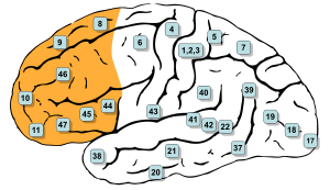
Brodmann areas, 8, 9, 10, 11, 12, 13, 14, 24, 25, 32, 44, 45, 46, and 47 are all in the prefrontal cortex
Details
Part ofFrontal lobe
PartsSuperior frontal gyrus
Middle frontal gyrus
Inferior frontal gyrus
ArteryAnterior cerebral
Middle cerebral
VeinSuperior sagittal sinus
Identifiers
LatinCortex praefrontalis
MeSHD017397
NeuroNames2429
NeuroLex IDnlx_anat_090801
FMA224850

In mammalian brain anatomy, the prefrontal cortex (PFC) is the cerebral cortex which covers the front part of the frontal lobe. The PFC contains the Brodmann areas BA8, BA9, BA10, BA11, BA12, BA13, BA14, BA24, BA25, BA32, BA44, BA45, BA46, and BA47.

Many authors have indicated an integral link between a person's will to live, personality, and the functions of the prefrontal cortex. This brain region has been implicated in executive functions, such as planning, decision making, short-term memory, personality expression, moderating social behavior and controlling certain aspects of speech and language. The basic activity of this brain region is considered to be orchestration of thoughts and actions in accordance with internal goals.

Executive function relates to abilities to differentiate among conflicting thoughts, determine good and bad, better and best, same and different, future consequences of current activities, working toward a defined goal, prediction of outcomes, expectation based on actions, and social "control" (the ability to suppress urges that, if not suppressed, could lead to socially unacceptable outcomes).

The frontal cortex supports concrete rule learning. More anterior regions along the rostro-caudal axis of frontal cortex support rule learning at higher levels of abstraction.

Structure

Definition

There are three possible ways to define the prefrontal cortex:

  • as the granular frontal cortex
  • as the projection zone of the medial dorsal nucleus of the thalamus
  • as that part of the frontal cortex whose electrical stimulation does not evoke movements

Granular frontal cortex

The prefrontal cortex has been defined based on cytoarchitectonics by the presence of a cortical granular layer IV. It is not entirely clear who first used this criterion. Many of the early cytoarchitectonic researchers restricted the use of the term prefrontal to a much smaller region of cortex including the gyrus rectus and the gyrus rostralis (Campbell, 1905; G. E. Smith, 1907; Brodmann, 1909; von Economo and Koskinas, 1925). In 1935, however, Jacobsen used the term prefrontal to distinguish granular prefrontal areas from agranular motor and premotor areas. In terms of Brodmann areas, the prefrontal cortex traditionally includes areas 8, 9, 10, 11, 12, 13, 14, 24, 25, 32, 44, 45, 46, and 47, however, not all of these areas are strictly granular – 44 is dysgranular, caudal 11 and orbital 47 are agranular. The main problem with this definition is that it works well only in primates but not in nonprimates, as the latter lack a granular layer IV.

Projection zone

To define the prefrontal cortex as the projection zone of the mediodorsal nucleus of the thalamus builds on the work of Rose and Woolsey, who showed that this nucleus projects to anterior and ventral parts of the brain in nonprimates, however, Rose and Woolsey termed this projection zone "orbitofrontal." It seems to have been Akert, who, for the first time in 1964, explicitly suggested that this criterion could be used to define homologues of the prefrontal cortex in primates and nonprimates. This allowed the establishment of homologies despite the lack of a granular frontal cortex in nonprimates.

The projection zone definition is still widely accepted today (e.g. Fuster), although its usefulness has been questioned. Modern tract tracing studies have shown that projections of the mediodorsal nucleus of the thalamus are not restricted to the granular frontal cortex in primates. As a result, it was suggested to define the prefrontal cortex as the region of cortex that has stronger reciprocal connections with the mediodorsal nucleus than with any other thalamic nucleus. Uylings et al. acknowledge, however, that even with the application of this criterion, it might be rather difficult to define the prefrontal cortex unequivocally.

Electrically silent area of frontal cortex

A third definition of the prefrontal cortex is the area of frontal cortex whose electrical stimulation does not lead to observable movements. For example, in 1890 David Ferrier used the term in this sense. One complication with this definition is that the electrically "silent" frontal cortex includes both granular and non-granular areas.

Subdivisions

Brodmann areas.jpg

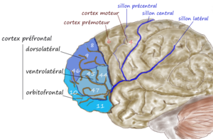
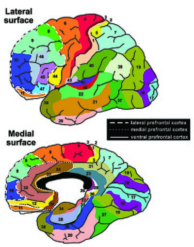
According to Striedter the PFC of humans can be delineated into two functionally, morphologically, and evolutionarily different regions: the ventromedial PFC (vmPFC) consisting of the ventral prefrontal cortex and the medial prefrontal cortex present in all mammals, and the lateral prefrontal cortex (LPFC), consisting of the dorsolateral prefrontal cortex and the ventrolateral prefrontal cortex, present only in primates.

The LPFC contains the Brodmann areas BA8, BA9, BA10, BA45, BA46, and BA47. Some researchers also include BA44. The vmPFC contains the Brodmann areas BA12, BA25, BA32, BA33, BA24, BA11, BA13, and BA14.

The table below shows different ways to subdivide parts of the human prefrontal cortex based upon Brodmann areas.

8
9
10
46
45
47
44
12
25
32
33
24
11
13
14
lateral
ventromedial
dorsolateral
ventrolateral
medial
ventral

Interconnections

The prefrontal cortex is highly interconnected with much of the brain, including extensive connections with other cortical, subcortical and brain stem sites. The dorsal prefrontal cortex is especially interconnected with brain regions involved with attention, cognition and action, while the ventral prefrontal cortex interconnects with brain regions involved with emotion. The prefrontal cortex also receives inputs from the brainstem arousal systems, and its function is particularly dependent on its neurochemical environment. Thus, there is coordination between our state of arousal and our mental state. The interplay between the prefrontal cortex and socioemotional system of the brain is relevant for adolescent development, as proposed by the Dual Systems Model.

The medial prefrontal cortex has been implicated in the generation of slow-wave sleep (SWS), and prefrontal atrophy has been linked to decreases in SWS. Prefrontal atrophy occurs naturally as individuals age, and it has been demonstrated that older adults experience impairments in memory consolidation as their medial prefrontal cortices degrade. In monkeys, significant atrophy has been found as a result of neuroleptic or antipsychotic psychiatric medication. In older adults, instead of being transferred and stored in the neocortex during SWS, memories start to remain in the hippocampus where they were encoded, as evidenced by increased hippocampal activation compared to younger adults during recall tasks, when subjects learned word associations, slept, and then were asked to recall the learned words.

The ventrolateral prefrontal cortex (VLPFC) has been implicated in various aspects of speech production and language comprehension. The VLPFC is richly connected to various regions of the brain including the lateral and medial temporal lobe, the superior temporal cortex, the infertemporal cortex, the perirhinal cortex, and the parahippoccampal cortex.These brain areas are implicated in memory retrieval and consolidation, language processing, and association of emotions. These connections allow the VLPFC to mediate explicit and implicit memory retrieval and integrate it with language stimulus to help plan coherent speech. In other words, choosing the correct words and staying “on topic” during conversation come from the VLPFC.

Function

Executive function

The original studies of Fuster and of Goldman-Rakic emphasized the fundamental ability of the prefrontal cortex to represent information not currently in the environment, and the central role of this function in creating the "mental sketch pad". Goldman-Rakic spoke of how this representational knowledge was used to intelligently guide thought, action, and emotion, including the inhibition of inappropriate thoughts, distractions, actions, and feelings. In this way, working memory can be seen as fundamental to attention and behavioral inhibition. Fuster speaks of how this prefrontal ability allows the wedding of past to future, allowing both cross-temporal and cross-modal associations in the creation of goal-directed, perception-action cycles. This ability to represent underlies all other higher executive functions.

Shimamura proposed Dynamic Filtering Theory to describe the role of the prefrontal cortex in executive functions. The prefrontal cortex is presumed to act as a high-level gating or filtering mechanism that enhances goal-directed activations and inhibits irrelevant activations. This filtering mechanism enables executive control at various levels of processing, including selecting, maintaining, updating, and rerouting activations. It has also been used to explain emotional regulation.

Miller and Cohen proposed an Integrative Theory of Prefrontal Cortex Function, that arises from the original work of Goldman-Rakic and Fuster. The two theorize that “cognitive control stems from the active maintenance of patterns of activity in the prefrontal cortex that represents goals and means to achieve them. They provide bias signals to other brain structures whose net effect is to guide the flow of activity along neural pathways that establish the proper mappings between inputs, internal states, and outputs needed to perform a given task”. In essence, the two theorize that the prefrontal cortex guides the inputs and connections, which allows for cognitive control of our actions.

The prefrontal cortex is of significant importance when top-down processing is needed. Top-down processing by definition is when behavior is guided by internal states or intentions. According to the two, “The PFC is critical in situations when the mappings between sensory inputs, thoughts, and actions either are weakly established relative to other existing ones or are rapidly changing”. An example of this can be portrayed in the Wisconsin Card Sorting Test (WCST). Subjects engaging in this task are instructed to sort cards according to the shape, color, or number of symbols appearing on them. The thought is that any given card can be associated with a number of actions and no single stimulus-response mapping will work. Human subjects with PFC damage are able to sort the card in the initial simple tasks, but unable to do so as the rules of classification change.

Miller and Cohen conclude that the implications of their theory can explain how much of a role the PFC has in guiding control of cognitive actions. In the researchers' own words, they claim that, “depending on their target of influence, representations in the PFC can function variously as attentional templates, rules, or goals by providing top-down bias signals to other parts of the brain that guide the flow of activity along the pathways needed to perform a task”.

Experimental data indicate a role for the prefrontal cortex in mediating normal sleep physiology, dreaming and sleep-deprivation phenomena.

When analyzing and thinking about attributes of other individuals, the medial prefrontal cortex is activated, however, it is not activated when contemplating the characteristics of inanimate objects.

Studies using fMRI have shown that the medial prefrontal cortex (mPFC), specifically the anterior medial prefrontal cortex (amPFC), may modulate mimicry behavior. Neuroscientists are suggesting that social priming influences activity and processing in the amPFC, and that this area of the prefrontal cortex modulates mimicry responses and behavior.

As of recent, researchers have used neuroimaging techniques to find that along with the basal ganglia, the prefrontal cortex is involved with learning exemplars, which is part of the exemplar theory, one of the three main ways our mind categorizes things. The exemplar theory states that we categorize judgements by comparing it to a similar past experience within our stored memories.

A 2014 meta-analysis by Professor Nicole P.Yuan from the University of Arizona found that larger prefrontal cortex volume and greater PFC cortical thickness were associated with better executive performance.

Attention and memory

Lebedev et al. experiment that dissociated representation of spatial attention from representation of spatial memory in prefrontal cortex 

A widely accepted theory regarding the function of the brain's prefrontal cortex is that it serves as a store of short-term memory. This idea was first formulated by Jacobsen, who reported in 1936 that damage to the primate prefrontal cortex caused short-term memory deficits. Karl Pribram and colleagues (1952) identified the part of the prefrontal cortex responsible for this deficit as area 46, also known as the dorsolateral prefrontal cortex (dlPFC). More recently, Goldman-Rakic and colleagues (1993) evoked short-term memory loss in localized regions of space by temporary inactivation of portions of the dlPFC. Once the concept of working memory was established in contemporary neuroscience by Alan Baddeley (1986), these neuropsychological findings contributed to the theory that the prefrontal cortex implements working memory and, in some extreme formulations, only working memory. In the 1990s this theory developed a wide following, and it became the predominant theory of PF function, especially for nonhuman primates. The concept of working memory used by proponents of this theory focused mostly on the short-term maintenance of information, and rather less on the manipulation or monitoring of such information or on the use of that information for decisions. Consistent with the idea that the prefrontal cortex functions predominantly in maintenance memory, delay-period activity in the PF has often been interpreted as a memory trace. (The phrase "delay-period activity" applies to neuronal activity that follows the transient presentation of an instruction cue and persists until a subsequent "go" or "trigger" signal.)

To explore alternative interpretations of delay-period activity in the prefrontal cortex, Lebedev et al. (2004) investigated the discharge rates of single prefrontal neurons as monkeys attended to a stimulus marking one location while remembering a different, unmarked location. Both locations served as potential targets of a saccadic eye movement. Although the task made intensive demands on short-term memory, the largest proportion of prefrontal neurons represented attended locations, not remembered ones. These findings showed that short-term memory functions cannot account for all, or even most, delay-period activity in the part of the prefrontal cortex explored. The authors suggested that prefrontal activity during the delay-period contributes more to the process of attentional selection (and selective attention) than to memory storage.

Speech production and language

Various areas of the prefrontal cortex have been implicated in a multitude of critical functions regarding speech production, language comprehension, and response planning before speaking. Cognitive neuroscience has shown that the left ventrolateral prefrontal cortex is vital in the processing of words and sentences.

The right prefrontal cortex has been found to be responsible for coordinating the retrieval of explicit memory for use in speech, whereas the deactivation of the left is responsible for mediating implicit memory retrieval to be used in verb generation. Impaired recollection of nouns (explicit memory) is impaired in some amnesic patients with damaged left prefrontal cortices, but verb generation remains intact because of its reliance on left prefrontal deactivation.

Many researchers now include BA45 in the prefrontal cortex because together with BA44 make up an area of the frontal lobe called Broca's Area. Broca's Area is the widely considered the output area of the language production pathway in the brain (as opposed to Wernike's area in the medial temporal lobe, which is seen as the language input area). BA45 has been shown to be implicated for the retrieval of relevant semantic knowledge to be used in conversation/speech. The right lateral prefrontal cortex (RLPFC) is implicated in the planning of complex behavior, and together with bilateral BA45, they act to maintain focus and coherence during speech production.  However, left BA45 has been shown to be activated significantly while maintaining speech coherence in young people. Older people have been shown to recruit the right BA45 more so than their younger counterparts.  This aligns with the evidence of decreased lateralization in other brain systems during aging.

In addition, this increase in BA45 and RLPFC activity in combination of BA47 in older patients has been shown to contribute to “off-topic utterances.” The BA47 area in the prefrontal cortex is implicated in “stimulus-driven” retrieval of less-salient knowledge than is required to contribute to a conversation. In other words, elevated activation of the BA47 together with altered activity in BA45 and the broader RLPFC has been shown to contribute to the inclusion of less relevant information and irrelevant tangential conversational speech patterns in older subjects.

Clinical significance

In the last few decades, brain imaging systems have been used to determine brain region volumes and nerve linkages. Several studies have indicated that reduced volume and interconnections of the frontal lobes with other brain regions is observed in patients diagnosed with mental disorders and prescribed potent antipsychotics; those subjected to repeated stressors; those who excessively consume sexually explicit materials; suicides; those incarcerated; criminals; sociopaths; those affected by lead poisoning; and daily male cannabis users (only 13 people were tested). It is believed that at least some of the human abilities to feel guilt or remorse, and to interpret reality, are dependent on a well-functioning prefrontal cortex. It is also widely believed that the size and number of connections in the prefrontal cortex relates directly to sentience, as the prefrontal cortex in humans occupies a far larger percentage of the brain than in any other animal. And it is theorized that, as the brain has tripled in size over five million years of human evolution, the prefrontal cortex has increased in size sixfold.

A review on executive functions in healthy exercising individuals noted that the left and right halves of the prefrontal cortex, which is divided by the medial longitudinal fissure, appears to become more interconnected in response to consistent aerobic exercise. Two reviews of structural neuroimaging research indicate that marked improvements in prefrontal and hippocampal gray matter volume occur in healthy adults that engage in medium intensity exercise for several months.

A functional neuroimaging review of meditation-based practices suggested that practicing mindfulness enhances prefrontal activation, which was noted to be correlated with increased well-being and reduced anxiety; however, the review noted the need for cohort studies in future research to better establish this.

Treatments with anti-cancer drugs often are toxic to the cells of the brain, leading to memory loss and cognitive dysfunction that can persist long after the period of exposure. Such a condition is referred to as chemo brain. To determine the basis of this condition, mice were treated with the chemotherapeutic agent mitomycin C. In the prefrontal cortex, this treatment resulted in an increase of the oxidative DNA damage 8-oxodG, a decrease in the enzyme OGG1 that ordinarily repairs such damage, and epigenetic alterations.

Chronic intake of alcohol leads to persistent alterations in brain function including altered decision making ability. The prefrontal cortex of chronic alcoholics has been shown to be vulnerable to oxidative DNA damage and neuronal cell death.

History

Perhaps the seminal case in prefrontal cortex function is that of Phineas Gage, whose left frontal lobe was destroyed when a large iron rod was driven through his head in an 1848 accident. The standard presentation (e.g.) is that, although Gage retained normal memory, speech and motor skills, his personality changed radically: He became irritable, quick-tempered, and impatient—characteristics he did not previously display — so that friends described him as "no longer Gage"; and, whereas he had previously been a capable and efficient worker, afterward he was unable to complete tasks. However, careful analysis of primary evidence shows that descriptions of Gage's psychological changes are usually exaggerated when held against the description given by Gage's doctor, the most striking feature being that changes described years after Gage's death are far more dramatic than anything reported while he was alive.

Subsequent studies on patients with prefrontal injuries have shown that the patients verbalized what the most appropriate social responses would be under certain circumstances. Yet, when actually performing, they instead pursued behavior aimed at immediate gratification, despite knowing the longer-term results would be self-defeating.

The interpretation of this data indicates that not only are skills of comparison and understanding of eventual outcomes harbored in the prefrontal cortex but the prefrontal cortex (when functioning correctly) controls the mental option to delay immediate gratification for a better or more rewarding longer-term gratification result. This ability to wait for a reward is one of the key pieces that define optimal executive function of the human brain.

There is much current research devoted to understanding the role of the prefrontal cortex in neurological disorders. Clinical trials have begun on certain drugs that have been shown to improve prefrontal cortex function, including guanfacine, which acts through the alpha-2A adrenergic receptor. A downstream target of this drug, the HCN channel, is one of the most recent areas of exploration in prefrontal cortex pharmacology.

Etymology

The term "prefrontal" as describing a part of the brain appears to have been introduced by Richard Owen in 1868. For him, the prefrontal area was restricted to the anterior-most part of the frontal lobe (approximately corresponding to the frontal pole). It has been hypothesized that his choice of the term was based on the prefrontal bone present in most amphibians and reptiles.
Limbic system

From Wikipedia, the free encyclopedia
 
Limbic system
Back Cover, STRESS R US.jpg
 
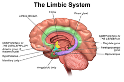
Cross section of the human brain showing parts of the limbic system from below.
Traité d'Anatomie et de Physiologie (1786)
 
1511 The Limbic Lobe.jpg
The limbic system largely consists of what was previously known as the limbic lobe.
 
Details
Identifiers
LatinSystema limbicum
MeSHD008032
NeuroNames2055
FMA242000
Anatomical terms of neuroanatomy

The limbic system, also known as the paleomammalian cortex, is a set of brain structures located on both sides of the thalamus, immediately beneath the medial temporal lobe of the cerebrum primarily in the forebrain.

It supports a variety of functions including emotion, behavior, long-term memory, and olfaction. Emotional life is largely housed in the limbic system, and it critically aids the formation of memories.

With a primordial structure, the limbic system is involved in lower order emotional processing of input from sensory systems and consists of the amygdaloid nuclear complex (amygdala), mammillary bodies, stria medullaris, central gray and dorsal and ventral nuclei of Gudden. This processed information is often relayed to a collection of structures from the telencephalon, diencephalon, and mesencephalon, including the prefrontal cortex, cingulate gyrus, limbic thalamus, hippocampus including the parahippocampal gyrus and subiculum, nucleus accumbens (limbic striatum), anterior hypothalamus, ventral tegmental area, midbrain raphe nuclei, habenular commissure, entorhinal cortex, and olfactory bulbs.

Structure

Anatomical components of the limbic system

The limbic system was originally defined by Paul D. MacLean as a series of cortical structures surrounding the boundary between the cerebral hemispheres and the brainstem. The name "limbic" comes from the Latin word for the border, limbus, and these structures were known together as the limbic lobe. Further studies began to associate these areas with emotional and motivational processes and linked them to subcortical components that were then grouped into the limbic system.

Currently, it is not considered an isolated entity responsible for the neurological regulation of emotion, but rather one of the many parts of the brain that regulate visceral autonomic processes. Therefore, the set of anatomical structures considered part of the limbic system is controversial. The following structures are, or have been considered, part of the limbic system:

Function

The structures and interacting areas of the limbic system are involved in motivation, emotion, learning, and memory. The limbic system is where the subcortical structures meet the cerebral cortex. The limbic system operates by influencing the endocrine system and the autonomic nervous system. It is highly interconnected with the nucleus accumbens, which plays a role in sexual arousal and the "high" derived from certain recreational drugs. These responses are heavily modulated by dopaminergic projections from the limbic system. In 1954, Olds and Milner found that rats with metal electrodes implanted into their nucleus accumbens, as well as their septal nuclei, repeatedly pressed a lever activating this region.

The limbic system also interacts with the basal ganglia. The basal ganglia are a set of subcortical structures that direct intentional movements. The basal ganglia are located near the thalamus and hypothalamus. They receive input from the cerebral cortex, which sends outputs to the motor centers in the brain stem. A part of the basal ganglia called the striatum controls posture and movement. Recent studies indicate that if there is an inadequate supply of dopamine in the striatum, this can lead to the symptoms of Parkinson's disease.

The limbic system is also tightly connected to the prefrontal cortex. Some scientists contend that this connection is related to the pleasure obtained from solving problems. To cure severe emotional disorders, this connection was sometimes surgically severed, a procedure of psychosurgery, called a prefrontal lobotomy (this is actually a misnomer). Patients having undergone this procedure often became passive and lacked all motivation.

The limbic system is often incorrectly classified as a cerebral structure, but simply interacts heavily with the cerebral cortex. These interactions are closely linked to olfaction, emotions, drives, autonomic regulation, memory, and pathologically to encephalopathy, epilepsy, psychotic symptoms, cognitive defects. The functional relevance of the limbic system has proven to serve many different functions such as affects/emotions, memory, sensory processing, time perception, attention, consciousness, instincts, autonomic/vegetative control, and actions/motor behavior. Some of the disorders associated with the limbic system and its interacting components are epilepsy and schizophrenia.

Hippocampus

Location and basic anatomy of the hippocampus, as a coronal section

The hippocampus is involved with various processes relating to cognition and is one of the most well understood and heavily involved limbic interacting structure.

Spatial memory

The first and most widely researched area concerns memory, particularly spatial memory. Spatial memory was found to have many sub-regions in the hippocampus, such as the dentate gyrus (DG) in the dorsal hippocampus, the left hippocampus, and the parahippocampal region. The dorsal hippocampus was found to be an important component for the generation of new neurons, called adult-born granules (GC), in adolescence and adulthood. These new neurons contribute to pattern separation in spatial memory, increasing the firing in cell networks, and overall causing stronger memory formations. This is thought to integrate spatial and episodic memories with the limbic system via a feedback loop that provides emotional context of a particular sensory input.

While the dorsal hippocampus is involved in spatial memory formation, the left hippocampus is a participant in the recall of these spatial memories. Eichenbaum and his team found, when studying the hippocampal lesions in rats, that the left hippocampus is “critical for effectively combining the ‘what, ‘when,’ and ‘where’ qualities of each experience to compose the retrieved memory.” This makes the left hippocampus a key component in the retrieval of spatial memory. However, Spreng found that the left hippocampus is a general concentrated region for binding together bits and pieces of memory composed not only by the hippocampus, but also by other areas of the brain to be recalled at a later time. Eichenbaum’s research in 2007 also demonstrates that the parahippocampal area of the hippocampus is another specialized region for the retrieval of memories just like the left hippocampus.

Learning

The hippocampus, over the decades, has also been found to have a huge impact in learning. Curlik and Shors examined the effects of neurogenesis in the hippocampus and its effects on learning. This researcher and his team employed many different types of mental and physical training on their subjects, and found that the hippocampus is highly responsive to these latter tasks. Thus, they discovered an upsurge of new neurons and neural circuits in the hippocampus as a result of the training, causing an overall improvement in the learning of the task. This neurogenesis contributes to the creation of adult-born granules cells (GC), cells also described by Eichenbaum in his own research on neurogenesis and its contributions to learning. The creation of these cells exhibited "enhanced excitability" in the dentate gyrus (DG) of the dorsal hippocampus, impacting the hippocampus and its contribution to the learning process.

Hippocampus damage

Damage related to the hippocampal region of the brain has reported vast effects on overall cognitive functioning, particularly memory such as spatial memory. As previously mentioned, spatial memory is a cognitive function greatly intertwined with the hippocampus. While damage to the hippocampus may be a result of a brain injury or other injuries of that sort, researchers particularly investigated the effects that high emotional arousal and certain types of drugs had on the recall ability in this specific memory type. In particular, in a study performed by Parkard, rats were given the task of correctly making their way through a maze. In the first condition, rats were stressed by shock or restraint which caused a high emotional arousal. When completing the maze task, these rats had an impaired effect on their hippocampal-dependent memory when compared to the control group. Then, in a second condition, a group of rats were injected with anxiogenic drugs. Like the former these results reported similar outcomes, in that hippocampal-memory was also impaired. Studies such as these reinforce the impact that the hippocampus has on memory processing, in particular the recall function of spatial memory. Furthermore, impairment to the hippocampus can occur from prolonged exposure to stress hormones such as glucocorticoids (GCs), which target the hippocampus and cause disruption in explicit memory.

In an attempt to curtail life-threatening epileptic seizures, 27-year-old Henry Gustav Molaison underwent bilateral removal of almost all of his hippocampus in 1953. Over the course of fifty years he participated in thousands of tests and research projects that provided specific information on exactly what he had lost. Semantic and episodic events faded within minutes, having never reached his long term memory, yet emotions, unconnected from the details of causation, were often retained. Dr. Suzanne Corkin, who worked with him for 46 years until his death, described the contribution of this tragic "experiment" in her 2013 book.

Amygdala

Episodic-autobiographical memory (EAM) networks

Another integrative part of the limbic system, the amygdala, which is the deepest part of the limbic system, is involved in many cognitive processes and is largely considered the most primordial and vital part of the limbic system. Like the hippocampus, processes in the amygdala seem to impact memory; however, it is not spatial memory as in the hippocampus but the semantic division of episodic-autobiographical memory (EAM) networks. Markowitsch's amygdala research shows it encodes, stores, and retrieves EAM memories. To delve deeper into these types of processes by the amygdala, Markowitsch and his team provided extensive evidence through investigations that the "amygdala's main function is to charge cues so that mnemonic events of a specific emotional significance can be successfully searched within the appropriate neural nets and re-activated." These cues for emotional events created by the amygdala encompass the EAM networks previously mentioned.

Attentional and emotional processes

Besides memory, the amygdala also seems to be an important brain region involved in attentional and emotional processes. First, to define attention in cognitive terms, attention is the ability to focus on some stimuli while ignoring others. Thus, the amygdala seems to be an important structure in this ability. Foremost, however, this structure was historically thought to be linked to fear, allowing the individual to take action in response to that fear. However, as time has gone by, researchers such as Pessoa, generalized this concept with help from evidence of EEG recordings, and concluded that the amygdala helps an organism to define a stimulus and therefore respond accordingly. However, when the amygdala was initially thought to be linked to fear, this gave way for research in the amygdala for emotional processes. Kheirbek demonstrated research that the amygdala is involved in emotional processes, in particular the ventral hippocampus. He described the ventral hippocampus as having a role in neurogenesis and the creation of adult-born granule cells (GC). These cells not only were a crucial part of neurogenesis and the strengthening of spatial memory and learning in the hippocampus but also appear to be an essential component to the function of the amygdala. A deficit of these cells, as Pessoa (2009) predicted in his studies, would result in low emotional functioning, leading to high retention rate of mental diseases, such as anxiety disorders.

Social processing

Social processing, specifically the evaluation of faces in social processing, is an area of cognition specific to the amygdala. In a study done by Todorov, fMRI tasks were performed with participants to evaluate whether the amygdala was involved in the general evaluation of faces. After the study, Todorov concluded from his fMRI results that the amygdala did indeed play a key role in the general evaluation of faces. However, in a study performed by researchers Koscik and his team, the trait of trustworthiness was particularly examined in the evaluation of faces. Koscik and his team demonstrated that the amygdala was involved in evaluating the trustworthiness of an individual. They investigated how brain damage to the amygdala played a role in trustworthiness, and found that individuals that suffered damage tended to confuse trust and betrayal, and thus placed trust in those having done them wrong. Furthermore, Rule, along with his colleagues, expanded on the idea of the amygdala in its critique of trustworthiness in others by performing a study in 2009 in which he examined the amygdala's role in evaluating general first impressions and relating them to real-world outcomes. Their study involved first impressions of CEOs. Rule demonstrated that while the amygdala did play a role in the evaluation of trustworthiness, as observed by Koscik in his own research two years later in 2011, the amygdala also played a generalized role in the overall evaluation of first impression of faces. This latter conclusion, along with Todorov's study on the amygdala's role in general evaluations of faces and Koscik's research on trustworthiness and the amygdala, further solidified evidence that the amygdala plays a role in overall social processing.

Klüver–Bucy syndrome

Based on experiments done on monkeys, the destruction of the temporal cortex almost always led to damage of the amygdala. This damage done to the amygdala led the physiologists Kluver and Bucy to pinpoint major changes in the behavior of the monkeys. The monkeys demonstrated the following changes:

  1. Monkeys were not afraid of anything.
  2. The animals (monkeys) had extreme curiosity about everything.
  3. The animal forgets rapidly.
  4. The animal has a tendency to place everything in its mouth.
  5. The animal often has a sexual drive so strong that it attempts to copulate with immature animals, animals of the opposite sex, or even animals of a different species.

This set of behavioral change came to be known as the Klüver–Bucy syndrome.

Evolution

Paul D. MacLean, as part of his triune brain theory, hypothesized that the limbic system is older than other parts of the forebrain, and that it developed to manage circuitry attributed to the fight or flight first identified by Hans Selye in his report of the General Adaptation Syndrome in 1936. It may be considered a part of survival adaptation in reptiles as well as mammals (including humans). MacLean postulated that the human brain has evolved three components, that evolved successively, with more recent components developing at the top/front. These components are, respectively:

  1. The archipallium or primitive ("reptilian") brain, comprising the structures of the brain stem – medulla, pons, cerebellum, mesencephalon, the oldest basal nuclei – the globus pallidus and the olfactory bulbs.
  2. The paleopallium or intermediate ("old mammalian") brain, comprising the structures of the limbic system.
  3. The neopallium, also known as the superior or rational ("new mammalian") brain, comprises almost the whole of the hemispheres (made up of a more recent type of cortex, called neocortex) and some subcortical neuronal groups. It corresponds to the brain of the superior mammals, thus including the primates and, as a consequence, the human species. Similar development of the neocortex in mammalian species unrelated to humans and primates has also occurred, for example in cetaceans and elephants; thus the designation of "superior mammals" is not an evolutionary one, as it has occurred independently in different species. The evolution of higher degrees of intelligence is an example of convergent evolution, and is also seen in non-mammals such as birds.

According to Maclean, each of the components, although connected with the others, retained "their peculiar types of intelligence, subjectivity, sense of time and space, memory, mobility and other less specific functions".

However, while the categorization into structures is reasonable, the recent studies of the limbic system of tetrapods, both living and extinct, have challenged several aspects of this hypothesis, notably the accuracy of the terms "reptilian" and "old mammalian". The common ancestors of reptiles and mammals had a well-developed limbic system in which the basic subdivisions and connections of the amygdalar nuclei were established. Further, birds, which evolved from the dinosaurs, which in turn evolved separately but around the same time as the mammals, have a well-developed limbic system. While the anatomic structures of the limbic system are different in birds and mammals, there are functional equivalents.

History

Etymology and history

The term limbic comes from the Latin limbus, for "border" or "edge", or, particularly in medical terminology, a border of an anatomical component. Paul Broca coined the term based on its physical location in the brain, sandwiched between two functionally different components.

The limbic system is a term that was introduced in 1949 by the American physician and neuroscientist, Paul D. MacLean. The French physician Paul Broca first called this part of the brain le grand lobe limbique in 1878. He examined the differentiation between deeply recessed cortical tissue and underlying, subcortical nuclei. However, most of its putative role in emotion was developed only in 1937 when the American physician James Papez described his anatomical model of emotion, the Papez circuit.

The first evidence that the limbic system was responsible for the cortical representation of emotions was discovered in 1939, by Heinrich Kluver and Paul Bucy. Kluver and Bucy, after much research, demonstrated that the bilateral removal of the temporal lobes in monkeys created an extreme behavioral syndrome. After performing a temporal lobectomy, the monkeys showed a decrease in aggression. The animals revealed a reduced threshold to visual stimuli, and were thus unable to recognize objects that were once familiar. MacLean expanded these ideas to include additional structures in a more dispersed "limbic system", more on the lines of the system described above. MacLean developed the intriguing theory of the "triune brain" to explain its evolution and to try to reconcile rational human behavior with its more primal and violent side. He became interested in the brain's control of emotion and behavior. After initial studies of brain activity in epileptic patients, he turned to cats, monkeys, and other models, using electrodes to stimulate different parts of the brain in conscious animals recording their responses.

In the 1950s, he began to trace individual behaviors like aggression and sexual arousal to their physiological sources. He analyzed the brain's center of emotions, the limbic system, and described an area that includes structures called the hippocampus and amygdala. Developing observations made by Papez, he determined that the limbic system had evolved in early mammals to control fight-or-flight responses and react to both emotionally pleasurable and painful sensations. The concept is now broadly accepted in neuroscience. Additionally, MacLean said that the idea of the limbic system leads to a recognition that its presence "represents the history of the evolution of mammals and their distinctive family way of life."

In the 1960s, Dr. MacLean enlarged his theory to address the human brain's overall structure and divided its evolution into three parts, an idea that he termed the triune brain. In addition to identifying the limbic system, he pointed to a more primitive brain called the R-complex, related to reptiles, which controls basic functions like muscle movement and breathing. The third part, the neocortex, controls speech and reasoning and is the most recent evolutionary arrival. The concept of the limbic system has since been further expanded and developed by Walle Nauta, Lennart Heimer, and others.

Academic dispute

There is controversy over the use of the term limbic system, with scientists such as LeDoux arguing that the term be considered obsolete and abandoned. Originally, the limbic system was believed to be the emotional center of the brain, with cognition being the business of the neocortex. However, cognition depends on acquisition and retention of memories, in which the hippocampus, a primary limbic interacting structure, is involved: hippocampus damage causes severe cognitive (memory) deficits. More important, the "boundaries" of the limbic system have been repeatedly redefined because of advances in neuroscience. Therefore, while it is true that limbic interacting structures are more closely related to emotion, the limbic system itself is best thought of as a component of a larger emotional processing plant. It is essentially responsible for sifting through and organizing lower order processing, and relaying sensory information to other brain areas for higher order emotional processing.

Dorsolateral prefrontal cortex

From Wikipedia, the free encyclopedia
 
Dorsolateral prefrontal cortex
Prefrontal1.png
An illustration of brain's prefrontal region
Details
Identifiers
LatinCortex praefrontalis dorsolateralis
FMA276189
Anatomical terms of neuroanatomy

The dorsolateral prefrontal cortex (DLPFC or DL-PFC) is an area in the prefrontal cortex of the brain of humans and other primates. It is one of the most recently derived parts of the human brain. It undergoes a prolonged period of maturation which lasts until adulthood. The DLPFC is not an anatomical structure, but rather a functional one. It lies in the middle frontal gyrus of humans (i.e., lateral part of Brodmann's area (BA) 9 and 46). In macaque monkeys, it is around the principal sulcus (i.e., in Brodmann's area 46). Other sources consider that DLPFC is attributed anatomically to BA 9 and 46 and BA 8, 9 and 10.

The DLPFC has connections with the orbitofrontal cortex, as well as the thalamus, parts of the basal ganglia (specifically, the dorsal caudate nucleus), the hippocampus, and primary and secondary association areas of neocortex (including posterior temporal, parietal, and occipital areas). The DLPFC is also the end point for the dorsal pathway (stream), which is concerned with how to interact with stimuli.

An important function of the DLPFC is the executive functions, such as working memory, cognitive flexibility, planning, inhibition, and abstract reasoning. However, the DLPFC is not exclusively responsible for the executive functions. All complex mental activity requires the additional cortical and subcortical circuits with which the DLPFC is connected. The DLPFC is also the highest cortical area that is involved in motor planning, organization and regulation.

Structure

As the DLPFC is composed of spatial selective neurons, it has a neural circuitry that encompasses the entire range of sub-functions necessary to carry out an integrated response, such as: sensory input, retention in short-term memory, and motor signaling. Historically, the DLPFC was defined by its connection to: the superior temporal cortex, the posterior parietal cortex, the anterior and posterior cingulate, the premotor cortex, the retrosplenial cortex, and the neocerebellum. These connections allow the DLPFC to regulate the activity of those regions, as well as to receive information from and be regulated by those regions.

Function

Primary functions

The DLPFC is known for its involvement in the executive functions, which is an umbrella term for the management of cognitive processes, including working memory, cognitive flexibility, and planning. A couple of tasks have been very prominent in the research on the DLPFC, such as the A-not-B task, the delayed response task and object retrieval tasks. The behavioral task that is most strongly linked to DLPFC is the combined A-not-B/delayed response task, in which the subject has to find a hidden object after a certain delay. This task requires holding information in mind (working memory), which is believed to be one of the functions of DLPFC. The importance of DLPFC for working memory was strengthened by studies with adult macaques. Lesions that destroyed DLPFC disrupted the macaques’ performance of the A-not-B/delayed response task, whereas lesions to other brain parts did not impair their performance on this task.

DLPFC is not required for the memory of a single item. Thus, damage to the dorsolateral prefrontal cortex does not impair recognition memory. Nevertheless, if two items must be compared from memory, the involvement of DLPFC is required. People with damaged DLPFC are not able to identify a picture they had seen, after some time, when given the opportunity to choose from two pictures. Moreover, these subjects also failed in Wisconsin Card-Sorting Test as they lose track of the currently correct rule and persistently organize their cards in the previously correct rule. In addition, as DLPFC deals with waking thought and reality testing, it is not active when one is asleep. Likewise, DLPFC is most frequently related to the dysfunction of drive, attention and motivation. Patients with minor DLPFC damage display disinterest in their surroundings and are deprived of spontaneity in language as well as behavior. Patients may also be less alert than normal to people and events they know. Damage to this region in a person also leads to the lack of motivation to do things for themselves and/or for others.

Decision making

The DLPFC is involved in both risky and moral decision making; when individuals have to make moral decisions like how to distribute limited resources, the DLPFC is activated. This region is also active when costs and benefits of alternative choices are of interest. Similarly, when options for choosing alternatives are present, the DLPFC evokes a preference towards the most equitable option and suppresses the temptation to maximize personal gain.

Working memory

Working memory is the system that actively holds multiple pieces of transitory information in the mind, where they can be manipulated. The DLPFC is important for working memory; reduced activity in this area correlates to poor performance on working memory tasks. However, other areas of the brain are involved in working memory as well.

There is an ongoing discussion if the DLPFC is specialized in a certain type of working memory, namely computational mechanisms for monitoring and manipulating items, or if it has a certain content, namely visuospatial information, which makes it possible to mentally represent coordinates within the spatial domain.

There have also been some suggestions that the function of the DLPFC in verbal and spatial working memory is lateralised into the left and right hemisphere, respectively. Smith, Jonides and Koeppe (1996) observed a lateralisation of DLPFC activations during verbal and visual working memory. Verbal working memory tasks mainly activated the left DLPFC and visual working memory tasks mainly activated the right DLPFC. Murphy et al. (1998) also found that verbal working memory tasks activated the right and left DLPFC, whereas spatial working memory tasks predominantly activated the left DLPFC. Reuter-Lorenz et al. (2000) found that activations of the DLPFC showed prominent lateralisation of verbal and spatial working memory in young adults, whereas in older adults this lateralisation was less noticeable. It was proposed that this reduction in lateralisation could be due to recruitment of neurons from the opposite hemisphere to compensate for neuronal decline with ageing.

Secondary functions

The DLPFC may also be involved in the act of deception and lying, which is thought to inhibit normal tendency to truth telling. Research also suggests that using TMS on the DLPFC can impede a person's ability to lie or to tell the truth.

Additionally, supporting evidence suggests that the DLPFC may also play a role in conflict-induced behavioral adjustment, for instance when an individual decides what to do when faced with conflicting rules. One way in which this has been tested is through the Stroop test, in which subjects are shown a name of a color printed in colored ink and then are asked to name the color of the ink as fast as possible. Conflict arises when the color of the ink does not match the name of the printed color. During this experiment, tracking of the subjects’ brain activity showed a noticeable activity within the DLPFC. The activation of the DLPFC correlated with the behavioral performance, which suggests that this region maintains the high demands of the task to resolve conflict, and thus in theory plays a role in taking control.

DLPFC may also be associated with human intelligence. However, even when correlations are found between the DLPFC and human intelligence, that does not mean that all human intelligence is a function of the DLPFC. In other words, this region may be attributed to general intelligence on a broader scale as well as very specific roles, but not all roles. For example, using imaging studies like PET and fMRI indicate DLPFC involvement in deductive, syllogistic reasoning. Specifically, when involved in activities that require syllogistic reasoning, left DLPFC areas are especially and consistently active.

The DLPFC may also be involved in threat-induced anxiety. In one experiment, participants were asked to rate themselves as behaviorally inhibited or not. Those who rated themselves as behaviorally inhibited, moreover, showed greater tonic (resting) activity in the right-posterior DLPFC. Such activity is able to be seen through Electroencephalogram (EEG) recordings. Individuals who are behaviorally inhibited are more likely to experience feelings of stress and anxiety when faced with a particularly threatening situation. In one theory, anxiety susceptibility may increase as a result of present vigilance. Evidence for this theory includes neuroimaging studies that demonstrate DLPFC activity when an individual experiences vigilance. More specifically, it is theorized that threat-induced anxiety may also be connected to deficits in resolving problems, which leads to uncertainty. When an individual experiences uncertainty, there is increased activity in the DLPFC. In other words, such activity can be traced back to threat-induced anxiety.

Social cognition

Among the prefrontal lobes, the DLPFC seems to be the one that has the least direct influence on social behavior, yet it does seem to give clarity and organization to social cognition. The DLPFC seems to contribute to social functions through the operation of its main speciality the executive functions, for instance when handling complex social situations. Social areas in which the role of the DLPFC is investigated are, amongst others, social perspective taking and inferring the intentions of other people, or theory of mind; the suppression of selfish behavior, and commitment in a relationship.

Relation to neurotransmitters

As the DLPFC undergoes long maturational changes, one change that has been attributed to the DLPFC for making early cognitive advances is the increasing level of the neurotransmitter dopamine in the DLPFC. In studies where adult macaques' dopamine receptors were blocked, it was seen that the adult macaques had deficits in the A-not-B task, as if the DFPLC was taken out altogether. A similar situation was seen when the macaques were injected with MPTP, which reduces the level of dopamine in the DLPFC. Even though there have been no physiological studies about involvement of cholinergic actions in sub-cortical areas, behavioral studies indicate that the neurotransmitter acetylcholine is essential for working memory function of the DLPFC.

Clinical significance

Schizophrenia

Schizophrenia may be partially attributed to a lack in activity in the frontal lobe. The dorsolateral prefrontal cortex is especially underactive when a person suffers from chronic schizophrenia. Schizophrenia is also related to lack of dopamine neurotransmitter in the frontal lobe. The DLPFC dysfunctions are unique among the schizophrenia patients as those that are diagnosed with depression do not tend to have the same abnormal activation in the DLPFC during working memory-related tasks. Working memory is dependent upon the DLPFC’s stability and functionality, thus reduced activation of the DLPFC causes schizophrenic patients to perform poorly on tasks involving working memory. The poor performance contributes to the added capacity limitations in working memory that is greater than the limits on normal patients. The cognitive processes that deal heavily with the DLPFC, such as memory, attention, and higher order processing, are the functions that once distorted contribute to the illness.

Depression

Along with regions of the brains such as the limbic system, the dorsolateral prefrontal cortex deals heavily with major depressive disorder (MDD). The DLPFC may contribute to depression due to being involved with the disorder on an emotional level during the suppression stage. While working memory tasks seem to activate the DLPFC normally, its decreased grey matter volume correlates to its decreased activity. The DLPFC may also have ties to the ventromedial prefrontal cortex in their functions with depression. This can be attributed to how the DLPFC’s cognitive functions can also involve emotions, and the VMPFC’s emotional effects can also involve self-awareness or self-reflection. Damage or lesion to the DLPFC can also lead to increased expression of depression symptoms.

Stress

Exposure to severe stress may also be linked to damage in the DLPFC. More specifically, acute stress has a negative impact on the higher cognitive function known as working memory (WM), which is also traced to be a function of the DLPFC. In an experiment, researchers used functional magnetic resonance imaging (fMRI) to record the neural activity in healthy individuals who participated in tasks while in a stressful environment. When stress successfully impacted the subjects, their neural activity showed reduced working memory related activity in the DLPFC. These findings not only demonstrate the importance of the DLPFC region in relation to stress, but they also suggest that the DLPFC may play a role in other psychiatric disorders. In patients with post-traumatic stress disorder (PTSD), for example, daily sessions of right dorsolateral prefrontal repetitive transcranial magnetic stimulation (rTMS) at a frequency of 10 Hz resulted in more effective therapeutic stimulation.

Substance abuse

Substance abuse of drugs, or substance use disorder (SUD), may correlate with dorsolateral prefrontal cortex dysfunction. Those who abuse drugs have been shown to engage in increased risky behavior, possibly correlating with a dysfunction of the DLPFC. The executive controlling functions of the DLPFC in individuals who display drug abuse may have a connection that is lessen from risk factoring areas such as the anterior cingulate cortex and insula. This weakened connection is even shown in healthy subjects, such as a patient who continued to make risky decisions with a disconnect between their DLPFC and insula. Lesions of the DLPFC may result in irresponsibility and freedom from inhibitions, and the abuse of drugs can invoke the same response of willingness or inspiration to engage in daring activity.

Alcohol

Alcohol creates deficits on the function of the prefrontal cortex. As the anterior cingulate cortex works to inhibit any inappropriate behaviors through processing information to the executive network of the DLPFC, as noted before this disruption in communication can lead to these actions being made. In a task known as Cambridge risk task, SUD participants have been shown to have a lower activation of their DLPFC. Specifically in a test related to alcoholism, a task called the Wheel of Fortune (WOF) had adolescents with a family history of alcoholism present lower DLPFC activation. Adolescents that have had no family members with a history of alcoholism did not exhibit the same decrease of activity.

Cooperative

From Wikipedia, the free encyclopedia ...