Search This Blog

Tuesday, June 29, 2021

Metabolic syndrome

From Wikipedia, the free encyclopedia
 
Metabolic syndrome
Other namesDysmetabolic syndrome X
Obesity6.JPG
A man with marked central obesity, a hallmark of metabolic syndrome. His weight is 182 kg (400 lbs), height 185 cm (6 ft 1 in), and body mass index (BMI) 53 (normal 18.5 to 25).
SpecialtyEndocrinology

Metabolic syndrome is a clustering of at least three of the following five medical conditions: abdominal obesity, high blood pressure, high blood sugar, high serum triglycerides, and low serum high-density lipoprotein (HDL).

Metabolic syndrome is associated with the risk of developing cardiovascular disease and type 2 diabetes. In the U.S., about 25% of the adult population has metabolic syndrome, a proportion increasing with age, particularly among racial and ethnic minorities.

Insulin resistance, metabolic syndrome, and prediabetes are closely related to one another and have overlapping aspects. The syndrome is thought to be caused by an underlying disorder of energy utilization and storage. The cause of the syndrome is an area of ongoing medical research.

Signs and symptoms

The key sign of metabolic syndrome is central obesity, also known as visceral, male-pattern or apple-shaped adiposity. It is characterized by adipose tissue accumulation predominantly around the waist and trunk. Other signs of metabolic syndrome include high blood pressure, decreased fasting serum HDL cholesterol, elevated fasting serum triglyceride level, impaired fasting glucose, insulin resistance, or prediabetes. Associated conditions include hyperuricemia; fatty liver (especially in concurrent obesity) progressing to nonalcoholic fatty liver disease; polycystic ovarian syndrome in women and erectile dysfunction in men; and acanthosis nigricans.

Causes and correlations

The mechanisms of the complex pathways of metabolic syndrome are under investigation. The pathophysiology is very complex and has been only partially elucidated. Most people affected by the condition are older, obese, sedentary, and have a degree of insulin resistance. Stress can also be a contributing factor. The most important risk factors are diet (particularly sugar-sweetened beverage consumption), genetics, aging, sedentary behavior or low physical activity, disrupted chronobiology/sleep, mood disorders/psychotropic medication use, and excessive alcohol use. The pathogenic role played in the syndrome by the excessive expansion of adipose tissue occurring under sustained overeating, and its resulting lipotoxicity was reviewed by Vidal-Puig.

There is debate regarding whether obesity or insulin resistance is the cause of the metabolic syndrome or if they are consequences of a more far-reaching metabolic derangement. Markers of systemic inflammation, including C-reactive protein, are often increased, as are fibrinogen, interleukin 6, tumor necrosis factor-alpha (TNF-α), and others. Some have pointed to a variety of causes, including increased uric acid levels caused by dietary fructose.

Research shows that Western diet habits are a factor in development of metabolic syndrome, with high consumption of food that is not biochemically suited to humans. Weight gain is associated with metabolic syndrome. Rather than total adiposity, the core clinical component of the syndrome is visceral and/or ectopic fat (i.e., fat in organs not designed for fat storage) whereas the principal metabolic abnormality is insulin resistance. The continuous provision of energy via dietary carbohydrate, lipid, and protein fuels, unmatched by physical activity/energy demand, creates a backlog of the products of mitochondrial oxidation, a process associated with progressive mitochondrial dysfunction and insulin resistance.

Stress

Recent research indicates prolonged chronic stress can contribute to metabolic syndrome by disrupting the hormonal balance of the hypothalamic-pituitary-adrenal axis (HPA-axis). A dysfunctional HPA-axis causes high cortisol levels to circulate, which results in raising glucose and insulin levels, which in turn cause insulin-mediated effects on adipose tissue, ultimately promoting visceral adiposity, insulin resistance, dyslipidemia and hypertension, with direct effects on the bone, causing "low turnover" osteoporosis. HPA-axis dysfunction may explain the reported risk indication of abdominal obesity to cardiovascular disease (CVD), type 2 diabetes and stroke. Psychosocial stress is also linked to heart disease.

Obesity

Central obesity is a key feature of the syndrome, being both a sign and a cause, in that the increasing adiposity often reflected in high waist circumference may both result from and contribute to insulin resistance. However, despite the importance of obesity, affected people who are of normal weight may also be insulin-resistant and have the syndrome.

Sedentary lifestyle

Physical inactivity is a predictor of CVD events and related mortality. Many components of metabolic syndrome are associated with a sedentary lifestyle, including increased adipose tissue (predominantly central); reduced HDL cholesterol; and a trend toward increased triglycerides, blood pressure, and glucose in the genetically susceptible. Compared with individuals who watched television or videos or used their computers for less than one hour daily, those who carried out these behaviors for greater than four hours daily have a two fold increased risk of metabolic syndrome.

Aging

Metabolic syndrome affects 60% of the U.S. population older than age 50. With respect to that demographic, the percentage of women having the syndrome is higher than that of men. The age dependency of the syndrome's prevalence is seen in most populations around the world.

Diabetes mellitus type 2

The metabolic syndrome quintuples the risk of type 2 diabetes mellitus. Type 2 diabetes is considered a complication of metabolic syndrome. In people with impaired glucose tolerance or impaired fasting glucose, presence of metabolic syndrome doubles the risk of developing type 2 diabetes. It is likely that prediabetes and metabolic syndrome denote the same disorder, defining it by the different sets of biological markers.

The presence of metabolic syndrome is associated with a higher prevalence of CVD than found in people with type 2 diabetes or impaired glucose tolerance without the syndrome. Hypoadiponectinemia has been shown to increase insulin resistance and is considered to be a risk factor for developing metabolic syndrome.

Coronary heart disease

The approximate prevalence of the metabolic syndrome in people with coronary artery disease (CAD) is 50%, with a prevalence of 37% in people with premature coronary artery disease (age 45), particularly in women. With appropriate cardiac rehabilitation and changes in lifestyle (e.g., nutrition, physical activity, weight reduction, and, in some cases, drugs), the prevalence of the syndrome can be reduced.

Lipodystrophy

Lipodystrophic disorders in general are associated with metabolic syndrome. Both genetic (e.g., Berardinelli-Seip congenital lipodystrophy, Dunnigan familial partial lipodystrophy) and acquired (e.g., HIV-related lipodystrophy in people treated with highly active antiretroviral therapy) forms of lipodystrophy may give rise to severe insulin resistance and many of metabolic syndrome's components.

Rheumatic diseases

There is research that associates comorbidity with rheumatic diseases. Both psoriasis and psoriatic arthritis have been found to be associated with metabolic syndrome.

Pathophysiology

It is common for there to be a development of visceral fat, after which the adipocytes (fat cells) of the visceral fat increase plasma levels of TNF-α and alter levels of other substances (e.g., adiponectin, resistin, and PAI-1). TNF-α has been shown to cause the production of inflammatory cytokines and also possibly trigger cell signaling by interaction with a TNF-α receptor that may lead to insulin resistance. An experiment with rats fed a diet with 33% sucrose has been proposed as a model for the development of metabolic syndrome. The sucrose first elevated blood levels of triglycerides, which induced visceral fat and ultimately resulted in insulin resistance. The progression from visceral fat to increased TNF-α to insulin resistance has some parallels to human development of metabolic syndrome. The increase in adipose tissue also increases the number of immune cells, which play a role in inflammation. Chronic inflammation contributes to an increased risk of hypertension, atherosclerosis and diabetes.

The involvement of the endocannabinoid system in the development of metabolic syndrome is indisputable. Endocannabinoid overproduction may induce reward system dysfunction and cause executive dysfunctions (e.g., impaired delay discounting), in turn perpetuating unhealthy behaviors. The brain is crucial in development of metabolic syndrome, modulating peripheral carbohydrate and lipid metabolism.

Metabolic syndrome can be induced by overfeeding with sucrose or fructose, particularly concomitantly with high-fat diet. The resulting oversupply of omega-6 fatty acids, particularly arachidonic acid (AA), is an important factor in the pathogenesis of metabolic syndrome. Arachidonic acid (with its precursor – linoleic acid) serves as a substrate to the production of inflammatory mediators known as eicosanoids, whereas the arachidonic acid-containing compound diacylglycerol (DAG) is a precursor to the endocannabinoid 2-arachidonoylglycerol (2-AG) while fatty acid amide hydrolase (FAAH) mediates the metabolism of anandamide into arachidonic acid. Anandamide can also be produced from N-acylphosphatidylethanolamine via several pathways. Anandamide and 2-AG can also be hydrolized into arachidonic acid, potentially leading to increased eicosanoid synthesis.

Diagnosis

A joint interim statement of the International Diabetes Federation Task Force on Epidemiology and Prevention; National Heart, Lung, and Blood Institute; American Heart Association; World Heart Federation; International Atherosclerosis Society; and International Association for the Study of Obesity published a guideline to harmonize the definition of the metabolic syndrome. This definition recognizes that the risk associated with a particular waist measurement will differ in different populations. Whether it is better at this time to set the level at which risk starts to increase or at which there is already substantially increased risk will be up to local decision-making groups. However, for international comparisons and to facilitate the etiology, it is critical that a commonly agreed-upon set of criteria be used worldwide, with agreed-upon cut points for different ethnic groups and sexes. There are many people in the world of mixed ethnicity, and in those cases, pragmatic decisions will have to be made. Therefore, an international criterion of overweight may be more appropriate than ethnic specific criteria of abdominal obesity for an anthropometric component of this syndrome which results from an excess lipid storage in adipose tissue, skeletal muscle and liver.

The previous definitions of the metabolic syndrome by the International Diabetes Federation (IDF) and the revised National Cholesterol Education Program (NCEP) are very similar, and they identify individuals with a given set of symptoms as having metabolic syndrome. There are two differences, however: the IDF definition states that if body mass index (BMI) is greater than 30 kg/m2, central obesity can be assumed, and waist circumference does not need to be measured. However, this potentially excludes any subject without increased waist circumference if BMI is less than 30. Conversely, the NCEP definition indicates that metabolic syndrome can be diagnosed based on other criteria. Also, the IDF uses geography-specific cut points for waist circumference, while NCEP uses only one set of cut points for waist circumference regardless of geography.

IDF

The International Diabetes Federation consensus worldwide definition of metabolic syndrome (2006) is: Central obesity (defined as waist circumference# with ethnicity-specific values) AND any two of the following:

  • Raised triglycerides: > 150 mg/dL (1.7 mmol/L), or specific treatment for this lipid abnormality
  • Reduced HDL cholesterol: < 40 mg/dL (1.03 mmol/L) in males, < 50 mg/dL (1.29 mmol/L) in females, or specific treatment for this lipid abnormality
  • Raised blood pressure (BP): systolic BP > 130 or diastolic BP >85 mm Hg, or treatment of previously diagnosed hypertension
  • Raised fasting plasma glucose (FPG): >100 mg/dL (5.6 mmol/L), or previously diagnosed type 2 diabetes

If FPG is >5.6 mmol/L or 100 mg/dL, an oral glucose tolerance test is strongly recommended, but is not necessary to define presence of the syndrome.
# If BMI is >30 kg/m2, central obesity can be assumed and waist circumference does not need to be measured

WHO

The World Health Organization (1999) requires the presence of any one of diabetes mellitus, impaired glucose tolerance, impaired fasting glucose or insulin resistance, AND two of the following:

  • Blood pressure ≥ 140/90 mmHg
  • Dyslipidemia: triglycerides (TG) ≥ 1.695 mmol/L and HDL cholesterol ≤ 0.9 mmol/L (male), ≤ 1.0 mmol/L (female)
  • Central obesity: waist:hip ratio > 0.90 (male); > 0.85 (female), or BMI > 30 kg/m2
  • Microalbuminuria: urinary albumin excretion ratio ≥ 20 µg/min or albumin:creatinine ratio ≥ 30 mg/g

EGIR

The European Group for the Study of Insulin Resistance (1999) requires insulin resistance defined as the top 25% of the fasting insulin values among nondiabetic individuals AND two or more of the following:

  • Central obesity: waist circumference ≥ 94 cm or 37 inches (male), ≥ 80 cm or 31.5 inches (female)
  • Dyslipidemia: TG ≥ 2.0 mmol/L and/or HDL-C < 1.0 mmol/L or treated for dyslipidemia
  • Blood pressure ≥ 140/90 mmHg or antihypertensive medication
  • Fasting plasma glucose ≥ 6.1 mmol/L

NCEP

The U.S. National Cholesterol Education Program Adult Treatment Panel III (2001) requires at least three of the following:

  • Central obesity: waist circumference ≥ 102 cm or 40 inches (male), ≥ 88 cm or 35 inches(female)
  • Dyslipidemia: TG ≥ 1.7 mmol/L (150 mg/dl)
  • Dyslipidemia: HDL-C < 40 mg/dL (male), < 50 mg/dL (female)
  • Blood pressure ≥ 130/85 mmHg (or treated for hypertension)
  • Fasting plasma glucose ≥ 6.1 mmol/L (110 mg/dl)

American Heart Association

There is confusion as to whether, in 2004, the American Heart Association and National Heart, Lung, and Blood Institute intended to create another set of guidelines or simply update the National Cholesterol Education Program definition.

  • Central obesity: waist circumference ≥ 102 cm or 40 inches (male), ≥ 88 cm or 35 inches(female)
  • Dyslipidemia: TG ≥ 1.7 mmol/L (150 mg/dL)
  • Dyslipidemia: HDL-C < 40 mg/dL (male), < 50 mg/dL (female)
  • Blood pressure ≥ 130/85 mmHg (or treated for hypertension)
  • Fasting plasma glucose ≥ 5.6 mmol/L (100 mg/dL), or use of medication for hyperglycemia

Other

High-sensitivity C-reactive protein has been developed and used as a marker to predict coronary vascular diseases in metabolic syndrome, and it was recently used as a predictor for nonalcoholic fatty liver disease (steatohepatitis) in correlation with serum markers that indicated lipid and glucose metabolism. Fatty liver disease and steatohepatitis can be considered manifestations of metabolic syndrome, indicative of abnormal energy storage as fat in ectopic distribution. Reproductive disorders (such as polycystic ovary syndrome in women of reproductive age), and erectile dysfunction or decreased total testosterone (low testosterone-binding globulin) in men can be attributed to metabolic syndrome.

Prevention

Various strategies have been proposed to prevent the development of metabolic syndrome. These include increased physical activity (such as walking 30 minutes every day), and a healthy, reduced calorie diet. Many studies support the value of a healthy lifestyle as above. However, one study stated these potentially beneficial measures are effective in only a minority of people, primarily because of a lack of compliance with lifestyle and diet changes. The International Obesity Taskforce states that interventions on a sociopolitical level are required to reduce development of the metabolic syndrome in populations.

The Caerphilly Heart Disease Study followed 2,375 male subjects over 20 years and suggested the daily intake of a pint (~568 mL) of milk or equivalent dairy products more than halved the risk of metabolic syndrome. Some subsequent studies support the authors' findings, while others dispute them. A systematic review of four randomized controlled trials said that, in the short term, a paleolithic nutritional pattern improved three of five measurable components of the metabolic syndrome in participants with at least one of the components.

Management

Medications

Generally, the individual disorders that compose the metabolic syndrome are treated separately. Diuretics and ACE inhibitors may be used to treat hypertension. Various cholesterol medications may be useful if LDL cholesterol, triglycerides, and/or HDL cholesterol is abnormal.

Diet

Dietary carbohydrate restriction reduces blood glucose levels, contributes to weight loss, and reduces the use of several medications that may be prescribed for metabolic syndrome.

Epidemiology

Approximately 20–25 percent of the world's adult population has the cluster of risk factors that is metabolic syndrome. In 2000, approximately 32% of U.S. adults had metabolic syndrome. In more recent years that figure has climbed to 34%.

In young children, there is no consensus on how to measure metabolic syndrome since age-specific cut points and reference values that would indicate "high risk" have not been well established. A continuous cardiometabolic risk summary score is often used for children instead of a dichotomous measure of metabolic syndrome.

History

  • In 1921, Joslin first reported the association of diabetes with hypertension and hyperuricemia.
  • In 1923, Kylin reported additional studies on the above triad.
  • In 1947, Vague observed that upper body obesity appeared to predispose to diabetes, atherosclerosis, gout and calculi.
  • In the late 1950s, the term metabolic syndrome was first used
  • In 1967, Avogadro, Crepaldi and coworkers described six moderately obese people with diabetes, hypercholesterolemia, and marked hypertriglyceridemia, all of which improved when the affected people were put on a hypocaloric, low-carbohydrate diet.
  • In 1977, Haller used the term "metabolic syndrome" for associations of obesity, diabetes mellitus, hyperlipoproteinemia, hyperuricemia, and hepatic steatosis when describing the additive effects of risk factors on atherosclerosis.
  • The same year, Singer used the term for associations of obesity, gout, diabetes mellitus, and hypertension with hyperlipoproteinemia.
  • In 1977 and 1978, Gerald B. Phillips developed the concept that risk factors for myocardial infarction concur to form a "constellation of abnormalities" (i.e., glucose intolerance, hyperinsulinemia, hypercholesterolemia, hypertriglyceridemia, and hypertension) associated not only with heart disease, but also with aging, obesity and other clinical states. He suggested there must be an underlying linking factor, the identification of which could lead to the prevention of cardiovascular disease; he hypothesized that this factor was sex hormones.
  • In 1988, in his Banting lecture, Gerald M. Reaven proposed insulin resistance as the underlying factor and named the constellation of abnormalities syndrome X. Reaven did not include abdominal obesity, which has also been hypothesized as the underlying factor, as part of the condition.
 

Insulin resistance

From Wikipedia, the free encyclopedia
 
Insulin resistance
SpecialtyEndocrinology

Insulin resistance (IR) is a pathological condition in which cells fail to respond normally to the hormone insulin.

Insulin is a hormone that allows glucose to enter cells which also reduces blood glucose (blood sugar). Insulin is released by the pancreas in response to carbohydrates consumed in the diet. In states of insulin resistance, the same amount of insulin does not have the same effect on glucose transport and blood sugar levels. There are many causes of insulin resistance and the underlying process is still not completely understood. Risk factors for insulin resistance include obesity, sedentary lifestyle, family history of diabetes, various health conditions, and certain medications. Insulin resistance is considered a component of the metabolic syndrome. There are multiple ways to measure insulin resistance such as fasting insulin levels or glucose tolerance tests but these are not often used in clinical practice. Insulin resistance can be improved or reversed with lifestyle approaches such as exercise and dietary changes.

Cause

Risk factors

There are a number of risk factors for insulin resistance, including being overweight or obese or having a sedentary lifestyle. Various genetic factors can increase risk, such as a family history of diabetes, and there are some specific medical conditions associated with insulin resistance, such as polycystic ovary syndrome.

The National Institute of Diabetes and Digestive and Kidney Diseases state specific risks that may predispose an individual to insulin resistance also include:

  • being aged 45 or older
  • having African American, Alaska Native, American Indian, Asian American, Hispanic/Latino, Native Hawaiian, or Pacific Islander American ethnicity
  • having health conditions such as high blood pressure and abnormal cholesterol levels
  • having a history of gestational diabetes
  • having a history of heart disease or stroke.

In addition some medications and other health conditions can raise the risk.

Lifestyle factors

Dietary factors likely contribute to insulin resistance, however, causative foods are difficult to determine given the limitations of nutrition research. Foods that have independently been linked to insulin resistance include those high in sugar with high glycemic indices, high in dietary fat and fructose, low in omega-3 and fiber, and which are hyper-palatable which increases risk of overeating. Overconsumption of fat- and sugar-rich meals and beverages have been proposed as a fundamental factor behind the metabolic syndrome epidemic.

Diet also has the potential to change the ratio of polyunsaturated to saturated phospholipids in cell membranes. The percentage of polyunsaturated fatty acids (PUFAs) is inversely correlated with insulin resistance. It is hypothesized that increasing cell membrane fluidity by increasing PUFA concentration might result in an enhanced number of insulin receptors, an increased affinity of insulin to its receptors, and reduced insulin resistance.

Vitamin D deficiency has also been associated with insulin resistance.

Sedentary lifestyle increases the likelihood of development of insulin resistance. In epidemiological studies, higher levels of physical activity (more than 90 minutes per day) reduce the risk of diabetes by 28%.

Studies have consistently shown that there is a link between insulin resistance and circadian rhythm, with insulin sensitivity being higher in the morning and lower in the evening. A mismatch between the circadian rhythm and the meals schedule, such as in circadian rhythm disorders, may increase insulin resistance.

Medications

Some medications are associated with insulin resistance including corticosteroids, protease inhibitors (type of HIV medication), and atypical antipsychotics.

Hormones

Many hormones can induce insulin resistance including cortisol, growth hormone, and human placental lactogen.

Cortisol counteracts insulin and can lead to increased hepatic gluconeogenesis, reduced peripheral utilization of glucose, and increased insulin resistance. It does this by decreasing the translocation of glucose transporters (especially GLUT4) to the cell membrane.

Based on the significant improvement in insulin sensitivity in humans after bariatric surgery and rats with surgical removal of the duodenum, it has been proposed that some substance is produced in the mucosa of that initial portion of the small intestine that signals body cells to become insulin resistant. If the producing tissue is removed, the signal ceases and body cells revert to normal insulin sensitivity. No such substance has been found as yet, and the existence of such a substance remains speculative.

Leptin, a hormone produced from the ob gene and adipocytes Its physiological role is to regulate hunger by alerting the body when it is full. Studies show that lack of leptin causes severe obesity and is strongly linked with insulin resistance.

Diseases

Polycystic ovary syndrome and non-alcoholic fatty liver disease (NAFLD) are associated with insulin resistance. Hepatitis C also makes people three to four times more likely to develop type 2 diabetes and insulin resistance.

Inflammation

Acute or chronic inflammation, such as in infections, can cause insulin resistance. TNF-α is a cytokine that may promote insulin resistance by promoting lipolysis, disrupting insulin signaling, and reducing the expression of GLUT4.

Genetics

Several genetic loci have been determined to be associated with insulin insensitivity. This includes variation in loci near the NAT2, GCKR, and IGFI genes associated with insulin resistance. Further research has shown that loci near the genes are linked to insulin resistance. However, these loci are estimated to only account for 25-44% of the genetic component of insulin resistance.

Pathophysiology

In normal metabolism, the elevated blood glucose instructs beta (β) cells in the Islets of Langerhans, located in the pancreas, to release insulin into the blood. The insulin makes insulin-sensitive tissues in the body (primarily skeletal muscle cells, adipose tissue, and liver) absorb glucose which provides energy as well as lowers blood glucose. The beta cells reduce insulin output as the blood glucose level falls, allowing blood glucose to settle at a constant of approximately 5 mmol/L (90 mg/dL). In an insulin-resistant person, normal levels of insulin do not have the same effect in controlling blood glucose levels.

When the body produces insulin under conditions of insulin resistance, the cells are unable to absorb or use it as effectively and it stays in the bloodstream. Certain cell types such as fat and muscle cells require insulin to absorb glucose and when these cells fail to respond adequately to circulating insulin, blood glucose levels rise. The liver normally helps regulate glucose levels by reducing its secretion of glucose in the presence of insulin. However, in insulin resistance, this normal reduction in the liver's glucose production may not occur, further contributing to elevated blood glucose.

Insulin resistance in fat cells results in reduced uptake of circulating lipids and increased hydrolysis of stored triglycerides. This leads to elevated free fatty acids in the blood plasma and can further worsen insulin resistance. Since insulin is the primary hormonal signal for energy storage into fat cells, which tend to retain their sensitivity in the face of hepatic and skeletal muscle resistance, insulin resistance stimulates the formation of new fatty tissue and accelerates weight gain.

In states of insulin resistance, beta cells in the pancreas increase their production of insulin. This causes high blood insulin (hyperinsulinemia) to compensate for the high blood glucose. During this compensated phase on insulin resistance, insulin levels are higher, and blood glucose levels are still maintained. If compensatory insulin secretion fails, then either fasting (impaired fasting glucose) or postprandial (impaired glucose tolerance) glucose concentrations increase. Eventually, type 2 diabetes occurs when glucose levels become higher as the resistance increases and compensatory insulin secretion fails. The inability of the β-cells to produce sufficient insulin in a condition of hyperglycemia is what characterizes the transition from insulin resistance to type 2 diabetes.

Insulin resistance is strongly associated with intestinal-derived apoB-48 production rate in insulin-resistant subjects and type 2 diabetic patients. Insulin resistance often is found in people with visceral adiposity, hypertension, hyperglycemia, and dyslipidemia involving elevated triglycerides, small dense low-density lipoprotein (sdLDL) particles, and decreased HDL cholesterol levels. With respect to visceral adiposity, a great deal of evidence suggests two strong links with insulin resistance. First, unlike subcutaneous adipose tissue, visceral adipose cells produce significant amounts of proinflammatory cytokines such as tumor necrosis factor-alpha (TNF-a), and Interleukins-1 and −6, etc. In numerous experimental models, these proinflammatory cytokines disrupt normal insulin action in fat and muscle cells and may be a major factor in causing the whole-body insulin resistance observed in patients with visceral adiposity. Much of the attention on production of proinflammatory cytokines has focused on the IKK-beta/NF-kappa-B pathway, a protein network that enhances transcription of inflammatory markers and mediators that may cause insulin resistance. Second, visceral adiposity is related to an accumulation of fat in the liver, a condition known as non-alcoholic fatty liver disease (NAFLD). The result of NAFLD is an excessive release of free fatty acids into the bloodstream (due to increased lipolysis), and an increase in hepatic glycogenolysis and hepatic glucose production, both of which have the effect of exacerbating peripheral insulin resistance and increasing the likelihood of Type 2 diabetes mellitus.

The excessive expansion of adipose tissue that tends to occur under sustainedly positive energy balance (as in overeating) has been postulated by Vidal-Puig to induce lipotoxic and inflammatory effects that may contribute to causing insulin resistance and its accompanying disease states.

Also, insulin resistance often is associated with a hypercoagulable state (impaired fibrinolysis) and increased inflammatory cytokine levels.

Molecular mechanism

At the molecular level, a cell senses insulin through insulin receptors, with the signal propagating through a signaling cascade collectively known as PI3K/Akt/mTOR signaling pathway. Recent studies suggested that the pathway may operate as a bistable switch under physiologic conditions for certain types of cells, and insulin response may well be a threshold phenomenon. The pathway's sensitivity to insulin may be blunted by many factors such as free fatty acids, causing insulin resistance. From a broader perspective, however, sensitivity tuning (including sensitivity reduction) is a common practice for an organism to adapt to the changing environment or metabolic conditions. Pregnancy, for example, is a prominent change of metabolic conditions, under which the mother has to reduce her muscles' insulin sensitivity to spare more glucose for the brains (the mother's brain and the fetal brain). This can be achieved through raising the response threshold (i.e., postponing the onset of sensitivity) by secreting placental growth factor to interfere with the interaction between insulin receptor substrate (IRS) and PI3K, which is the essence of the so-called adjustable threshold hypothesis of insulin resistance.

Insulin resistance has been proposed to be a reaction to excess nutrition by superoxide dismutase in cell mitochondria that acts as an antioxidant defense mechanism. This link seems to exist under diverse causes of insulin resistance. It also is based on the finding that insulin resistance may be reversed rapidly by exposing cells to mitochondrial uncouplers, electron transport chain inhibitors, or mitochondrial superoxide dismutase mimetics.

Diagnosis

Fasting insulin levels

A fasting serum insulin level greater than 25 mU/L or 174 pmol/L indicates insulin resistance. The same levels apply three hours after the last meal.

Glucose tolerance testing

During a glucose tolerance test (GTT), which may be used to diagnose diabetes mellitus, a fasting patient takes a 75 gram oral dose of glucose. Then blood glucose levels are measured over the following two hours.

Interpretation is based on WHO guidelines. After two hours a glycemia less than 7.8 mmol/L (140 mg/dL) is considered normal, a glycemia of between 7.8 and 11.0 mmol/L (140 to 197 mg/dL) is considered as impaired glucose tolerance (IGT), and a glycemia of greater than or equal to 11.1 mmol/L (200 mg/dL) is considered diabetes mellitus.

An oral glucose tolerance test (OGTT) may be normal or mildly abnormal in simple insulin resistance. Often, there are raised glucose levels in the early measurements, reflecting the loss of a postprandial peak (after the meal) in insulin production. Extension of the testing (for several more hours) may reveal a hypoglycemic "dip," that is a result of an overshoot in insulin production after the failure of the physiologic postprandial insulin response.

Hyperinsulinemic euglycemic clamp

The gold standard for investigating and quantifying insulin resistance is the "hyperinsulinemic euglycemic clamp," so-called because it measures the amount of glucose necessary to compensate for an increased insulin level without causing hypoglycemia. It is a type of glucose clamp technique. The test is rarely performed in clinical care, but is used in medical research, for example, to assess the effects of different medications. The rate of glucose infusion commonly is referred to in diabetes literature as the GINF value.

The procedure takes about two hours. Through a peripheral vein, insulin is infused at 10–120 mU per m2 per minute. In order to compensate for the insulin infusion, glucose 20% is infused to maintain blood sugar levels between 5 and 5.5 mmol/L. The rate of glucose infusion is determined by checking the blood sugar levels every five to ten minutes.

The rate of glucose infusion during the last thirty minutes of the test determines insulin sensitivity. If high levels (7.5 mg/min or higher) are required, the patient is insulin-sensitive. Very low levels (4.0 mg/min or lower) indicate that the body is resistant to insulin action. Levels between 4.0 and 7.5 mg/min are not definitive, and suggest "impaired glucose tolerance," an early sign of insulin resistance.

This basic technique may be enhanced significantly by the use of glucose tracers. Glucose may be labeled with either stable or radioactive atoms. Commonly used tracers are 3-3H glucose (radioactive), 6,6 2H-glucose (stable) and 1-13C Glucose (stable). Prior to beginning the hyperinsulinemic period, a 3h tracer infusion enables one to determine the basal rate of glucose production. During the clamp, the plasma tracer concentrations enable the calculation of whole-body insulin-stimulated glucose metabolism, as well as the production of glucose by the body (i.e., endogenous glucose production).

Modified insulin suppression test

Another measure of insulin resistance is the modified insulin suppression test developed by Gerald Reaven at Stanford University. The test correlates well with the euglycemic clamp, with less operator-dependent error. This test has been used to advance the large body of research relating to the metabolic syndrome.

Patients initially receive 25 μg of octreotide (Sandostatin) in 5 mL of normal saline over 3 to 5 minutes via intravenous infusion (IV) as an initial bolus, and then, are infused continuously with an intravenous infusion of somatostatin (0.27 μg/m2/min) to suppress endogenous insulin and glucose secretion. Next, insulin and 20% glucose are infused at rates of 32 and 267 mg/m2/min, respectively. Blood glucose is checked at zero, 30, 60, 90, and 120 minutes, and thereafter, every 10 minutes for the last half-hour of the test. These last four values are averaged to determine the steady-state plasma glucose level (SSPG). Subjects with an SSPG greater than 150 mg/dL are considered to be insulin-resistant.

Alternatives

Given the complicated nature of the "clamp" technique (and the potential dangers of hypoglycemia in some patients), alternatives have been sought to simplify the measurement of insulin resistance. The first was the Homeostatic Model Assessment (HOMA), and a more recent method is the Quantitative insulin sensitivity check index (QUICKI). Both employ fasting insulin and glucose levels to calculate insulin resistance, and both correlate reasonably with the results of clamping studies.

Prevention and management

Maintaining a healthy body weight and being physically active can help reduce the risk of developing insulin resistance.

The primary treatment for insulin resistance is exercise and weight loss. Both metformin and thiazolidinediones improve insulin resistance. Metformin is approved for prediabetes and type 2 diabetes and has become one of the more commonly prescribed medications for insulin resistance.

The Diabetes Prevention Program (DPP) showed that exercise and diet were nearly twice as effective as metformin at reducing the risk of progressing to type 2 diabetes. However, the participants in the DPP trial regained about 40% of the weight that they had lost at the end of 2.8 years, resulting in a similar incidence of diabetes development in both the lifestyle intervention and the control arms of the trial. In epidemiological studies, higher levels of physical activity (more than 90 minutes per day) reduce the risk of diabetes by 28%.

Resistant starch from high-amylose corn, amylomaize, has been shown to reduce insulin resistance in healthy individuals, in individuals with insulin resistance, and in individuals with type 2 diabetes.

Some types of polyunsaturated fatty acids (omega-3) may moderate the progression of insulin resistance into type 2 diabetes, however, omega-3 fatty acids appear to have limited ability to reverse insulin resistance, and they cease to be efficacious once type 2 diabetes is established.

History

The concept that insulin resistance may be the underlying cause of diabetes mellitus type 2 was first advanced by Professor Wilhelm Falta and published in Vienna in 1931, and confirmed as contributory by Sir Harold Percival Himsworth of the University College Hospital Medical Centre in London in 1936, however, type 2 diabetes does not occur unless there is concurrent failure of compensatory insulin secretion.

Adaptive explanations

Some scholars go as far as to claim that neither insulin resistance, nor obesity really are metabolic disorders per se, but simply adaptive responses to sustained caloric surplus, intended to protect bodily organs from lipotoxicity (unsafe levels of lipids in the bloodstream and tissues): "Obesity should therefore not be regarded as a pathology or disease, but rather as the normal, physiologic response to sustained caloric surplus... As a consequence of the high level of lipid accumulation in insulin target tissues including skeletal muscle and liver, it has been suggested that exclusion of glucose from lipid-laden cells is a compensatory defense against further accumulation of lipogenic substrate."

Other prevailing thoughts that insulin resistance can be an evolutionary adaptation include the thrifty gene hypothesis. This hypothesis raises the point that if there is a genetic component to insulin resistance and Type 2 diabetes, these phenotypes should be selected against. Yet, there has been an increase in mean insulin resistance in both the normoglycemic population as well as the diabetic population.

J.V. Neel postulates that originally in times of increased famine in ancient humans ancestors, that genes conferring a mechanism for increased glucose storage would be advantageous. In the modern environment today however this is not the case.

Evidence is contradictory to Neel in studies of the Pima Indians, which indicate that the people with higher insulin sensitives tended to weigh the most and conversely people with insulin resistance tended to weigh less on average in this demographic.

Modern hypotheses suggest that insulin metabolism is a socio-ecological adaptation with insulin being the means for differentiating energy allocation to various components of the body and insulin sensitivity an adaptation to manipulate where the energy is diverted to. The Behavioral Switch Hypothesis posits that insulin resistance results in two methods to alter reproductive strategies and behavioral methods. The two strategies are coined as “r to K” and “soldier to diplomat.” The r to K strategy involves diverting insulin via placenta to the fetus. This has demonstrated weight gain in the fetus, but not the mother indicating a method of increased parental investment (K strategy). In the “soldier to diplomat” the insensitivity of skeletal muscle to insulin could divert the glucose to the brain, which doesn't require insulin receptors. This has shown increased in cognitive development across various studies.

Pancreas

From Wikipedia, the free encyclopedia

Pancreas
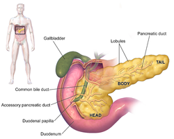
Blausen 0699 PancreasAnatomy2.png
Anatomy of the pancreas
Details
Pronunciation/ˈpæŋkriəs/
PrecursorPancreatic buds
SystemDigestive system and endocrine system
ArteryInferior pancreaticoduodenal artery, anterior superior pancreaticoduodenal artery, posterior superior pancreaticoduodenal artery, splenic artery
VeinPancreaticoduodenal veins, pancreatic veins
NervePancreatic plexus, celiac ganglia, vagus nerve
LymphSplenic lymph nodes, celiac lymph nodes and superior mesenteric lymph nodes
Identifiers
LatinPancreas
GreekΠάγκρεας (Pánkreas)
MeSHD010179
TA98A05.9.01.001
TA23114
FMA7198

The pancreas is an organ of the digestive system and endocrine system of vertebrates. In humans, it is located in the abdomen behind the stomach and functions as a gland. The pancreas is a mixed or heterocrine gland, i.e. it has both an endocrine and a digestive exocrine function. 99% part of pancreas is exocrine and 1% part is endocrine. As an endocrine gland, it functions mostly to regulate blood sugar levels, secreting the hormones insulin, glucagon, somatostatin, and pancreatic polypeptide. As a part of the digestive system, it functions as an exocrine gland secreting pancreatic juice into the duodenum through the pancreatic duct. This juice contains bicarbonate, which neutralizes acid entering the duodenum from the stomach; and digestive enzymes, which break down carbohydrates, proteins, and fats in food entering the duodenum from the stomach.

Inflammation of the pancreas is known as pancreatitis, with common causes including chronic alcohol use and gallstones. Because of its role in the regulation of blood sugar, the pancreas is also a key organ in diabetes mellitus. Pancreatic cancer can arise following chronic pancreatitis or due to other reasons, and carries a very poor prognosis, as it is often identified when it has spread to other areas of the body.

The word pancreas comes from the Greek πᾶν (pân, “all”) & κρέας (kréas, “flesh”). The function of the pancreas in diabetes has been known since at least 1889, with its role in insulin production identified in 1921.

Structure

The pancreas (shown here in pink) sits behind the stomach, with the body near the curvature of the duodenum, and the tail stretching to touch the spleen.
 
Diagram showing different functional parts of the pancreas

The pancreas is an organ that in humans lies in the abdomen, stretching from behind the stomach to the left upper abdomen near the spleen. In adults, it is about 12–15 centimetres (4.7–5.9 in) long, lobulated, and salmon-coloured in appearance.

Anatomically, the pancreas is divided into a head, neck, body, and tail. The pancreas stretches from the inner curvature of the duodenum, where the head surrounds two blood vessels: the superior mesenteric artery, and vein. The longest part of the pancreas, the body, stretches across behind the stomach, and the tail of the pancreas ends adjacent to the spleen.

Two ducts, the main pancreatic duct and a smaller accessory pancreatic ductrun through the body of the pancreas. The main pancreatic duct joins with the common bile duct forming a small ballooning called the ampulla of Vater (hepatopancreatic ampulla). This ampulla is surrounded by a muscle, the sphincter of Oddi. This ampulla opens into the descending part of the duodenum. The opening of the common bile duct into main pancreatic duct is controlled by sphincter of Boyden. The accessory pancreatic duct opens into duodenum with separate openings located above the opening of the main pancreatic duct.

Parts

The head of the pancreas sits within the curvature of the duodenum, and wraps around the superior mesenteric artery and vein. To the right sits the descending part of the duodenum, and between these travel the superior and inferior pancreaticoduodenal arteries. Behind rests the inferior vena cava, and the common bile duct. In front sits the peritoneal membrane and the transverse colon.[7] A small uncinate process emerges from below the head, situated behind the superior mesenteric vein and sometimes artery.

The neck of the pancreas separates the head of the pancreas, located in the curvature of the duodenum, from the body. The neck is about 2 cm (0.79 in) wide, and sits in front of where the portal vein is formed. The neck lies mostly behind the pylorus of the stomach, and is covered with peritoneum. The anterior superior pancreaticoduodenal artery travels in front of the neck of the pancreas.

The body is the largest part of the pancreas, and mostly lies behind the stomach, tapering along its length. The peritoneum sits on top of the body of the pancreas, and the transverse colon in front of the peritoneum. Behind the pancreas are several blood vessels, including the aorta, the splenic vein, and the left renal vein, as well as the beginning of the superior mesenteric artery. Below the body of the pancreas sits some of the small intestine, specifically the last part of the duodenum and the jejunum to which it connects, as well as the suspensory ligament of the duodenum which falls between these two. In front of the pancreas sits the transverse colon.

The pancreas narrows towards the tail, which sits near to the spleen. It is usually between 1.3–3.5 cm (0.51–1.38 in) long, and sits between the layers of the ligament between the spleen and the left kidney. The splenic artery and vein, which also passes behind the body of the pancreas, pass behind the tail of the pancreas.

Blood supply

The pancreas has a rich blood supply, with vessels originating as branches of both the coeliac artery and superior mesenteric artery. The splenic artery runs along the top of the pancreas, and supplies the left part of the body and the tail of the pancreas through its pancreatic branches, the largest of which is called the greater pancreatic artery. The superior and inferior pancreaticoduodenal arteries run along the back and front surfaces of the head of the pancreas adjacent to the duodenum. These supply the head of the pancreas. These vessels join together (anastamose) in the middle.

The body and neck of the pancreas drain into the splenic vein, which sits behind the pancreas. The head drains into, and wraps around, the superior mesenteric and portal veins, via the pancreaticoduodenal veins.

The pancreas drains into lymphatic vessels that travel alongside its arteries, and has a rich lymphatic supply. The lymphatic vessels of the body and tail drain into splenic lymph nodes, and eventually into lymph nodes that lie in front of the aorta, between the coeliac and superior mesenteric arteries. The lymphatic vessels of the head and neck drain into intermediate lymphatic vessels around the pancreaticoduodenal, mesenteric and hepatic arteries, and from there into the lymph nodes that lie in front of the aorta.

Microanatomy

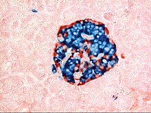
This image shows a pancreatic islet when pancreatic tissue is stained and viewed under a microscope. Parts of the digestive ("exocrine") pancreas can be seen around the islet, more darkly. These contain hazy dark purple granules of inactive digestive enzymes (zymogens).
 
A pancreatic islet that uses fluorescent antibodies to show the location of different cell types in the pancreatic islet. Antibodies against glucagon, secreted by alpha cells, show their peripheral position. Antibodies against insulin, secreted by beta cells, show the more widespread and central position that these cells tend to have.

The pancreas contains tissue with an endocrine and exocrine role, and this division is also visible when the pancreas is viewed under a microscope.

The majority of pancreatic tissue has a digestive role. The cells with this role form clusters (Latin: acini) around small ducts, and are arranged in lobes that have thin fibrous walls. The cells of each acinus secrete inactive digestive enzymes called zymogens into the small intercalated ducts which they surround. In each acinus, the cells are pyramid-shaped and situated around the intercalated ducts, with the nuclei resting on the basement membrane, a large endoplasmic reticulum, and a number of zymogen granules visible within the cytoplasm. The intercalated ducts drain into larger intralobular ducts within the lobule, and finally interlobular ducts. The ducts are lined by a single layer of column-shaped cells. There is more than one layer of cells as the diameter of the ducts increases.

The tissues with an endocrine role within the pancreas exist as clusters of cells called pancreatic islets (also called islets of Langerhans) that are distributed throughout the pancreas. Pancreatic islets contain alpha cells, beta cells, and delta cells, each of which releases a different hormone. These cells have characteristic positions, with alpha cells (secreting glucagon) tending to be situated around the periphery of the islet, and beta cells (secreting insulin) more numerous and found throughout the islet. Enterochromaffin cells are also scattered throughout the islets. Islets are composed of up to 3,000 secretory cells, and contain several small arterioles to receive blood, and venules that allow the hormones secreted by the cells to enter the systemic circulation.

Variation

The size of the pancreas varies considerably. Several anatomical variations exist, relating to the embryological development of the two pancreatic buds. The pancreas develops from these buds on either side of the duodenum. The ventral bud rotates to lie next to the dorsal bud, eventually fusing. In about 10% of adults, an accessory pancreatic duct may be present if the main duct of the dorsal bud of the pancreas does not regress; this duct opens into the minor duodenal papilla. If the two buds themselves, each having a duct, do not fuse, a pancreas may exist with two separate ducts, a condition known as a pancreas divisum. This condition has no physiologic consequence. If the ventral bud does not fully rotate, an annular pancreas may exist, where part or all of the duodenum is encircled by the pancreas. This may be associated with duodenal atresia.

Gene and protein expression

10,000 protein coding genes (50% of all genes) are expressed in the normal human pancreas. Less than 100 of these genes are specifically expressed in the pancreas. Similar to the salivary glands, most pancreas-specific genes encode for secreted proteins. Corresponding pancreas-specific proteins are either expressed in the exocrine cellular compartment and have functions related to digestion or food uptake such as digestive chymotrypsinogen enzymes and pancreatic lipase PNLIP, or are expressed in the various cells of the endocrine pancreatic islets and have functions related to secreted hormones such as insulin, glucagon, somatostatin and pancreatic polypeptide.

Development

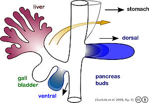
The pancreas originates from the foregut, a precursor tube to part of the digestive tract, as a dorsal and ventral bud. As it develops, the ventral bud rotates to the other side and the two buds fuse together.

The pancreas forms during development from two buds that arise from the duodenal part of the foregut, an embryonic tube that is a precursor to the gastrointestinal tract. It is of endodermal origin. Pancreatic development begins with the formation of a dorsal and ventral pancreatic bud. Each joins with the foregut through a duct. The dorsal pancreatic bud forms the neck, body, and tail of the developed pancreas, and the ventral pancreatic bud forms the head and uncinate process.

The definitive pancreas results from rotation of the ventral bud and the fusion of the two buds. During development, the duodenum rotates to the right, and the ventral bud rotates with it, moving to a position that becomes more dorsal. Upon reaching its final destination, the ventral pancreatic bud is below the larger dorsal bud, and eventually fuses with it. At this point of fusion, the main ducts of the ventral and dorsal pancreatic buds fuse, forming the main pancreatic duct. Usually, the duct of the dorsal bud regresses, leaving the main pancreatic duct.

Cellular development

Pancreatic progenitor cells are precursor cells that differentiate into the functional pancreatic cells, including exocrine acinar cells, endocrine islet cells, and ductal cells. These progenitor cells are characterised by the co-expression of the transcription factors PDX1 and NKX6-1.

The cells of the exocrine pancreas differentiate through molecules that induce differentiation including follistatin, fibroblast growth factors, and activation of the Notch receptor system. Development of the exocrine acini progresses through three successive stages. These are the predifferentiated, protodifferentiated, and differentiated stages, which correspond to undetectable, low, and high levels of digestive enzyme activity, respectively.

Pancreatic progenitor cells differentiate into endocrine islet cells under the influence of neurogenin-3 and ISL1, but only in the absence of notch receptor signaling. Under the direction of a Pax gene, the endocrine precursor cells differentiate to form alpha and gamma cells. Under the direction of Pax-6, the endocrine precursor cells differentiate to form beta and delta cells. The pancreatic islets form as the endocrine cells migrate from the duct system to form small clusters around capillaries. This occurs around the third month of development, and insulin and glucagon can be detected in the human fetal circulation by the fourth or fifth month of development.

Function

The pancreas is involved in blood sugar control and metabolism within the body, and also in the secretion of substances (collectively pancreatic juice) that help digestion. These are divided into an "endocrine" role, relating to the secretion of insulin and other substances within pancreatic islets that help control blood sugar levels and metabolism within the body, and an "exocrine" role, relating to the secretion of enzymes involved in digesting substances in the digestive tract.

Blood glucose regulation

The pancreas maintains constant blood glucose levels (shown as the waving line). When the blood glucose level is too high, the pancreas secretes insulin and when the level is too low, the pancreas secretes glucagon.

Cells within the pancreas help to maintain blood glucose levels (homeostasis). The cells that do this are located within the pancreatic islets that are present throughout the pancreas. When blood glucose levels are low, alpha cells secrete glucagon, which increases blood glucose levels. When blood glucose levels are high beta cells secrete insulin to decrease glucose in blood. Delta cells in the islet also secrete somatostatin which decreases the release of insulin and glucagon.

Glucagon acts to increase glucose levels by promoting the creation of glucose and the breakdown of glycogen to glucose in the liver. It also decreases the uptake of glucose in fat and muscle. Glucagon release is stimulated by low blood glucose or insulin levels, and during exercise. Insulin acts to decrease blood glucose levels by facilitating uptake by cells (particularly skeletal muscle), and promoting its use in the creation of proteins, fats and carbohydrates. Insulin is initially created as a precursor form called preproinsulin. This is converted to proinsulin and cleaved by C-peptide to insulin which is then stored in granules in beta cells. Glucose is taken into the beta cells and degraded. The end effect of this is to cause depolarisation of the cell membrane which stimulates the release of the insulin.

The main factor influencing the secretion of insulin and glucagon are the levels of glucose in blood plasma. Low blood sugar stimulates glucagon release, and high blood sugar stimulates insulin release. Other factors also influence the secretion of these hormones. Some amino acids, that are byproducts of the digestion of protein, stimulate insulin and glucagon release. Somatostatin acts as an inhibitor of both insulin and glucagon. The autonomic nervous system also plays a role. Activation of Beta-2 receptors of the sympathetic nervous system by catecholamines secreted from sympathetic nerves stimulates secretion of insulin and glucagon, whereas activation of Alpha-1 receptors inhibits secretion. M3 receptors of the parasympathetic nervous system act when stimulated by the right vagus nerve to stimulate release of insulin from beta cells.

Digestion

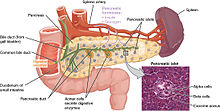
The pancreas has a role in digestion, highlighted here. Ducts in the pancreas (green) conduct digestive enzymes into the duodenum. This image also shows a pancreatic islet, part of the endocrine pancreas, which contains cells responsible for secretion of insulin and glucagon.

The pancreas plays a vital role in the digestive system. It does this by secreting a fluid that contains digestive enzymes into the duodenum, the first part of the small intestine that receives food from the stomach. These enzymes help to break down carbohydrates, proteins and lipids (fats). This role is called the "exocrine" role of the pancreas. The cells that do this are arranged in clusters called acini. Secretions into the middle of the acinus accumulate in intralobular ducts, which drain to the main pancreatic duct, which drains directly into the duodenum. About 1.5 - 3 liters of fluid are secreted in this manner every day.

The cells in each acinus are filled with granules containing the digestive enzymes. These are secreted in an inactive form termed zymogens or proenzymes. When released into the duodenum, they are activated by the enzyme enterokinase present in the lining of the duodenum. The proenzymes are cleaved, creating a cascade of activating enzymes.

These enzymes are secreted in a fluid rich in bicarbonate. Bicarbonate helps maintain an alkaline pH for the fluid, a pH in which most of the enzymes act most efficiently, and also helps to neutralise the stomach acids that enter the duodenum. Secretion is influenced by hormones including secretin, cholecystokinin, and VIP, as well as acetylcholine stimulation from the vagus nerve. Secretin is released from the S cells which form part of the lining of the duodenum in response to stimulation by gastric acid. Along with VIP, it increases the secretion of enzymes and bicarbonate. Cholecystokinin is released from Ito cells of the lining of the duodenum and jejunum mostly in response to long chain fatty acids, and increases the effects of secretin. At a cellular level, bicarbonate is secreted from the acinar cells through a sodium and bicarbonate cotransporter that acts because of membrane depolarisation caused by the cystic fibrosis transmembrane conductance regulator. Secretin and VIP act to increase the opening of the cystic fibrosis transmembrane conductance regulator, which leads to more membrane depolarisation and more secretion of bicarbonate.

A variety of mechanisms act to ensure that the digestive action of the pancreas does not act to digest pancreatic tissue itself. These include the secretion of inactive enzymes (zymogens), the secretion of the protective enzyme trypsin inhibitor, which inactivates trypsin, the changes in pH that occur with bicarbonate secretion that stimulate digestion only when the pancreas is stimulated, and the fact that the low calcium within cells causes inactivation of trypsin.

Additional functions

The pancreas also secretes vasoactive intestinal peptide and pancreatic polypeptide. Enterochromaffin cells of the pancreas secrete the hormones motilin, serotonin, and substance P.

Clinical significance

Inflammation

Inflammation of the pancreas is known as pancreatitis. Pancreatitis is most often associated with recurrent gallstones or chronic alcohol use, with other common causes including traumatic damage, damage following an ERCP, some medications, infections such as mumps and very high blood triglyceride levels. Acute pancreatitis is likely to cause intense pain in the central abdomen, that often radiates to the back, and may be associated with nausea or vomiting. Severe pancreatitis may lead to bleeding or perforation of the pancreas resulting in shock or a systemic inflammatory response syndrome, bruising of the flanks or the region around the belly button. These severe complications are often managed in an intensive care unit.

In pancreatitis, enzymes of the exocrine pancreas damage the structure and tissue of the pancreas. Detection of some of these enzymes, such as amylase and lipase in the blood, along with symptoms and findings on medical imaging such as ultrasound or a CT scan, are often used to indicate that a person has pancreatitis. Pancreatitis is often managed medically with pain reliefs, and monitoring to prevent or manage shock, and management of any identified underlying causes. This may include removal of gallstones, lowering of blood triglyceride or glucose levels, the use of corticosteroids for autoimmune pancreatitis, and the cessation of any medication triggers.

Chronic pancreatitis refers to the development of pancreatitis over time. It shares many similar causes, with the most common being chronic alcohol use, with other causes including recurrent acute episodes and cystic fibrosis. Abdominal pain, characteristically relieved by sitting forward or drinking alcohol, is the most common symptom. When the digestive function of the pancreas is severely affected, this may lead to problems with fat digestion and the development of steatorrhoea; when the endocrine function is affected, this may lead to diabetes. Chronic pancreatitis is investigated in a similar way to acute pancreatitis. In addition to management of pain and nausea, and management of any identified causes (which may include alcohol cessation), because of the digestive role of the pancreas, enzyme replacement may be needed to prevent malabsorption.

Cancer

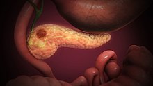
Pancreatic cancer, shown here, most commonly occurs as an adenocarcinoma in the head of the pancreas. Because symptoms (such as skin yellowing, pain, or itch) do not occur until later in the disease, it often presents at a later stage and has limited treatment options.

Pancreatic cancers, particularly the most common type, pancreatic adenocarcinoma, remain very difficult to treat, and are mostly diagnosed only at a stage that is too late for surgery, which is the only curative treatment. Pancreatic cancer is rare in people younger than 40 and the median age of diagnosis is 71. Risk factors include chronic pancreatitis, older age, smoking, obesity, diabetes, and certain rare genetic conditions including multiple endocrine neoplasia type 1, hereditary nonpolyposis colon cancer and dysplastic nevus syndrome among others. About 25% of cases are attributable to tobacco smoking, while 5–10% of cases are linked to inherited genes.

Pancreatic adenocarcinoma is the most common form of pancreatic cancer, and is cancer arising from the exocrine digestive part of the pancreas. Most occur in the head of the pancreas. Symptoms tend to arise late in the course of the cancer, when it causes abdominal pain, weight loss, or yellowing of the skin (jaundice). Jaundice occurs when the outflow of bile is blocked by the cancer. Other less common symptoms include nausea, vomiting, pancreatitis, diabetes or recurrent venous thrombosis. Pancreatic cancer is usually diagnosed by medical imaging in the form of an ultrasound or CT scan with contrast enhancement. An endoscopic ultrasound may be used if a tumour is being considered for surgical removal, and biopsy guided by ERCP or ultrasound can be used to confirm an uncertain diagnosis.

Because of the late development of symptoms, most cancer presents at an advanced stage. Only 10 to 15% of tumours are suitable for surgical resection. As of 2018, when chemotherapy is given the FOLFIRINOX regimen containing fluorouracil, irinotecan, oxaliplatin and leucovorin has been shown to extend survival beyond traditional gemcitabine regimens. For the most part, treatment is palliative, focus on the management of symptoms that develop. This may include management of itch, a choledochojejunostomy or the insertion of stents with ERCP to facilitate the drainage of bile, and medications to help control pain. In the United States pancreatic cancer is the fourth most common cause of deaths due to cancer. The disease occurs more often in the developed world, which had 68% of new cases in 2012. Pancreatic adenocarcinoma typically has poor outcomes with the average percentage alive for at least one and five years after diagnosis being 25% and 5% respectively. In localized disease where the cancer is small (< 2 cm) the number alive at five years is approximately 20%.

There are several types of pancreatic cancer, involving both the endocrine and exocrine tissue. The many types of pancreatic endocrine tumors are all uncommon or rare, and have varied outlooks. However the incidence of these cancers has been rising sharply; it is not clear to what extent this reflects increased detection, especially through medical imaging, of tumors that would be very slow to develop. Insulinomas (largely benign) and gastrinomas are the most common types. For those with neuroendocrine cancers the number alive after five years is much better at 65%, varying considerably with type.

A solid pseudopapillary tumour is a low-grade malignant tumour of the pancreas of papillary architecture that typically afflicts young women.

Diabetes mellitus

Type 1 diabetes

Diabetes mellitus type 1 is a chronic autoimmune disease in which the immune system attacks the insulin-secreting beta cells of the pancreas. Insulin is needed to keep blood sugar levels within optimal ranges, and its lack can lead to high blood sugar. As an untreated chronic condition, complications including accelerated vascular disease, diabetic retinopathy, kidney disease and neuropathy can result. In addition, if there is not enough insulin for glucose to be used within cells, the medical emergency diabetic ketoacidosis, which is often the first symptom that a person with type 1 diabetes may have, can result. Type 1 diabetes can develop at any age but is most often diagnosed before age 40. For people living with type 1 diabetes, insulin injections are critical for survival. An experimental procedure to treat type 1 diabetes is pancreas transplantation or isolated transplantation of islet cells to supply a person with functioning beta cells.

Type 2 diabetes

Diabetes mellitus type 2 is the most common form of diabetes. The causes for high blood sugar in this form of diabetes usually are a combination of insulin resistance and impaired insulin secretion, with both genetic and environmental factors playing a role in the development of the disease. Over time, pancreatic beta cells may become "exhausted" and less functional. The management of type 2 diabetes involves a combination of lifestyle measures, medications if required and potentially insulin. With relevance to the pancreas, several medications act to enhance the secretion of insulin from beta cells, particularly sulphonylureas, which act directly on beta cells; incretins which replicate the action of the hormones glucagon-like peptide 1, increasing the secretion of insulin from beta cells after meals, and are more resistant to breakdown; and DPP-4 inhibitors, which slow the breakdown of incretins.

Removal

It is possible for a person to live without a pancreas, provided that the person takes insulin for proper regulation of blood glucose concentration and pancreatic enzyme supplements to aid digestion.

History

The pancreas was first identified by Herophilus (335–280 BC), a Greek anatomist and surgeon. A few hundred years later, Rufus of Ephesus, another Greek anatomist, gave the pancreas its name. Etymologically, the term "pancreas", a modern Latin adaptation of Greek πάγκρεας, [πᾶν ("all", "whole"), and κρέας ("flesh")], originally means sweetbread, although literally meaning all-flesh, presumably because of its fleshy consistency. It was only in 1889 when Oskar Minkowski discovered that removing the pancreas from a dog caused it to become diabetic. Insulin was later isolated from pancreatic islets by Frederick Banting and Charles Herbert Best in 1921.

The way the tissue of the pancreas has been viewed has also changed. Previously, it was viewed using simple staining methods such as H&E stains. Now, immunohistochemistry can be used to more easily differentiate cell types. This involves visible antibodies to the products of certain cell types, and helps identify with greater ease cell types such as alpha and beta cells.

Other animals

Pancreatic tissue is present in all vertebrates, but its precise form and arrangement varies widely. There may be up to three separate pancreases, two of which arise from ventral buds, and the other dorsally. In most species (including humans), these "fuse" in the adult, but there are several exceptions. Even when a single pancreas is present, two or three pancreatic ducts may persist, each draining separately into the duodenum (or equivalent part of the foregut). Birds, for example, typically have three such ducts.

In teleost fish, and a few other species (such as rabbits), there is no discrete pancreas at all, with pancreatic tissue being distributed diffusely across the mesentery and even within other nearby organs, such as the liver or spleen. In a few teleost species, the endocrine tissue has fused to form a distinct gland within the abdominal cavity, but otherwise it is distributed among the exocrine components. The most primitive arrangement, however, appears to be that of lampreys and lungfish, in which pancreatic tissue is found as a number of discrete nodules within the wall of the gut itself, with the exocrine portions being little different from other glandular structures of the intestine.

Social privilege

From Wikipedia, the free encyclopedia https://en.wikipedi...