Search This Blog

Saturday, September 18, 2021

Deep brain stimulation

From Wikipedia, the free encyclopedia
 
Deep brain stimulation
Tiefe Hirnstimulation - Sonden RoeSchaedel ap.jpg
DBS-probes shown in X-ray of the skull (white areas around maxilla and mandible represent metal dentures and are unrelated to DBS devices)
MeSHD046690
MedlinePlus007453

Deep brain stimulation (DBS) is a neurosurgical procedure involving the placement of a medical device called a neurostimulator (sometimes referred to as a "brain pacemaker"), which sends electrical impulses, through implanted electrodes, to specific targets in the brain (brain nuclei) for the treatment of movement disorders, including Parkinson's disease, essential tremor, and dystonia. and other conditions such as obsessive-compulsive disorder and epilepsy. While its underlying principles and mechanisms are not fully understood, DBS directly changes brain activity in a controlled manner.

DBS has been approved by the Food and Drug Administration as a treatment for essential tremor and Parkinson's disease (PD) since 1997. DBS was approved for dystonia in 2003, obsessive–compulsive disorder (OCD) in 2009, and epilepsy in 2018. DBS has been studied in clinical trials as a potential treatment for chronic pain for various affective disorders, including major depression. It is one of few neurosurgical procedures that allow blinded studies.

Medical use

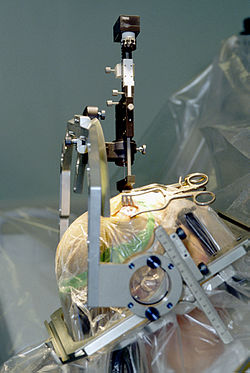
Insertion of electrode during surgery using a stereotactic frame

Parkinson's disease

DBS is used to manage some of the symptoms of Parkinson's disease that cannot be adequately controlled with medications. PD is treated by applying high-frequency (> 100 Hz) stimulation to three target structures namely to entrolateral thalamus, internal pallidum, and subthalamic nucleus (STN) to mimic the clinical effects of lesioning. It is recommended for people who have PD with motor fluctuations and tremor inadequately controlled by medication, or to those who are intolerant to medication, as long as they do not have severe neuropsychiatric problems. Four areas of the brain have been treated with neural stimulators in PD. These are the globus pallidus internus, thalamus, subthalamic nucleus and the pedunculopontine nucleus. However, most DBS surgeries in routine practice target either the globus pallidus internus, or the Subthalamic nucleus.

  • DBS of the globus pallidus internus reduces uncontrollable shaking movements called dyskinesias. This enables a patient to take adequate quantities of medications (especially levodopa), thus leading to better control of symptoms.
  • DBS of the subthalamic nucleus directly reduces symptoms of Parkinson's. This enables a decrease in the dose of anti-parkinsonian medications.
  • DBS of the PPN may help with freezing of gait, while DBS of the thalamus may help with tremor. These targets are not routinely utilized.

Selection of the correct DBS target is a complicated process. Multiple clinical characteristics are used to select the target including – identifying the most troublesome symptoms, the dose of levodopa that the patient is currently taking, the effects and side-effects of current medications and concurrent problems. For example, subthalamic nucleus DBS may worsen depression and hence is not preferred in patients with uncontrolled depression.

Generally DBS is associated with 30–60% improvement in motor score evaluations.

Tourette syndrome

DBS has been used experimentally in treating adults with severe Tourette syndrome that does not respond to conventional treatment. Despite widely publicized early successes, DBS remains a highly experimental procedure for the treatment of Tourette's, and more study is needed to determine whether long-term benefits outweigh the risks. The procedure is well tolerated, but complications include "short battery life, abrupt symptom worsening upon cessation of stimulation, hypomanic or manic conversion, and the significant time and effort involved in optimizing stimulation parameters". As of 2006, five people with TS had been reported on; all experienced reduction in tics and the disappearance of obsessive-compulsive behaviors.

The procedure is invasive and expensive, and requires long-term expert care. Benefits for severe Tourette's are not conclusive, considering less robust effects of this surgery seen in the Netherlands. Tourette's is more common in pediatric populations, tending to remit in adulthood, so in general this would not be a recommended procedure for use on children. Because diagnosis of Tourette's is made based on a history of symptoms rather than analysis of neurological activity, it may not always be clear how to apply DBS for a particular person. Due to concern over the use of DBS in Tourette syndrome treatment, the Tourette Association of America convened a group of experts to develop recommendations guiding the use and potential clinical trials of DBS for TS.

Robertson reported that DBS had been used on 55 adults by 2011, remained an experimental treatment at that time, and recommended that the procedure "should only be conducted by experienced functional neurosurgeons operating in centres which also have a dedicated Tourette syndrome clinic". According to Malone et al. (2006), "Only patients with severe, debilitating, and treatment-refractory illness should be considered; while those with severe personality disorders and substance-abuse problems should be excluded." Du et al. (2010) say, "As an invasive therapy, DBS is currently only advisable for severely affected, treatment-refractory TS adults". Singer (2011) says, "pending determination of patient selection criteria and the outcome of carefully controlled clinical trials, a cautious approach is recommended". Viswanathan et al. (2012) say DBS should be used for people with "severe functional impairment that cannot be managed medically".

Adverse effects

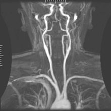
Arteriogram of the arterial supply that can hemorrhage during DBS implantation.

DBS carries the risks of major surgery, with a complication rate related to the experience of the surgical team. The major complications include hemorrhage (1–2%) and infection (3–5%).

The potential exists for neuropsychiatric side effects after DBS, including apathy, hallucinations, hypersexuality, cognitive dysfunction, depression, and euphoria. However, these effects may be temporary and related to (1) correct placement of electrodes, (2) open-loop VS closed loop stimulation , meaning a constant stimulation or an A.I. monitoring delivery system and (3) calibration of the stimulator, so these side effects are potentially reversible.

Because the brain can shift slightly during surgery, the electrodes can become displaced or dislodged from the specific location. This may cause more profound complications such as personality changes, but electrode misplacement is relatively easy to identify using CT scan. Also, complications of surgery may occur, such as bleeding within the brain. After surgery, swelling of the brain tissue, mild disorientation, and sleepiness are normal. After 2–4 weeks, a follow-up visit is used to remove sutures, turn on the neurostimulator, and program it.

Impaired swimming skills surfaced as an unexpected risk of the procedure; several Parkinson's disease patients lost their ability to swim after receiving deep brain stimulation.

Mechanisms

The exact mechanism of action of DBS is not known. A variety of hypotheses try to explain the mechanisms of DBS:

  1. Depolarization blockade: Electrical currents block the neuronal output at or near the electrode site.
  2. Synaptic inhibition: This causes an indirect regulation of the neuronal output by activating axon terminals with synaptic connections to neurons near the stimulating electrode.
  3. Desynchronization of abnormal oscillatory activity of neurons
  4. Antidromic activation either activating/blockading distant neurons or blockading slow axons

DBS represents an advance on previous treatments which involved pallidotomy (i.e., surgical ablation of the globus pallidus) or thalamotomy (i.e., surgical ablation of the thalamus). Instead, a thin lead with multiple electrodes is implanted in the globus pallidus, nucleus ventralis intermedius thalami, or subthalamic nucleus, and electric pulses are used therapeutically. The lead from the implant is extended to the neurostimulator under the skin in the chest area.

Its direct effect on the physiology of brain cells and neurotransmitters is currently debated, but by sending high-frequency electrical impulses into specific areas of the brain, it can mitigate symptoms and directly diminish the side effects induced by PD medications, allowing a decrease in medications, or making a medication regimen more tolerable.

Components and placement

The DBS system consists of three components: the implanted pulse generator (IPG), the lead, and an extension. The IPG is a battery-powered neurostimulator encased in a titanium housing, which sends electrical pulses to the brain that interfere with neural activity at the target site. The lead is a coiled wire insulated in polyurethane with four platinum-iridium electrodes and is placed in one or two different nuclei of the brain. The lead is connected to the IPG by an extension, an insulated wire that runs below the skin, from the head, down the side of the neck, behind the ear, to the IPG, which is placed subcutaneously below the clavicle, or in some cases, the abdomen. The IPG can be calibrated by a neurologist, nurse, or trained technician to optimize symptom suppression and control side effects.

DBS leads are placed in the brain according to the type of symptoms to be addressed. For non-Parkinsonian essential tremor, the lead is placed in either the ventrointermediate nucleus of the thalamus or the zona incerta; for dystonia and symptoms associated with PD (rigidity, bradykinesia/akinesia, and tremor), the lead may be placed in either the globus pallidus internus or the subthalamic nucleus; for OCD and depression to the nucleus accumbens; for incessant pain to the posterior thalamic region or periaqueductal gray; and for epilepsy treatment to the anterior thalamic nucleus.

All three components are surgically implanted inside the body. Lead implantation may take place under local anesthesia or under general anesthesia ("asleep DBS") such as for dystonia. A hole about 14 mm in diameter is drilled in the skull and the probe electrode is inserted stereotactically, using either frame-based or frameless stereotaxis. During the awake procedure with local anesthesia, feedback from the person is used to determine the optimal placement of the permanent electrode. During the asleep procedure, intraoperative MRI guidance is used for direct visualization of brain tissue and device. The installation of the IPG and extension leads occurs under general anesthesia. The right side of the brain is stimulated to address symptoms on the left side of the body and vice versa.

Research

Chronic pain

Stimulation of the periaqueductal gray and periventricular gray for nociceptive pain, and the internal capsule, ventral posterolateral nucleus, and ventral posteromedial nucleus for neuropathic pain has produced impressive results with some people, but results vary. One study of 17 people with intractable cancer pain found that 13 were virtually pain free and only four required opioid analgesics on release from hospital after the intervention. Most ultimately did resort to opioids, usually in the last few weeks of life. DBS has also been applied for phantom limb pain.

Major depression and obsessive-compulsive disorder

Lateral X-ray of the head: Deep brain stimulation in obsessive–compulsive disorder (OCD). 42 year old man, surgery in 2013.

DBS has been used in a small number of clinical trials to treat people with severe treatment-resistant depression (TRD). A number of neuroanatomical targets have been used for DBS for TRD including the subgenual cingulate gyrus, posterior gyrus rectus, nucleus accumbens, ventral capsule/ventral striatum, inferior thalamic peduncle, and the lateral habenula. A recently proposed target of DBS intervention in depression is the superolateral branch of the medial forebrain bundle; its stimulation lead to surprisingly rapid antidepressant effects.

The small numbers in the early trials of DBS for TRD currently limit the selection of an optimal neuroanatomical target. Evidence is insufficient to support DBS as a therapeutic modality for depression; however, the procedure may be an effective treatment modality in the future. In fact, beneficial results have been documented in the neurosurgical literature, including a few instances in which people who were deeply depressed were provided with portable stimulators for self treatment.

A systematic review of DBS for TRD and OCD identified 23 cases, nine for OCD, seven for TRD, and one for both. "[A]bout half the patients did show dramatic improvement" and adverse events were "generally trivial" given the younger age of the psychiatric population relative to the age of people with movement disorders. The first randomized, controlled study of DBS for the treatment of TRD targeting the ventral capsule/ventral striatum area did not demonstrate a significant difference in response rates between the active and sham groups at the end of a 16-week study. However, a second randomized controlled study of ventral capsule DBS for TRD did demonstrate a significant difference in response rates between active DBS (44% responders) and sham DBS (0% responders). Efficacy of DBS is established for OCD, with on average 60% responders in severely ill and treatment-resistant patients. Based on these results the FDA has approved DBS for treatment-resistant OCD under a Humanitarian Device Exemption (HDE), requiring that the procedure be performed only in a hospital with specialist qualifications to do so.

DBS for TRD can be as effective as antidepressants and can have good response and remission rates, but adverse effects and safety must be more fully evaluated. Common side effects include "wound infection, perioperative headache, and worsening/irritable mood [and] increased suicidality".

Other clinical applications

Results of DBS in people with dystonia, where positive effects often appear gradually over a period of weeks to months, indicate a role of functional reorganization in at least some cases. The procedure has been tested for effectiveness in people with epilepsy that is resistant to medication. DBS may reduce or eliminate epileptic seizures with programmed or responsive stimulation.

DBS of the septal areas of persons with schizophrenia have resulted in enhanced alertness, cooperation, and euphoria. Persons with narcolepsy and complex-partial seizures also reported euphoria and sexual thoughts from self-elicited DBS of the septal nuclei.

Orgasmic ecstasy was reported with the electrical stimulation of the brain with depth electrodes in the left hippocampus at 3mA, and the right hippocampus at 1 mA.

In 2015, a group of Brazilian researchers led by neurosurgeon Erich Fonoff [pt] described a new technique that allows for simultaneous implants of electrodes called bilateral stereotactic procedure for DBS. The main benefits are less time spent on the procedure and greater accuracy.

In 2016, DBS was found to improve learning and memory in a mouse model of Rett syndrome. More recent (2018) work showed, that forniceal DBS upregulates genes involved in synaptic function, cell survival, and neurogenesis, making some first steps at explaining the restoration of hippocampal circuit function.

See also
 

Neural engineering

From Wikipedia, the free encyclopedia
https://en.wikipedia.org/wiki/Neural_engineering

Neural engineering (also known as neuroengineering) is a discipline within biomedical engineering that uses engineering techniques to understand, repair, replace, or enhance neural systems. Neural engineers are uniquely qualified to solve design problems at the interface of living neural tissue and non-living constructs (Hetling, 2008).

Overview

The field of neural engineering draws on the fields of computational neuroscience, experimental neuroscience, clinical neurology, electrical engineering and signal processing of living neural tissue, and encompasses elements from robotics, cybernetics, computer engineering, neural tissue engineering, materials science, and nanotechnology.

Prominent goals in the field include restoration and augmentation of human function via direct interactions between the nervous system and artificial devices.

Much current research is focused on understanding the coding and processing of information in the sensory and motor systems, quantifying how this processing is altered in the pathological state, and how it can be manipulated through interactions with artificial devices including brain–computer interfaces and neuroprosthetics.

Other research concentrates more on investigation by experimentation, including the use of neural implants connected with external technology.

Neurohydrodynamics is a division of neural engineering that focuses on hydrodynamics of the neurological system.

History

As neural engineering is a relatively new field, information and research relating to it is comparatively limited, although this is changing rapidly. The first journals specifically devoted to neural engineering, The Journal of Neural Engineering and The Journal of NeuroEngineering and Rehabilitation both emerged in 2004. International conferences on neural engineering have been held by the IEEE since 2003, from 29 April until 2 May 2009 in Antalya, Turkey 4th Conference on Neural Engineering, the 5th International IEEE EMBS Conference on Neural Engineering in April/May 2011 in Cancún, Mexico, and the 6th conference in San Diego, California in November 2013. The 7th conference was held in April 2015 in Montpellier. The 8th conference was held in May 2017 in Shanghai.

Fundamentals

The fundamentals behind neuroengineering involve the relationship of neurons, neural networks, and nervous system functions to quantifiable models to aid the development of devices that could interpret and control signals and produce purposeful responses.

Neuroscience

Messages that the body uses to influence thoughts, senses, movements, and survival are directed by nerve impulses transmitted across brain tissue and to the rest of the body. Neurons are the basic functional unit of the nervous system and are highly specialized cells that are capable of sending these signals that operate high and low level functions needed for survival and quality of life. Neurons have special electro-chemical properties that allow them to process information and then transmit that information to other cells. Neuronal activity is dependent upon neural membrane potential and the changes that occur along and across it. A constant voltage, known as the membrane potential, is normally maintained by certain concentrations of specific ions across neuronal membranes. Disruptions or variations in this voltage create an imbalance, or polarization, across the membrane. Depolarization of the membrane past its threshold potential generates an action potential, which is the main source of signal transmission, known as neurotransmission of the nervous system. An action potential results in a cascade of ion flux down and across an axonal membrane, creating an effective voltage spike train or "electrical signal" which can transmit further electrical changes in other cells. Signals can be generated by electrical, chemical, magnetic, optical, and other forms of stimuli that influence the flow of charges, and thus voltage levels across neural membranes (He 2005).

Engineering

Engineers employ quantitative tools that can be used for understanding and interacting with complex neural systems. Methods of studying and generating chemical, electrical, magnetic, and optical signals responsible for extracellular field potentials and synaptic transmission in neural tissue aid researchers in the modulation of neural system activity (Babb et al. 2008). To understand properties of neural system activity, engineers use signal processing techniques and computational modeling (Eliasmith & Anderson 2003). To process these signals, neural engineers must translate the voltages across neural membranes into corresponding code, a process known as neural coding. Neural coding studies on how the brain encodes simple commands in the form of central pattern generators (CPGs), movement vectors, the cerebellar internal model, and somatotopic maps to understand movement and sensory phenomena. Decoding of these signals in the realm of neuroscience is the process by which neurons understand the voltages that have been transmitted to them. Transformations involve the mechanisms that signals of a certain form get interpreted and then translated into another form. Engineers look to mathematically model these transformations (Eliasmith & Anderson 2003). There are a variety of methods being used to record these voltage signals. These can be intracellular or extracellular. Extracellular methods involve single-unit recordings, extracellular field potentials, and amperometry; more recently, multielectrode arrays have been used to record and mimic signals.

Scope

Neuromechanics

Neuromechanics is the coupling of neurobiology, biomechanics, sensation and perception, and robotics (Edwards 2010). Researchers are using advanced techniques and models to study the mechanical properties of neural tissues and their effects on the tissues' ability to withstand and generate force and movements as well as their vulnerability to traumatic loading (Laplaca & Prado 2010). This area of research focuses on translating the transformations of information among the neuromuscular and skeletal systems to develop functions and governing rules relating to operation and organization of these systems (Nishikawa et al. 2007). Neuromechanics can be simulated by connecting computational models of neural circuits to models of animal bodies situated in virtual physical worlds (Edwards 2010). Experimental analysis of biomechanics including the kinematics and dynamics of movements, the process and patterns of motor and sensory feedback during movement processes, and the circuit and synaptic organization of the brain responsible for motor control are all currently being researched to understand the complexity of animal movement. Dr. Michelle LaPlaca's lab at Georgia Institute of Technology is involved in the study of mechanical stretch of cell cultures, shear deformation of planar cell cultures, and shear deformation of 3D cell containing matrices. Understanding of these processes is followed by development of functioning models capable of characterizing these systems under closed loop conditions with specially defined parameters. The study of neuromechanics is aimed at improving treatments for physiological health problems which includes optimization of prostheses design, restoration of movement post injury, and design and control of mobile robots. By studying structures in 3D hydrogels, researchers can identify new models of nerve cell mechanoproperties. For example, LaPlaca et al. developed a new model showing that strain may play a role in cell culture (LaPlaca et al. 2005).

Neuromodulation

Neuromodulation aims to treat disease or injury by employing medical device technologies that would enhance or suppress activity of the nervous system with the delivery of pharmaceutical agents, electrical signals, or other forms of energy stimulus to re-establish balance in impaired regions of the brain. Researchers in this field face the challenge of linking advances in understanding neural signals to advancements in technologies delivering and analyzing these signals with increased sensitivity, biocompatibility, and viability in closed loops schemes in the brain such that new treatments and clinical applications can be created to treat those suffering from neural damage of various kinds. Neuromodulator devices can correct nervous system dysfunction related to Parkinson's disease, dystonia, tremor, Tourette's, chronic pain, OCD, severe depression, and eventually epilepsy. Neuromodulation is appealing as treatment for varying defects because it focuses in on treating highly specific regions of the brain only, contrasting that of systemic treatments that can have side effects on the body. Neuromodulator stimulators such as microelectrode arrays can stimulate and record brain function and with further improvements are meant to become adjustable and responsive delivery devices for drugs and other stimuli.

Neural regrowth and repair

Neural engineering and rehabilitation applies neuroscience and engineering to investigating peripheral and central nervous system function and to finding clinical solutions to problems created by brain damage or malfunction. Engineering applied to neuroregeneration focuses on engineering devices and materials that facilitate the growth of neurons for specific applications such as the regeneration of peripheral nerve injury, the regeneration of the spinal cord tissue for spinal cord injury, and the regeneration of retinal tissue. Genetic engineering and tissue engineering are areas developing scaffolds for spinal cord to regrow across thus helping neurological problems (Schmidt & Leach 2003).

Research and applications

Research focused on neural engineering utilizes devices to study how the nervous system functions and malfunctions (Schmidt & Leach 2003).

Neural imaging

Neuroimaging techniques are used to investigate the activity of neural networks, as well as the structure and function of the brain. Neuroimaging technologies include functional magnetic resonance imaging (fMRI), magnetic resonance imaging (MRI), positron emission tomography (PET) and computed axial tomography (CAT) scans. Functional neuroimaging studies are interested in which areas of the brain perform specific tasks. fMRI measures hemodynamic activity that is closely linked to neural activity. It is used to map metabolic responses in specific regions of the brain to a given task or stimulus. PET, CT scanners, and electroencephalography (EEG) are currently being improved and used for similar purposes.

Neural networks

Scientists can use experimental observations of neuronal systems and theoretical and computational models of these systems to create Neural networks with the hopes of modeling neural systems in as realistic a manner as possible. Neural networks can be used for analyses to help design further neurotechnological devices. Specifically, researchers handle analytical or finite element modeling to determine nervous system control of movements and apply these techniques to help patients with brain injuries or disorders. Artificial neural networks can be built from theoretical and computational models and implemented on computers from theoretically devices equations or experimental results of observed behavior of neuronal systems. Models might represent ion concentration dynamics, channel kinetics, synaptic transmission, single neuron computation, oxygen metabolism, or application of dynamic system theory (LaPlaca et al. 2005). Liquid-based template assembly was used to engineer 3D neural networks from neuron-seeded microcarrier beads.

Neural interfaces

Neural interfaces are a major element used for studying neural systems and enhancing or replacing neuronal function with engineered devices. Engineers are challenged with developing electrodes that can selectively record from associated electronic circuits to collect information about the nervous system activity and to stimulate specified regions of neural tissue to restore function or sensation of that tissue (Cullen et al. 2011). The materials used for these devices must match the mechanical properties of neural tissue in which they are placed and the damage must be assessed. Neural interfacing involves temporary regeneration of biomaterial scaffolds or chronic electrodes and must manage the body's response to foreign materials. Microelectrode arrays are recent advances that can be used to study neural networks (Cullen & Pfister 2011). Optical neural interfaces involve optical recordings and optogenetics stimulation that makes brain cells light sensitive. Fiber optics can be implanted in the brain to stimulate and record this photon activity instead of electrodes. Two-photon excitation microscopy can study living neuronal networks and the communicatory events among neurons.

Brain–computer interfaces

Brain–computer interfaces seek to directly communicate with human nervous system to monitor and stimulate neural circuits as well as diagnose and treat intrinsic neurological dysfunction. Deep brain stimulation is a significant advance in this field that is especially effective in treating movement disorders such as Parkinson's disease with high frequency stimulation of neural tissue to suppress tremors (Lega et al. 2011).

Microsystems

Neural microsystems can be developed to interpret and deliver electrical, chemical, magnetic, and optical signals to neural tissue. They can detect variations in membrane potential and measure electrical properties such as spike population, amplitude, or rate by using electrodes, or by assessment of chemical concentrations, fluorescence light intensity, or magnetic field potential. The goal of these systems is to deliver signals that would influence neuronal tissue potential and thus stimulate the brain tissue to evoke a desired response (He 2005).

Microelectrode arrays

Microelectrode arrays are specific tools used to detect the sharp changes in voltage in the extracellular environments that occur from propagation of an action potential down an axon. Dr. Mark Allen and Dr. LaPlaca have microfabricated 3D electrodes out of cytocompatible materials such as SU-8 and SLA polymers which have led to the development of in vitro and in vivo microelectrode systems with the characteristics of high compliance and flexibility to minimize tissue disruption.

Neural prostheses

Neuroprosthetics are devices capable of supplementing or replacing missing functions of the nervous system by stimulating the nervous system and recording its activity. Electrodes that measure firing of nerves can integrate with prosthetic devices and signal them to perform the function intended by the transmitted signal. Sensory prostheses use artificial sensors to replace neural input that might be missing from biological sources (He 2005). Engineers researching these devices are charged with providing a chronic, safe, artificial interface with neuronal tissue. Perhaps the most successful of these sensory prostheses is the cochlear implant which has restored hearing abilities to the deaf. Visual prosthesis for restoring visual capabilities of blind persons is still in more elementary stages of development. Motor prosthesics are devices involved with electrical stimulation of biological neural muscular system that can substitute for control mechanisms of the brain or spinal cord. Smart prostheses can be designed to replace missing limbs controlled by neural signals by transplanting nerves from the stump of an amputee to muscles. Sensory prosthetics provide sensory feedback by transforming mechanical stimuli from the periphery into encoded information accessible by the nervous system. Electrodes placed on the skin can interpret signals and then control the prosthetic limb. These prosthetics have been very successful. Functional electrical stimulation (FES) is a system aimed at restoring motor processes such as standing, walking, and hand grasp.

Neurorobotics

Neurorobotics is the study of how neural systems can be embodied and movements emulated in mechanical machines. Neurorobots are typically used to study motor control and locomotion, learning and memory selection, and value systems and action selection. By studying neurorobots in real-world environments, they are more easily observed and assessed to describe heuristics of robot function in terms of its embedded neural systems and the reactions of these systems to its environment (Krichmar 2008). For instance, making use of a computational model of epilectic spike-wave dynamics, it has been already proven the effectiveness of a method to simulate seizure abatement through a pseudospectral protocol. The computational model emulates the brain connectivity by using a magnetic imaging resonance from a patient suffering of idiopathic generalized epilepsy. The method was able to generate stimuli able to lessen the seizures.

Neural tissue regeneration

Neural tissue regeneration, or neuroregeneration looks to restore function to those neurons that have been damaged in small injuries and larger injuries like those caused by traumatic brain injury. Functional restoration of damaged nerves involves re-establishment of a continuous pathway for regenerating axons to the site of innervation. Researchers like Dr. LaPlaca at Georgia Institute of Technology are looking to help find treatment for repair and regeneration after traumatic brain injury and spinal cord injuries by applying tissue engineering strategies. Dr. LaPlaca is looking into methods combining neural stem cells with an extracellular matrix protein based scaffold for minimally invasive delivery into the irregular shaped lesions that form after a traumatic insult. By studying the neural stem cells in vitro and exploring alternative cell sources, engineering novel biopolymers that could be utilized in a scaffold, and investigating cell or tissue engineered construct transplants in vivo in models of traumatic brain and spinal cord injury, Dr. LaPlaca's lab aims to identify optimal strategies for nerve regeneration post injury.

Current approaches to clinical treatment

End to end surgical suture of damaged nerve ends can repair small gaps with autologous nerve grafts. For larger injuries, an autologous nerve graft that has been harvested from another site in the body might be used, though this process is time consuming, costly and requires two surgeries (Schmidt & Leach 2003). Clinical treatment for CNS is minimally available and focuses mostly on reducing collateral damage caused by bone fragments near the site of injury or inflammation. After swelling surrounding injury lessens, patients undergo rehabilitation so that remaining nerves can be trained to compensate for the lack of nerve function in injured nerves. No treatment currently exists to restore nerve function of CNS nerves that have been damaged (Schmidt & Leach 2003).

Engineering strategies for repair

Engineering strategies for the repair of spinal cord injury are focused on creating a friendly environment for nerve regeneration. Only PNS nerve damage has been clinically possible so far, but advances in research of genetic techniques and biomaterials demonstrate the potential for SC nerves to regenerate in permissible environments.

Grafts

Advantages of autologous tissue grafts are that they come from natural materials which have a high likelihood of biocompatibility while providing structural support to nerves that encourage cell adhesion and migration (Schmidt & Leach 2003). Nonautologous tissue, acellular grafts, and extracellular matrix based materials are all options that may also provide ideal scaffolding for nerve regeneration. Some come from allogenic or xenogenic tissues that must be combined with immunosuppressants. while others include small intestinal submucosa and amniotic tissue grafts (Schmidt & Leach 2003). Synthetic materials are attractive options because their physical and chemical properties can typically be controlled. A challenge that remains with synthetic materials is biocompatibility (Schmidt & Leach 2003). Methylcellulose-based constructs have been shown to be a biocompatible option serving this purpose (Tate et al. 2001). AxoGen uses a cell graft technology AVANCE to mimic a human nerve. It has been shown to achieve meaningful recovery in 87 percent of patients with peripheral nerve injuries.

Nerve guidance channels

Nerve guidance channels, Nerve guidance conduit are innovative strategies focusing on larger defects that provide a conduit for sprouting axons directing growth and reducing growth inhibition from scar tissue. Nerve guidance channels must be readily formed into a conduit with the desired dimensions, sterilizable, tear resistant, and easy to handle and suture (Schmidt & Leach 2003). Ideally they would degrade over time with nerve regeneration, be pliable, semipermeable, maintain their shape, and have a smooth inner wall that mimics that of a real nerve (Schmidt & Leach 2003).

Biomolecular therapies

Highly controlled delivery systems are needed to promote neural regeneration. Neurotrophic factors can influence development, survival, outgrowth, and branching. Neurotrophins include nerve growth factor (NGF), brain derived neurotrophic factor (BDNF), neurotrophin-3 (NT-3) and neurotrophin-4/5 (NT-4/5). Other factors are ciliary neurotrophic factor (CNTF), glial cell line-derived growth factor (GDNF) and acidic and basic fibroblast growth factor (aFGF, bFGF) that promote a range of neural responses.(Schmidt & Leach 2003) Fibronectin has also been shown to support nerve regeneration following TBI in rats (Tate et al. 2002). Other therapies are looking into regeneration of nerves by upregulating regeneration associated genes (RAGs), neuronal cytoskeletal components, and antiapoptosis factors. RAGs include GAP-43 and Cap-23, adhesion molecules such as L1 family, NCAM, and N-cadherin (Schmidt & Leach 2003). There is also the potential for blocking inhibitory biomolecules in the CNS due to glial scarring. Some currently being studied are treatments with chondroitinase ABC and blocking NgR, ADP-ribose (Schmidt & Leach 2003).

Delivery techniques

Delivery devices must be biocompatible and stable in vivo. Some examples include osmotic pumps, silicone reservoirs, polymer matrices, and microspheres. Gene therapy techniques have also been studied to provide long-term production of growth factors and could be delivered with viral or non-viral vectors such as lipoplexes. Cells are also effective delivery vehicles for ECM components, neurotrophic factors and cell adhesion molecules. Olfactory ensheathing cells (OECs) and stem cells as well as genetically modified cells have been used as transplants to support nerve regeneration (LaPlaca et al. 2005, Schmidt & Leach 2003, Tate et al. 2002).

Advanced therapies

Advanced therapies combine complex guidance channels and multiple stimuli that focus on internal structures that mimic the nerve architecture containing internal matrices of longitudinally aligned fibers or channels. Fabrication of these structures can use a number of technologies: magnetic polymer fiber alignment, injection molding, phase separation, solid free-form fabrication, and ink jet polymer printing (Schmidt & Leach 2003).

Neural enhancement

Augmentation of human neural systems, or human enhancement using engineering techniques is another possible application of neuroengineering. Deep brain stimulation has already been shown to enhance memory recall as noted by patients currently using this treatment for neurological disorders. Brain stimulation techniques are postulated to be able to sculpt emotions and personalities as well as enhance motivation, reduce inhibitions, etc. as requested by the individual. Ethical issues with this sort of human augmentation are a new set of questions that neural engineers have to grapple with as these studies develop.

See also


Neuroprosthetics

From Wikipedia, the free encyclopedia

Neuroprosthetics (also called neural prosthetics) is a discipline related to neuroscience and biomedical engineering concerned with developing neural prostheses. They are sometimes contrasted with a brain–computer interface, which connects the brain to a computer rather than a device meant to replace missing biological functionality.

Neural prostheses are a series of devices that can substitute a motor, sensory or cognitive modality that might have been damaged as a result of an injury or a disease. Cochlear implants provide an example of such devices. These devices substitute the functions performed by the eardrum and stapes while simulating the frequency analysis performed in the cochlea. A microphone on an external unit gathers the sound and processes it; the processed signal is then transferred to an implanted unit that stimulates the auditory nerve through a microelectrode array. Through the replacement or augmentation of damaged senses, these devices intend to improve the quality of life for those with disabilities.

These implantable devices are also commonly used in animal experimentation as a tool to aid neuroscientists in developing a greater understanding of the brain and its functioning. By wirelessly monitoring the brain's electrical signals sent out by electrodes implanted in the subject's brain, the subject can be studied without the device affecting the results.

Accurately probing and recording the electrical signals in the brain would help better understand the relationship among a local population of neurons that are responsible for a specific function.

Neural implants are designed to be as small as possible in order to be minimally invasive, particularly in areas surrounding the brain, eyes or cochlea. These implants typically communicate with their prosthetic counterparts wirelessly. Additionally, power is currently received through wireless power transmission through the skin. The tissue surrounding the implant is usually highly sensitive to temperature rise, meaning that power consumption must be minimal in order to prevent tissue damage.

The neuroprosthetic currently undergoing the most widespread use is the cochlear implant, with over 300,000 in use worldwide as of 2012.

History

The first known cochlear implant was created in 1957. Other milestones include the first motor prosthesis for foot drop in hemiplegia in 1961, the first auditory brainstem implant in 1977 and a peripheral nerve bridge implanted into the spinal cord of an adult rat in 1981. In 1988, the lumbar anterior root implant and functional electrical stimulation (FES) facilitated standing and walking, respectively, for a group of paraplegics.

Regarding the development of electrodes implanted in the brain, an early difficulty was reliably locating the electrodes, originally done by inserting the electrodes with needles and breaking off the needles at the desired depth. Recent systems utilize more advanced probes, such as those used in deep brain stimulation to alleviate the symptoms of Parkinson's disease. The problem with either approach is that the brain floats free in the skull while the probe does not, and relatively minor impacts, such as a low speed car accident, are potentially damaging. Some researchers, such as Kensall Wise at the University of Michigan, have proposed tethering 'electrodes to be mounted on the exterior surface of the brain' to the inner surface of the skull. However, even if successful, tethering would not resolve the problem in devices meant to be inserted deep into the brain, such as in the case of deep brain stimulation (DBS).

Visual prosthetics

A visual prosthesis can create a sense of image by electrically stimulating neurons in the visual system. A camera would wirelessly transmit to an implant, the implant would map the image across an array of electrodes. The array of electrodes has to effectively stimulate 600–1000 locations, stimulating these optic neurons in the retina thus will create an image. The stimulation can also be done anywhere along the optic signal's path way. The optical nerve can be stimulated in order to create an image, or the visual cortex can be stimulated, although clinical tests have proven most successful for retinal implants.

A visual prosthesis system consists of an external (or implantable) imaging system which acquires and processes the video. Power and data will be transmitted to the implant wirelessly by the external unit. The implant uses the received power/data to convert the digital data to an analog output which will be delivered to the nerve via micro electrodes.

Photoreceptors are the specialized neurons that convert photons into electrical signals. They are part of the retina, a multilayer neural structure about 200 um thick that lines the back of the eye. The processed signal is sent to the brain through the optical nerve. If any part of this pathway is damaged blindness can occur.

Blindness can result from damage to the optical pathway (cornea, aqueous humor, crystalline lens, and vitreous). This can happen as a result of accident or disease. The two most common retinal degenerative diseases that result in blindness secondary to photoreceptor loss is age related macular degeneration (AMD) and retinitis pigmentosa (RP).

The first clinical trial of a permanently implanted retinal prosthesis was a device with a passive microphotodiode array with 3500 elements. This trial was implemented at Optobionics, Inc., in 2000. In 2002, Second Sight Medical Products, Inc. (Sylmar, CA) began a trial with a prototype epiretinal implant with 16 electrodes. The subjects were six individuals with bare light perception secondary to RP. The subjects demonstrated their ability to distinguish between three common objects (plate, cup, and knife) at levels statistically above chance. An active sub retinal device developed by Retina Implant GMbH (Reutlingen, Germany) began clinical trials in 2006. An IC with 1500 microphotodiodes was implanted under the retina. The microphotodiodes serve to modulate current pulses based on the amount of light incident on the photo diode.

The seminal experimental work towards the development of visual prostheses was done by cortical stimulation using a grid of large surface electrodes. In 1968 Giles Brindley implanted an 80 electrode device on the visual cortical surface of a 52-year-old blind woman. As a result of the stimulation the patient was able to see phosphenes in 40 different positions of the visual field. This experiment showed that an implanted electrical stimulator device could restore some degree of vision. Recent efforts in visual cortex prosthesis have evaluated efficacy of visual cortex stimulation in a non-human primate. In this experiment after a training and mapping process the monkey is able to perform the same visual saccade task with both light and electrical stimulation.

The requirements for a high resolution retinal prosthesis should follow from the needs and desires of blind individuals who will benefit from the device. Interactions with these patients indicate that mobility without a cane, face recognition and reading are the main necessary enabling capabilities.

The results and implications of fully functional visual prostheses are exciting. However, the challenges are grave. In order for a good quality image to be mapped in the retina a high number of micro-scale electrode arrays are needed. Also, the image quality is dependent on how much information can be sent over the wireless link. Also this high amount of information must be received and processed by the implant without much power dissipation which can damage the tissue. The size of the implant is also of great concern. Any implant would be preferred to be minimally invasive.

With this new technology, several scientists, including Karen Moxon at Drexel, John Chapin at SUNY, and Miguel Nicolelis at Duke University, started research on the design of a sophisticated visual prosthesis. Other scientists have disagreed with the focus of their research, arguing that the basic research and design of the densely populated microscopic wire was not sophisticated enough to proceed.

Auditory prosthetics

(For receiving sound)

Cochlear implants (CIs), auditory brain stem implants (ABIs), and auditory midbrain implants (AMIs) are the three main categories for auditory prostheses. CI electrode arrays are implanted in the cochlea, ABI electrode arrays stimulate the cochlear nucleus complex in the lower brain stem, and AMIs stimulates auditory neurons in the inferior colliculus. Cochlear implants have been very successful among these three categories. Today the Advanced Bionics Corporation, the Cochlear Corporation and the Med-El Corporation are the major commercial providers of cochlea implants.

In contrast to traditional hearing aids that amplify sound and send it through the external ear, cochlear implants acquire and process the sound and convert it into electrical energy for subsequent delivery to the auditory nerve. The microphone of the CI system receives sound from the external environment and sends it to processor. The processor digitizes the sound and filters it into separate frequency bands that are sent to the appropriate tonotonic region in the cochlea that approximately corresponds to those frequencies.

In 1957, French researchers A. Djourno and C. Eyries, with the help of D. Kayser, provided the first detailed description of directly stimulation the auditory nerve in a human subject. The individuals described hearing chirping sounds during simulation. In 1972, the first portable cochlear implant system in an adult was implanted at the House Ear Clinic. The U.S. Food and Drug Administration (FDA) formally approved the marketing of the House-3M cochlear implant in November 1984.

Improved performance on cochlear implant not only depends on understanding the physical and biophysical limitations of implant stimulation but also on an understanding of the brain's pattern processing requirements. Modern signal processing represents the most important speech information while also providing the brain the pattern recognition information that it needs. Pattern recognition in the brain is more effective than algorithmic preprocessing at identifying important features in speech. A combination of engineering, signal processing, biophysics, and cognitive neuroscience was necessary to produce the right balance of technology to maximize the performance of auditory prosthesis.

Cochlear implants have been also used to allow acquiring of spoken language development in congenitally deaf children, with remarkable success in early implantations (before 2–4 years of life have been reached). There have been about 80,000 children implanted worldwide.

The concept of combining simultaneous electric-acoustic stimulation (EAS) for the purposes of better hearing was first described by C. von Ilberg and J. Kiefer, from the Universitätsklinik Frankfurt, Germany, in 1999. That same year the first EAS patient was implanted. Since the early 2000s FDA has been involved in a clinical trial of device termed the "Hybrid" by Cochlear Corporation. This trial is aimed at examining the usefulness of cochlea implantation in patients with residual low-frequency hearing. The "Hybrid" utilizes a shorter electrode than the standard cochlea implant, since the electrode is shorter it stimulates the basil region of the cochlea and hence the high-frequency tonotopic region. In theory these devices would benefit patients with significant low-frequency residual hearing who have lost perception in the speech frequency range and hence have decreased discrimination scores.

For producing sound see Speech synthesis.

Prosthetics for pain relief

The SCS (Spinal Cord Stimulator) device has two main components: an electrode and a generator. The technical goal of SCS for neuropathic pain is to mask the area of a patient's pain with a stimulation induced tingling, known as "paresthesia", because this overlap is necessary (but not sufficient) to achieve pain relief. Paresthesia coverage depends upon which afferent nerves are stimulated. The most easily recruited by a dorsal midline electrode, close to the pial surface of spinal cord, are the large dorsal column afferents, which produce broad paresthesia covering segments caudally.

In ancient times the electrogenic fish was used as a shocker to subside pain. Healers had developed specific and detailed techniques to exploit the generative qualities of the fish to treat various types of pain, including headache. Because of the awkwardness of using a living shock generator, a fair level of skill was required to deliver the therapy to the target for the proper amount of time. (Including keeping the fish alive as long as possible) Electro analgesia was the first deliberate application of electricity. By the nineteenth century, most western physicians were offering their patients electrotherapy delivered by portable generator. In the mid-1960s, however, three things converged to ensure the future of electro stimulation.

  1. Pacemaker technology, which had it start in 1950, became available.
  2. Melzack and Wall published their gate control theory of pain, which proposed that the transmission of pain could be blocked by stimulation of large afferent fibers.
  3. Pioneering physicians became interested in stimulating the nervous system to relieve patients from pain.

The design options for electrodes include their size, shape, arrangement, number, and assignment of contacts and how the electrode is implanted. The design option for the pulse generator include the power source, target anatomic placement location, current or voltage source, pulse rate, pulse width, and number of independent channels. Programming options are very numerous (a four-contact electrode offers 50 functional bipolar combinations). The current devices use computerized equipment to find the best options for use. This reprogramming option compensates for postural changes, electrode migration, changes in pain location, and suboptimal electrode placement.

Motor prosthetics

Devices which support the function of autonomous nervous system include the implant for bladder control. In the somatic nervous system attempts to aid conscious control of movement include Functional electrical stimulation and the lumbar anterior root stimulator.

Bladder control implants

Where a spinal cord lesion leads to paraplegia, patients have difficulty emptying their bladders and this can cause infection. From 1969 onwards Brindley developed the sacral anterior root stimulator, with successful human trials from the early 1980s onwards. This device is implanted over the sacral anterior root ganglia of the spinal cord; controlled by an external transmitter, it delivers intermittent stimulation which improves bladder emptying. It also assists in defecation and enables male patients to have a sustained full erection.

The related procedure of sacral nerve stimulation is for the control of incontinence in able-bodied patients.

Motor prosthetics for conscious control of movement

Researchers are currently investigating and building motor neuroprosthetics that will help restore movement and the ability to communicate with the outside world to persons with motor disabilities such as tetraplegia or amyotrophic lateral sclerosis. Research has found that the striatum plays a crucial role in motor sensory learning. This was demonstrated by an experiment in which lab rats' firing rates of the striatum was recorded at higher rates after performing a task consecutively.

To capture electrical signals from the brain, scientists have developed microelectrode arrays smaller than a square centimeter that can be implanted in the skull to record electrical activity, transducing recorded information through a thin cable. After decades of research in monkeys, neuroscientists have been able to decode neuronal signals into movements. Completing the translation, researchers have built interfaces that allow patients to move computer cursors, and they are beginning to build robotic limbs and exoskeletons that patients can control by thinking about movement.

The technology behind motor neuroprostheses is still in its infancy. Investigators and study participants continue to experiment with different ways of using the prostheses. Having a patient think about clenching a fist, for example, produces a different result than having him or her think about tapping a finger. The filters used in the prostheses are also being fine-tuned, and in the future, doctors hope to create an implant capable of transmitting signals from inside the skull wirelessly, as opposed to through a cable.

Prior to these advancements, Philip Kennedy (Emory and Georgia Tech) had an operable if somewhat primitive system which allowed an individual with paralysis to spell words by modulating their brain activity. Kennedy's device used two neurotrophic electrodes: the first was implanted in an intact motor cortical region (e.g. finger representation area) and was used to move a cursor among a group of letters. The second was implanted in a different motor region and was used to indicate the selection.

Developments continue in replacing lost arms with cybernetic replacements by using nerves normally connected to the pectoralis muscles. These arms allow a slightly limited range of motion, and reportedly are slated to feature sensors for detecting pressure and temperature.

Dr. Todd Kuiken at Northwestern University and Rehabilitation Institute of Chicago has developed a method called targeted reinnervation for an amputee to control motorized prosthetic devices and to regain sensory feedback.

In 2002 a Multielectrode array of 100 electrodes, which now forms the sensor part of a Braingate, was implanted directly into the median nerve fibers of scientist Kevin Warwick. The recorded signals were used to control a robot arm developed by Warwick's colleague, Peter Kyberd and was able to mimic the actions of Warwick's own arm. Additionally, a form of sensory feedback was provided via the implant by passing small electrical currents into the nerve. This caused a contraction of the first lumbrical muscle of the hand and it was this movement that was perceived.

In June 2014, Juliano Pinto, a paraplegic athlete, performed the ceremonial first kick at the 2014 FIFA World Cup using a powered exoskeleton with a brain interface. The exoskeleton was developed by the Walk Again Project at the laboratory of Miguel Nicolelis, funded by the government of Brazil. Nicolelis says that feedback from replacement limbs (for example, information about the pressure experienced by a prosthetic foot touching the ground) is necessary for balance. He has found that as long as people can see the limbs being controlled by a brain interface move at the same time as issuing the command to do so, with repeated use the brain will assimilate the externally powered limb and it will start to perceive it (in terms of position awareness and feedback) as part of the body.

Amputation techniques

The MIT Biomechatronics Group has designed a novel amputation paradigm that enables biological muscles and myoelectric prostheses to interface neurally with high reliability. This surgical paradigm, termed the agonist-antagonist myoneural interface (AMI), provides the user with the ability to sense and control their prosthetic limb as an extension of their own body, rather than using a prosthetic that merely resembles an appendage. In a normal agonist-antagonist muscle pair relationship (e.g. bicep-tricep), when the agonist muscle contracts, the antagonist muscle is stretched, and vice versa, providing one with the knowledge of the position of one's limb without even having to look at it. During a standard amputation, agonist-antagonist muscles (e.g. bicep-tricep) are isolated from each other, preventing the ability to have the dynamic contract-extend mechanism that generates sensory feedback. Therefore, current amputees have no way of feeling the physical environment their prosthetic limb encounters. Moreover, with the current amputation surgery which has been in place for over 200 years, 1/3 patients undergo revision surgeries due to pain in their stumps.

An AMI is composed of two muscles that originally shared an agonist-antagonist relationship. During the amputation surgery, these two muscles are mechanically linked together within the amputated stump. One AMI muscle pair can be created for each joint degree of freedom in a patient in order to establish control and sensation of multiple prosthetic joints. In preliminary testing of this new neural interface, patients with an AMI have demonstrated and reported greater control over the prosthesis. Additionally, more naturally reflexive behavior during stair walking was observed compared to subjects with a traditional amputation. An AMI can also be constructed through the combination of two devascularized muscle grafts. These muscle grafts (or flaps) are spare muscle that is denervated (detached from original nerves) and removed from one part of the body to be re-innervated by severed nerves found in the limb to be amputated. Through the use of regenerated muscle flaps, AMIs can be created for patients with muscle tissue that has experienced extreme atrophy or damage or for patients who are undergoing revision of an amputated limb for reasons such as neuroma pain, bone spurs, etc.

Obstacles

Mathematical modelling

Accurate characterization of the nonlinear input/output (I/O) parameters of the normally functioning tissue to be replaced is paramount to designing a prosthetic that mimics normal biologic synaptic signals. Mathematical modeling of these signals is a complex task "because of the nonlinear dynamics inherent in the cellular/molecular mechanisms comprising neurons and their synaptic connections". The output of nearly all brain neurons are dependent on which post-synaptic inputs are active and in what order the inputs are received. (spatial and temporal properties, respectively).

Once the I/O parameters are modeled mathematically, integrated circuits are designed to mimic the normal biologic signals. For the prosthetic to perform like normal tissue, it must process the input signals, a process known as transformation, in the same way as normal tissue.

Size

Implantable devices must be very small to be implanted directly in the brain, roughly the size of a quarter. One of the example of microimplantable electrode array is the Utah array.

Wireless controlling devices can be mounted outside of the skull and should be smaller than a pager.

Power consumption

Power consumption drives battery size. Optimization of the implanted circuits reduces power needs. Implanted devices currently need on-board power sources. Once the battery runs out, surgery is needed to replace the unit. Longer battery life correlates to fewer surgeries needed to replace batteries. One option that could be used to recharge implant batteries without surgery or wires is being used in powered toothbrushes. These devices make use of inductive charging to recharge batteries. Another strategy is to convert electromagnetic energy into electrical energy, as in radio-frequency identification tags.

Biocompatibility

Cognitive prostheses are implanted directly in the brain, so biocompatibility is a very important obstacle to overcome. Materials used in the housing of the device, the electrode material (such as iridium oxide), and electrode insulation must be chosen for long term implantation. Subject to Standards: ISO 14708-3 2008-11-15, Implants for Surgery - Active implantable medical devices Part 3: Implantable neurostimulators.

Crossing the blood–brain barrier can introduce pathogens or other materials that may cause an immune response. The brain has its own immune system that acts differently from the immune system of the rest of the body.

Questions to answer: How does this affect material choice? Does the brain have unique phages that act differently and may affect materials thought to be biocompatible in other areas of the body?

Data transmission

Wireless Transmission is being developed to allow continuous recording of neuronal signals of individuals in their daily life. This allows physicians and clinicians to capture more data, ensuring that short term events like epileptic seizures can be recorded, allowing better treatment and characterization of neural disease.

A small, light weight device has been developed that allows constant recording of primate brain neurons at Stanford University. This technology also enables neuroscientists to study the brain outside of the controlled environment of a lab.

Methods of data transmission between neural prosthetics and external systems must be robust and secure. Wireless neural implants can have the same cybersecurity vulnerabilities as any other IT system, giving rise to the term neurosecurity. A neurosecurity breach can be considered a violation of medical privacy.

Correct implantation

Implantation of the device presents many problems. First, the correct presynaptic inputs must be wired to the correct postsynaptic inputs on the device. Secondly, the outputs from the device must be targeted correctly on the desired tissue. Thirdly, the brain must learn how to use the implant. Various studies in brain plasticity suggest that this may be possible through exercises designed with proper motivation.

Technologies involved

Local field potentials

Local field potentials (LFPs) are electrophysiological signals that are related to the sum of all dendritic synaptic activity within a volume of tissue. Recent studies suggest goals and expected value are high-level cognitive functions that can be used for neural cognitive prostheses. Also, Rice University scientists have discovered a new method to tune the light-induced vibrations of nanoparticles through slight alterations to the surface to which the particles are attached. According to the university, the discovery could lead to new applications of photonics from molecular sensing to wireless communications. They used ultrafast laser pulses to induce the atoms in gold nanodisks to vibrate.

Automated movable electrical probes

One hurdle to overcome is the long term implantation of electrodes. If the electrodes are moved by physical shock or the brain moves in relation to electrode position, the electrodes could be recording different nerves. Adjustment to electrodes is necessary to maintain an optimal signal. Individually adjusting multi electrode arrays is a very tedious and time consuming process. Development of automatically adjusting electrodes would mitigate this problem. Anderson's group is currently collaborating with Yu-Chong Tai's lab and the Burdick lab (all at Caltech) to make such a system that uses electrolysis-based actuators to independently adjust electrodes in a chronically implanted array of electrodes.

Imaged guided surgical techniques

Image-guided surgery is used to precisely position brain implants.

See also


Butane

From Wikipedia, the free encyclopedia ...