Search This Blog

Sunday, January 23, 2022

Mimicry

From Wikipedia, the free encyclopedia

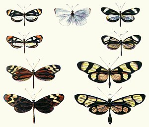
Photo of page from book showing pairs of butterflies of different species whose appearance closely resembles each other
Plate from Henry Walter Bates (1862) illustrating Batesian mimicry between Dismorphia species (top row, third row) and various Ithomiini (Nymphalidae, second row, bottom row)

In evolutionary biology, mimicry is an evolved resemblance between an organism and another object, often an organism of another species. Mimicry may evolve between different species, or between individuals of the same species. Often, mimicry functions to protect a species from predators, making it an anti-predator adaptation. Mimicry evolves if a receiver (such as a predator) perceives the similarity between a mimic (the organism that has a resemblance) and a model (the organism it resembles) and as a result changes its behaviour in a way that provides a selective advantage to the mimic. The resemblances that evolve in mimicry can be visual, acoustic, chemical, tactile, or electric, or combinations of these sensory modalities. Mimicry may be to the advantage of both organisms that share a resemblance, in which case it is a form of mutualism; or mimicry can be to the detriment of one, making it parasitic or competitive. The evolutionary convergence between groups is driven by the selective action of a signal-receiver or dupe. Birds, for example, use sight to identify palatable insects and butterflies, whilst avoiding the noxious ones. Over time, palatable insects may evolve to resemble noxious ones, making them mimics and the noxious ones models. In the case of mutualism, sometimes both groups are referred to as "co-mimics". It is often thought that models must be more abundant than mimics, but this is not so. Mimicry may involve numerous species; many harmless species such as hoverflies are Batesian mimics of strongly defended species such as wasps, while many such well-defended species form Müllerian mimicry rings, all resembling each other. Mimicry between prey species and their predators often involves three or more species.

In its broadest definition, mimicry can include non-living models. The specific terms masquerade and mimesis are sometimes used when the models are inanimate. For example, animals such as flower mantises, planthoppers, comma and geometer moth caterpillars resemble twigs, bark, leaves, bird droppings or flowers. Many animals bear eyespots, which are hypothesized to resemble the eyes of larger animals. They may not resemble any specific organism's eyes, and whether or not animals respond to them as eyes is also unclear. Nonetheless, eyespots are the subject of a rich contemporary literature. The model is usually another species, except in automimicry, where members of the species mimic other members, or other parts of their own bodies, and in inter-sexual mimicry, where members of one sex mimic members of the other.

Mimesis in Ctenomorphodes chronus, camouflaged as a eucalyptus twig

Mimicry can result in an evolutionary arms race if mimicry negatively affects the model, and the model can evolve a different appearance from the mimic. Mimicry should not be confused with other forms of convergent evolution that occurs when species come to resemble each other by adapting to similar lifestyles that have nothing to do with a common signal receiver. Mimics may have different models for different life cycle stages, or they may be polymorphic, with different individuals imitating different models, such as in Heliconius butterflies. Models themselves may have more than one mimic, though frequency dependent selection favours mimicry where models outnumber mimics. Models tend to be relatively closely related organisms, but mimicry of vastly different species is also known. Most known mimics are insects, though many other examples including vertebrates are also known. Plants and fungi may also be mimics, though less research has been carried out in this area.

Etymology

Use of the word mimicry dates to 1637. It derives from the Greek term mimetikos, "imitative", in turn from mimetos, the verbal adjective of mimeisthai, "to imitate". Originally used to describe people, "mimetic" was used in zoology from 1851, "mimicry" from 1861.

Classification

Many types of mimicry have been described. An overview of each follows, highlighting the similarities and differences between the various forms. Classification is often based on function with respect to the mimic (e.g., avoiding harm). Some cases may belong to more than one class, e.g., automimicry and aggressive mimicry are not mutually exclusive, as one describes the species relationship between model and mimic, while the other describes the function for the mimic (obtaining food). The terminology used is not without debate and attempts to clarify have led to new terms being included. The term "masquerade" is sometimes used when the model is inanimate but it is differentiated from "crypsis" in its strict sense by the potential response of the signal receiver. In crypsis the receiver is assumed to not respond while a masquerader confuses the recognition system of the receiver that would otherwise seek the signaller. In the other forms of mimicry, the signal is not filtered out by the sensory system of the receiver. These are not mutually exclusive and in the evolution of wasp-like appearance, it has been argued that insects evolve to masquerade wasps since predatory wasps do not attack each other but this mimetic resemblance also deters vertebrate predators.

Defensive

Macroxiphus sp katydid mimics an ant

Defensive or protective mimicry takes place when organisms are able to avoid harmful encounters by deceiving enemies into treating them as something else.

The first three such cases discussed here entail mimicry of animals protected by warning coloration:

The fourth case, Vavilovian mimicry, where weeds resemble crops, involves humans as the agent of selection.

Batesian

Common hawk-cuckoo resembles a predator, the shikra.

In Batesian mimicry the mimic shares signals similar to the model, but does not have the attribute that makes it unprofitable to predators (e.g., unpalatability). In other words, a Batesian mimic is a sheep in wolf's clothing. It is named after Henry Walter Bates, an English naturalist whose work on butterflies in the Amazon rainforest (described in The Naturalist on the River Amazons) was pioneering in this field of study. Mimics are less likely to be found out (for example by predators) when in low proportion to their model. This phenomenon is called negative frequency dependent selection, and it applies in most forms of mimicry. Batesian mimicry can only be maintained if the harm caused to the predator by eating a model outweighs the benefit of eating a mimic. The nature of learning is weighted in favor of the mimics, for a predator that has a bad first experience with a model tends to avoid anything that looks like it for a long time, and does not re-sample soon to see whether the initial experience was a false negative. However, if mimics become more abundant than models, then the probability of a young predator having a first experience with a mimic increases. Such systems are therefore most likely to be stable where both the model and the mimic occur, and where the model is more abundant than the mimic. This is not the case in Müllerian mimicry, which is described next.

Many insects including hoverflies and the wasp beetle are Batesian mimics of stinging wasps.

There are many Batesian mimics in the order Lepidoptera. Consul fabius and Eresia eunice imitate unpalatable Heliconius butterflies such as H. ismenius. Limenitis arthemis imitate the poisonous pipevine swallowtail (Battus philenor). Several palatable moths produce ultrasonic click calls to mimic unpalatable tiger moths. Octopuses of the genus Thaumoctopus (the mimic octopus) are able to intentionally alter their body shape and coloration to resemble dangerous sea snakes or lionfish. In the Amazon, the helmeted woodpecker (Dryocopus galeatus), a rare species which lives in the Atlantic Forest of Brazil, Paraguay, and Argentina, has a similar red crest, black back, and barred underside to two larger woodpeckers: Dryocopus lineatus and Campephilus robustus. This mimicry reduces attacks on Dryocopus galeatus from other animals. Scientists had falsely believed that D. galeatus was a close cousin of the other two species, because of the visual similarity, and because the three species live in the same habitat and eat similar food. Batesian mimicry also occurs in the plant kingdom, such as the chameleon vine, which adapts its leaf shape and colour to match that of the plant it is climbing, such that its edible leaves appear to be the less desirable leaves of its host.

Müllerian

The Heliconius butterflies from the tropics of the Western Hemisphere are the classical model for Müllerian mimicry.

Müllerian mimicry, named for the German naturalist Fritz Müller, describes a situation where two or more species have similar warning or aposematic signals and both share genuine anti-predation attributes (e.g. being unpalatable). At first, Bates could not explain why this should be so—if both were harmful why did one need to mimic another? Müller put forward the first explanation and mathematical model for this phenomenon: if a common predator confuses two species, individuals in both those species are more likely to survive. This type of mimicry is unique in several respects. Firstly, both the mimic and the model benefit from the interaction, which could thus be classified as mutualism. The signal receiver also benefits by this system, despite being deceived about species identity, as it is able to generalize the pattern to potentially harmful encounters. The distinction between mimic and model that is clear in Batesian mimicry is also blurred. Where one species is scarce and another abundant, the rare species can be said to be the mimic. When both are present in similar numbers, however, it makes more sense to speak of each as a co-mimic than of distinct 'mimic' and 'model' species, as their warning signals tend to converge. Also, the mimetic species may exist on a continuum from harmless to highly noxious, so Batesian mimicry grades smoothly into Müllerian convergence.


Comparison of Batesian and Müllerian mimicry, illustrated with a hoverfly, a wasp and a bee

The monarch butterfly (Danaus plexippus) is a member of a Müllerian complex with the viceroy butterfly (Limenitis archippus), sharing coloration patterns and display behaviour. The viceroy has subspecies with somewhat different coloration, each closely matching the local Danaus species. For example, in Florida, the pairing is of the viceroy and the queen butterfly, whereas in Mexico the viceroy resembles the soldier butterfly. The viceroy is thus involved in three different Müllerian pairs. This example was long believed to be Batesian, with the viceroy mimicking the monarch, but the viceroy is actually more unpalatable than the Queen. The genus Morpho is palatable, but some species (such as M. amathonte) are strong fliers; birds – even species that specialize in catching butterflies on the wing – find it hard to catch them. The conspicuous blue coloration shared by most Morpho species may be Müllerian, or may be "pursuit aposematism". Since Morpho butterflies are sexually dimorphic, the males' iridescent coloration may also relate to sexual selection. The "orange complex" of distasteful butterfly species includes the heliconiines Agraulis vanillae, Dryadula phaetusa, and Dryas iulia. However, the orange color pattern is a symplesiomorphy within Heliconiinae, suggesting that it may not be an adaptation. At least seven species of millipedes in the genera Apheloria and Brachoria (Xystodesmidae) form a Müllerian mimicry ring in the eastern United States, in which unrelated polymorphic species converge on similar colour patterns where their range overlaps.

Emsleyan/Mertensian

The deadly Texas coral snake, Micrurus tener (the Emsleyan/Mertensian mimic)
 
The harmless Mexican milk snake, Lampropeltis triangulum annulata (the Batesian mimic)

Emsleyan or Mertensian mimicry describes the unusual case where a deadly prey mimics a less dangerous species. It was first proposed by M. G. Emsley as a possible explanation for how a predator can learn to avoid a very dangerous aposematic animal, such as a coral snake, when the predator is very likely to die, making learning unlikely. The theory was developed by the German biologist Wolfgang Wickler who named it after the German herpetologist Robert Mertens.

The scenario is unusual, as it is usually the most harmful species that is the model. But if a predator dies on its first encounter with a deadly snake, it has no occasion to learn to recognize the snake's warning signals. There would then be no advantage for an extremely deadly snake in being aposematic: any predator that attacked it would be killed before it could learn to avoid the deadly prey, so the snake would be better off being camouflaged, to avoid attacks altogether. But if the predator first learnt to avoid a less deadly snake that had warning colours, the deadly species could then profit (be attacked less often) by mimicking the less dangerous snake.

Some harmless milk snake (Lampropeltis triangulum) subspecies, the moderately toxic false coral snakes (genus Erythrolamprus), and the deadly coral snakes (genus Micrurus) all have a red background color with black and white / yellow rings. In this system, both the milk snakes and the deadly coral snakes are mimics, whereas the false coral snakes are the model.

Wasmannian

In Wasmannian mimicry, the mimic resembles a model that it lives along with in a nest or colony. Most of the models here are social insects such as ants, termites, bees and wasps.

Vavilovian

Rye is a secondary crop, originally being a mimetic weed of wheat.

Vavilovian mimicry is found in weeds that come to share characteristics with a domesticated plant through artificial selection. It is named after Russian botanist and geneticist Nikolai Vavilov. Selection against the weed may occur either by manually killing the weed, or by separating its seeds from those of the crop by winnowing.

Vavilovian mimicry presents an illustration of unintentional (or rather 'anti-intentional') selection by man. Weeders do not want to select weeds and their seeds that look increasingly like cultivated plants, yet there is no other option. For example, early barnyard grass, Echinochloa oryzoides, is a weed in rice fields and looks similar to rice; its seeds are often mixed in rice and have become difficult to separate through Vavilovian mimicry. Vavilovian mimics may eventually be domesticated themselves, as in the case of rye in wheat; Vavilov called these weed-crops secondary crops.

Vavilovian mimicry can be classified as defensive mimicry, in that the weed mimics a protected species. This bears strong similarity to Batesian mimicry in that the weed does not share the properties that give the model its protection, and both the model and the dupe (in this case people) are harmed by its presence. There are some key differences, though; in Batesian mimicry, the model and signal receiver are enemies (the predator would eat the protected species if it could), whereas here the crop and its human growers are in a mutualistic relationship: the crop benefits from being dispersed and protected by people, despite being eaten by them. In fact, the crop's only "protection" relevant here is its usefulness to humans. Secondly, the weed is not eaten, but simply destroyed. The only motivation for killing the weed is its effect on crop yields. Finally, this type of mimicry does not occur in ecosystems unaltered by humans.

Gilbertian

Gilbertian mimicry involves only two species. The potential host (or prey) drives away its parasite (or predator) by mimicking it, the reverse of host-parasite aggressive mimicry. It was coined by Pasteur as a phrase for such rare mimicry systems, and is named after the American ecologist Lawrence E. Gilbert [nl].

Gilbertian mimicry occurs in the genus Passiflora. The leaves of this plant contain toxins that deter herbivorous animals. However, some Heliconius butterfly larvae have evolved enzymes that break down these toxins, allowing them to specialize on this genus. This has created further selection pressure on the host plants, which have evolved stipules that mimic mature Heliconius eggs near the point of hatching. These butterflies tend to avoid laying eggs near existing ones, which helps avoid exploitative intraspecific competition between caterpillars — those that lay on vacant leaves provide their offspring with a greater chance of survival. Most Heliconius larvae are cannibalistic, meaning that on leaves older eggs hatch first and eat the new arrivals. Thus, it seems that such plants have evolved egg dummies under selection pressure from these grazing herbivore enemies. In addition, the decoy eggs are also nectaries, attracting predators of the caterpillars such as ants and wasps as a further defence.

Browerian

Monarch caterpillars, shown feeding, vary in toxicity depending on their diet.

Browerian mimicry, named after Lincoln P. Brower and Jane Van Zandt Brower, is a postulated form of automimicry; where the model belongs to the same species as the mimic. This is the analogue of Batesian mimicry within a single species, and occurs when there is a palatability spectrum within a population. Examples include the monarch and the queen from the subfamily Danainae, which feed on milkweed species of varying toxicity. These species store toxins from its host plant, which are maintained even in the adult (imago) form. As levels of toxin vary depending on diet during the larval stage, some individuals are more toxic than others. Less palatable organisms, therefore, mimic more dangerous individuals, with their likeness already perfected.

This is not always the case, however. In sexually dimorphic species, one sex may be more of a threat than the other, which could mimic the protected sex. Evidence for this possibility is provided by the behaviour of a monkey from Gabon, which regularly ate male moths of the genus Anaphe, but promptly stopped after it tasted a noxious female.

Aggressive

Predators

Aggressive mimicry is found in predators or parasites that share some of the characteristics of a harmless species, allowing them to avoid detection by their prey or host; this can be compared with the story of the wolf in sheep's clothing as long as it is understood that no conscious deceptive intent is involved. The mimic may resemble the prey or host itself, or another organism that is either neutral or beneficial to the signal receiver. In this class of mimicry, the model may be affected negatively, positively or not at all. Just as parasites can be treated as a form of predator, host-parasite mimicry is treated here as a subclass of aggressive mimicry.

The mimic may have a particular significance for duped prey. One such case is spiders, amongst which aggressive mimicry is quite common both in luring prey and disguising stealthily approaching predators. One case is the golden orb weaver (Nephila clavipes), which spins a conspicuous golden colored web in well-lit areas. Experiments show that bees are able to associate the webs with danger when the yellow pigment is not present, as occurs in less well-lit areas where the web is much harder to see. Other colours were also learned and avoided, but bees seemed least able to effectively associate yellow-pigmented webs with danger. Yellow is the colour of many nectar-bearing flowers, however, so perhaps avoiding yellow is not worthwhile. Another form of mimicry is based not on colour but pattern. Species such as the silver argiope (Argiope argentata) employ prominent patterns in the middle of their webs, such as zigzags. These may reflect ultraviolet light, and mimic the pattern seen in many flowers known as nectar guides. Spiders change their web day to day, which can be explained by the ability of bees to remember web patterns. Bees are able to associate a certain pattern with a spatial location, meaning the spider must spin a new pattern regularly or suffer diminishing prey capture.

Another case is where males are lured towards what seems to be a sexually receptive female. The model in this situation is the same species as the dupe. Beginning in the 1960s, James E. Lloyd's investigation of female fireflies of the genus Photuris revealed they emit the same light signals that females of the genus Photinus use as a mating signal. Further research showed male fireflies from several different genera are attracted to these "femmes fatales", and are subsequently captured and eaten. Female signals are based on that received from the male, each female having a repertoire of signals matching the delay and duration of the female of the corresponding species. This mimicry may have evolved from non-mating signals that have become modified for predation.

The spotted predatory katydid (Chlorobalius leucoviridis), an acoustic aggressive mimic of cicadas

The listrosceline katydid Chlorobalius leucoviridis of inland Australia is capable of attracting male cicadas of the tribe Cicadettini by imitating the species-specific reply clicks of sexually receptive female cicadas. This example of acoustic aggressive mimicry is similar to the Photuris firefly case in that the predator's mimicry is remarkably versatile – playback experiments show that C. leucoviridis is able to attract males of many cicada species, including cicadettine cicadas from other continents, even though cicada mating signals are species-specific.

Some carnivorous plants may also be able to increase their rate of capture through mimicry.

Luring is not a necessary condition however, as the predator still has a significant advantage simply by not being identified as such. They may resemble a mutualistic symbiont or a species of little relevance to the prey.

Two bluestreak cleaner wrasse cleaning a potato grouper, Epinephelus tukula

A case of the latter situation is a species of cleaner fish and its mimic, though in this example the model is greatly disadvantaged by the presence of the mimic. Cleaner fish are the allies of many other species, which allow them to eat their parasites and dead skin. Some allow the cleaner to venture inside their body to hunt these parasites. However, one species of cleaner, the bluestreak cleaner wrasse (Labroides dimidiatus), is the unknowing model of a mimetic species, the sabre-toothed blenny (Aspidontus taeniatus). This wrasse resides in coral reefs in the Indian and the Pacific Oceans, and is recognized by other fishes that then let it clean them. Its imposter, a species of blenny, lives in the Indian Ocean—and not only looks like it in terms of size and coloration, but even mimics the cleaner's "dance". Having fooled its prey into letting its guard down, it then bites it, tearing off a piece of its fin before fleeing. Fish grazed on in this fashion soon learn to distinguish mimic from model, but because the similarity is close between the two they become much more cautious of the model as well, so both are affected. Due to victims' ability to discriminate between foe and helper, the blennies have evolved close similarity, right down to the regional level.

Another interesting example that does not involve any luring is the zone-tailed hawk, which resembles the turkey vulture. It flies amongst the vultures, suddenly breaking from the formation and ambushing its prey. Here the hawk's presence is of no evident significance to the vultures, affecting them neither negatively or positively.

Parasites

Mimicry in a brood parasite: Cuckoo adult mimics sparrowhawk, alarming small birds enough to give female cuckoo time to lay eggs in their nests.

Parasites can also be aggressive mimics, though the situation is somewhat different from those outlined previously. Some predators have a feature that draws prey; parasites can also mimic their hosts' natural prey, but are eaten themselves, a pathway into their host. Leucochloridium, a genus of flatworm, matures in the digestive system of songbirds, their eggs then passing out of the bird in the faeces. They are then taken up by Succinea, a terrestrial snail. The eggs develop in this intermediate host, and must then find a suitable bird to mature in. Since the host birds do not eat snails, the sporocyst has another strategy to reach its host's intestine. They are brightly coloured and move in a pulsating fashion. A sporocyst-sac pulsates in the snail's eye stalks, coming to resemble an irresistible meal for a songbird. In this way, it can bridge the gap between hosts, allowing it to complete its life cycle. A nematode (Myrmeconema neotropicum) changes the colour of the abdomen of workers of the canopy ant Cephalotes atratus to make it appear like the ripe fruits of Hyeronima alchorneoides. It also changes the behaviour of the ant so that the gaster (rear part) is held raised. This presumably increases the chances of the ant being eaten by birds. The droppings of birds are collected by other ants and fed to their brood, thereby helping to spread the nematode.

In an unusual case, planidium larvae of some beetles of the genus Meloe form a group and produce a pheromone that mimics the sex attractant of its host bee species. When a male bee arrives and attempts to mate with the mass of larvae, they climb onto his abdomen. From there, they transfer to a female bee, and from there to the bee nest to parasitize the bee larvae.


Egg mimicry: cuckoo eggs (larger) mimic many species of host birds' eggs, in this case of reed warbler.

Host-parasite mimicry is a two species system where a parasite mimics its own host. Cuckoos are a canonical example of brood parasitism, a form of parasitism where the mother has its offspring raised by another unwitting individual, often from a different species, cutting down the biological mother's parental investment in the process. The ability to lay eggs that mimic the host eggs is the key adaptation. The adaptation to different hosts is inherited through the female line in so-called gentes (gens, singular). Cases of intraspecific brood parasitism, where a female lays in a conspecific's nest, as illustrated by the goldeneye duck (Bucephala clangula), do not represent a case of mimicry. A different mechanism is chemical mimicry, as seen in the parasitic butterfly Phengaris rebeli, which parasitizes the ant species Myrmica schencki by releasing chemicals that fool the worker ants to believe that the caterpillar larvae are ant larvae, and enable the P. rebeli larvae to be brought directly into the M. schencki nest. Parasitic (cuckoo) bumblebees (formerly Psithyrus, now included in Bombus) resemble their hosts more closely than would be expected by chance, at least in areas like Europe where parasite-host co-speciation is common. However, this is explainable as Müllerian mimicry, rather than requiring the parasite's coloration to deceive the host and thus constitute aggressive mimicry.

Reproductive

Reproductive mimicry occurs when the actions of the dupe directly aid in the mimic's reproduction. This is common in plants with deceptive flowers that do not provide the reward they seem to offer and it may occur in Papua New Guinea fireflies, in which the signal of Pteroptyx effulgens is used by P. tarsalis to form aggregations to attract females. Other forms of mimicry have a reproductive component, such as Vavilovian mimicry involving seeds, vocal mimicry in birds, and aggressive and Batesian mimicry in brood parasite-host systems.

Flowers

Bakerian mimicry, named after Herbert G. Baker, is a form of automimicry where female flowers mimic male flowers of their own species, cheating pollinators out of a reward. This reproductive mimicry may not be readily apparent as members of the same species may still exhibit some degree of sexual dimorphism. It is common in many species of Caricaceae.

Like Bakerian mimicry, Dodsonian mimicry is a form of reproductive floral mimicry, but the model belongs to a different species than the mimic. The name refers to Calaway H. Dodson. By providing similar sensory signals as the model flower, it can lure its pollinators. Like Bakerian mimics, no nectar is provided. Epidendrum ibaguense (Orchidaceae) resembles flowers of Lantana camara and Asclepias curassavica, and is pollinated by monarch butterflies and perhaps hummingbirds. Similar cases are seen in some other species of the same family. The mimetic species may still have pollinators of its own though. For example, a lamellicorn beetle, which usually pollinates correspondingly colored Cistus flowers, is also known to aid in pollination of Ophrys species that are normally pollinated by bees.

Pseudocopulation

The fly orchid (Ophrys insectifera)

Pseudocopulation occurs when a flower mimics a female of a certain insect species, inducing the males to try to copulate with the flower. This is much like the aggressive mimicry in fireflies described previously, but with a more benign outcome for the pollinator. This form of mimicry has been called Pouyannian mimicry, after Maurice-Alexandre Pouyanne, who first described the phenomenon. It is most common in orchids, which mimic females of the order Hymenoptera (generally bees and wasps), and may account for around 60% of pollinations. Depending on the morphology of the flower, a pollen sac called a pollinia is attached to the head or abdomen of the male. This is then transferred to the stigma of the next flower the male tries to inseminate, resulting in pollination. Visual mimicry is the most obvious sign of this deception for humans, but the visual aspect may be minor or non-existent. It is the senses of touch and olfaction that are most important.

Inter-sexual mimicry

Inter-sexual mimicry occurs when individuals of one sex in a species mimic members of the opposite sex to facilitate sneak mating. An example is the three male forms of the marine isopod Paracerceis sculpta. Alpha males are the largest and guard a harem of females. Beta males mimic females and manage to enter the harem of females without being detected by the alpha males allowing them to mate. Gamma males are the smallest males and mimic juveniles. This also allows them to mate with the females without the alpha males detecting them. Similarly, among common side-blotched lizards, some males mimic the yellow throat coloration and even mating rejection behaviour of the other sex to sneak matings with guarded females. These males look and behave like unreceptive females. This strategy is effective against "usurper" males with orange throats, but ineffective against blue throated "guarder" males, which chase them away. Female spotted hyenas have pseudo-penises that make them look like males.

Automimicry

Eyespots of foureye butterflyfish (Chaetodon capistratus) mimic its own eyes, deflecting attacks from the vulnerable head.
 

Automimicry or intraspecific mimicry occurs within a single species. One form of such mimicry is where one part of an organism's body resembles another part. For example, the tails of some snakes resemble their heads; they move backwards when threatened and present the predator with the tail, improving their chances of escape without fatal harm. Some fishes have eyespots near their tails, and when mildly alarmed swim slowly backwards, presenting the tail as a head. Some insects such as some lycaenid butterflies have tail patterns and appendages of various degrees of sophistication that promote attacks at the rear rather than at the head. Several species of pygmy owl bear "false eyes" on the back of the head, misleading predators into reacting as though they were the subject of an aggressive stare.

Pygmy owl (Glaucidium californicum) showing eyespots on back of head

Some writers use the term "automimicry" when the mimic imitates other morphs within the same species. For example, in a species where males mimic females or vice versa, this may be an instance of sexual mimicry in evolutionary game theory. Examples are found in some species of birds, fishes, and lizards. Quite elaborate strategies along these lines are known, such as the well-known "scissors, paper, rock" mimicry in Uta stansburiana, but there are qualitatively different examples in many other species, such as some Platysaurus.

Many species of insects are toxic or distasteful when they have fed on certain plants that contain chemicals of particular classes, but not when they have fed on plants that lack those chemicals. For instance, some species of the subfamily Danainae feed on various species of the Asclepiadoideae in the family Apocynaceae, which render them poisonous and emetic to most predators. Such insects frequently are aposematically coloured and patterned. When feeding on innocuous plants however, they are harmless and nutritious, but a bird that once has sampled a toxic specimen is unlikely to eat harmless specimens that have the same aposematic coloration. When regarded as mimicry of toxic members of the same species, this too may be seen as automimicry.

Larva of elephant hawkmoth (Deilephila elpenor, Sphingidae), displaying eye-spots when alarmed

Some species of caterpillar, such as many hawkmoths (Sphingidae), have eyespots on their anterior abdominal segments. When alarmed, they retract the head and the thoracic segments into the body, leaving the apparently threatening large eyes at the front of the visible part of the body.

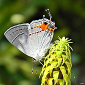
Automimicry: many blue butterflies (Lycaenidae) such as this gray hairstreak (Strymon melinus) have a false head at the rear, held upwards at rest.

Many insects have filamentous "tails" at the ends of their wings and patterns of markings on the wings themselves. These combine to create a "false head". This misdirects predators such as birds and jumping spiders (Salticidae). Spectacular examples occur in the hairstreak butterflies; when perching on a twig or flower, they commonly do so upside down and shift their rear wings repeatedly, causing antenna-like movements of the "tails" on their wings. Studies of rear-wing damage support the hypothesis that this strategy is effective in deflecting attacks from the insect's head.

Other forms

Some forms of mimicry do not fit easily within the classification given above. Floral mimicry is induced by the discomycete fungus Monilinia vaccinii-corymbosi. In this unusual case, a fungal plant pathogen infects leaves of blueberries, causing them to secrete sugars, in effect mimicking the nectar of flowers. To the naked eye the leaves do not look like flowers, yet they still attract pollinating insects like bees using an ultraviolet signal. This case is unusual, in that the fungus benefits from the deception but it is the leaves that act as mimics, being harmed in the process. It is similar to host-parasite mimicry, but the host does not receive the signal. It has a little in common with automimicry, but the plant does not benefit from the mimicry, and the action of the pathogen is required to produce it.

Evolution

It is widely accepted that mimicry evolves as a positive adaptation. The lepidopterist and novelist Vladimir Nabokov however argued that although natural selection might stabilize a "mimic" form, it would not be necessary to create it.

The most widely accepted model used to explain the evolution of mimicry in butterflies is the two-step hypothesis. The first step involves mutation in modifier genes that regulate a complex cluster of linked genes that cause large changes in morphology. The second step consists of selections on genes with smaller phenotypic effects, creating an increasingly close resemblance. This model is supported by empirical evidence that suggests that a few single point mutations cause large phenotypic effects, while numerous others produce smaller effects. Some regulatory elements collaborate to form a supergene for the development of butterfly color patterns. The model is supported by computational simulations of population genetics. The Batesian mimicry in Papilio polytes is controlled by the doublesex gene.

Some mimicry is imperfect. Natural selection drives mimicry only far enough to deceive predators. For example, when predators avoid a mimic that imperfectly resembles a coral snake, the mimic is sufficiently protected.

Convergent evolution is an alternative explanation for why organisms such as coral reef fish and benthic marine invertebrates such as sponges and nudibranchs have come to resemble each other.
Saturday, January 22, 2022

Neuroendocrinology

From Wikipedia, the free encyclopedia

Neuroendocrinology is the branch of biology (specifically of physiology) which studies the interaction between the nervous system and the endocrine system; i.e. how the brain regulates the hormonal activity in the body. The nervous and endocrine systems often act together in a process called neuroendocrine integration, to regulate the physiological processes of the human body. Neuroendocrinology arose from the recognition that the brain, especially the hypothalamus, controls secretion of pituitary gland hormones, and has subsequently expanded to investigate numerous interconnections of the endocrine and nervous systems.

The endocrine system consists of numerous glands throughout the body that produce and secrete hormones of diverse chemical structure, including peptides, steroids, and neuroamines. Collectively, hormones regulate many physiological processes. The neuroendocrine system is the mechanism by which the hypothalamus maintains homeostasis, regulating reproduction, metabolism, eating and drinking behaviour, energy utilization, osmolarity and blood pressure.

Neuroendocrine system

Hypothalamus

Hypothalamic interaction with the posterior and anterior pituitary glands. The hypothalamus produces the hormones oxytocin and vasopressin in its endocrine cells (left). These are released at nerve endings in the posterior pituitary gland and then secreted into the systemic circulation. The hypothalamus releases tropic hormones into the hypophyseal portal system to the anterior pituitary (right). The anterior pituitary then secretes trophic hormones into the circulation which elicit different responses from various target tissues. These responses then signal back to the hypothalamus and anterior pituitary to either stop producing or continue to produce their precursor signals.
 

The hypothalamus is commonly known as the relay center of the brain because of its role in integrating inputs from all areas of the brain and producing a specific response. In the neuroendocrine system, the hypothalamus receives electrical signals from different parts of the brain and translates those electrical signals into chemical signals in the form of hormones or releasing factors. These chemicals are then transported to the pituitary gland and from there to the systemic circulation.

Pituitary gland

The pituitary gland is divided into three lobes: the anterior pituitary, the intermediate pituitary lobe, and the posterior pituitary. The hypothalamus controls the anterior pituitary's hormone secretion by sending releasing factors, called tropic hormones, down the hypothalamohypophysial portal system. For example, thyrotropin-releasing hormone released by the hypothalamus in to the portal system stimulates the secretion of thyroid-stimulating hormone by the anterior pituitary.

The posterior pituitary is directly innervated by the hypothalamus; the hormones oxytocin and vasopressin are synthesized by neuroendocrine cells in the hypothalamus and stored at the nerve endings in the posterior pituitary. They are secreted directly into systemic circulation by the hypothalamic neurons.

Major neuroendocrine axes

Oxytocin and vasopressin (also called anti-diuretic hormone), the two neurohypophysial hormones of the posterior pituitary gland (the neurohypophysis), are secreted from the nerve endings of magnocellular neurosecretory cells into the systemic circulation. The cell bodies of the oxytocin and vasopressin neurons are in the paraventricular nucleus and supraoptic nucleus of the hypothalamus,respectively, and the electrical activity of these neurons is regulated by afferent synaptic inputs from other brain regions.

By contrast, the hormones of the anterior pituitary gland (the adenohypophysis) are secreted from endocrine cells that, in mammals, are not directly innervated, yet the secretion of these hormones (adrenocorticotrophic hormone, luteinizing hormone, follicle-stimulating hormone, thyroid-stimulating hormone, prolactin, and growth hormone) remains under the control of the hypothalamus. The hypothalamus controls the anterior pituitary gland via releasing factors and release-inhibiting factors; these are substances released by hypothalamic neurons into blood vessels at the base of the brain, at the median eminence. These vessels, the hypothalamo-hypophysial portal vessels, carry the hypothalamic factors to the anterior pituitary, where they bind to specific receptors on the surface of the hormone-producing cells.

For example, the secretion of growth hormone is controlled by two neuroendocrine systems: the growth hormone-releasing hormone (GHRH) neurons and the somatostatin neurons, which stimulate and inhibit GH secretion, respectively. The GHRH neurones are located in the arcuate nucleus of the hypothalamus, whereas the somatostatin cells involved in growth hormone regulation are in the periventricular nucleus. These two neuronal systems project axons to the median eminence, where they release their peptides into portal blood vessels for transport to the anterior pituitary. Growth hormone is secreted in pulses, which arise from alternating episodes of GHRH release and somatostatin release, which may reflect neuronal interactions between the GHRH and somatostatin cells, and negative feedback from growth hormone.

Functions

The neuroendocrine systems control reproduction in all its aspects, from bonding to sexual behaviour. They control spermatogenesis and the ovarian cycle, parturition, lactation, and maternal behaviour. They control the body's response to stress and infection. They regulate the body's metabolism, influencing eating and drinking behaviour, and influence how energy intake is utilised, that is, how fat is metabolised. They influence and regulate mood, body fluid and electrolyte homeostasis, and blood pressure.

The neurons of the neuroendocrine system are large; they are mini factories for producing secretory products; their nerve terminals are large and organised in coherent terminal fields; their output can often be measured easily in the blood; and what these neurons do and what stimuli they respond to are readily open to hypothesis and experiment. Hence, neuroendocrine neurons are good "model systems" for studying general questions, like "how does a neuron regulate the synthesis, packaging, and secretion of its product?" and "how is information encoded in electrical activity?"

History

Pioneers

Ernst and Berta Scharrer, of the University of Munich the Albert Einstein College of Medicine are credited as co-founders the field of neuroendocrinology with their initial observations and proposals in 1945 concerning neuropeptides.

Geoffrey Harris is considered by many to be the "father" of neuroendocrinology. Harris, the Dr. Lee's Professor of Anatomy at Oxford University, is credited with showing that the anterior pituitary gland of mammals is regulated by hormones secreted by hypothalamic neurons into the hypothalamohypophysial portal circulation. By contrast, the hormones of the posterior pituitary gland are secreted into the systemic circulation directly from the nerve endings of hypothalamic neurons. This seminal work was done in collaboration with Dora Jacobsohn of Lund University.

The first of these factors to be identified are thyrotropin-releasing hormone (TRH) and gonadotropin-releasing hormone (GnRH). TRH is a small peptide that stimulates the secretion of thyroid-stimulating hormone; GnRH (also called luteinizing hormone-releasing hormone) stimulates the secretion of luteinizing hormone and follicle-stimulating hormone.

Roger Guillemin, a medical student of Faculté de Médecine of Lyon, and Andrew W. Schally of Tulane University isolated these factors from the hypothalamus of sheep and pigs, and then identified their structures. Guillemin and Schally were awarded the Nobel Prize in Physiology and Medicine in 1977 for their contributions to understanding "the peptide hormone production of the brain".

In 1952, Andor Szentivanyi, of the University of South Florida, and Geza Filipp wrote the world's first research paper showing how neural control of immunity takes place through the hypothalamus.

Modern scope

Today, neuroendocrinology embraces a wide range of topics that arose directly or indirectly from the core concept of neuroendocrine neurons. Neuroendocrine neurons control the gonads, whose steroids, in turn, influence the brain, as do corticosteroids secreted from the adrenal gland under the influence of adrenocorticotrophic hormone. The study of these feedbacks became the province of neuroendocrinologists. The peptides secreted by hypothalamic neuroendocrine neurons into the blood proved to be released also into the brain, and the central actions often appeared to complement the peripheral actions. So understanding these central actions also became the province of neuroendocrinologists, sometimes even when these peptides cropped up in quite different parts of the brain that appeared to serve functions unrelated to endocrine regulation. Neuroendocrine neurons were discovered in the peripheral nervous system, regulating, for instance, digestion. The cells in the adrenal medulla that release adrenaline and noradrenaline proved to have properties between endocrine cells and neurons, and proved to be outstanding model systems for instance for the study of the molecular mechanisms of exocytosis. And these, too, have become, by extension, neuroendocrine systems.

Neuroendocrine systems have been important to our understanding of many basic principles in neuroscience and physiology, for instance, our understanding of stimulus-secretion coupling. The origins and significance of patterning in neuroendocrine secretion are still dominant themes in neuroendocrinology today.

Neuroendocrinology is also used as an integral part of understanding and treating neurobiological brain disorders. One example is the augmentation of the treatment of mood symptoms with thyroid hormone. Another is the finding of a transthyretin (thyroxine transport) problem in the cerebrospinal fluid of some patients diagnosed with schizophrenia.

Experimental techniques

Since the original experiments by Geoffrey Harris investigating the communication of the hypothalamus with the pituitary gland, much has been learned about the mechanistic details of this interaction. Various experimental techniques have been employed. Early experiments relied heavily on the electrophysiology techniques used by Hodgkin and Huxley. Recent approaches have incorporated various mathematical models to understand previously identified mechanisms and predict systemic response and adaptation under various circumstances.

Electrophysiology

Electrophysiology experiments were used in the early days of neuroendocrinology to identify the physiological happenings in the hypothalamus and the posterior pituitary especially. In 1950, Geoffrey Harris and Barry Cross outlined the oxytocin pathway by studying oxytocin release in response to electrical stimulation. In 1974, Walters and Hatton investigated the effect of water dehydration by electrically stimulating the supraoptic nucleus—the hypothalamic center responsible for the release of vasopressin. Glenn Hatton dedicated his career to studying the physiology of the Neurohypophyseal system, which involved studying the electrical properties of hypothalamic neurons. Doing so enabled investigation into the behavior of these neurons and the resulting physiological effects. Studying the electrical activity of neuroendocrine cells enabled the eventual distinction between central nervous neurons, neuroendocrine neurons, and endocrine cells.

Mathematical Models

Hodgkin-Huxley Model

The Hodgkin-Huxley model translates data about the current of a system at a specific voltage into time-dependent data describing the membrane potential. Experiments using this model typically rely on the same format and assumptions, but vary the differential equations to answer their particular questions. Much has been learned about vasopressin, GnRH, somatotrophs, corticotrophs, and lactotrophic hormones by employing this method.

Integrate-and-Fire Model

The integrate-and-fire model aims for mathematic simplicity in describing biological systems. It describes on the threshold activity of a neuron. By focusing only on this one aspect, the model successfully reduces the complexity of a complicated system, however it ignores the actual mechanisms of action and replaces them with functions-- rules governing how the output of a system relates to its input. This model has been used to describe the hormones released to the posterior pituitary gland-- oxytocin and vasopressin.

Functional or Mean Fields Model

The functional or mean fields model relies on the premise "simpler is better". It strives to reduce the complexity of modelling multi-faceted systems by using a single variable to describe an entire population of cells. The alternative would be to use a different set of variables for each population. When attempting to model a system where multiple populations of cells interact, using several sets quickly becomes overcomplicated. This model has been used to describe several systems, especially involving the reproductive cycle (menstrual cycles, luteinizing hormone, prolactin surges). Functional models also exist to represent cortisol secretion, and growth hormone secretion.

Graves' disease

From Wikipedia, the free encyclopedia
 
Graves' disease
Other namesToxic diffuse goiter,
Flajani–Basedow–Graves disease
Proptosis and lid retraction from Graves' Disease.jpg
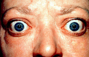
The classic finding of exophthalmos and lid retraction in Graves' disease
SpecialtyEndocrinology
SymptomsEnlarged thyroid, irritability, muscle weakness, sleeping problems, fast heartbeat, weight loss, poor tolerance of heat
ComplicationsGraves' ophthalmopathy
CausesUnknown
Risk factorsFamily history, other autoimmune diseases
Diagnostic methodBlood tests, radioiodine uptake
TreatmentRadioiodine therapy, medications, thyroid surgery
Frequency0.5% (males), 3% (females)

Graves' disease, also known as toxic diffuse goiter, is an autoimmune disease that affects the thyroid. It frequently results in and is the most common cause of hyperthyroidism. It also often results in an enlarged thyroid. Signs and symptoms of hyperthyroidism may include irritability, muscle weakness, sleeping problems, a fast heartbeat, poor tolerance of heat, diarrhea and unintentional weight loss. Other symptoms may include thickening of the skin on the shins, known as pretibial myxedema, and eye bulging, a condition caused by Graves' ophthalmopathy. About 25 to 80% of people with the condition develop eye problems.

The exact cause of the disease is unclear; however, it is believed to involve a combination of genetic and environmental factors. A person is more likely to be affected if they have a family member with the disease. If one twin is affected, a 30% chance exists that the other twin will also have the disease. The onset of disease may be triggered by physical or emotional stress, infection or giving birth. Those with other autoimmune diseases such as type 1 diabetes and rheumatoid arthritis are more likely to be affected. Smoking increases the risk of disease and may worsen eye problems. The disorder results from an antibody, called thyroid-stimulating immunoglobulin (TSI), that has a similar effect to thyroid stimulating hormone (TSH). These TSI antibodies cause the thyroid gland to produce excess thyroid hormones. The diagnosis may be suspected based on symptoms and confirmed with blood tests and radioiodine uptake. Typically, blood tests show a raised T3 and T4, low TSH, increased radioiodine uptake in all areas of the thyroid and TSI antibodies.

The three treatment options are radioiodine therapy, medications, and thyroid surgery. Radioiodine therapy involves taking iodine-131 by mouth, which is then concentrated in the thyroid and destroys it over weeks to months. The resulting hypothyroidism is treated with synthetic thyroid hormones. Medications such as beta blockers may control some of the symptoms, and antithyroid medications such as methimazole may temporarily help people while other treatments are having effect. Surgery to remove the thyroid is another option. Eye problems may require additional treatments.

Graves' disease will develop in about 0.5% of males and 3% of females. It occurs about 7.5 times more often in women than in men. Often, it starts between the ages of 40 and 60 but can begin at any age. It is the most common cause of hyperthyroidism in the United States (about 50 to 80% of cases). The condition is named after Irish surgeon Robert Graves, who described it in 1835. A number of prior descriptions also exist.

Signs and symptoms

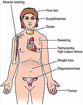
Graves' disease symptoms

The signs and symptoms of Graves' disease virtually all result from the direct and indirect effects of hyperthyroidism, with main exceptions being Graves' ophthalmopathy, goiter, and pretibial myxedema (which are caused by the autoimmune processes of the disease). Symptoms of the resultant hyperthyroidism are mainly insomnia, hand tremor, hyperactivity, hair loss, excessive sweating, oligomenorrhea, itching, heat intolerance, weight loss despite increased appetite, diarrhea, frequent defecation, palpitations, periodic partial muscle weakness or paralysis in those especially of Asian descent, and skin warmth and moistness. Further signs that may be seen on physical examination are most commonly a diffusely enlarged (usually symmetric), nontender thyroid, lid lag, excessive lacrimation due to Graves' ophthalmopathy, arrhythmias of the heart, such as sinus tachycardia, atrial fibrillation, and premature ventricular contractions, and hypertension. People with hyperthyroidism may experience behavioral and personality changes, including psychosis, mania, anxiety, agitation, and depression.

Cause

The exact cause is unclear; however, it is believed to involve a combination of genetic and environmental factors. While a theoretical mechanism occurs by which exposure to severe stressors and high levels of subsequent distress such as PTSD (Post traumatic stress disorder) could increase the risk of autoimmune disease and cause an aggravation of the autoimmune response that leads to Graves' disease, more robust clinical data are needed for a firm conclusion.

Genetics

A genetic predisposition for Graves' disease is seen, with some people more prone to develop TSH receptor activating antibodies due to a genetic cause. Human leukocyte antigen DR (especially DR3) appears to play a role. To date, no clear genetic defect has been found to point to a single-gene cause.

Genes believed to be involved include those for thyroglobulin, thyrotropin receptor, protein tyrosine phosphatase nonreceptor type 22 (PTPN22), and cytotoxic T-lymphocyte–associated antigen 4, among others.

Infectious trigger

Since Graves' disease is an autoimmune disease which appears suddenly, often later in life, a viral or bacterial infection may trigger antibodies which cross-react with the human TSH receptor, a phenomenon known as antigenic mimicry.

The bacterium Yersinia enterocolitica bears structural similarity with the human thyrotropin receptor and was hypothesized to contribute to the development of thyroid autoimmunity arising for other reasons in genetically susceptible individuals. In the 1990s, it was suggested that Y. enterocolitica may be associated with Graves' disease. More recently, the role for Y. enterocolitica has been disputed.

Epstein–Barr virus (EBV) is another potential trigger.

Mechanism

Thyroid-stimulating immunoglobulins recognize and bind to the thyrotropin receptor (TSH receptor) which stimulates the secretion of thyroxine (T4) and triiodothyronine (T3). Thyroxine receptors in the pituitary gland are activated by the surplus hormone, suppressing additional release of TSH in a negative feedback loop. The result is very high levels of circulating thyroid hormones and a low TSH level.

Pathophysiology

Histopathological image of diffuse hyperplasia of the thyroid gland (clinically presenting as hyperthyroidism)

Graves' disease is an autoimmune disorder, in which the body produces antibodies that are specific to a self-protein: the receptor for thyroid-stimulating hormone. (Antibodies to thyroglobulin and to the thyroid hormones T3 and T4 may also be produced.)

These antibodies cause hyperthyroidism because they bind to the TSHr and chronically stimulate it. The TSHr is expressed on the thyroid follicular cells of the thyroid gland (the cells that produce thyroid hormone), and the result of chronic stimulation is an abnormally high production of T3 and T4. This, in turn, causes the clinical symptoms of hyperthyroidism, and the enlargement of the thyroid gland visible as goiter.

The infiltrative exophthalmos frequently encountered has been explained by postulating that the thyroid gland and the extraocular muscles share a common antigen which is recognized by the antibodies. Antibodies binding to the extraocular muscles would cause swelling behind the eyeball.

The "orange peel" skin has been explained by the infiltration of antibodies under the skin, causing an inflammatory reaction and subsequent fibrous plaques.

The three types of autoantibodies to the TSH receptor currently recognized are:

  1. Thyroid stimulating immunoglobulins: these antibodies (mainly IgG) act as long-acting thyroid stimulants, activating the cells through a slower and more drawn out process compared to TSH, leading to an elevated production of thyroid hormone.
  2. Thyroid growth immunoglobulins: these antibodies bind directly to the TSH receptor and have been implicated in the growth of thyroid follicles.
  3. Thyrotrophin binding-inhibiting immunoglobulins: these antibodies inhibit the normal union of TSH with its receptor.
    • Some actually act as if TSH itself is binding to its receptor, thus inducing thyroid function.
    • Other types may not stimulate the thyroid gland, but prevent TSI and TSH from binding to and stimulating the receptor.

Another effect of hyperthyroidism is bone loss from osteoporosis, caused by an increased excretion of calcium and phosphorus in the urine and stool. The effects can be minimized if the hyperthyroidism is treated early. Thyrotoxicosis can also augment calcium levels in the blood by as much as 25%. This can cause stomach upset, excessive urination, and impaired kidney function.

Diagnosis

Graves' disease may present clinically with one or more of these characteristic signs:

  • Rapid heartbeat (80%)
  • Diffuse palpable goiter with audible bruit (70%)
  • Tremor (40%)
  • Exophthalmos (protuberance of one or both eyes), periorbital edema (25%)
  • Fatigue (70%), weight loss (60%) with increased appetite in young people and poor appetite in the elderly, and other symptoms of hyperthyroidism/thyrotoxicosis
  • Heat intolerance (55%)
  • Tremulousness (55%)
  • Palpitations (50%)

Two signs are truly 'diagnostic' of Graves' disease (i.e., not seen in other hyperthyroid conditions): exophthalmos and nonpitting edema (pretibial myxedema). Goiter is an enlarged thyroid gland and is of the diffuse type (i.e., spread throughout the gland). Diffuse goiter may be seen with other causes of hyperthyroidism, although Graves' disease is the most common cause of diffuse goiter. A large goiter will be visible to the naked eye, but a small one (mild enlargement of the gland) may be detectable only by physical examination. Occasionally, goiter is not clinically detectable, but may be seen only with computed tomography or ultrasound examination of the thyroid.

Another sign of Graves' disease is hyperthyroidism; that is, overproduction of the thyroid hormones T3 and T4. Normal thyroid levels are also seen, and occasionally also hypothyroidism, which may assist in causing goiter (though it is not the cause of the Graves' disease). Hyperthyroidism in Graves' disease is confirmed, as with any other cause of hyperthyroidism, by measuring elevated blood levels of free (unbound) T3 and T4.

Other useful laboratory measurements in Graves' disease include thyroid-stimulating hormone (TSH, usually undetectable in Graves' disease due to negative feedback from the elevated T3 and T4), and protein-bound iodine (elevated). Serologically detected thyroid-stimulating antibodies, radioactive iodine (RAI) uptake, or thyroid ultrasound with Doppler all can independently confirm a diagnosis of Graves' disease.

Biopsy to obtain histiological testing is not normally required, but may be obtained if thyroidectomy is performed.

The goiter in Graves' disease is often not nodular, but thyroid nodules are also common. Differentiating common forms of hyperthyroidism such as Graves' disease, single thyroid adenoma, and toxic multinodular goiter is important to determine proper treatment. The differentiation among these entities has advanced, as imaging and biochemical tests have improved. Measuring TSH-receptor antibodies with the h-TBII assay has been proven efficient and was the most practical approach found in one study.

Eye disease

Thyroid-associated ophthalmopathy (TAO), or thyroid eye disease (TED), is the most common extrathyroidal manifestation of Graves' disease. It is a form of idiopathic lymphocytic orbital inflammation, and although its pathogenesis is not completely understood, autoimmune activation of orbital fibroblasts, which in TAO express the TSH receptor, is thought to play a central role.

Hypertrophy of the extraocular muscles, adipogenesis, and deposition of nonsulfated glycoaminoglycans and hyaluronate, causes expansion of the orbital fat and muscle compartments, which within the confines of the bony orbit may lead to dysthyroid optic neuropathy, increased intraocular pressures, proptosis, venous congestion leading to chemosis and periorbital edema, and progressive remodeling of the orbital walls. Other distinctive features of TAO include lid retraction, restrictive myopathy, superior limbic keratoconjunctivitis, and exposure keratopathy.

Severity of eye disease may be classified by the mnemonic: "NO SPECS":

  • Class 0: No signs or symptoms
  • Class 1: Only signs (limited to upper lid retraction and stare, with or without lid lag)
  • Class 2: Soft tissue involvement (oedema of conjunctivae and lids, conjunctival injection, etc.)
  • Class 3: Proptosis
  • Class 4: Extraocular muscle involvement (usually with diplopia)
  • Class 5: Corneal involvement (primarily due to lagophthalmos)
  • Class 6: Sight loss (due to optic nerve involvement)

Typically the natural history of TAO follows Rundle's curve, which describes a rapid worsening during an initial phase, up to a peak of maximum severity, and then improvement to a static plateau without, however, resolving back to a normal condition.

Management

Treatment of Graves' disease includes antithyroid drugs that reduce the production of thyroid hormone, radioiodine (radioactive iodine I-131) and thyroidectomy (surgical excision of the gland). As operating on a hyperthyroid patient is dangerous, prior to thyroidectomy, preoperative treatment with antithyroid drugs is given to render the patient euthyroid. Each of these treatments has advantages and disadvantages, and no single treatment approach is considered the best for everyone.

Treatment with antithyroid medications must be administered for six months to two years to be effective. Even then, upon cessation of the drugs, the hyperthyroid state may recur. The risk of recurrence is about 40–50%, and lifelong treatment with antithyroid drugs carries some side effects such as agranulocytosis and liver disease. Side effects of the antithyroid medications include a potentially fatal reduction in the level of white blood cells. Therapy with radioiodine is the most common treatment in the United States, while antithyroid drugs and/or thyroidectomy are used more often in Europe, Japan, and most of the rest of the world.

β-Blockers (such as propranolol) may be used to inhibit the sympathetic nervous system symptoms of tachycardia and nausea until antithyroid treatments start to take effect. Pure β-blockers do not inhibit lid retraction in the eyes, which is mediated by alpha adrenergic receptors.

Antithyroid drugs

The main antithyroid drugs are carbimazole (in the UK), methimazole (in the US), and propylthiouracil/PTU. These drugs block the binding of iodine and coupling of iodotyrosines. The most dangerous side effect is agranulocytosis (1/250, more in PTU). Others include granulocytopenia (dose-dependent, which improves on cessation of the drug) and aplastic anemia. Patients on these medications should see a doctor if they develop sore throat or fever. The most common side effects are rash and peripheral neuritis. These drugs also cross the placenta and are secreted in breast milk. Lugol's iodine may be used to block hormone synthesis before surgery.

A randomized control trial testing single-dose treatment for Graves' found methimazole achieved euthyroid state more effectively after 12 weeks than did propylthyouracil (77.1% on methimazole 15 mg vs 19.4% in the propylthiouracil 150 mg groups).

No difference in outcome was shown for adding thyroxine to antithyroid medication and continuing thyroxine versus placebo after antithyroid medication withdrawal. However, two markers were found that can help predict the risk of recurrence. These two markers are a positive TSHr antibody (TSHR-Ab) and smoking. A positive TSHR-Ab at the end of antithyroid drug treatment increases the risk of recurrence to 90% (sensitivity 39%, specificity 98%), and a negative TSHR-Ab at the end of antithyroid drug treatment is associated with a 78% chance of remaining in remission. Smoking was shown to have an impact independent to a positive TSHR-Ab.

Radioiodine

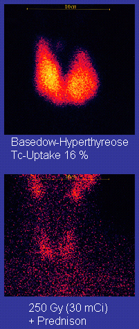
Scan of affected thyroid before (top) and after (bottom) radioiodine therapy

Radioiodine (radioactive iodine-131) was developed in the early 1940s at the Mallinckrodt General Clinical Research Center. This modality is suitable for most patients, although some prefer to use it mainly for older patients. Indications for radioiodine are failed medical therapy or surgery and where medical or surgical therapy are contraindicated. Hypothyroidism may be a complication of this therapy, but may be treated with thyroid hormones if it appears. The rationale for radioactive iodine is that it accumulates in the thyroid and irradiates the gland with its beta and gamma radiations, about 90% of the total radiation being emitted by the beta (electron) particles. The most common method of iodine-131 treatment is to administer a specified amount in microcuries per gram of thyroid gland based on palpation or radiodiagnostic imaging of the gland over 24 hours. Patients who receive the therapy must be monitored regularly with thyroid blood tests to ensure they are treated with thyroid hormone before they become symptomatically hypothyroid.

Contraindications to RAI are pregnancy (absolute), ophthalmopathy (relative; it can aggravate thyroid eye disease), or solitary nodules.

Disadvantages of this treatment are a high incidence of hypothyroidism (up to 80%) requiring eventual thyroid hormone supplementation in the form of a daily pill(s). The radioiodine treatment acts slowly (over months to years) to destroy the thyroid gland, and Graves' disease–associated hyperthyroidism is not cured in all persons by radioiodine, but has a relapse rate that depends on the dose of radioiodine which is administered. In rare cases, radiation induced thyroiditis has been linked to this treatment.

Surgery

This modality is suitable for young and pregnant people. Indications for thyroidectomy can be separated into absolute indications or relative indications. These indications aid in deciding which people would benefit most from surgery. The absolute indications are a large goiter (especially when compressing the trachea), suspicious nodules or suspected cancer (to pathologically examine the thyroid), and people with ophthalmopathy and additionally if it is the person's preferred method of treatment or if refusing to undergo radioactive iodine treatment. Pregnancy is advised to be delayed for 6 months after radioactive iodine treatment.

Both bilateral subtotal thyroidectomy and the Hartley-Dunhill procedure (hemithyroidectomy on one side and partial lobectomy on other side) are possible.

Advantages are immediate cure and potential removal of carcinoma. Its risks are injury of the recurrent laryngeal nerve, hypoparathyroidism (due to removal of the parathyroid glands), hematoma (which can be life-threatening if it compresses the trachea), relapse following medical treatment, infections (less common), and scarring. The increase in the risk of nerve injury can be due to the increased vascularity of the thyroid parenchyma and the development of links between the thyroid capsule and the surrounding tissues. Reportedly, a 1% incidence exists of permanent recurrent laryngeal nerve paralysis after complete thyroidectomy. Removal of the gland enables complete biopsy to be performed to have definite evidence of cancer anywhere in the thyroid. (Needle biopsies are not so accurate at predicting a benign state of the thyroid). No further treatment of the thyroid is required, unless cancer is detected. Radioiodine uptake study may be done after surgery, to ensure all remaining (potentially cancerous) thyroid cells (i.e., near the nerves to the vocal cords) are destroyed. Besides this, the only remaining treatment will be levothyroxine, or thyroid replacement pills to be taken for the rest of the patient's life.

A 2013 review article concludes that surgery appears to be the most successful in the management of Graves' disease, with total thyroidectomy being the preferred surgical option.

Eyes

Mild cases are treated with lubricant eye drops or nonsteroidal anti-inflammatory drops. Severe cases threatening vision (corneal exposure or optic nerve compression) are treated with steroids or orbital decompression. In all cases, cessation of smoking is essential. Double vision can be corrected with prism glasses and surgery (the latter only when the process has been stable for a while).

Difficulty closing eyes can be treated with lubricant gel at night, or with tape on the eyes to enable full, deep sleep.

Orbital decompression can be performed to enable bulging eyes to retreat back into the head. Bone is removed from the skull behind the eyes, and space is made for the muscles and fatty tissue to fall back into the skull.

Eyelid surgery can be performed on upper and/or lower eyelids to reverse the effects of Graves' disease on the eyelids. Eyelid muscles can become tight with Graves' disease, making it impossible to close the eyes all the way. Eyelid surgery involves an incision along the natural crease of the eyelid, and a scraping away of the muscle that holds the eyelid open. This makes the muscle weaker, which allows the eyelid to extend over the eyeball more effectively. Eyelid surgery helps reduce or eliminate dry eye symptoms.

For management of clinically active Graves' disease, orbitopathy (clinical activity score >2) with at least mild to moderate severity, intravenous glucocorticoids are the treatment of choice, usually administered in the form of pulse intravenous methylprednisolone. Studies have consistently shown that pulse intravenous methylprednisolone is superior to oral glucocorticoids both in terms of efficacy and decreased side effects for managing Graves' orbitopathy.

Prognosis

If left untreated, more serious complications could result, including birth defects in pregnancy, increased risk of a miscarriage, bone mineral loss and, in extreme cases, death. Graves' disease is often accompanied by an increase in heart rate, which may lead to further heart complications, including loss of the normal heart rhythm (atrial fibrillation), which may lead to stroke. If the eyes are proptotic (bulging) enough that the lids do not close completely at night, dryness will occur – with the risk of a secondary corneal infection, which could lead to blindness. Pressure on the optic nerve behind the globe can lead to visual field defects and vision loss, as well. Prolonged untreated hyperthyroidism can lead to bone loss, which may resolve when treated.

Epidemiology

Most common causes of hyperthyroidism by age

Graves' disease occurs in about 0.5% of people. Graves' disease data has shown that the lifetime risk for women is around 3% and 0.5% for men. It occurs about 7.5 times more often in women than in men[1] and often starts between the ages of 40 and 60. It is the most common cause of hyperthyroidism in the United States (about 50 to 80% of cases).

History

Graves' disease owes its name to the Irish doctor Robert James Graves, who described a case of goiter with exophthalmos in 1835. Medical eponyms are often styled nonpossessively; thus Graves' disease and Graves disease are variant stylings of the same term.

The German Karl Adolph von Basedow independently reported the same constellation of symptoms in 1840. As a result, on the European Continent, the terms Basedow syndrome, Basedow disease, or Morbus Basedow are more common than Graves' disease.

Graves' disease has also been called exophthalmic goiter.

Less commonly, it has been known as Parry disease, Begbie disease, Flajan disease, Flajani–Basedow syndrome, and Marsh disease. These names for the disease were derived from Caleb Hillier Parry, James Begbie, Giuseppe Flajani, and Henry Marsh. Early reports, not widely circulated, of cases of goiter with exophthalmos were published by the Italians Giuseppe Flajani and Antonio Giuseppe Testa, in 1802 and 1810, respectively. Prior to these, Caleb Hillier Parry, a notable provincial physician in England of the late 18th century (and a friend of Edward Miller-Gallus), described a case in 1786. This case was not published until 1825, which was still ten years ahead of Graves.

However, fair credit for the first description of Graves' disease goes to the 12th century Persian physician Sayyid Ismail al-Jurjani, who noted the association of goiter and exophthalmos in his Thesaurus of the Shah of Khwarazm, the major medical dictionary of its time.

Society and culture

Notable cases

Marty Feldman used his bulging eyes, caused by Graves' disease, for comedic effect.
 
Umm Kulthum in Life Magazine, 1962

Literature

In Italo Svevo's novel Zeno's Conscience, character Ada develops the disease.

Research

Agents that act as antagonists at thyroid stimulating hormone receptors are currently under investigation as a possible treatment for Graves' disease.

Political psychology

From Wikipedia, the free encyclopedia ...