Search This Blog

Monday, February 6, 2023

Venom

From Wikipedia, the free encyclopedia
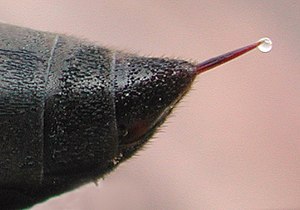
Wasp sting with a droplet of venom

Venom or zootoxin is a type of toxin produced by an animal that is actively delivered through a wound by means of a bite, sting, or similar action. The toxin is delivered through a specially evolved venom apparatus, such as fangs or a stinger, in a process called envenomation. Venom is often distinguished from poison, which is a toxin that is passively delivered by being ingested, inhaled, or absorbed through the skin, and toxungen, which is actively transferred to the external surface of another animal via a physical delivery mechanism.

Venom has evolved in terrestrial and marine environments and in a wide variety of animals: both predators and prey, and both vertebrates and invertebrates. Venoms kill through the action of at least four major classes of toxin, namely necrotoxins and cytotoxins, which kill cells; neurotoxins, which affect nervous systems; myotoxins, which damage muscles; and haemotoxins, which disrupt blood clotting. Venomous animals cause tens of thousands of human deaths per year.

Venoms are often complex mixtures of toxins of differing types. Toxins from venom are used to treat a wide range of medical conditions including thrombosis, arthritis, and some cancers. Studies in venomics are investigating the potential use of venom toxins for many other conditions.

Evolution

The use of venom across a wide variety of taxa is an example of convergent evolution. It is difficult to conclude exactly how this trait came to be so intensely widespread and diversified. The multigene families that encode the toxins of venomous animals are actively selected, creating more diverse toxins with specific functions. Venoms adapt to their environment and victims and accordingly evolve to become maximally efficient on a predator's particular prey (particularly the precise ion channels within the prey). Consequently, venoms become specialized to an animal's standard diet.

Mechanisms

Phospholipase A2, an enzyme in bee venom, releases fatty acids, affecting calcium signalling.

Venoms cause their biological effects via the many toxins that they contain; some venoms are complex mixtures of toxins of differing types. Major classes of toxin in venoms include:

Taxonomic range

Venom is widely distributed taxonomically, being found in both invertebrates and vertebrates, in aquatic and terrestrial animals, and among both predators and prey. The major groups of venomous animals are described below.

Arthropods

Venomous arthropods include spiders, which use fangs on their chelicerae to inject venom; and centipedes, which use forcipules, modified legs, to deliver venom; while scorpions and stinging insects inject venom with a sting. In bees and wasps, the sting is a modified egg-laying device – the ovipositor. In Polistes fuscatus, the female continuously releases a venom that contains a sex pheromone that induces copulatory behavior in males. In wasps such as Polistes exclamans, venom is used as an alarm pheromone, coordinating a response with from the nest and attracting nearby wasps to attack the predator. In some species, such as Parischnogaster striatula, venom is applied all over the body as an antimicrobial protection.

Many caterpillars have defensive venom glands associated with specialized bristles on the body called urticating hairs. These are usually merely irritating, but those of the Lonomia moth can be fatal to humans.

Bees synthesize and employ an acidic venom (apitoxin) to defend their hives and food stores, whereas wasps use a chemically different venom to paralyse prey, so their prey remains alive to provision the food chambers of their young. The use of venom is much more widespread than just these examples; many other insects, such as true bugs and many ants, also produce venom. The ant species Polyrhachis dives uses venom topically for the sterilisation of pathogens.

Other invertebrates

The fingernail-sized box jellyfish Malo kingi has among the most dangerous venom of any animal, causing Irukandji syndrome – severe pain, vomiting, and rapid rise in blood pressure.

There are venomous invertebrates in several phyla, including jellyfish such as the dangerous box jellyfish, the Portuguese man-of-war (a siphonophore) and sea anemones among the Cnidaria, sea urchins among the Echinodermata, and cone snails and cephalopods, including octopuses, among the Molluscs.

Vertebrates

Fish

Venom is found in some 200 cartilaginous fishes, including stingrays, sharks, and chimaeras; the catfishes (about 1000 venomous species); and 11 clades of spiny-rayed fishes (Acanthomorpha), containing the scorpionfishes (over 300 species), stonefishes (over 80 species), gurnard perches, blennies, rabbitfishes, surgeonfishes, some velvetfishes, some toadfishes, coral crouchers, red velvetfishes, scats, rockfishes, deepwater scorpionfishes, waspfishes, weevers, and stargazers.

Amphibians

Some salamanders can extrude sharp venom-tipped ribs. Two frog species in Brazil have tiny spines around the crown of their skulls which, on impact, deliver venom into their targets.

Reptiles

The venom of the prairie rattlesnake, Crotalus viridis (left), includes metalloproteinases (example on the bottom) which help digest prey before eating.

Some 450 species of snake are venomous. Snake venom is produced by glands below the eye (the mandibular glands) and delivered to the target through tubular or channeled fangs. Snake venoms contain a variety of peptide toxins, including proteases, which hydrolyze protein peptide bonds; nucleases, which hydrolyze the phosphodiester bonds of DNA; and neurotoxins, which disrupt signalling in the nervous system. Snake venom causes symptoms including pain, swelling, tissue necrosis, low blood pressure, convulsions, haemorrhage (varying by species of snake), respiratory paralysis, kidney failure, coma, and death. Snake venom may have originated with duplication of genes that had been expressed in the salivary glands of ancestors.

Venom is found in a few other reptiles such as the Mexican beaded lizard, the gila monster, and some monitor lizards, including the Komodo dragon. Mass spectrometry showed that the mixture of proteins present in their venom is as complex as the mixture of proteins found in snake venom. Some lizards possess a venom gland; they form a hypothetical clade, Toxicofera, containing the suborders Serpentes and Iguania and the families Varanidae, Anguidae, and Helodermatidae.

Mammals

Euchambersia, an extinct genus of therocephalians, is hypothesized to have had venom glands attached to its canine teeth.

A few species of living mammals are venomous, including solenodons, shrews, vampire bats, male platypuses, and slow lorises. Shrews have venomous saliva and most likely evolved their trait similarly to snakes. The presence of tarsal spurs akin to those of the platypus in many non-therian Mammaliaformes groups suggests that venom was an ancestral characteristic among mammals.

Extensive research on platypuses shows that their toxin was initially formed from gene duplication, but data provides evidence that the further evolution of platypus venom does not rely as much on gene duplication as was once thought. Modified sweat glands are what evolved into platypus venom glands. Although it is proven that reptile and platypus venom have independently evolved, it is thought that there are certain protein structures that are favored to evolve into toxic molecules. This provides more evidence of why venom has become a homoplastic trait and why very different animals have convergently evolved.

Venom and humans

Envenomation resulted in 57,000 human deaths in 2013, down from 76,000 deaths in 1990. Venoms, found in over 173,000 species, have potential to treat a wide range of diseases, explored in over 5,000 scientific papers.

In medicine, snake venom proteins are used to treat conditions including thrombosis, arthritis, and some cancers. Gila monster venom contains exenatide, used to treat type 2 diabetes. Solenopsins extracted from fire ant venom has demonstrated biomedical applications, ranging from cancer treatment to psoriasis. A branch of science, venomics, has been established to study the proteins associated with venom and how individual components of venom can be used for pharmaceutical means.

Resistance

The California ground squirrel is resistant to the Northern Pacific rattlesnake's powerful venom.

Venom is used as a trophic weapon by many predator species. The coevolution between predators and prey is the driving force of venom resistance, which has evolved multiple times throughout the animal kingdom. The coevolution between venomous predators and venom-resistant prey has been described as a chemical arms race. Predator/prey pairs are expected to coevolve over long periods of time. As the predator capitalizes on susceptible individuals, the surviving individuals are limited to those able to evade predation. Resistance typically increases over time as the predator becomes increasingly unable to subdue resistant prey. The cost of developing venom resistance is high for both predator and prey. The payoff for the cost of physiological resistance is an increased chance of survival for prey, but it allows predators to expand into underutilised trophic niches.

The California ground squirrel has varying degrees of resistance to the venom of the Northern Pacific rattlesnake. The resistance involves toxin scavenging and depends on the population. Where rattlesnake populations are denser, squirrel resistance is higher. Rattlesnakes have responded locally by increasing the effectiveness of their venom.

The kingsnakes of the Americas are constrictors that prey on many venomous snakes. They have evolved resistance which does not vary with age or exposure. They are immune to the venom of snakes in their immediate environment, like copperheads, cottonmouths, and North American rattlesnakes, but not to the venom of, for example, king cobras or black mambas.

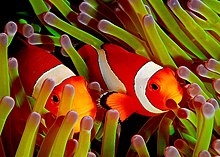
Ocellaris clownfish always live among venomous sea anemone tentacles and are resistant to the venom.

Among marine animals, eels are resistant to sea snake venoms, which contain complex mixtures of neurotoxins, myotoxins, and nephrotoxins, varying according to species. Eels are especially resistant to the venom of sea snakes that specialise in feeding on them, implying coevolution; non-prey fishes have little resistance to sea snake venom.

Clownfish always live among the tentacles of venomous sea anemones (an obligatory symbiosis for the fish), and are resistant to their venom. Only 10 known species of anemones are hosts to clownfish and only certain pairs of anemones and clownfish are compatible. All sea anemones produce venoms delivered through discharging nematocysts and mucous secretions. The toxins are composed of peptides and proteins. They are used to acquire prey and to deter predators by causing pain, loss of muscular coordination, and tissue damage. Clownfish have a protective mucus that acts as a chemical camouflage or macromolecular mimicry preventing "not self" recognition by the sea anemone and nematocyst discharge. Clownfish may acclimate their mucus to resemble that of a specific species of sea anemone.
Antivenom

From Wikipedia, the free encyclopedia

Antivenom
Snake Milking.jpg
Milking a snake for the production of antivenom
 
Clinical data
Other namesantivenin, antivenene
AHFS/Drugs.comMonograph
Routes of
administration
injection
ATC code
Identifiers
ChemSpider
  • none

Antivenom, also known as antivenin, venom antiserum, and antivenom immunoglobulin, is a specific treatment for envenomation. It is composed of antibodies and used to treat certain venomous bites and stings. Antivenoms are recommended only if there is significant toxicity or a high risk of toxicity. The specific antivenom needed depends on the species involved. It is given by injection.

Side effects may be severe. They include serum sickness, shortness of breath, and allergic reactions including anaphylaxis. Antivenom is traditionally made by collecting venom from the relevant animal and injecting small amounts of it into a domestic animal. The antibodies that form are then collected from the domestic animal's blood and purified.

Versions are available for spider bites, snake bites, fish stings, and scorpion stings. Due to the high cost of producing antibody-based antivenoms and their short shelf lives when not refrigerated, alternative methods of production of antivenoms are being actively explored. One such different method of production involves production from bacteria. Another approach is to develop targeted drugs (which, unlike antibodies, are usually synthetic and easier to manufacture at scale).

Antivenom was first developed in the late 19th century and came into common use in the 1950s. It is on the World Health Organization's List of Essential Medicines.

Medical uses

Antivenom is used to treat certain venomous bites and stings. They are recommended only if there is significant toxicity or a high risk of toxicity. The specific antivenom needed depends on the venomous species involved.

In the US, approved antivenom, including for pit viper (rattlesnake, copperhead and water moccasin) snakebite, is based on a purified product made in sheep known as CroFab. It was approved by the FDA in October, 2000. U.S. coral snake antivenom is no longer manufactured, and remaining stocks of in-date antivenom for coral snakebite expired in the Fall of 2009, leaving the U.S. without a coral snake antivenom. Efforts are being made to obtain approval for a coral snake antivenom produced in Mexico which would work against U.S. coral snakebite, but such approval remains speculative.

As an alternative when conventional antivenom is not available, hospitals sometimes use an intravenous version of the antiparalytic drug neostigmine to delay the effects of neurotoxic envenomation through snakebite. Some promising research results have also been reported for administering the drug nasally as a "universal antivenom" for neurotoxic snakebite treatment.

A monovalent antivenom is specific for one toxin or species, while a polyvalent one is effective against multiple toxins or species.

The majority of antivenoms (including all snake antivenoms) are administered intravenously; however, stonefish and redback spider antivenoms are given intramuscularly. The intramuscular route has been questioned in some situations as not uniformly effective.

Antivenoms bind to and neutralize the venom, halting further damage, but do not reverse damage already done. Thus, they should be given as soon as possible after the venom has been injected, but are of some benefit as long as venom is present in the body. Since the advent of antivenoms, some bites which were previously invariably fatal have become only rarely fatal provided that the antivenom is given soon enough.

Side effects

Antivenoms are purified from animal serum by several processes and may contain other serum proteins that can act as immunogens. Some individuals may react to the antivenom with an immediate hypersensitivity reaction (anaphylaxis) or a delayed hypersensitivity (serum sickness) reaction, and antivenom should, therefore, be used with caution. Although rare, severe hypersensitivity reactions including anaphylaxis to antivenom are possible. Despite this caution, antivenom is typically the sole effective treatment for a life-threatening condition, and once the precautions for managing these reactions are in place, an anaphylactoid reaction is not grounds to refuse to give antivenom if otherwise indicated. Although it is a popular myth that a person allergic to horses "cannot" be given antivenom, the side effects are manageable, and antivenom should be given rapidly as the side effects can be managed.

Method of preparation

Most antivenoms are prepared by freeze drying (synonym, cryodesiccation, lyophilization). The process involves freezing the antisera, followed by application of high vacuum. This causes frozen water to sublimate. Sera is reduced to powder with no water content. In such an environment, microorganisms and enzymes cannot degrade the antivenom, and it can be stored for up to 5 years [at normal temperatures]. Liquid antivenoms may also be stored for 5 years, but they must be stored at low temperatures [<8 degrees Celsius (or 46 degrees Fahrenheit)].

Mechanism

Antivenoms act by binding to and neutralizing venoms. The principle of antivenom is based on that of vaccines, developed by Edward Jenner; however, instead of inducing immunity in the person directly, it is induced in a host animal and the hyperimmunized serum is transfused into the person. The host animals may include horses, donkeys, goats, sheep, rabbits, chickens, llamas, and camels. In addition, opossums are being studied for antivenom production. Antivenoms for medical use are often preserved as freeze-dried ampoules, but some are available only in liquid form and must be kept refrigerated. They are not immediately inactivated by heat, however, so a minor gap in the cold chain is not disastrous.

History

Surgeon-Major Edward Nicholson wrote in the November 1870 Madras Medical Journal that he had witnessed a Burmese snake-catcher inoculating himself with cobra venom. However, the snake-catcher was unsure whether this was actually effective and therefore continued to treat his snakes with care. Nicholson, along with other Britons, began to consider that venom might provide its own cure. Although Scottish surgeon Patrick Russell had noted in the late 18th century that snakes were not affected by their own venom, it was not until the late 19th century that Joseph Frayer, Lawrence Waddell, and others began to consider venom-based remedies again. However, they and other naturalists working in India did not have the funding to fully develop their theories. Not until 1895 did Sir Thomas Fraser, Professor of Medicine at the University of Edinburgh, pick up Fayrer and Waddell's research to produce a serum to act against cobra venom. His 'Antivenin' was effective, but failed to make an impact as the public were focused on contemporary Pasteurian discoveries.

Another anti-ophidic serum was developed by Albert Calmette, a French scientist of the Pasteur Institute working at its Indochine branch in 1895, to treat the bites of the Indian Cobra (Naja naja).

In 1901, Vital Brazil, working at the Instituto Butantan in São Paulo, Brazil, developed the first monovalent and polyvalent antivenoms for Central and South American Crotalus and Bothrops genera, as well as for certain species of venomous spiders, scorpions, and frogs.

In Australia, the Commonwealth Serum Laboratories (CSL) began antivenom research in the 1920s. CSL has developed antivenoms for the redback spider, funnel-web spiders and all deadly Australian snakes.

Availability

There is an overall shortage of antivenom to treat snakebites. Because of this shortage, clinical researchers are considering whether lower doses may be as effective as higher doses in severe neurotoxic snake envenoming.

Snake antivenom is complicated and expensive for manufacturers to produce. When weighed against profitability (especially for sale in poorer regions), the result is that many snake antivenoms, world-wide, are very expensive. Availability, from region to region, also varies.

Internationally, antivenoms must conform to the standards of pharmacopoeia and the World Health Organization (WHO). Antivenoms have been developed for the venoms associated with the following animals:

Spiders

Antivenom Species Country
Funnel web spider antivenom Sydney funnel-web spider Australia
Soro antiaracnidico Brazilian wandering spider Brazil
Soro antiloxoscelico Recluse spider Brazil
Suero antiloxoscelico Chilean recluse Chile
Aracmyn All species of Loxosceles and Latrodectus Mexico
Redback spider antivenom Redback spider Australia
Black widow spider (Latrodectus Mactans) antivenin (equine origin) Southern black widow spider United States
SAIMR spider antivenom Button spider South Africa
Anti-Latrodectus antivenom Black widow spider Argentina

Acarids

Antivenom Species Country
Tick antivenom Paralysis tick Australia

Insects

Antivenom Species Country
soro antilonomico Lonomia obliqua caterpillar Brazil

Scorpions

Antivenom Species Country
Scorpion Venom Anti Serum (India) Purified lyophilized enzyme refined Equine Immunoglobulins Buthus tamulus India
ANTISCORP - Premium (Scorpion Venom Antiserum North Africa) Purified lyophilized enzyme refined Equine Immunoglobulins Androctonus amoerexi and Leiurus quinquestraiatus India
INOSCORPI MENA (Middle East and North Africa) Androctonus australis, Androctonus mauritanicus, Androctonus crassicauda, Buthus occitanus mardochei, Buthus occitanus occitanus, Leiurus quinquestriatus quinquestriatus, Leiurus quinquestriatus hebreus Spain
Alacramyn Centruroides limpidus, C. noxius, C. suffusus Mexico
Suero Antialacran Centruroides limpidus, C. noxius, C. suffusus Mexico
Tunisian polyvalent antivenom All Iranian scorpions Tunisia
Anti-Scorpion Venom Serum I.P. (AScVS) Indian red scorpion India
Anti-scorpionique Androctonus spp., Buthus spp. Algeria
Scorpion antivenom Black scorpion, Buthus occitanus Morocco
Soro antiscorpionico Tityus spp. Brazil
SAIMR scorpion antivenin Parabuthus spp. South Africa
Purified prevalent Anti-Scorpion Serum (equine source) Leiurus spp. and Androctonus scorpions Egypt

Marine animals

Antivenom Species Country
CSL box jellyfish antivenom Box jellyfish Australia
CSL stonefish antivenom Stonefish Australia

Snakes

Antivenom Species Country
PANAF PREMIUM (Sub-Sahara Africa) Purified lyophilized enzyme refined Equine Immunoglobulins  Echis ocellatus, Echis leucogaster, Echis carinatus, Bitis arietans, Bitis rhinoceros, Bitis nasicornis, Bitis gabonica, Dendroaspis polylepis, Dendroaspis viridis, Dendroaspis angusticeps, Dendroaspis jamesoni, Naja nigricollis, Naja melanoleuca and Naja haje India
Snake Venom Antiserum (India) Purified lyophilized enzyme refined Equine Immunoglobulins Naja naja, Vipera russelii and Echis carinatus India
INOSERP MENA (Middle East and North Africa) Bitis arietans, Cerastes cerastes, Cerastes gasperettii,Cerastes vipera, Daboia deserti, Daboia mauritanica, Daboia palaestinae, Echis carinatus sochureki, Echis coloratus, Echis khosatzkii, Echis leucogaster, Echis megalocephalus, Echis omanensis, Echis pyramidum, Macrovipera lebetina obtusa, Macrovipera lebetina transmediterranea, Macrovipera lebetina turanica, Montivipera bornmuelleri, Montivipera raddei kurdistanica, Pseuocerastes fieldi, Pseudocerastes persicus, Vipera latastei, Naja haje, Naja nubiae, Naja pallida and Walterinnesia aegyptia Spain
INOSERP PAN-AFRICA (Sub-Sahara Africa) Echis ocellatus, Bitis arietans, Dendroaspis polylepis and Naja nigricollis Spain
EchiTAbG (Sub-Sahara Africa)[33] Echis ocellatus, Echis pyramidum Wales, UK
Polyvalent snake antivenom ANAVIP South American rattlesnake Crotalus durissus and fer-de-lance Bothrops asper Mexico (Instituto Bioclon); South America
Polyvalent snake antivenom Saw-scaled viper Echis carinatus, Russell's viper Daboia russelli, spectacled cobra Naja naja, common krait Bungarus caeruleus India
Death adder antivenom Death adder Australia
Taipan antivenom Taipan Australia
Black snake antivenom Pseudechis spp. Australia
Tiger snake antivenom Australian copperheads, tiger snakes, Pseudechis spp., rough-scaled snake Australia
Brown snake antivenom Brown snakes Australia
Polyvalent snake antivenom Australian snakes as listed above Australia
Sea snake antivenom Sea snakes Australia
Vipera tab Vipera spp. UK
Polyvalent crotalid antivenin (CroFab—Crotalidae Polyvalent Immune Fab (Ovine)) North American pit vipers (all rattlesnakes, copperheads, and cottonmouths) North America
Soro antibotropicocrotalico Pit vipers and rattlesnakes Brazil
Antielapidico Coral snakes Brazil
SAIMR polyvalent antivenom Mambas, cobras, Rinkhalses, puff adders (Unsuitable small adders: B. worthingtoni, B. atropos, B. caudalis, B. cornuta, B. heraldica, B. inornata, B. peringueyi, B. schneideri, B. xeropaga) South Africa
SAIMR echis antivenom Saw-scaled vipers South Africa
SAIMR Boomslang antivenom Boomslang South Africa
Panamerican serum Coral snakes Costa Rica
Anticoral Coral snakes Costa Rica
Anti-mipartitus antivenom Coral snakes Costa Rica
Anticoral monovalent Coral snakes Costa Rica
Antimicrurus Coral snakes Argentina
Coralmyn Coral snakes Mexico
Anti-micruricoscorales Coral snakes Colombia
crotalidae immune F(ab')2 (equine)) (Anavip) North American species of Crotalinae US

Terminology

The name "antivenin" comes from the French word venin, meaning venom, which in turn was derived from Latin venenum, meaning poison.

Historically, the term antivenin was predominant around the world, its first published use being in 1895. In 1981, the World Health Organization decided that the preferred terminology in the English language would be venom and antivenom rather than venin and antivenin or venen and antivenene.

Recombinant antibodies

From Wikipedia, the free encyclopedia

Recombinant antibodies are antibody fragments produced by using recombinant antibody coding genes. They mostly consist of a heavy and light chain of the variable region of immunoglobulin. Recombinant antibodies have many advantages in both medical and research applications, which make them a popular subject of exploration and new production against specific targets. The most commonly used form is the single chain variable fragment (scFv), which has shown the most promising traits exploitable in human medicine and research. In contrast to monoclonal antibodies produced by hybridoma technology, which may lose the capacity to produce the desired antibody over time or the antibody may undergo unwanted changes, which affect its functionality, recombinant antibodies produced in phage display maintain high standard of specificity and low immunogenicity.

Structure and characterization

Formats

There are several known formats of recombinant antibodies which are commonly produced. These are the Fab recombinant antibodies, scFv and diabodies. Each of the formats has a slightly different potential in applications and may be used in various fields of research as well as human and animal medicine. Another researched possibility is the development of anti-idiotypic antibodies. Anti-idiotypic antibodies bind to a paratope of another specific antibody. Therefore, it can be used for measuring presence of antibodies and drug loads in patients' sera. Based on their binding specificity 3 types of anti-idiotypic antibodies can be distinguished, which partially overlap with the previously mentioned formats: the classical ones, a group including Fab fragment antibodies, antibodies binding to idiotope outside of the drug binding site and antibodies, which only bind to the already assembled complex of drug bound to the target. The most commonly used are the scFv, Fab fragments and bispecific antibodies.

Single chain variable fragment (scFv)

scFv is the smallest of the recombinant antibody formats, which is capable of antigen binding. They have a molecular weight of approximately 27kDa. They are formed by light and heavy chain of the variable region of an immunoglobulin. The two chains are linked by a flexible peptide linker. The flexible peptide linker usually consists of short sequence repetition. The sequence is made up of four glycines and a serine and it serves the purpose of stabilization of the fragment. The functionality may be enhanced by site-specific chemical modifications, adding a peptide-tag or by fusion with a gene to achieve production of bifunctional recombinant antibodies. It is important to establish the binding activity in order to ensure good functionality of the product. To determine the binding activity, ELISA assay is routinely performed.

Fab fragments

Structurally Fab fragments consist of two sets of variable and constant components, which create two polypetide chains. Together they form a stable structure. As a member of the anti-idiotypic antibodies, Fab fragment recombinant antibodies bind directly to the paratope of the target antibody. That means that they compete with the drug for binding site and have an inhibitory function. Fab fragment antibodies can be used for detection of not bound drugs or free drugs in the serum. Fab antibodies have also been used to avoid the adverse effects caused by unspecific binding of the Fc portion of the antibody, which is missing in the Fab fragment. In case the IgG immunoglobulin was more suitable for the treatment or some other particular application, experiments have also been conducted, in which the recombinant Fab fragments were converted into recombinant IgG form. This possibility further broadens the pool of potential target structures.

Bispecific recombinant antibodies

Along scFv and Fab fragments, diabodies or bispecific recombinant antibodies are the third major format. Bispecific antibodies combine two different antigen binding specificities within one molecule. The bispecific antibodies are used to crosslink the target molecules with two different cells and mediate direct cytotoxicity.

Production and development

Production of recombinant antibodies

The production of recombinant antibodies follows principally similar workflow. It consists of determining the sequence of the desired product followed by refinement of the codon, then gene synthesis and construct generation. Once the construct is delivered to the laboratory, expression constructs are produced, then they are transferred to a cell culture in the process called transfection and once the cell culture produces the desired recombinant antibody, it is regularly collected, purified and analyzed or used for further experimentation. For recombinant antibody production the stable cell lines such as CHO and HEK293 are used. Optimizations of mammalian cell cultures have led to increase the yield of antibodies from HEK293 or CHO cell lines to over 12g/liter. In the beginning phases of the recombinant antibody production it was important to achieve the assembly of a functional Fv fragment in Escherichia coli. The correct fold is essential for functionality of the antibody. Second essential prerequisite for the modern day production of scFv was the successful assembly of recombinant antibodies from heavy and light chain of immunoglobulin. These two experiments allowed for further development and refinement of the recombinant antibodies until modern day form. Today's in vitro production process eliminates the need for laboratory animals. Using a synthetic or human Ab library, as opposed to immunization of animals and the subsequent generation of stable hybridoma cell lines, requires fewer resources and produces less waste, making the entire process more sustainable. 

Hybridoma

Monoclonal antibodies are essential for many therapies applied today in human medicine. The first successful technology which was robust and led to a stable production of desired antibodies was hybridoma technology. The hybridoma cell lines, which produced large quantities of relatively pure and predictable antibodies was first introduced in 1975. Since then, it has been used for various purposes scaling from diagnostic and therapeutic to research applications. Despite its indisputable role in scientific discoveries and numerous treatment strategies, the hybridoma technology presents researchers with some obstacles such as ethical issues, potential to lose expression of the target protein or lengthy production and most importantly the development of HAMA in patients as mentioned previously. Therefore, different methods need to complement or even partially replace the hybridoma. Hybridomas are an essential part of the recombinant antibody generation even today as they are still used to produce the monoclonal antibodies, from which the Fab fragments, scFv or somatically fused antibodies create a bispecific antibody.

Phage display

The most commonly applied technology to produce recombinant antibodies in the laboratory settings today is the phage display. Phage display is a method, in which the target recombinant antibody is produced on the surface of a bacteriophage. This allows for a fast recombinant antibody production and easy manipulation in the laboratory conditions. Both scFv and Fab fragment recombinant antibodies are routinely produced using the antibody phage display. From all the possible phage display systems, the most common is the Escherichia coli, due to its rapid growth and division rate and cheap set up and maintenance.

Engineering and development

Two main strategies have been described to engineer the scFv fragments. The first one is the so-called non-colinear approach. It works on the principle of heterodimerization of two chains. Non-colinear approach leads to production of diabodies and recombinant antibodies, which combine two specificities. The second approach is called colinear and it described the process of fusion of two different scFv with a biologically active protein.

Medical and research applications

Recombinant antibodies fulfill a large spectrum of functions spanning from research to diagnosis and treatment therapies for various diseases. Their specificity and low immunogenicity make them a great alternative to traditional forms of treatment, increasing the accuracy of targeting specific molecules and avoiding adverse side effects.

Recombinant antibodies have been explored as a treatment for cancer, HIV, herpes simplex virus (HSV) and more. ScFv have been a part of the highly promising therapeutic approach of universal chimeric antigen receptors (uniCAR) technology, which shows promising results. The scFv are part of the technology in the form of target modules, which direct the immune response to specific cancer cells, expressing the target antigen. In case of research into HIV treatment, the recombinant antibodies are rather used for their neutralizing quality. The same goes for HSV infection. Specific recombinant antibodies are designed to bind with to surface heparin sulphate proteoglycan (HSP), which complicates or even disables the entry of the HSV into the host cell. This is a method which significantly decreases the severity of HSV infection.

As was mentioned in the beginning of this section, recombinant antibodies can also be used in diagnosis, an example of such diagnostic application is the detection of rabies virus. Since the current diagnostic antibodies are not as accurate as would be desired, the recombinant antibodies offer a promising alternative. In case of rabies infection, which is only treatable shortly after exposure, accurate and precise diagnosis is vital for survival of the patient. In comparison to commercially produced and commonly available antibodies, the recombinant antibodies are cheaper to produce and more accurate in determining the infection. Another advantage of the recombinant antibody is the potential application as a neutralizing antibody as part of the subsequent treatment.

The potential of recombinant antibodies in human and animal medicine is immense as shown even by the few selected examples. As mentioned previously the recombinant antibodies and especially those, which have been developed in phage display are highly specific, have great pharmacokinetics and could be used in wide range of treatments. However, it is important to realize that it is not expected or desired for the recombinant antibodies created in phage display to completely replace the hybridoma antibody production but rather to complement it.

Advantages of using recombinant antibodies

Recombinant antibodies bring many advantages with their application in human medicine and research. The first one is the complete elimination of ethical issues because there is no need for animal immunization. The cultivation of CHO cells for recombinant antibody expression is a popular strategy for antibody producers since the cell structure is similar to that of the human body. Thanks to their size, which is smaller than complete antibody and particularly than 2000 nm, yet not smaller than 8 nm they are cleared from the organism with ease and in a timely manner, through the renal pathway, which is the desirable clearance. Another great advantage is their monovalency, which means that they are highly specific and bind to a single antigen. Researchers have managed to produce antibodies carrying no other activity than the antigen binding. Since the recombinant antibodies are sequence defined they are more reliable as well as reproducible. In combination with their small size the great specificity can be exploited to deliver highly specific drug to a specific site precisely because the small size predisposes the recombinant antibodies to penetrate tissues more easily. It has been reported that the recombinant antibodies penetrate tumor tissue better than the full-length IgG immunoglobulins. The small size also adds to better biodistribution in the patient. In comparison to antibodies derived from hybridoma cell lines the recombinant antibodies do not cause immunogenicity, the infamous human anti-mouse antibody (HAMA). Further advantages show afucosylated recombinant antibodies which are used successfully in the fight against cancer.

These were the top advantages for use in patients. However, the use of recombinant antibodies is also advantageous compared to traditional monoclonal antibodies derived from hybridoma cell lines during their production as well. The production is much faster and we have better control over the process than in hybridoma technology. Moreover, the recombinant antibodies may be designed virtually against any antigen, of the proper size and shape, but they are not solely limited to the peptide nature of an antigen. The recombinant antibodies may also be used in fused form with drugs and/or toxins, which may be further exploited in the medical applications. Last but not least of their advantages during production is the possibility to optimize and genetically engineer the recombinant antibodies based on the current demand of the patient or researcher. An experienced technician is required to perform the phage display and third it is almost inevitable to include outsource companies in the process for the gene synthesis and construct generation. However, in a systematic comparison of animal derived antibodies verus phage display derived recombinant antibodies used for research and diagnostic applications, the EU Reference Laboratory for Alternatives to Animal Testing (EURL ECVAM) released a recommendation in favor of on non-animal derived antibodies in May 2020, mainly based on the fact that in contrast to animal derived antibodies, recombinant antibodies are always sequence defined protein reagents, allowing to eliminate some of the quality issues attributed to current research antibodies when made in animals.

Sunday, February 5, 2023

Affimer

From Wikipedia, the free encyclopedia
The Affimer protein scaffold – showing the two loops where random peptides can be inserted to create a protein binding surface

Affimer molecules are small proteins that bind to target proteins with affinity in the nanomolar range. These engineered non-antibody binding proteins are designed to mimic the molecular recognition characteristics of monoclonal antibodies in different applications. These affinity reagents have been optimized to increase their stability, make them tolerant to a range of temperatures and pH, reduce their size, and to increase their expression in E.coli and mammalian cells.

Development

Affimer proteins were developed initially at the MRC Cancer Cell Unit in Cambridge then across two laboratories at the University of Leeds. Derived from the cysteine protease inhibitor family of cystatins, which function in nature as cysteine protease inhibitors, these 12–14 kDa proteins share the common tertiary structure of an alpha-helix lying on top of an anti-parallel beta-sheet.

Affimer proteins display two peptide loops that can all be randomized to bind to desired target proteins, in a similar manner to monoclonal antibodies. Stabilization of the two peptides by the protein scaffold constrains the possible conformations that the peptides can take. This increases the binding affinity and specificity compared to libraries of free peptides, though can limit the target repertoire of Affimers.

Production

Phage display libraries of 109 randomized sequences are used to screen for Affimer proteins that exhibit high-specificity binding to the target protein with binding affinities in the nM range. The ability to direct in vitro screening techniques allows the identification of specific, high affinity Affimers. In vitro screening and development also mean that the target space for Affimers is not limited by the animal immune system. Affimers are generated using recombinant systems, so their generation is more rapid and reproducible compared to the production of polyclonal antibodies.

Multimeric forms Affimers have been generated and shown to yield titres in the range of 200–400 mg/L under small-scale culture using bacterial host systems. Multimeric forms of Affimers with the same target specificity provide avidity effects in target binding.

Many different tags and fusion proteins, such as fluorophores, single-stranded DNA, His, and c-Myc tags can be conjugated to Affimers. Specific cysteine residues can be introduced to the protein to allow thiol chemistry to uniformly orient Affimers on a solid support eg ELISA plates. This flexible functionalisation of the Affimer molecule allows functionality across multiple applications and assay formats.

Properties

Affimers are recombinant proteins. As they are manufactured using recombinant bacterial production processes, the batch-to-batch consistency for Affimers is improved compared to polyclonal antibodies, overcoming some of the issues of reproducibility and security of supply.

These synthetic antibodies were engineered to be stable, non-toxic, biologically neutral and contain no post-translational modifications or disulfide bridges. Two separate loop sequences, incorporating a total of 12 to 36 amino acids, form the target interaction surface so interaction surfaces can range form 650–1000 Å. The large interaction surface results allows binding to target proteins.

Applications

Affimer technology has been commercialised and developed by Avacta, who are developing these affinity reagents as tools for diagnostics and as biotherapeutics.

Reagents and Diagnostics

Affimer binders have been used across a number of platforms, including ELISA, surface plasmon resonance, affinity purification. Affimers that inhibit protein-protein interactions can be produced with the potential to express these inhibitors in mammalian cells modify signalling pathways as cell therapies.

Therapeutics

The small size and stability profile of Affimers combined with their human origin confer drug-like properties. This may represent advantages over antibodies in terms of tissue penetration, for example in solid tumours where Avacta are developing PD-L1 inhibitors as alternatives to Opdivo and Yervoy, though requires half life modification to prevent rapid excretion through the kidney.

Affimers can be conjugated to form multimers for the design of therapeutics. Examples include the production of multi-specific Affimer molecules to albumin binders to increase their half-life in vivo and for use as the targeting moiety in chimeric receptors or modified to carry a toxin in Affimer-drug conjugates.

Affimers as therapeutics are in discovery and preclinical development to tackle cancer, both via CAR-T cell therapy and as immune checkpoint inhibitors. Early studies using ex vivo human samples showed low immunogenicity associated with the Affimer scaffold, at levels comparable to a marketed antibody therapeutic. Furthermore, initial preclinical studies showed good efficacy and tolerability of the anti-PDL1 immuno-oncology Affimers in mice. It is anticipated that IND filing for the first Affimer therapeutic will occur in 2023.

Butane

From Wikipedia, the free encyclopedia ...