Search This Blog

Tuesday, June 20, 2023

Psychophysics

From Wikipedia, the free encyclopedia

Psychophysics quantitatively investigates the relationship between physical stimuli and the sensations and perceptions they produce. Psychophysics has been described as "the scientific study of the relation between stimulus and sensation" or, more completely, as "the analysis of perceptual processes by studying the effect on a subject's experience or behaviour of systematically varying the properties of a stimulus along one or more physical dimensions".

Psychophysics also refers to a general class of methods that can be applied to study a perceptual system. Modern applications rely heavily on threshold measurement, ideal observer analysis, and signal detection theory.

Psychophysics has widespread and important practical applications. For example, in the study of digital signal processing, psychophysics has informed the development of models and methods of lossy compression. These models explain why humans perceive very little loss of signal quality when audio and video signals are formatted using lossy compression.

History

Many of the classical techniques and theories of psychophysics were formulated in 1860 when Gustav Theodor Fechner in Leipzig published Elemente der Psychophysik (Elements of Psychophysics). He coined the term "psychophysics", describing research intended to relate physical stimuli to the contents of consciousness such as sensations (Empfindungen). As a physicist and philosopher, Fechner aimed at developing a method that relates matter to the mind, connecting the publicly observable world and a person's privately experienced impression of it. His ideas were inspired by experimental results on the sense of touch and light obtained in the early 1830s by the German physiologist Ernst Heinrich Weber in Leipzig, most notably those on the minimum discernible difference in intensity of stimuli of moderate strength (just noticeable difference; jnd) which Weber had shown to be a constant fraction of the reference intensity, and which Fechner referred to as Weber's law. From this, Fechner derived his well-known logarithmic scale, now known as Fechner scale. Weber's and Fechner's work formed one of the bases of psychology as a science, with Wilhelm Wundt founding the first laboratory for psychological research in Leipzig (Institut für experimentelle Psychologie). Fechner's work systematised the introspectionist approach (psychology as the science of consciousness), that had to contend with the Behaviorist approach in which even verbal responses are as physical as the stimuli.

Fechner's work was studied and extended by Charles S. Peirce, who was aided by his student Joseph Jastrow, who soon became a distinguished experimental psychologist in his own right. Peirce and Jastrow largely confirmed Fechner's empirical findings, but not all. In particular, a classic experiment of Peirce and Jastrow rejected Fechner's estimation of a threshold of perception of weights. In their experiment, Peirce and Jastrow in fact invented randomized experiments: They randomly assigned volunteers to a blinded, repeated-measures design to evaluate their ability to discriminate weights. On the basis of their results they argued that the underlying functions were continuous, and that there is no threshold below which a difference in physical magnitude would be undetected. Peirce's experiment inspired other researchers in psychology and education, which developed a research tradition of randomized experiments in laboratories and specialized textbooks in the 1900s.

The Peirce–Jastrow experiments were conducted as part of Peirce's application of his pragmaticism program to human perception; other studies considered the perception of light, etc. Jastrow wrote the following summary: "Mr. Peirce’s courses in logic gave me my first real experience of intellectual muscle. Though I promptly took to the laboratory of psychology when that was established by Stanley Hall, it was Peirce who gave me my first training in the handling of a psychological problem, and at the same time stimulated my self-esteem by entrusting me, then fairly innocent of any laboratory habits, with a real bit of research. He borrowed the apparatus for me, which I took to my room, installed at my window, and with which, when conditions of illumination were right, I took the observations. The results were published over our joint names in the Proceedings of the National Academy of Sciences. The demonstration that traces of sensory effect too slight to make any registry in consciousness could none the less influence judgment, may itself have been a persistent motive that induced me years later to undertake a book on The Subconscious." This work clearly distinguishes observable cognitive performance from the expression of consciousness.

Modern approaches to sensory perception, such as research on vision, hearing, or touch, measure what the perceiver's judgment extracts from the stimulus, often putting aside the question what sensations are being experienced. One leading method is based on signal detection theory, developed for cases of very weak stimuli. However, the subjectivist approach persists among those in the tradition of Stanley Smith Stevens (1906–1973). Stevens revived the idea of a power law suggested by 19th century researchers, in contrast with Fechner's log-linear function (cf. Stevens' power law). He also advocated the assignment of numbers in ratio to the strengths of stimuli, called magnitude estimation. Stevens added techniques such as magnitude production and cross-modality matching. He opposed the assignment of stimulus strengths to points on a line that are labeled in order of strength. Nevertheless, that sort of response has remained popular in applied psychophysics. Such multiple-category layouts are often misnamed Likert scaling after the question items used by Likert to create multi-item psychometric scales, e.g., seven phrases from "strongly agree" through "strongly disagree".

Omar Khaleefa has argued that the medieval scientist Alhazen should be considered the founder of psychophysics. Although al-Haytham made many subjective reports regarding vision, there is no evidence that he used quantitative psychophysical techniques and such claims have been rebuffed.

Thresholds

Psychophysicists usually employ experimental stimuli that can be objectively measured, such as pure tones varying in intensity, or lights varying in luminance. All the senses have been studied: vision, hearing, touch (including skin and enteric perception), taste, smell and the sense of time. Regardless of the sensory domain, there are three main areas of investigation: absolute thresholds, discrimination thresholds and scaling.

A threshold (or limen) is the point of intensity at which the participant can just detect the presence of a stimulus (absolute threshold) or the presence of a difference between two stimuli (difference threshold). Stimuli with intensities below the threshold are considered not detectable (hence: sub-liminal). Stimuli at values close enough to a threshold will often be detectable some proportion of occasions; therefore, a threshold is considered to be the point at which a stimulus, or change in a stimulus, is detected some proportion p of occasions.

Detection

An absolute threshold is the level of intensity of a stimulus at which the subject is able to detect the presence of the stimulus some proportion of the time (a p level of 50% is often used). An example of an absolute threshold is the number of hairs on the back of one's hand that must be touched before it can be felt – a participant may be unable to feel a single hair being touched, but may be able to feel two or three as this exceeds the threshold. Absolute threshold is also often referred to as detection threshold. Several different methods are used for measuring absolute thresholds (as with discrimination thresholds; see below).

Discrimination

A difference threshold (or just-noticeable difference, JND) is the magnitude of the smallest difference between two stimuli of differing intensities that the participant is able to detect some proportion of the time (the percentage depending on the kind of task). To test this threshold, several different methods are used. The subject may be asked to adjust one stimulus until it is perceived as the same as the other (method of adjustment), may be asked to describe the direction and magnitude of the difference between two stimuli, or may be asked to decide whether intensities in a pair of stimuli are the same or not (forced choice). The just-noticeable difference (JND) is not a fixed quantity; rather, it depends on how intense the stimuli being measured are and the particular sense being measured. Weber's Law states that the just-noticeable difference of a stimulus is a constant proportion despite variation in intensity.

In discrimination experiments, the experimenter seeks to determine at what point the difference between two stimuli, such as two weights or two sounds, is detectable. The subject is presented with one stimulus, for example a weight, and is asked to say whether another weight is heavier or lighter (in some experiments, the subject may also say the two weights are the same). At the point of subjective equality (PSE), the subject perceives the two weights to be the same. The just-noticeable difference, or difference limen (DL), is the magnitude of the difference in stimuli that the subject notices some proportion p of the time (50% is usually used for p in the comparison task). In addition, a two-alternative forced choice (2-afc) paradigm can be used to assess the point at which performance reduces to chance on a discrimination between two alternatives (p will then typically be 75% since p=50% corresponds to chance in the 2-afc task).

Absolute and difference thresholds are sometimes considered similar in principle because there is always background noise interfering with our ability to detect stimuli.

Experimentation

In psychophysics, experiments seek to determine whether the subject can detect a stimulus, identify it, differentiate between it and another stimulus, or describe the magnitude or nature of this difference. Software for psychophysical experimentation is overviewed by Strasburger.

Classical psychophysical methods

Psychophysical experiments have traditionally used three methods for testing subjects' perception in stimulus detection and difference detection experiments: the method of limits, the method of constant stimuli and the method of adjustment.

Method of limits

In the ascending method of limits, some property of the stimulus starts out at a level so low that the stimulus could not be detected, then this level is gradually increased until the participant reports that they are aware of it. For example, if the experiment is testing the minimum amplitude of sound that can be detected, the sound begins too quietly to be perceived, and is made gradually louder. In the descending method of limits, this is reversed. In each case, the threshold is considered to be the level of the stimulus property at which the stimuli are just detected.

In experiments, the ascending and descending methods are used alternately and the thresholds are averaged. A possible disadvantage of these methods is that the subject may become accustomed to reporting that they perceive a stimulus and may continue reporting the same way even beyond the threshold (the error of habituation). Conversely, the subject may also anticipate that the stimulus is about to become detectable or undetectable and may make a premature judgment (the error of anticipation).

To avoid these potential pitfalls, Georg von Békésy introduced the staircase procedure in 1960 in his study of auditory perception. In this method, the sound starts out audible and gets quieter after each of the subject's responses, until the subject does not report hearing it. At that point, the sound is made louder at each step, until the subject reports hearing it, at which point it is made quieter in steps again. This way the experimenter is able to "zero in" on the threshold.

Method of constant stimuli

Instead of being presented in ascending or descending order, in the method of constant stimuli the levels of a certain property of the stimulus are not related from one trial to the next, but presented randomly. This prevents the subject from being able to predict the level of the next stimulus, and therefore reduces errors of habituation and expectation. For 'absolute thresholds' again the subject reports whether they are able to detect the stimulus. For 'difference thresholds' there has to be a constant comparison stimulus with each of the varied levels. Friedrich Hegelmaier described the method of constant stimuli in an 1852 paper. This method allows for full sampling of the psychometric function, but can result in a lot of trials when several conditions are interleaved.

Method of adjustment

In the method of adjustment, the subject is asked to control the level of the stimulus and to alter it until it is just barely detectable against the background noise, or is the same as the level of another stimulus. The adjustment is repeated many times. This is also called the method of average error. In this method, the observers themselves control the magnitude of the variable stimulus, beginning with a level that is distinctly greater or lesser than a standard one and vary it until they are satisfied by the subjective equality of the two. The difference between the variable stimuli and the standard one is recorded after each adjustment, and the error is tabulated for a considerable series. At the end, the mean is calculated giving the average error which can be taken as a measure of sensitivity.

Adaptive psychophysical methods

The classic methods of experimentation are often argued to be inefficient. This is because, in advance of testing, the psychometric threshold is usually unknown and most of the data are collected at points on the psychometric function that provide little information about the parameter of interest, usually the threshold. Adaptive staircase procedures (or the classical method of adjustment) can be used such that the points sampled are clustered around the psychometric threshold. Data points can also be spread in a slightly wider range, if the psychometric function's slope is also of interest. Adaptive methods can thus be optimized for estimating the threshold only, or both threshold and slope. Adaptive methods are classified into staircase procedures (see below) and Bayesian, or maximum-likelihood, methods. Staircase methods rely on the previous response only, and are easier to implement. Bayesian methods take the whole set of previous stimulus-response pairs into account and are generally more robust against lapses in attention. Practical examples are found here.

Staircase procedures

Diagram showing a specific staircase procedure: Transformed Up/Down Method (1 up/ 2 down rule). Until the first reversal (which is neglected) the simple up/down rule and a larger step size is used.

Staircases usually begin with a high intensity stimulus, which is easy to detect. The intensity is then reduced until the observer makes a mistake, at which point the staircase 'reverses' and intensity is increased until the observer responds correctly, triggering another reversal. The values for the last of these 'reversals' are then averaged. There are many different types of staircase procedures, using different decision and termination rules. Step-size, up/down rules and the spread of the underlying psychometric function dictate where on the psychometric function they converge. Threshold values obtained from staircases can fluctuate wildly, so care must be taken in their design. Many different staircase algorithms have been modeled and some practical recommendations suggested by Garcia-Perez.

One of the more common staircase designs (with fixed-step sizes) is the 1-up-N-down staircase. If the participant makes the correct response N times in a row, the stimulus intensity is reduced by one step size. If the participant makes an incorrect response the stimulus intensity is increased by the one size. A threshold is estimated from the mean midpoint of all runs. This estimate approaches, asymptotically, the correct threshold.

Bayesian and maximum-likelihood procedures

Bayesian and maximum-likelihood (ML) adaptive procedures behave, from the observer's perspective, similar to the staircase procedures. The choice of the next intensity level works differently, however: After each observer response, from the set of this and all previous stimulus/response pairs the likelihood is calculated of where the threshold lies. The point of maximum likelihood is then chosen as the best estimate for the threshold, and the next stimulus is presented at that level (since a decision at that level will add the most information). In a Bayesian procedure, a prior likelihood is further included in the calculation. Compared to staircase procedures, Bayesian and ML procedures are more time-consuming to implement but are considered to be more robust. Well-known procedures of this kind are Quest, ML-PEST, and Kontsevich & Tyler's method.

Magnitude estimation

In the prototypical case, people are asked to assign numbers in proportion to the magnitude of the stimulus. This psychometric function of the geometric means of their numbers is often a power law with stable, replicable exponent. Although contexts can change the law & exponent, that change too is stable and replicable. Instead of numbers, other sensory or cognitive dimensions can be used to match a stimulus and the method then becomes "magnitude production" or "cross-modality matching". The exponents of those dimensions found in numerical magnitude estimation predict the exponents found in magnitude production. Magnitude estimation generally finds lower exponents for the psychophysical function than multiple-category responses, because of the restricted range of the categorical anchors, such as those used by Likert as items in attitude scales.

Physiological psychology

From Wikipedia, the free encyclopedia

Physiological psychology is a subdivision of behavioral neuroscience (biological psychology) that studies the neural mechanisms of perception and behavior through direct manipulation of the brains of nonhuman animal subjects in controlled experiments. This field of psychology takes an empirical and practical approach when studying the brain and human behavior. Most scientists in this field believe that the mind is a phenomenon that stems from the nervous system. By studying and gaining knowledge about the mechanisms of the nervous system, physiological psychologists can uncover many truths about human behavior. Unlike other subdivisions within biological psychology, the main focus of psychological research is the development of theories that describe brain-behavior relationships.

Physiological psychology studies many topics relating to the body's response to a behavior or activity in an organism. It concerns the brain cells, structures, components, and chemical interactions that are involved in order to produce actions. Psychologists in this field usually focus their attention to topics such as sleep, emotion, ingestion, senses, reproductive behavior, learning/memory, communication, psychopharmacology, and neurological disorders. The basis for these studies all surround themselves around the notion of how the nervous system intertwines with other systems in the body to create a specific behavior.

Nervous system

The nervous system can be described as a control system that interconnects the other body systems. It consists of the brain, spinal cord, and other nerve tissues throughout the body. The system's primary function is to react to internal and external stimuli in the human body. It uses electrical and chemical signals to send out responses to different parts of the body, and it is made up of nerve cells called neurons. Through the system, messages are transmitted to body tissues such as a muscle. There are two major subdivisions in the nervous system known as the central and peripheral nervous system. The central nervous system is composed of the brain and spinal cord. The brain is the control center of the body and contains millions of neural connections. This organ is responsible for sending and receiving messages from the body and its environment. Each part of the brain is specialized for different aspects of the human being. For example, the temporal lobe has a major role in vision and audition, whereas the frontal lobe is significant for motor function and problem solving. The spinal cord is attached to the brain and serves as the main connector of nerves and the brain. The nerve tissue that lies outside of the central nervous system is collectively known as the peripheral nervous system. This system can be further divided into the autonomic and somatic nervous system. The autonomic system can be referred to as the involuntary component that regulates bodily organs and mechanisms, such as digestion and respiration. The somatic system is responsible for relaying messages back and forth from the brain to various parts of the body, whether it is taking in sensory stimuli and sending it to the brain or sending messages from the brain in order for muscles to contract and relax.

Emotion

Emotion constitutes a major influence for determining human behaviors. It is thought that emotions are predictable and are rooted in different areas in our brains, depending on what emotion it evokes. An emotional response can be divided into three major categories including behavioral, autonomic, and hormonal.

  • The behavioral component is explained by the muscular movements that accompany the emotion. For example, if a person is experiencing fear, a possible behavioral mechanism would be to run away from the fear factor.
  • The autonomic aspect of an emotion provides the ability to react to the emotion. This would be the fight-or-flight response that the body automatically receives from the brain signals.
  • Lastly, hormones released facilitate the autonomic response. For example, the autonomic response, which has sent out the fight-or-flight response, would be aided by the release of such chemicals like epinephrine and norepinephrine, both secreted by the adrenal gland, in order to further increase blood flow to aid in muscular rejuvenation of oxygen and nutrients.

Emotion activates several areas of the brain inside the limbic system and varies per emotion:

  • Fear: the amygdala is the main component for acquisition, storage, and expression of fear.
    • Lesions on the central amygdaloid can lead to disruptions in the behavioral and autonomic emotional responses of fear.
  • Anger/aggression: the hypothalamus and amygdala work together to send inhibitory/excitatory impulses to the periaqueductal gray which then carries out usually defensive behaviors.
  • Happiness: the ventral tegmental area works closely with the prefrontal cortex to produce emotions of happiness as they lie upon the same dopamine pathways.

Several hormones are secreted in response to emotions and vary from general emotional tuning to specific hormones released from certain emotions alone:

  • Emotions are seen as a positive feedback cycle in the brain. Oxytocin acts to over-sensitize the limbic system to emotional responses leading to even larger emotional responses. Under the response to emotions, even more oxytocin is secreted therefore increasing the response further. In addition to the general effects oxytocin has on the limbic system, it provides a more specific purpose as well in the body. It acts as an anxiety suppressant mainly found in stressful and social situations. It provides a calming effect to the body during these high stress situations. Oxytocin is also seen as a strong hormone in maternal attachment and aggression found in new mothers. This hormone also plays a slight part in the female desire to pair and mate.
  • Another hormone found in the direct response from emotion is adrenocorticotropic hormone (ACTH) secreted in response to fearful stimuli. ACTH is secreted by the posterior pituitary in response to fear and plays a role in the facilitation or inhibition of behaviors and actions to follow. In most cases, a high ACTH secretion will lead to the inhibition of actions that would produce the same fearful response that just occurred.
  • Happiness is primarily controlled by the levels of dopamine and serotonin in the body. Both are monoamine neurotransmitters that act on different sites in the body. Serotonin acts on receptors in the gastrointestinal tract while dopamine acts on receptors in the brain, while both performing similar functions. Dopamine is known to be the primary hormone acting on the brain's reward system, while this has recently begun to be a point of debate in the research community. Serotonin has less known on how it carries out its function in reducing depression, but only that it works. Specific-serotonin reuptake inhibitors (SSRI) are the type of drug given to patients with depression in which the serotonin is left in the synapse to continue to be absorbed in the body.

Sleep

Sleep is a behavior that is provoked by the body initiating the feeling of sleepiness in order for people to rest for usually several hours at a time. During sleep, there is a reduction of awareness, responsiveness, and movement. On average, an adult human sleeps between seven and eight hours per night. There is a minute percentage that sleeps less than five to six hours, which is also a symptom of sleep deprivation, and an even smaller percentage of people who sleep more than ten hours a day. Oversleeping has been shown to have a correlation with higher mortality. There are no benefits to oversleeping and it can result in sleep inertia, which is the feeling of drowsiness for a period of time after waking. There are two phases of sleep: rapid eye movement (REM) and Non-REM sleep (NREM).

REM sleep is the less restful stage in which you dream and experience muscle movements or twitches. Also during this stage in sleep, a person's heart rate and breathing are typically irregular. Non-REM sleep, also sometimes referred to as slow-wave sleep, is associated with deep sleep. The body's blood pressure, heart rate, and breathing are generally significantly decreased compared to an alert state. Dreaming can occur in this state; however a person is not able to remember them due to how deep in sleep they are and the inability for consolidation to occur in memory. REM cycles typically occur in 90 minute intervals and increase in length as the amount of sleep in one session progresses. In a typical night's rest, a person will have about four to six cycles of REM and Non-REM sleep.

Sleep is important for the body in order to restore itself from the depletion of energy during wakefulness and allows for recovery since cell division occurs the fastest during the Non-REM cycle. Sleep is also important for maintaining the functioning of the immune system, as well as helping with the consolidation of information previously learned and experienced into the memory. If sleep deprived, recall of information is typically decreased. Dreams that occur during sleep have been shown to increase mental creativity and problem solving skills.

As the period of time since the last Non-REM cycle has occurred increases, the body's drive towards sleep also increases. Physical and environmental factors can have a great influence over the body's drive towards sleep. Mental stimulation, pain and discomfort, higher/lower than normal environmental temperatures, exercise, light exposure, noise, hunger, and overeating all result in an increase in wakefulness. On the contrary, sexual activity and some foods such as carbohydrates and dairy products promote sleep.

Careers in the field

In the past, physiological psychologists received a good portion of their training in psychology departments of major universities. Currently, physiological psychologists are also being trained in behavioral neuroscience or biological psychology programs that are affiliated with psychology departments, or in interdisciplinary neuroscience programs. Most physiological psychologists receive PhDs in neuroscience or a related subject and either teach and carry out research at colleges or universities, are employed for research for government laboratories or other private organizations, or are hired by pharmaceutical companies to study the effects that various drugs have on an individual's behavior.

Cognitive neuroscience

From Wikipedia, the free encyclopedia
https://en.wikipedia.org/wiki/Cognitive_neuroscience

Cognitive neuroscience is the scientific field that is concerned with the study of the biological processes and aspects that underlie cognition, with a specific focus on the neural connections in the brain which are involved in mental processes. It addresses the questions of how cognitive activities are affected or controlled by neural circuits in the brain. Cognitive neuroscience is a branch of both neuroscience and psychology, overlapping with disciplines such as behavioral neuroscience, cognitive psychology, physiological psychology and affective neuroscience. Cognitive neuroscience relies upon theories in cognitive science coupled with evidence from neurobiology, and computational modeling.

Parts of the brain play an important role in this field. Neurons play the most vital role, since the main point is to establish an understanding of cognition from a neural perspective, along with the different lobes of the cerebral cortex.

Methods employed in cognitive neuroscience include experimental procedures from psychophysics and cognitive psychology, functional neuroimaging, electrophysiology, cognitive genomics, and behavioral genetics.

Studies of patients with cognitive deficits due to brain lesions constitute an important aspect of cognitive neuroscience. The damages in lesioned brains provide a comparable starting point on regards to healthy and fully functioning brains. These damages change the neural circuits in the brain and cause it to malfunction during basic cognitive processes, such as memory or learning. People have learning disabilities and such damage, can be compared with how the healthy neural circuits are functioning, and possibly draw conclusions about the basis of the affected cognitive processes. Some examples of learning disabilities in the brain include places in Wernicke's area, the left side of the temporal lobe, and Brocca's area close to the frontal lobe.

Also, cognitive abilities based on brain development are studied and examined under the subfield of developmental cognitive neuroscience. This shows brain development over time, analyzing differences and concocting possible reasons for those differences.

Theoretical approaches include computational neuroscience and cognitive psychology.

Historical origins

Timeline of development of field of cognitive neuroscience
Timeline showing major developments in science that led to the emergence of the field cognitive neuroscience.

Cognitive neuroscience is an interdisciplinary area of study that has emerged from neuroscience and psychology. There are several stages in these disciplines that have changed the way researchers approached their investigations and that led to the field becoming fully established.

Although the task of cognitive neuroscience is to describe the neural mechanisms associated with the mind, historically it has progressed by investigating how a certain area of the brain supports a given mental faculty. However, early efforts to subdivide the brain proved to be problematic. The phrenologist movement failed to supply a scientific basis for its theories and has since been rejected. The aggregate field view, meaning that all areas of the brain participated in all behavior, was also rejected as a result of brain mapping, which began with Hitzig and Fritsch's experiments and eventually developed through methods such as positron emission tomography (PET) and functional magnetic resonance imaging (fMRI). Gestalt theory, neuropsychology, and the cognitive revolution were major turning points in the creation of cognitive neuroscience as a field, bringing together ideas and techniques that enabled researchers to make more links between behavior and its neural substrates.

Origins in philosophy

Philosophers have always been interested in the mind: "the idea that explaining a phenomenon involves understanding the mechanism responsible for it has deep roots in the History of Philosophy from atomic theories in 5th century B.C. to its rebirth in the 17th and 18th century in the works of Galileo, Descartes, and Boyle. Among others, it's Descartes' idea that machines humans build could work as models of scientific explanation." For example, Aristotle thought the brain was the body's cooling system and the capacity for intelligence was located in the heart. It has been suggested that the first person to believe otherwise was the Roman physician Galen in the second century AD, who declared that the brain was the source of mental activity, although this has also been accredited to Alcmaeon. However, Galen believed that personality and emotion were not generated by the brain, but rather by other organs. Andreas Vesalius, an anatomist and physician, was the first to believe that the brain and the nervous system are the center of the mind and emotion. Psychology, a major contributing field to cognitive neuroscience, emerged from philosophical reasoning about the mind.

19th century

Phrenology

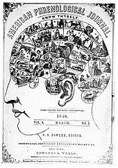
A page from the American Phrenological Journal
 

One of the predecessors to cognitive neuroscience was phrenology, a pseudoscientific approach that claimed that behavior could be determined by the shape of the scalp. In the early 19th century, Franz Joseph Gall and J. G. Spurzheim believed that the human brain was localized into approximately 35 different sections. In his book, The Anatomy and Physiology of the Nervous System in General, and of the Brain in Particular, Gall claimed that a larger bump in one of these areas meant that that area of the brain was used more frequently by that person. This theory gained significant public attention, leading to the publication of phrenology journals and the creation of phrenometers, which measured the bumps on a human subject's head. While phrenology remained a fixture at fairs and carnivals, it did not enjoy wide acceptance within the scientific community. The major criticism of phrenology is that researchers were not able to test theories empirically.

Localizationist view

The localizationist view was concerned with mental abilities being localized to specific areas of the brain rather than on what the characteristics of the abilities were and how to measure them. Studies performed in Europe, such as those of John Hughlings Jackson, supported this view. Jackson studied patients with brain damage, particularly those with epilepsy. He discovered that the epileptic patients often made the same clonic and tonic movements of muscle during their seizures, leading Jackson to believe that they must be caused by activity in the same place in the brain every time. Jackson proposed that specific functions were localized to specific areas of the brain, which was critical to future understanding of the brain lobes.

Aggregate field view

According to the aggregate field view, all areas of the brain participate in every mental function.

Pierre Flourens, a French experimental psychologist, challenged the localizationist view by using animal experiments. He discovered that removing the cerebellum (brain) in rabbits and pigeons affected their sense of muscular coordination, and that all cognitive functions were disrupted in pigeons when the cerebral hemispheres were removed. From this he concluded that the cerebral cortex, cerebellum, and brainstem functioned together as a whole. His approach has been criticised on the basis that the tests were not sensitive enough to notice selective deficits had they been present.

Emergence of neuropsychology

Perhaps the first serious attempts to localize mental functions to specific locations in the brain was by Broca and Wernicke. This was mostly achieved by studying the effects of injuries to different parts of the brain on psychological functions. In 1861, French neurologist Paul Broca came across a man with a disability who was able to understand the language but unable to speak. The man could only produce the sound "tan". It was later discovered that the man had damage to an area of his left frontal lobe now known as Broca's area. Carl Wernicke, a German neurologist, found a patient who could speak fluently but non-sensibly. The patient had been the victim of a stroke, and could not understand spoken or written language. This patient had a lesion in the area where the left parietal and temporal lobes meet, now known as Wernicke's area. These cases, which suggested that lesions caused specific behavioral changes, strongly supported the localizationist view. Additionally, Aphasia is a learning disorder which was also discovered by Paul Broca. According to, Johns Hopkins School of Medicine, Aphasia is a language disorder caused by damage in a specific area of the brain that controls language expression and comprehension. This can often lead to the person speaking words with no sense known as "word salad" 

Mapping the brain

In 1870, German physicians Eduard Hitzig and Gustav Fritsch published their findings of the behavior of animals. Hitzig and Fritsch ran an electric current through the cerebral cortex of a dog, causing different muscles to contract depending on which areas of the brain were electrically stimulated. This led to the proposition that individual functions are localized to specific areas of the brain rather than the cerebrum as a whole, as the aggregate field view suggests. Brodmann was also an important figure in brain mapping; his experiments based on Franz Nissl's tissue staining techniques divided the brain into fifty-two areas.

20th century

Cognitive revolution

At the start of the 20th century, attitudes in America were characterized by pragmatism, which led to a preference for behaviorism as the primary approach in psychology. J.B. Watson was a key figure with his stimulus-response approach. By conducting experiments on animals he was aiming to be able to predict and control behavior. Behaviorism eventually failed because it could not provide realistic psychology of human action and thought – it focused primarily on stimulus-response associations at the expense of explaining phenomena like thought and imagination. This led to what is often termed as the "cognitive revolution".

Neuron doctrine

In the early 20th century, Santiago Ramón y Cajal and Camillo Golgi began working on the structure of the neuron. Golgi developed a silver staining method that could entirely stain several cells in a particular area, leading him to believe that neurons were directly connected with each other in one cytoplasm. Cajal challenged this view after staining areas of the brain that had less myelin and discovering that neurons were discrete cells. Cajal also discovered that cells transmit electrical signals down the neuron in one direction only. Both Golgi and Cajal were awarded a Nobel Prize in Physiology or Medicine in 1906 for this work on the neuron doctrine.

Mid-late 20th century

Several findings in the 20th century continued to advance the field, such as the discovery of ocular dominance columns, recording of single nerve cells in animals, and coordination of eye and head movements. Experimental psychology was also significant in the foundation of cognitive neuroscience. Some particularly important results were the demonstration that some tasks are accomplished via discrete processing stages, the study of attention, and the notion that behavioural data do not provide enough information by themselves to explain mental processes. As a result, some experimental psychologists began to investigate neural bases of behaviour. Wilder Penfield created maps of primary sensory and motor areas of the brain by stimulating the cortices of patients during surgery. The work of Sperry and Gazzaniga on split brain patients in the 1950s was also instrumental in the progress of the field. The term cognitive neuroscience itself was coined by Gazzaniga and cognitive psychologist George Armitage Miller while sharing a taxi in 1976.

Brain mapping

New brain mapping technology, particularly fMRI and PET, allowed researchers to investigate experimental strategies of cognitive psychology by observing brain function. Although this is often thought of as a new method (most of the technology is relatively recent), the underlying principle goes back as far as 1878 when blood flow was first associated with brain function. Angelo Mosso, an Italian psychologist of the 19th century, had monitored the pulsations of the adult brain through neurosurgically created bony defects in the skulls of patients. He noted that when the subjects engaged in tasks such as mathematical calculations the pulsations of the brain increased locally. Such observations led Mosso to conclude that blood flow of the brain followed function.

Emergence of a new discipline

Birth of cognitive science

On September 11, 1956, a large-scale meeting of cognitivists took place at the Massachusetts Institute of Technology. George A. Miller presented his "The Magical Number Seven, Plus or Minus Two" paper while Noam Chomsky and Newell & Simon presented their findings on computer science. Ulric Neisser commented on many of the findings at this meeting in his 1967 book Cognitive Psychology. The term "psychology" had been waning in the 1950s and 1960s, causing the field to be referred to as "cognitive science". Behaviorists such as Miller began to focus on the representation of language rather than general behavior. David Marr concluded that one should understand any cognitive process at three levels of analysis. These levels include computational, algorithmic/representational, and physical levels of analysis.

Combining neuroscience and cognitive science

Before the 1980s, interaction between neuroscience and cognitive science was scarce. Cognitive neuroscience began to integrate the newly laid theoretical ground in cognitive science, that emerged between the 1950s and 1960s, with approaches in experimental psychology, neuropsychology and neuroscience. (Neuroscience was not established as a unified discipline until 1971). In the very late 20th century new technologies evolved that are now the mainstay of the methodology of cognitive neuroscience, including TMS (1985) and fMRI (1991). Earlier methods used in cognitive neuroscience include EEG (human EEG 1920) and MEG (1968). Occasionally cognitive neuroscientists utilize other brain imaging methods such as PET and SPECT. An upcoming technique in neuroscience is NIRS which uses light absorption to calculate changes in oxy- and deoxyhemoglobin in cortical areas. In some animals Single-unit recording can be used. Other methods include microneurography, facial EMG, and eye tracking. Integrative neuroscience attempts to consolidate data in databases, and form unified descriptive models from various fields and scales: biology, psychology, anatomy, and clinical practice.

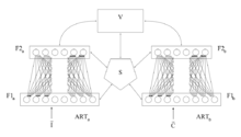
ARTMAP overview
 

Adaptive resonance theory (ART) is a cognitive neuroscience theory developed by Gail Carpenter and Stephen Grossberg in the late 1970s on aspects of how the brain processes information. It describes a number of neural network models which use supervised and unsupervised learning methods, and address problems such as pattern recognition and prediction.

In 2014, Stanislas Dehaene, Giacomo Rizzolatti and Trevor Robbins, were awarded the Brain Prize "for their pioneering research on higher brain mechanisms underpinning such complex human functions as literacy, numeracy, motivated behaviour and social cognition, and for their efforts to understand cognitive and behavioural disorders". Brenda Milner, Marcus Raichle and John O'Keefe received the Kavli Prize in Neuroscience "for the discovery of specialized brain networks for memory and cognition" and O'Keefe shared the Nobel Prize in Physiology or Medicine in the same year with May-Britt Moser and Edvard Moser "for their discoveries of cells that constitute a positioning system in the brain".

In 2017, Wolfram Schultz, Peter Dayan and Ray Dolan were awarded the Brain Prize "for their multidisciplinary analysis of brain mechanisms that link learning to reward, which has far-reaching implications for the understanding of human behaviour, including disorders of decision-making in conditions such as gambling, drug addiction, compulsive behaviour and schizophrenia".,

Recent trends

Recently the focus of research had expanded from the localization of brain area(s) for specific functions in the adult brain using a single technology. Studies have been diverging in several different directions: exploring the interactions between different brain areas, using multiple technologies and approaches to understand brain functions, and using computational approaches. Advances in non-invasive functional neuroimaging and associated data analysis methods have also made it possible to use highly naturalistic stimuli and tasks such as feature films depicting social interactions in cognitive neuroscience studies.

Another very recent trend in cognitive neuroscience is the use of optogenetics to explore circuit function and its behavioral consequences.

Topics

Methods

Experimental methods include:

Cranial nerves

From Wikipedia, the free encyclopedia
 
Cranial nerves
Skull brain human normal.svg
Left View of the human brain from below, showing origins of cranial nerves.
Right Juxtaposed skull base with foramina in which many nerves exit the skull.
 
Skull and brainstem inner ear.svg
Cranial nerves as they pass through the skull base to the brain
Details
Identifiers
Latinnervus cranialis
(pl: nervi craniales)
MeSHD003391
TA98A14.2.01.001
A14.2.00.038
TA26142, 6178
FMA5865

Cranial nerves are the nerves that emerge directly from the brain (including the brainstem), of which there are conventionally considered twelve pairs. Cranial nerves relay information between the brain and parts of the body, primarily to and from regions of the head and neck, including the special senses of vision, taste, smell, and hearing.

The cranial nerves emerge from the central nervous system above the level of the first vertebra of the vertebral column. Each cranial nerve is paired and is present on both sides. There are conventionally twelve pairs of cranial nerves, which are described with Roman numerals I–XII. Some considered there to be thirteen pairs of cranial nerves, including cranial nerve zero. The numbering of the cranial nerves is based on the order in which they emerge from the brain and brainstem, from front to back.

The terminal nerves (0), olfactory nerves (I) and optic nerves (II) emerge from the cerebrum, and the remaining ten pairs arise from the brainstem, which is the lower part of the brain.

The cranial nerves are considered components of the peripheral nervous system (PNS), although on a structural level the olfactory (I), optic (II), and trigeminal (V) nerves are more accurately considered part of the central nervous system (CNS).

The cranial nerves are in contrast to spinal nerves, which emerge from segments of the spinal cord.

Anatomy

View of the human brain from below showing the cranial nerves on an autopsy specimen
 
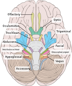
View from below of the brain and brainstem showing the cranial nerves, numbered from olfactory to hypoglossal after the order in which they emerge
 
The brainstem, with cranial nerve nuclei and tracts shown in red

Most typically, humans are considered to have twelve pairs of cranial nerves (I–XII), with the terminal nerve (0) more recently canonized. The nerves are: the olfactory nerve (I), the optic nerve (II), oculomotor nerve (III), trochlear nerve (IV), trigeminal nerve (V), abducens nerve (VI), facial nerve (VII), vestibulocochlear nerve (VIII), glossopharyngeal nerve (IX), vagus nerve (X), accessory nerve (XI), and the hypoglossal nerve (XII).

Terminology

Cranial nerves are generally named according to their structure or function. For example, the olfactory nerve (I) supplies smell, and the facial nerve (VII) supplies the muscles of the face. Because Latin was the lingua franca of the study of anatomy when the nerves were first documented, recorded, and discussed, many nerves maintain Latin or Greek names, including the trochlear nerve (IV), named according to its structure, as it supplies a muscle that attaches to a pulley (Greek: trochlea). The trigeminal nerve (V) is named in accordance with its three components (Latin: trigeminus meaning triplets), and the vagus nerve (X) is named for its wandering course (Latin: vagus).

Cranial nerves are numbered based on their position from front to back (rostral-caudal) of their position on the brain, as, when viewing the forebrain and brainstem from below, they are often visible in their numeric order. For example, the olfactory nerves (I) and optic nerves (II) arise from the base of the forebrain, and the other nerves, III to XII, arise from the brainstem.

Cranial nerves have paths within and outside the skull. The paths within the skull are called "intracranial" and the paths outside the skull are called "extracranial". There are many holes in the skull called "foramina" by which the nerves can exit the skull. All cranial nerves are paired, which means they occur on both the right and left sides of the body. The muscle, skin, or additional function supplied by a nerve, on the same side of the body as the side it originates from, is an ipsilateral function. If the function is on the opposite side to the origin of the nerve, this is known as a contralateral function.

Intracranial course

Nuclei

Grossly, all cranial nerves have a Nucleus. With the exception of the olfactory nerve (I) and optic nerve (II), all the nuclei are present in the brainstem.

The midbrain of the brainstem has the nuclei of the oculomotor nerve (III) and trochlear nerve (IV); the pons has the nuclei of the trigeminal nerve (V), abducens nerve (VI), facial nerve (VII) and vestibulocochlear nerve (VIII); and the medulla has the nuclei of the glossopharyngeal nerve (IX), vagus nerve (X), accessory nerve (XI) and hypoglossal nerve (XII). The olfactory nerve (I) emerges from the olfactory bulb, and depending slightly on division the optic nerve (II) is considered to emerge from the lateral geniculate nuclei.

Because each nerve may have several functions, the nerve fibres that make up the nerve may collect in more than one nucleus. For example, the trigeminal nerve (V), which has a sensory and a motor role, has at least four nuclei.

Exiting the brainstem

With the exception of the olfactory nerve (I) and optic nerve (II), the cranial nerves emerge from the brainstem. The oculomotor nerve (III) and trochlear nerve (IV) emerge from the midbrain, the trigeminal (V), abducens (VI), facial (VII) and vestibulocochlear (VIII) from the pons, and the glossopharyngeal (IX), vagus (X), accessory (XI) and hypoglossal (XII) emerge from the medulla.

The olfactory nerve (I) and optic nerve (II) emerge separately. The olfactory nerves emerge from the olfactory bulbs on either side of the crista galli, a bony projection below the frontal lobe, and the optic nerves (II) emerge from the lateral colliculus, swellings on either side of the temporal lobes of the brain.

Ganglia

The cranial nerves give rise to a number of ganglia, collections of the cell bodies of neurons in the nerves that are outside of the brain. These ganglia are both parasympathetic and sensory ganglia.

The sensory ganglia of the cranial nerves, directly correspond to the dorsal root ganglia of spinal nerves and are known as cranial nerve ganglia. Sensory ganglia exist for nerves with sensory function: V, VII, VIII, IX, X. There are also a number of parasympathetic cranial nerve ganglia. Sympathetic ganglia supplying the head and neck reside in the upper regions of the sympathetic trunk, and do not belong to the cranial nerves.

The ganglion of the sensory nerves, which are similar in structure to the dorsal root ganglion of the spinal cord, include:

Additional ganglia for nerves with parasympathetic function exist, and include the ciliary ganglion of the oculomotor nerve (III), the pterygopalatine ganglion of the maxillary nerve (V2), the submandibular ganglion of the lingual nerve, a branch of the facial nerve (VII), and the otic ganglion of the glossopharyngeal nerve (IX).

Exiting the skull and extracranial course

Exits of cranial nerves from the skull.
Location Nerve
cribriform plate Terminal nerve (0)
cribriform plate Olfactory nerve (I)
optic foramen Optic nerve (II)
superior orbital fissure Oculomotor (III)
Trochlear (IV)
Abducens (VI)
Trigeminal V1
(ophthalmic)
foramen rotundum Trigeminal V2
(maxillary)
foramen ovale Trigeminal V3
(mandibular)
stylomastoid foramen Facial nerve (VII)
internal auditory canal Vestibulocochlear (VIII)
jugular foramen Glossopharyngeal (IX)
Vagus (X)
Accessory (XI)
hypoglossal canal Hypoglossal (XII)

After emerging from the brain, the cranial nerves travel within the skull, and some must leave it in order to reach their destinations. Often the nerves pass through holes in the skull, called foramina, as they travel to their destinations. Other nerves pass through bony canals, longer pathways enclosed by bone. These foramina and canals may contain more than one cranial nerve and may also contain blood vessels.

  • The terminal nerve (0), is a thin network of fibers associated with the dura and lamina terminalis running rostral to the olfactory nerve, with projections through the cribriform plate.
  • The olfactory nerve (I), passes through perforations in the cribriform plate part of the ethmoid bone. The nerve fibres end in the upper nasal cavity.
  • The optic nerve (II) passes through the optic foramen in the sphenoid bone as it travels to the eye.
  • The oculomotor nerve (III), trochlear nerve (IV), abducens nerve (VI) and the ophthalmic branch of the trigeminal nerve (V1) travel through the cavernous sinus into the superior orbital fissure, passing out of the skull into the orbit.
  • The maxillary division of the trigeminal nerve (V2) passes through foramen rotundum in the sphenoid bone.
  • The mandibular division of the trigeminal nerve (V3) passes through foramen ovale of the sphenoid bone.
  • The facial nerve (VII) and vestibulocochlear nerve (VIII) both enter the internal auditory canal in the temporal bone. The facial nerve then reaches the side of the face by using the stylomastoid foramen, also in the temporal bone. Its fibers then spread out to reach and control all of the muscles of facial expression. The vestibulocochlear nerve reaches the organs that control balance and hearing in the temporal bone and therefore does not reach the external surface of the skull.
  • The glossopharyngeal (IX), vagus (X) and accessory nerve (XI) all leave the skull via the jugular foramen to enter the neck. The glossopharyngeal nerve provides sensation to the upper throat and the back of the tongue, the vagus supplies the muscles in the larynx and continues downward to supply parasympathetic supply to the chest and abdomen. The accessory nerve controls the trapezius and sternocleidomastoid muscles in the neck and shoulder.
  • The hypoglossal nerve (XII) exits the skull using the hypoglossal canal in the occipital bone.

Development

The cranial nerves are formed from the contribution of two specialized embryonic cell populations, cranial neural crest and ectodermal placodes. The components of the sensory nervous system of the head are derived from the neural crest and from an embryonic cell population developing in close proximity, the cranial sensory placodes (the olfactory, lens, otic, trigeminal, epibranchial and paratympanic placodes). The dual origin cranial nerves are summarized in the following Table:

Contributions of neural crest cells and placodes to ganglia and cranial nerves

Cranial nerve Ganglion and type Origin of neurons
CNI – olfactory

(Ensheating glia of olfactory nerves)


Telencephalon/olfactory placode; NCCs at forebrain
CNIII – oculomotor

(m)

Ciliary, visceral efferent NCCs at forebrain-midbrain junction (caudal diencephalon and the anterior mesencephalon)
CNV – trigeminal

(mix)

Trigeminal, general afferent NCCs at forebrain-midbrain junction (from r2 into 1st PA), trigeminal placode
CNVII – facial

(mix)

-Superior, general and special afferent

-Inferior: geniculate, general and special afferent

-Sphenopalatine, visceral efferent

-Submandibular, visceral efferent

-Hindbrain NCCs (from r4 into 2nd PA), 1st epibranchial placode

-1st epibranchial placode (geniculate)

-Hindbrain NCCs (2nd PA)

-Hindbrain NCCs (2nd PA)

CNVIII – Vestibulocochlear

(s)

-Acoustic: cochlear, special afferent; and vestibular, special afferent -Otic placode and hindbrain (from r4) NCCs
CNIX – glossopharyngeal

(mix)

-Superior, general and special afferent

-Inferior, petrosal, general and special afferent

-Otic, visceral efferent

-Hindbrain NCCs (from r6 into 3rd PA)

-2nd epibranchial placode (petrosal)

-Hindbrain NCCs (from r6 into 3rd PA)

CNX – vagus

(mix)

Superior laryngeal branch; and recurrent laryngeal branch

-Superior, general afferent

-Inferior: nodose, general and special afferent

-Vagal: parasympathetic, visceral efferent

-Hindbrain NCCs (from r7-r8 to 4th & 6th PA)

-Hindbrain NCCs (4th& 6th PA); 3rd (nodose) and 4th epibranchial placodes

-Hindbrain NCCs (4th & 6th PA)

CNXI – accessory

(m)

No ganglion * Hindbrain (from r7-r8 to PA 4); NCCs (4th PA)

Abbreviations: CN, cranial nerve; m, purely motor nerve; mix, mixed nerve (sensory and motor); NC, neural crest; PA, pharyngeal (branchial) arch; r, rhombomere; s, purely sensory nerve. * There is no known ganglion of the accessory nerve. The cranial part of the accessory nerve sends occasional branches to the superior ganglion of the vagus nerve.

Function

The cranial nerves provide motor and sensory supply mainly to the structures within the head and neck. The sensory supply includes both "general" sensation such as temperature and touch, and "special" senses such as taste, vision, smell, balance and hearing. The vagus nerve (X) provides sensory and autonomic (parasympathetic) supply to structures in the neck and also to most of the organs in the chest and abdomen.

Terminal nerve (0)

The terminal nerve (0) may not have a role in humans, although it has been implicated in hormonal responses to smell, sexual response and mate selection.

Smell (I)

The olfactory nerve (I) conveys information giving rise to the sense of smell.

Damage to the olfactory nerve (I) can cause an inability to smell (anosmia), a distortion in the sense of smell (parosmia), or a distortion or lack of taste.

Vision (II)

The optic nerve (II) transmits visual information.

Damage to the optic nerve (II) affects specific aspects of vision that depend on the location of the damage. A person may not be able to see objects on their left or right sides (homonymous hemianopsia), or may have difficulty seeing objects from their outer visual fields (bitemporal hemianopsia) if the optic chiasm is involved. Inflammation (optic neuritis) may impact the sharpness of vision or colour detection.

Eye movement (III, IV, VI)

The oculomotor (III), troclear (IV) and abducens (VI) nerves supply the muscle of the eye. Damage will affect the movement of the eye in various ways, shown here.

The oculomotor nerve (III), trochlear nerve (IV) and abducens nerve (VI) coordinate eye movement. The oculomotor nerve controls all muscles of the eye except for the superior oblique muscle controlled by the trochlear nerve (IV), and the lateral rectus muscle controlled by the abducens nerve (VI). This means the ability of the eye to look down and inwards is controlled by the trochlear nerve (IV), the ability to look outwards is controlled by the abducens nerve (VI), and all other movements are controlled by the oculomotor nerve (III)

Damage to these nerves may affect the movement of the eye. Damage may result in double vision (diplopia) because the movements of the eyes are not synchronized. Abnormalities of visual movement may also be seen on examination, such as jittering (nystagmus).

Damage to the oculomotor nerve (III) can cause double vision and inability to coordinate the movements of both eyes (strabismus), also eyelid drooping (ptosis) and pupil dilation (mydriasis). Lesions may also lead to inability to open the eye due to paralysis of the levator palpebrae muscle. Individuals suffering from a lesion to the oculomotor nerve may compensate by tilting their heads to alleviate symptoms due to paralysis of one or more of the eye muscles it controls.

Damage to the trochlear nerve (IV) can also cause double vision with the eye adducted and elevated. The result will be an eye which can not move downwards properly (especially downwards when in an inward position). This is due to impairment in the superior oblique muscle.

Damage to the abducens nerve (VI) can also result in double vision. This is due to impairment in the lateral rectus muscle, supplied by the abducens nerve.

Trigeminal nerve (V)

The trigeminal nerve (V) and its three main branches the ophthalmic (V1), maxillary (V2), and mandibular (V3) provide sensation to the skin of the face and also controls the muscles of chewing.

Damage to the trigeminal nerve leads to loss of sensation in an affected area. Other conditions affecting the trigeminal nerve (V) include trigeminal neuralgia, herpes zoster, sinusitis pain, presence of a dental abscess, and cluster headaches.

The facial nerve (VII) supplies the muscles of facial expression. Damage to the nerve causes a lack of muscle tone on the affected side, as can be seen on the right side of the face here.

Facial expression (VII)

The facial nerve (VII) controls most muscles of facial expression, supplies the sensation of taste from the front two-thirds of the tongue, and controls the stapedius muscle. Most muscles are supplied by the cortex on the opposite side of the brain; the exception is the frontalis muscle of the forehead, in which the left and the right side of the muscle both receive inputs from both sides of the brain.

Damage to the facial nerve (VII) may cause facial palsy. This is where a person is unable to move the muscles on one or both sides of their face. The most common cause of this is Bell's palsy, the ultimate cause of which is unknown. Patients with Bell's palsy often have a drooping mouth on the affected side and often have trouble chewing because the buccinator muscle is affected. The facial nerve is also the most commonly affected cranial nerve in blunt trauma.

Hearing and balance (VIII)

The vestibulocochlear nerve (VIII) supplies information relating to balance and hearing via its two branches, the vestibular and cochlear nerves. The vestibular part is responsible for supplying sensation from the vestibules and semicircular canal of the inner ear, including information about balance, and is an important component of the vestibuloocular reflex, which keeps the head stable and allows the eyes to track moving objects. The cochlear nerve transmits information from the cochlea, allowing sound to be heard.

When damaged, the vestibular nerve may give rise to the sensation of spinning and dizziness (vertigo). Function of the vestibular nerve may be tested by putting cold and warm water in the ears and watching eye movements caloric stimulation. Damage to the vestibulocochlear nerve can also present as repetitive and involuntary eye movements (nystagmus), particularly when the eye is moving horizontally. Damage to the cochlear nerve will cause partial or complete deafness in the affected ear.

Oral sensation, taste, and salivation (IX)

A damaged glossopharyngeal nerve (IX) may cause the uvula to deviate to the affected side.

The glossopharyngeal nerve (IX) supplies the stylopharyngeus muscle and provides sensation to the oropharynx and back of the tongue. The glossopharyngeal nerve also provides parasympathetic input to the parotid gland.

Damage to the nerve may cause failure of the gag reflex; a failure may also be seen in damage to the vagus nerve (X).

Vagus nerve (X)

The vagus nerve (X) provides sensory and parasympathetic supply to structures in the neck and also to most of the organs in the chest and abdomen.

Loss of function of the vagus nerve (X) will lead to a loss of parasympathetic supply to a very large number of structures. Major effects of damage to the vagus nerve may include a rise in blood pressure and heart rate. Isolated dysfunction of only the vagus nerve is rare, but – if the lesion is located above the point at which the vagus first branches off – can be indicated by a hoarse voice, due to dysfunction of one of its branches, the recurrent laryngeal nerve.

Damage to this nerve may result in difficulties swallowing.

Shoulder elevation and head-turning (XI)

The accessory nerve (XI) supplies the sternocleidomastoid and trapezius muscles. Damage to the nerve may cause a winged scapula, shown here.
 
The hypoglossal nerve (XII) supplies the muscles of the tongue. A damaged hypoglossal nerve will result in an inability to stick the tongue out straight; here seen in an injury resulting from branchial cyst surgery. 

The accessory nerve (XI) supplies the sternocleidomastoid and trapezius muscles.

Damage to the accessory nerve (XI) will lead to weakness in the trapezius muscle on the same side as the damage. The trapezius lifts the shoulder when shrugging, so the affected shoulder will not be able to shrug and the shoulder blade (scapula) will protrude into a winged position. Depending on the location of the lesion there may also be weakness present in the sternocleidomastoid muscle, which acts to turn the head so that the face points to the opposite side.

Tongue movement (XII)

The hypoglossal nerve (XII) supplies the intrinsic muscles of the tongue, controlling tongue movement. The hypoglossal nerve (XII) is unique in that it is supplied by the motor cortices of both hemispheres of the brain.

Damage to the nerve may lead to fasciculations or wasting (atrophy) of the muscles of the tongue. This will lead to weakness of tongue movement on that side. When damaged and extended, the tongue will move towards the weaker or damaged side, as shown in the image. The fasciculations of the tongue are sometimes said to look like a "bag of worms". Damage to the nerve tract or nucleus will not lead to atrophy or fasciculations, but only weakness of the muscles on the same side as the damage.

Clinical significance

Examination

Doctors, neurologists and other medical professionals may conduct a cranial nerve examination as part of a neurological examination to examine the cranial nerves. This is a highly formalised series of steps involving specific tests for each nerve. Dysfunction of a nerve identified during testing may point to a problem with the nerve or of a part of the brain.

A cranial nerve exam starts with observation of the patient, as some cranial nerve lesions may affect the symmetry of the eyes or face. Vision may be tested by examining the visual fields, or by examining the retina with an ophthalmoscope, using a process known as funduscopy. Visual field testing may be used to pin-point structural lesions in the optic nerve, or further along the visual pathways. Eye movement is tested and abnormalities such as nystagmus are observed for. The sensation of the face is tested, and patients are asked to perform different facial movements, such as puffing out of the cheeks. Hearing is checked by voice and tuning forks. The patient's uvula is examined. After performing a shrug and head turn, the patient's tongue function is assessed by various tongue movements.

Smell is not routinely tested, but if there is suspicion of a change in the sense of smell, each nostril is tested with substances of known odors such as coffee or soap. Intensely smelling substances, for example ammonia, may lead to the activation of pain receptors of the trigeminal nerve (V) located in the nasal cavity and this can confound olfactory testing.

Damage

Compression

Nerves may be compressed because of increased intracranial pressure, a mass effect of an intracerebral haemorrhage, or tumour that presses against the nerves and interferes with the transmission of impulses along the nerve. Loss of function of a cranial nerve may sometimes be the first symptom of an intracranial or skull base cancer.

An increase in intracranial pressure may lead to impairment of the optic nerves (II) due to compression of the surrounding veins and capillaries, causing swelling of the eyeball (papilloedema). A cancer, such as an optic nerve glioma, may also impact the optic nerve (II). A pituitary tumour may compress the optic tracts or the optic chiasm of the optic nerve (II), leading to visual field loss. A pituitary tumour may also extend into the cavernous sinus, compressing the oculomotor nerve (III), trochlear nerve (IV) and abducens nerve (VI), leading to double-vision and strabismus. These nerves may also be affected by herniation of the temporal lobes of the brain through the falx cerebri.

The cause of trigeminal neuralgia, in which one side of the face is exquisitely painful, is thought to be compression of the nerve by an artery as the nerve emerges from the brain stem. An acoustic neuroma, particularly at the junction between the pons and medulla, may compress the facial nerve (VII) and vestibulocochlear nerve (VIII), leading to hearing and sensory loss on the affected side.

Stroke

Occlusion of blood vessels that supply the nerves or their nuclei, an ischemic stroke, may cause specific signs and symptoms relating to the damaged area. If there is a stroke of the midbrain, pons or medulla, various cranial nerves may be damaged, resulting in dysfunction and symptoms of a number of different syndromes. Thrombosis, such as a cavernous sinus thrombosis, refers to a clot (thrombus) affecting the venous drainage from the cavernous sinus, affects the optic (II), oculomotor (III), trochlear (IV), opthalmic branch of the trigeminal nerve (V1) and the abducens nerve (VI).

Inflammation

Inflammation of a cranial nerve can occur as a result of infection, such as viral causes like reactivated herpes simplex virus, or can occur spontaneously. Inflammation of the facial nerve (VII) may result in Bell's palsy.

Multiple sclerosis, an inflammatory process resulting in a loss of the myelin sheathes which surround the cranial nerves, may cause a variety of shifting symptoms affecting multiple cranial nerves. Inflammation may also affect other cranial nerves. Other rarer inflammatory causes affecting the function of multiple cranial nerves include sarcoidosis, miliary tuberculosis, and inflammation of arteries, such as granulomatosis with polyangiitis.

Other

Trauma to the skull, disease of bone, such as Paget's disease, and injury to nerves during surgery are other causes of nerve damage.

History

The Graeco-Roman anatomist Galen (AD 129–210) named seven pairs of cranial nerves. Much later, in 1664, English anatomist Sir Thomas Willis suggested that there were actually 9 pairs of nerves. Finally, in 1778, German anatomist Samuel Soemmering named the 12 pairs of nerves that are generally accepted today. However, because many of the nerves emerge from the brain stem as rootlets, there is continual debate as to how many nerves there actually are, and how they should be grouped. For example, there is reason to consider both the olfactory (I) and optic (II) nerves to be brain tracts, rather than cranial nerves.

Other animals

Dog-fish brain in two projections.
top; ventral bottom; lateral
The accessory nerve (XI) and hypoglossal nerve (XII) cannot be seen, as they are not always present in all vertebrates.

Cranial nerves are also present in other vertebrates. Other amniotes (non-amphibian tetrapods) have cranial nerves similar to those of humans. In anamniotes (fishes and amphibians), the accessory nerve (XI) and hypoglossal nerve (XII) do not exist, with the accessory nerve (XI) being an integral part of the vagus nerve (X); the hypoglossal nerve (XII) is represented by a variable number of spinal nerves emerging from vertebral segments fused into the occiput. These two nerves only became discrete nerves in the ancestors of amniotes. The very small terminal nerve (nerve N or O) exists in humans but may not be functional. In other animals, it appears to be important to sexual receptivity based on perceptions of pheromones.

Politics of Europe

From Wikipedia, the free encyclopedia ...