Search This Blog

Saturday, September 30, 2023

Sample-return mission

From Wikipedia, the free encyclopedia
The Genesis Rock, returned by the Apollo 15 lunar mission in 1971.
Apollo astronauts work on the Moon to collect samples and explore. Near Shorty Crater they found some orange lunar regolith.

A sample-return mission is a spacecraft mission to collect and return samples from an extraterrestrial location to Earth for analysis. Sample-return missions may bring back merely atoms and molecules or a deposit of complex compounds such as loose material and rocks. These samples may be obtained in a number of ways, such as soil and rock excavation or a collector array used for capturing particles of solar wind or cometary debris. Nonetheless, concerns have been raised that the return of such samples to planet Earth may endanger Earth itself.

To date, samples of Moon rock from Earth's Moon have been collected by robotic and crewed missions; the comet Wild 2 and the asteroids 25143 Itokawa, 162173 Ryugu, and 101955 Bennu have been visited by robotic spacecraft which returned samples to Earth; and samples of the solar wind have been returned by the robotic Genesis mission.

In addition to sample-return missions, samples from three identified non-terrestrial bodies have been collected by other means: samples from the Moon in the form of Lunar meteorites, samples from Mars in the form of Martian meteorites, and samples from Vesta in the form of HED meteorites.

Scientific use

A meteorite thought to be from Mars

Samples available on Earth can be analyzed in laboratories, so we can further our understanding and knowledge as part of the discovery and exploration of the Solar System. Until now, many important scientific discoveries about the Solar System were made remotely with telescopes, and some Solar System bodies were visited by orbiting or even landing spacecraft with instruments capable of remote sensing or sample analysis. While such an investigation of the Solar System is technically easier than a sample-return mission, the scientific tools available on Earth to study such samples are far more advanced and diverse than those that can go on spacecraft. Further, analysis of samples on Earth allows follow up of any findings with different tools, including tools that can tell intrinsic extraterrestrial material from terrestrial contamination, and those that have yet to be developed; in contrast, a spacecraft can carry only a limited set of analytic tools, and these have to be chosen and built long before launch.

Samples analyzed on Earth can be matched against findings of remote sensing for more insight into the processes that formed the Solar System. This was done, for example, with findings by the Dawn spacecraft, which visited the asteroid Vesta from 2011 to 2012 for imaging, and samples from HED meteorites (collected on Earth until then), which were compared to data gathered by Dawn. These meteorites could then be identified as material ejected from the large impact crater Rheasilvia on Vesta. This allowed deducing the composition of the crust, mantle and core of Vesta. Similarly, some differences in the composition of asteroids (and, to a lesser extent, different compositions of comets) can be discerned by imaging alone. However, for a more precise inventory of the material on these different bodies, more samples will be collected and returned in the future, to match their compositions with the data gathered through telescopes and astronomical spectroscopy.

One further focus of such investigation—besides the basic composition and geologic history of the various Solar System bodies—is the presence of the building blocks of life on comets, asteroids, Mars or the moons of the gas giants. Several sample-return missions to asteroids and comets are currently in the works. More samples from asteroids and comets will help determine whether life formed in space and was carried to Earth by meteorites. Another question under investigation is whether extraterrestrial life formed on other Solar System bodies like Mars or on the moons of the gas giants, and whether life might even exist there. The result of NASA's last "Decadal Survey" was to prioritize a Mars sample-return mission, as Mars has a special importance: it is comparatively "nearby", might have harbored life in the past, and might even continue to sustain life. Jupiter's moon Europa is another important focus in the search for life in the Solar System. However, due to the distance and other constraints, Europa might not be the target of a sample-return mission in the foreseeable future.

Planetary protection

Planetary protection aims to prevent biological contamination of both the target celestial body and the Earth in the case of sample-return missions. A sample return from Mars or other location with the potential to host life is a category V mission under COSPAR, which directs to the containment of any unsterilized sample returned to Earth. This is because it is unknown what the effects such hypothetical life would be on humans or the biosphere of Earth. For this reason, Carl Sagan and Joshua Lederberg argued in the 1970s that we should do sample-return missions classified as category V missions with extreme caution, and later studies by the NRC and ESF agreed.

Sample-return missions

First missions

Apollo 11 was the first mission to return extraterrestrial samples.
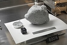
Lunar sample 60016 on display at Space Center Houston Lunar Samples Vault, at NASA's Johnson Space Center

The Apollo program returned over 382 kg (842 lb) of lunar rocks and regolith (including lunar 'soil') to the Lunar Receiving Laboratory in Houston. Today, 75% of the samples are stored at the Lunar Sample Laboratory Facility built in 1979. In July 1969, Apollo 11 achieved the first successful sample return from another Solar System body. It returned approximately 22 kilograms (49 lb) of Lunar surface material. This was followed by 34 kilograms (75 lb) of material and Surveyor 3 parts from Apollo 12, 42.8 kilograms (94 lb) of material from Apollo 14, 76.7 kilograms (169 lb) of material from Apollo 15, 94.3 kilograms (208 lb) of material from Apollo 16, and 110.4 kilograms (243 lb) of material from Apollo 17.

One of the most significant advances in sample-return missions occurred in 1970 when the robotic Soviet mission known as Luna 16 successfully returned 101 grams (3.6 oz) of lunar soil. Likewise, Luna 20 returned 55 grams (1.9 oz) in 1974, and Luna 24 returned 170 grams (6.0 oz) in 1976. Although they recovered far less than the Apollo missions, they did this fully automatically. Apart from these three successes, other attempts under the Luna programme failed. The first two missions were intended to outstrip Apollo 11 and were undertaken shortly before them in June and July 1969: Luna E-8-5 No. 402 failed at start, and Luna 15 crashed on the Moon. Later, other sample-return missions failed: Kosmos 300 and Kosmos 305 in 1969, Luna E-8-5 No. 405 in 1970, Luna E-8-5M No. 412 in 1975 had unsuccessful launches, and Luna 18 in 1971 and Luna 23 in 1974 had unsuccessful landings on the Moon.

In 1970, the Soviet Union planned for a 1975 first Mars sample-return mission in the Mars 5NM project. This mission was planned to use an N1 rocket, but as this rocket never flew successfully, the mission evolved into the Mars 5M project, which would use a double launch with the smaller Proton rocket and an assembly at a Salyut space station. This Mars 5M mission was planned for 1979, but was canceled in 1977 due to technical problems and complexity; all hardware was ordered destroyed.

1990s

The Orbital Debris Collection (ODC) experiment deployed on the Mir space station for 18 months in 1996–97 used aerogel to capture particles from low Earth orbit, including both interplanetary dust and man-made particles.

2000s
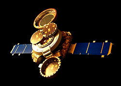
An artist's rendering of Genesis collecting solar wind.

The next mission to return extraterrestrial samples was the Genesis mission, which returned solar wind samples to Earth from beyond Earth orbit in 2004. Unfortunately, the Genesis capsule failed to open its parachute while re-entering the Earth's atmosphere and crash-landed in the Utah desert. There were fears of severe contamination or even total mission loss, but scientists managed to save many of the samples. They were the first to be collected from beyond lunar orbit. Genesis used a collector array made of wafers of ultra-pure silicon, gold, sapphire, and diamond. Each different wafer was used to collect a different part of the solar wind.

Sample-return capsule from the Stardust mission

Genesis was followed by NASA's Stardust spacecraft, which returned comet samples to Earth on January 15, 2006. It safely passed by Comet Wild 2 and collected dust samples from the comet's coma while imaging the comet's nucleus. Stardust used a collector array made of low-density aerogel (99% of which is space), which has about 1/1000 of the density of glass. This enables the collection of cometary particles without damaging them due to high impact velocities. Particle collisions with even slightly porous solid collectors would result in the destruction of those particles and damage to the collection apparatus. During the cruise, the array collected at least seven interstellar dust particles.

2010s and 2020s

In June 2010 the Japan Aerospace Exploration Agency (JAXA) Hayabusa probe returned asteroid samples to Earth after a rendezvous with (and a landing on) S-type asteroid 25143 Itokawa. In November 2010, scientists at the agency confirmed that, despite failure of the sampling device, the probe retrieved micrograms of dust from the asteroid, the first brought back to Earth in pristine condition.

The Russian Fobos-Grunt was a failed sample-return mission designed to return samples from Phobos, one of the moons of Mars. It was launched on November 8, 2011, but failed to leave Earth orbit and crashed after several weeks into the southern Pacific Ocean.

OSIRIS-REx collecting a sample from asteroid 101955 Bennu
OSIRIS-REx Sample Return Capsule in Utah

The Japan Aerospace Exploration Agency (JAXA) launched the improved Hayabusa2 space probe on December 3, 2014. Hayabusa2 arrived at the target near-Earth C-type asteroid 162173 Ryugu (previously designated 1999 JU3) on 27 June 2018. It surveyed the asteroid for a year and a half and took samples. It left the asteroid in November 2019 and returned to Earth on December 6, 2020.

The OSIRIS-REx mission was launched in September 2016 on a mission to return samples from the asteroid 101955 Bennu. The samples are expected to enable scientists to learn more about the time before the birth of the Solar System, initial stages of planet formation, and the source of organic compounds that led to the formation of life. It reached the proximity of Bennu on 3 December 2018, where it began analyzing its surface for a target sample area over the next several months. It collected its sample on 20 October 2020, and landed back on Earth again on 24 September 2023, making Osiris Rex the fifth successful sample return mission for mankind, in its return of samples from an extra-terrestrial body. Shortly after the sample container was retrieved and transferred to an "airtight chamber at the Johnson Space Center in Houston, Texas", the lid on the container was opened. Scientists commented that they "found black dust and debris on the avionics deck of the OSIRIS-REx science canister" on the initial opening. Later study was planned. A news conference on the asteroid sample is scheduled for 11 October 2023.

Lunar soil sample collected by China's Chang'e 5 mission displayed at Airshow China 2021.

China launched the Chang'e 5 lunar sample return mission on November 23, 2020, which returned to Earth with 2 kilograms of lunar soil on December 16, 2020. It was first lunar sample return in over 40 years.

Future missions

JAXA is developing the MMX mission, a sample-return mission to Phobos that will be launched in 2024. MMX will study both moons of Mars, but the landing and the sample collection will be on Phobos. This selection was made because of the two moons, Phobos's orbit is closer to Mars and its surface may have particles blasted from Mars. Thus the sample may contain material originating on Mars itself. A propulsion module carrying the sample is expected to return to Earth in approximately September 2029.

China will launch the Chang'e 6 lunar sample return mission in 2023. China is also planning a mission called Tianwen-2 to return samples from 469219 Kamoʻoalewa which is planned to launch in 2024. China plans for a Mars sample return mission by 2030. Also, the Chinese Space Agency is designing a sample-retrieval mission from Ceres that would take place during the 2020s.

NASA and ESA has long planned a Mars Sample-Return Mission, which is only now coming to fruition. The Perseverance rover, deployed in 2020, is collecting drill core samples and stashing them on the Mars surface. A joint NASA-ESA mission to return them is planned for the late twenties, consisting of a lander to retrieve the samples and raise them into orbit, and an orbiter to return them to Earth.

Comet sample-return missions also continue to be a NASA priority. Comet Surface Sample Return was one of the six themes for proposals for NASA's fourth New Frontiers mission in 2017.

Russia has plans for Luna-Glob missions to return samples from the Moon by 2027 and Mars-Grunt to return samples from Mars in the late 2020s.

Methods of sample return

Animation of TAGSAM arm moving

Sample-return methods include, but are not restricted to the following:

A Genesis collector array consisting of a grid of ultra-pure wafers of silicon, gold, sapphire, and diamond

Collector array

A collector array may be used to collect millions or billions of atoms, molecules, and fine particulates by using wafers made of different elements. The molecular structure of these wafers allows the collection of various sizes of particles. Collector arrays, such as those flown on Genesis, are ultra-pure in order to ensure maximal collection efficiency, durability, and analytical distinguishability.

Collector arrays are useful for collecting tiny, fast-moving atoms such as those expelled by the Sun through the solar wind, but can also be used for collection of larger particles such as those found in the coma of a comet. The NASA spacecraft known as Stardust implemented this technique. However, due to the high speeds and size of the particles that make up the coma and the area nearby, a dense solid-state collector array was not viable. As a result, another means for collecting samples had to be designed to preserve the safety of the spacecraft and the samples themselves.

Aerogel

A particle captured in aerogel

Aerogel is a silica-based porous solid with a sponge-like structure, 99.8% of whose volume is empty space. Aerogel has about 1/1000 of the density of glass. An aerogel was used in the Stardust spacecraft because the dust particles the spacecraft was to collect would have an impact speed of about 6 km/s. A collision with a dense solid at that speed could alter their chemical composition or vaporize them completely.

Since the aerogel is mostly transparent, and the particles leave a carrot-shaped path once they penetrate the surface, scientists can easily find and retrieve them. Since its pores are on the nanometer scale, particles, even ones smaller than a grain of sand, do not merely pass through the aerogel completely. Instead, they slow to a stop and then are embedded within it. The Stardust spacecraft has a tennis-racket-shaped collector with aerogel fitted to it. The collector is retracted into its capsule for safe storage and delivery back to Earth. Aerogel is quite strong and easily survives both launching and space environments.

Robotic excavation and return

Some of the riskiest and most difficult types of sample-return missions are those that require landing on an extraterrestrial body such as an asteroid, moon, or planet. It takes a great deal of time, money, and technical ability to even initiate such plans. It is a difficult feat that requires that everything from launch to landing to retrieval and launch back to Earth is planned out with high precision and accuracy.

This type of sample return, although having the most risks, is the most rewarding for planetary science. Furthermore, such missions carry a great deal of public outreach potential, which is an important attribute for space exploration when it comes to public support. The only successful robotic sample-return missions of this type have been the Soviet Luna landers and Chinese Chang'e 5.

Frontotemporal dementia

From Wikipedia, the free encyclopedia
 
Frontotemporal dementia
Brain MRI of a 65-year-old female person with frontotemporal dementia. Cortical and white matter atrophy of the frontal lobes is clear in all images.
SpecialtyPsychiatry, neurology
Causesfrontotemporal lobar degeneration

Frontotemporal dementia (FTD), or frontotemporal degeneration disease, or frontotemporal neurocognitive disorder, encompasses several types of dementia involving the progressive degeneration of frontal and temporal lobes. FTDs broadly present as behavioral or language disorders with gradual onsets. Common signs and symptoms include significant changes in social and personal behavior, apathy, blunting of emotions, and deficits in both expressive and receptive language. Currently, there is no cure for FTD, but there are treatments that help alleviate symptoms.

Frontotemporal dementias are mostly early-onset syndromes that are linked to frontotemporal lobar degeneration (FTLD), which is characterized by progressive neuronal loss predominantly involving the frontal or temporal lobes, and a typical loss of more than 70% of spindle neurons, while other neuron types remain intact. The three main subtypes or variant syndromes are a behavioral variant (bvFTD) previously known as Pick's disease, and two variants of primary progressive aphasiasemantic variant (svPPA), and nonfluent variant (nfvPPA). Two rare distinct subtypes of FTD are neuronal intermediate filament inclusion disease (NIFID), and basophilic inclusion body disease. Other related disorders include corticobasal syndrome and FTD with amyotrophic lateral sclerosis (ALS), FTD-ALS, also called FTD-MND.

Signs and symptoms tend to appear in late adulthood, typically between the ages of 45 and 65. Men and women appear to be equally affected. FTD is the second most prevalent type of early onset dementia after Alzheimer's disease. Considered individually, each of the subtypes is relatively rare.

Features of FTD were first described by Arnold Pick between 1892 and 1906. The name Pick's disease was coined in 1922. This term is now reserved only for the behavioral variant of FTD which shows the presence of the characteristic Pick bodies and Pick cells first described by Alois Alzheimer in 1911.

Signs and symptoms

Frontotemporal dementia is an early-onset disorder that mostly occurs between the ages of 45 and 65, but can begin earlier, and in 20–25% of cases onset is later. It is the most common early presenting dementia.

The International Classification of Diseases recognizes the disease as causative to disorder affecting mental and behavioural aspects of the human organism. Dissociation from family, compulsive buying disorder (oniomania), vulgar speech characteristics, screaming, inability to control emotions, behavior, personality, and temperament are characteristic social display patterns. A gradual onset and progression of changes in behavior or language deficits are reported to have begun several years prior to presentation to a neurologist.

Subtypes and related disorders

The main subtypes of frontotemporal dementia are behavioral variant FTD, semantic dementia, progressive nonfluent aphasia, and FTD associated with amyotrophic lateral sclerosis (FTD–ALS). Two distinct rare subtypes are neuronal intermediate filament inclusion disease, and basophilic inclusion body disease. Related disorders are corticobasal syndrome, and progressive supranuclear palsy.

Behavioral variant frontotemporal dementia

Behavioral variant frontotemporal dementia (BvFTD) was previously known as Pick's disease, and is the most common of the FTD types. BvFTD is diagnosed four times as often as the PPA variants. Behavior can change in BvFTD in either of two ways—it can change to being impulsive and disinhibited, acting in socially unacceptable ways; or it can change to being listless and apathetic. About 12–13% of people with bvFTD develop motor neuron disease.

The Pick bodies in behavioral variant FTD are spherical inclusion bodies found in the cytoplasm of affected cells. They consist of tau fibrils as a major component together with a number of other protein products including ubiquitin and tubulin.

Semantic dementia

Semantic dementia (SD) is characterized by the loss of semantic understanding, resulting in impaired word comprehension. However, speech remains fluent and grammatical.

Progressive nonfluent aphasia

Progressive nonfluent aphasia (PNFA) is characterized by progressive difficulties in speech production.

Neuronal intermediate filament inclusion disease

Neuronal intermediate filament inclusion disease (NIFID) is a rare distinct variant. The inclusion bodies that are present in NIFID are cytoplasmic and made up of type IV intermediate filaments. NIFID has an early age of onset between 23 and 56. Symptoms can include behavioural, and personality changes, memory and cognitive impairments, language difficulties, motor weakness, and extrapyramidal symptoms. NIFID is one of the FTLD-FUS proteopathies. Imaging commonly shows atrophy in the frontotemporal region, and in part of the striatum in the basal ganglia. Post-mortem studies show a marked reduction in the caudate nucleus of the striatum; frontotemporal gyri are narrowed, with widened intervening sulci, and the lateral ventricles are enlarged.

Basophilic inclusion body disease

Another rare FTD variant, also a FTLD-FUS proteopathy, is basophilic inclusion body disease (BIBD).

Other characteristics

In later stages of FTD, the clinical phenotypes may overlap. People with FTD tend to struggle with binge eating and compulsive behaviors. Binge eating habits are often associated with changes in food preferences (cravings for more sweets, carbohydrates), eating inedible objects and snatching food from others. Recent findings from structural MRI research have indicated that eating changes in FTD are associated with atrophy (wasting) in the right ventral insula, striatum, and orbitofrontal cortex.

People with FTD show marked deficiencies in executive functioning and working memory. Most become unable to perform skills that require complex planning or sequencing. In addition to the characteristic cognitive dysfunction, a number of primitive reflexes known as frontal release signs are often able to be elicited. Usually the first of these frontal release signs to appear is the palmomental reflex which appears relatively early in the disease course whereas the palmar grasp reflex and rooting reflex appear late in the disease course.

In rare cases, FTD can occur in people with amyotrophic lateral sclerosis (ALS), a motor neuron disease. The prognosis for people with ALS is worse when combined with FTD, shortening survival by about a year.

Genetics

A higher proportion of frontotemporal dementias seem to have a familial component than other neurodegenerative diseases such as Alzheimer's disease. More and more mutations and genetic variants are being identified all the time, needing constant updating of genetic influences.

  • Tau-positive frontotemporal dementia and parkinsonism linked to chromosome 17 (FTDP-17) is caused by mutations in the MAPT gene on chromosome 17 that encodes the tau protein. It has been determined that there is a direct relationship between the type of tau mutation and the neuropathology of gene mutations. The mutations at the splice junction of exon 10 of tau lead to the selective deposition of the repetitive tau in neurons and glia. The pathological phenotype associated with mutations elsewhere in tau is less predictable, with both typical neurofibrillary tangles (consisting of both 3-repeat and 4-repeat tau) and Pick bodies (consisting of 3-repeat tau) having been described. The presence of tau deposits within glia is also variable in families with mutations outside of exon 10. This disease is now informally designated FTDP-17T. FTD shows a linkage to the region of the tau locus on chromosome 17, but it is believed that there are two loci leading to FTD within megabases of each other on chromosome 17. The only other known autosomal dominant genetic cause of FTLD-tau is a hypomorphic mutation in VCP which is associated with a unique neuropathology called vacuolar tauopathy.
  • FTD caused by FTLD-TDP43 has numerous genetic causes. Some cases are due to mutations in the GRN gene, also located on chromosome 17. Others are caused by hypomorphic VCP mutations, although these patients present with a complex picture of multisystem proteinopathy that can include amyotrophic lateral sclerosis, inclusion body myopathy, Paget's disease of bone, and FTD. The most recent addition to the list (as of 2019) was a hexanucleotide repeat expansion in intron 1 of C9ORF72. Only one or two cases have been reported describing TARDBP (the TDP-43 gene) mutations in a clinically-pure FTD (FTD without MND).
  • Several other genes have been linked to this condition. These include CYLD, OPTN, SQSTM1 and TBK1. These genes have been implicated in the autophagy pathway.
  • No genetic causes of FUS pathology in FTD have yet been reported.
  • Major alleles of TMEM106B SNPs have been found to be associated with risk of FTLD.

Pathology

There are three main histological subtypes found at post-mortem: FTLD-tau, FTLD-TDP, and FTLD-FUS. In rare cases, patients with clinical FTD were found to have changes consistent with Alzheimer's disease on autopsy. The most severe brain atrophy appears to be associated with behavioral variant FTD, and corticobasal degeneration.

With regard to the genetic defects that have been found, repeat expansion in the C9orf72 gene is considered a major contribution to frontotemporal lobar degeneration, although defects in the GRN and MAPT genes are also associated with it.

DNA damage and the defective repair of such damages have been etiologically linked to various neurodegenerative diseases including FTD.

Diagnosis

FTD is traditionally difficult to diagnose owing to the diverse nature of the associated symptoms. Signs and symptoms are classified into three groups based on the affected functions of the frontal and temporal lobes: These are behavioural variant frontotemporal dementia, semantic dementia, and progressive nonfluent aphasia. An overlap between symptoms can occur as the disease progresses and spreads through the brain regions. Structural MRI scans often reveal frontal lobe and/or anterior temporal lobe atrophy but in early cases the scan may seem normal. Atrophy can be either bilateral or asymmetric. Registration of images at different points of time (e.g., one year apart) can show evidence of atrophy that otherwise (at individual time points) may be reported as normal. Many research groups have begun using techniques such as magnetic resonance spectroscopy, functional imaging and cortical thickness measurements in an attempt to offer an earlier diagnosis to the FTD patient. Fluorine-18-fluorodeoxyglucose positron emission tomography (FDG-PET) scans classically show frontal and/or anterior temporal hypometabolism, which helps differentiate the disease from Alzheimer's disease. The PET scan in Alzheimer's disease classically shows biparietal hypometabolism. Meta-analyses based on imaging methods have shown that frontotemporal dementia mainly affects a frontomedial network discussed in the context of social cognition or 'theory of mind'. This is entirely in keeping with the notion that on the basis of cognitive neuropsychological evidence, the ventromedial prefrontal cortex is a major locus of dysfunction early on in the course of the behavioural variant of frontotemporal degeneration. The language subtypes of frontotemporal lobar degeneration (semantic dementia and progressive nonfluent aphasia) can be regionally dissociated by imaging approaches in vivo.

The confusion between Alzheimer's and FTD is justifiable due to the similarities between their initial symptoms. Patients do not have difficulty with movement and other motor tasks. As FTD symptoms appear, it is difficult to differentiate between a diagnosis of Alzheimer's disease and FTD. There are distinct differences in the behavioral and emotional symptoms of the two dementias – notably, the blunting of emotions seen in FTD patients.[10] In the early stages of FTD, anxiety and depression are common, which may result in an ambiguous diagnosis. However, over time, these ambiguities fade away as this dementia progresses and defining symptoms of apathy, unique to FTD, start to appear.

Recent studies over several years have developed new criteria for the diagnosis of behavioral variant frontotemporal dementia (bvFTD). The confirmatory diagnosis is made by brain biopsy, but other tests can be used to help, such as MRI, EEG, CT, and physical examination and history. Six distinct clinical features have been identified as symptoms of bvFTD.

  1. Disinhibition
  2. Apathy / Inertia
  3. Loss of Sympathy / Empathy
  4. Perseverative / Compulsive behaviors
  5. Hyperorality
  6. Dysexecutive neuropsychological profile

Of the six features, three must be present in a patient to diagnose one with possible bvFTD. Similar to standard FTD, the primary diagnosis stems from clinical trials that identify the associated symptoms, instead of imaging studies. The above criteria are used to distinguish bvFTD from disorders such as Alzheimer's and other causes of dementia. In addition, the new criteria allow for a diagnostic hierarchy distinguished possible, probable, and definite bvFTD based on the number of symptoms present.

A 2021 study, led by researchers at the University of Pennsylvania, determined that using cerebrospinal fluid (CSF) biomarkers of pathologic amyloid plaques, tangles, and neurodegeneration – collectively called ATN – can be useful in diagnosing FTD.

Neuropsychological tests

The progression of the degeneration caused by bvFTD may follow a predictable course. The degeneration begins in the orbitofrontal cortex and medial aspects such as ventromedial cortex. In later stages, it gradually expands its area to the dorsolateral cortex and the temporal lobe. Thus, the detection of dysfunction of the orbitofrontal cortex and ventromedial cortex is important in the detection of early stage bvFTD. As stated above, a behavioural change may occur before the appearance of any atrophy in the brain in the course of the disease. Because of that, image scanning such as MRI can be insensitive to the early degeneration and it is difficult to detect early-stage bvFTD.

In neuropsychology, there is an increasing interest in using neuropsychological tests such as the Iowa gambling task or Faux Pas Recognition test as an alternative to imaging for the diagnosis of bvFTD. Both the Iowa gambling task and the Faux Pas test are known to be sensitive to dysfunction of the orbitofrontal cortex.

Faux Pas Recognition test is intended to measure one's ability to detect faux pas types of social blunders (accidentally make a statement or an action that offends others). It is suggested that people with orbitofrontal cortex dysfunction show a tendency to make social blunders due to a deficit in self-monitoring. Self-monitoring is the ability of individuals to evaluate their behaviour to make sure that their behaviour is appropriate in particular situations. The impairment in self-monitoring leads to a lack of social emotion signals. The social emotions such as embarrassment are important in the way that they signal the individual to adapt social behaviour in an appropriate manner to maintain relationships with others. Though patients with damage to the OFC retain intact knowledge of social norms, they fail to apply it to actual behaviour because they fail to generate social emotions that promote adaptive social behaviour.

The other test, the Iowa gambling task, is a psychological test intended to simulate real-life decision making. The underlying concept of this test is the somatic marker hypothesis. This hypothesis argues that when people have to make complex uncertain decisions, they employ both cognitive and emotional processes to assess the values of the choices available to them. Each time a person makes a decision, both physiological signals and evoked emotion (somatic marker) are associated with their outcomes and it accumulates as experience. People tend to choose the choice which might produce the outcome reinforced with positive stimuli, thus it biases decision-making towards certain behaviours while avoiding others. It is thought that somatic markers are processed in orbitofrontal cortex.

The symptoms observed in bvFTD are caused by dysfunction of the orbitofrontal cortex, thus these two neuropsychological tests might be useful in detecting early-stage bvFTD. However, as self-monitoring and somatic marker processes are so complex, it likely involves other brain regions. Therefore, neuropsychological tests are sensitive to the dysfunction of orbitofrontal cortex, yet not specific to it. The weakness of these tests is that they do not necessarily show dysfunction of the orbitofrontal cortex.

In order to solve this problem, some researchers combined neuropsychological tests which detect the dysfunction of orbitofrontal cortex into one, so that it increases its specificity to the degeneration of the frontal lobe, in order to detect the early-stage bvFTD. They invented the Executive and Social Cognition Battery which comprises five neuropsychological tests:

The result has shown that this combined test is more sensitive in detecting the deficits in early bvFTD.

Management

Currently, there is no cure for FTD. Treatments are available to manage the behavioral symptoms. Disinhibition and compulsive behaviors can be controlled by selective serotonin reuptake inhibitors (SSRIs). Although Alzheimer's and FTD share certain symptoms, they cannot be treated with the same pharmacological agents because the cholinergic systems are not affected in FTD.

Because FTD often occurs in relatively younger adults (i.e. in their 40s or 50s), it can severely affect families. Patients often still have children living in the home.

Prognosis

Symptoms of frontotemporal dementia progress at a rapid, steady rate. Patients with the disease can survive for 2–20 years. Eventually patients will need 24-hour care for daily function.

Cerebrospinal fluid leaks are a known cause of reversible frontotemporal dementia.

History

Features of FTD were first described by the Czech psychiatrist Arnold Pick between 1892 and 1906. The name Pick's disease was coined in 1922. This term is now reserved only for behavioral variant FTD which shows the presence of the characteristic Pick bodies and Pick cells, which were first described by Alois Alzheimer in 1911.

In 1989, Snowden suggested the term "semantic dementia" to describe the patient with predominant left temporal atrophy and aphasia that Pick described. The first research criteria for FTD, "Clinical and neuropathological criteria for frontotemporal dementia. The Lund and Manchester Groups", was developed in 1994. The clinical diagnostic criteria were revised in the late 1990s, when the FTD spectrum was divided into a behavioral variant, a nonfluent aphasia variant and a semantic dementia variant. The most recent revision of the clinical research criteria was by International Behavioural Variant FTD Criteria Consortium (FTDC) in 2011.

Palpitations

From Wikipedia, the free encyclopedia
https://en.wikipedia.org/wiki/Palpitations

Palpitation
Artistic impression of a woman experiencing syncope, which may be accompanied by heart palpitations
SpecialtyCardiology
Differential diagnosisTachycardia

Palpitations are perceived abnormalities of the heartbeat characterized by awareness of cardiac muscle contractions in the chest, which is further characterized by the hard, fast and/or irregular beatings of the heart.

Symptoms include a rapid pulsation, an abnormally rapid or irregular beating of the heart. Palpitations are a sensory symptom and are often described as a skipped beat, rapid fluttering in the chest, pounding sensation in the chest or neck, or a flip-flopping in the chest.

Palpitation can be associated with anxiety and does not necessarily indicate a structural or functional abnormality of the heart, but it can be a symptom arising from an objectively rapid or irregular heartbeat. Palpitation can be intermittent and of variable frequency and duration, or continuous. Associated symptoms include dizziness, shortness of breath, sweating, headaches and chest pain.

Palpitation may be associated with coronary heart disease, hyperthyroidism, diseases affecting cardiac muscle such as hypertrophic cardiomyopathy, diseases causing low blood oxygen such as asthma and emphysema; previous chest surgery; kidney disease; blood loss and pain; anemia; drugs such as antidepressants, statins, alcohol, nicotine, caffeine, cocaine and amphetamines; electrolyte imbalances of magnesium, potassium and calcium; and deficiencies of nutrients such as taurine, arginine, iron, vitamin B12.

Signs and symptoms

Three common descriptions of palpitation are "flip-flopping" (or "stop and start"), often caused by premature contraction of the atrium or ventricle, with the perceived "stop" from the pause following the contraction, and the "start" from the subsequent forceful contraction; rapid "fluttering in the chest", with regular "fluttering" suggesting supraventricular or ventricular arrhythmias (including sinus tachycardia) and irregular "fluttering" suggesting atrial fibrillation, atrial flutter, or tachycardia with variable block; and "pounding in the neck" or neck pulsations, often due to cannon A waves in the jugular venous, pulsations that occur when the right atrium contracts against a closed tricuspid valve.

Palpitation associated with chest pain suggests coronary artery disease, or if the chest pain is relieved by leaning forward, pericardial disease is suspected. Palpitation associated with light-headedness, fainting or near fainting suggest low blood pressure and may signify a life-threatening abnormal heart rhythm. Palpitation that occurs regularly with exertion suggests a rate-dependent bypass tract or hypertrophic cardiomyopathy. If a benign cause for these concerning symptoms cannot be found at the initial visit, then ambulatory monitoring or prolonged heart monitoring in the hospital might be warranted. Noncardiac symptoms should also be elicited since the palpitations may be caused by a normal heart responding to a metabolic or inflammatory condition. Weight loss suggests hyperthyroidism. Palpitation can be precipitated by vomiting or diarrhea that leads to electrolyte disorders and hypovolemia. Hyperventilation, hand tingling, and nervousness are common when anxiety or panic disorder is the cause of the palpitations.

Causes

The responsibility for the perception of heartbeat by neural pathways is not clear. It has been hypothesized that these pathways include different structures located both at the intra-cardiac and extra-cardiac level. Palpitations are a widely diffuse complaint and particularly in subjects affected by structural heart disease. The list of causes of palpitations is long, and in some cases, the etiology is unable to be determined. In one study reporting the etiology of palpitations, 43% were found to be cardiac, 31% psychiatric, and approximately 10% were classified as miscellaneous (medication induced, thyrotoxicosis, caffeine, cocaine, anemia, amphetamine, mastocytosis).

The cardiac etiologies of palpitations are the most life-threatening and include ventricular sources (premature ventricular contractions (PVC), ventricular tachycardia and ventricular fibrillation), atrial sources (atrial fibrillation, atrial flutter) high output states (anemia, AV fistula, Paget's disease of bone or pregnancy), structural abnormalities (congenital heart disease, cardiomegaly, aortic aneurysm, or acute left ventricular failure), and miscellaneous sources (postural orthostatic tachycardia syndrome abbreviated as POTS, Brugada syndrome, and sinus tachycardia).

Palpitation can be attributed to one of five main causes:

  1. Extra-cardiac stimulation of the sympathetic nervous system (inappropriate stimulation of the sympathetic and parasympathetic, particularly the vagus nerve, (which innervates the heart), can be caused by anxiety and stress due to acute or chronic elevations in glucocorticoids and catecholamines. Gastrointestinal distress such as bloating or indigestion, along with muscular imbalances and poor posture, can also irritate the vagus nerve causing palpitations)
  2. Sympathetic overdrive (panic disorder, low blood sugar, hypoxia, antihistamines (levocetirizine), low red blood cell count, heart failure, mitral valve prolapse).
  3. Hyperdynamic circulation (valvular incompetence, thyrotoxicosis, hypercapnia, high body temperature, low red blood cell count, pregnancy).
  4. Abnormal heart rhythms (ectopic beat, premature atrial contraction, junctional escape beat, premature ventricular contraction, atrial fibrillation, supraventricular tachycardia, ventricular tachycardia, ventricular fibrillation, heart block).
  5. Pectus Excavatum, also known as funnel chest, is a chest wall deformity. When the breastbone (sternum) and attached ribs are sunken in enough to put excess pressure on the heart and lungs which can cause tachycardia and skipped beats.


Palpitations can occur during times of catecholamine excess, such as during exercise or at times of stress. The cause of the palpitations during these conditions is often a sustained supraventricular tachycardia or ventricular tachyarrhythmia. Supraventricular tachycardias can also be induced at the termination of exercise when the withdrawal of catecholamines is coupled with a surge in the vagal tone. Palpitations secondary to catecholamine excess may also occur during emotionally startling experiences, especially in patients with a long QT syndrome.

Psychiatric problems

Adrenaline, a natural hormone released during periods of emotional and physical stress, can cause palpitations as a result of its effects on the parasympathetic nervous system.

Anxiety and stress elevate the body's level of cortisol and adrenaline, which in turn can interfere with the normal functioning of the parasympathetic nervous system resulting in overstimulation of the vagus nerve. Vagus nerve induced palpitation is felt as a thud, a hollow fluttery sensation, or a skipped beat, depending on at what point during the heart's normal rhythm the vagus nerve fires. In many cases, the anxiety and panic of experiencing palpitations cause a patient to experience further anxiety and increased vagus nerve stimulation. The link between anxiety and palpitation may also explain why many panic attacks involve an impending sense of cardiac arrest. Similarly, physical and mental stress may contribute to the occurrence of palpitation, possibly due to the depletion of certain micronutrients involved in maintaining healthy psychological and physiological function. Gastrointestinal bloating, indigestion and hiccups have also been associated with overstimulation of the vagus nerve causing palpitations, due to branches of the vagus nerve innervating the GI tract, diaphragm, and lungs.

Many psychiatric conditions can result in palpitations including depression, generalized anxiety disorder, panic attacks, and somatization. However one study noted that up to 67% of patients diagnosed with a mental health condition had an underlying arrhythmia. There are many metabolic conditions that can result in palpitations including, hyperthyroidism, hypoglycemia, hypocalcemia, hyperkalemia, hypokalemia, hypermagnesemia, hypomagnesemia, and pheochromocytoma.

Medication

The medications most likely to result in palpitations include sympathomimetic agents, anticholinergic drugs, vasodilators and withdrawal from beta blockers.

Common etiologies also include excess caffeine, or marijuana. Cocaine, amphetamines, 3-4 methylenedioxymethamphetamine (Ecstasy or MDMA) can also cause palpitations.


Pathophysiology

The sensation of palpitations can arise from extra-systoles or tachyarrhythmia. It is very rarely noted due to bradycardia. Palpitations can be described in many ways. The most common descriptions include a flip-flopping in the chest, a rapid fluttering in the chest, or pounding in the neck. The description of the symptoms may provide a clue regarding the etiology of the palpitations, and the pathophysiology of each of these descriptions is thought to be different. In patients who describe the palpitations as a brief flip-flopping in the chest, the palpitations are thought to be caused by extra- systoles such as supraventricular or ventricular premature contractions. The flip-flop sensation is thought to result from the forceful contraction following the pause, and the sensation that the heart is stopped results from the pause. The sensation of rapid fluttering in the chest is thought to result from a sustained ventricular or supraventricular arrhythmia. Furthermore, the sudden cessation of this arrythmia can suggest paroxysmal supraventricular tachycardia. This is further supported if the patient can stop the palpitations by using Valsalva maneuvers. The rhythm of the palpitations may indicate the etiology of the palpitations (irregular palpitations indicate atrial fibrillation as a source of the palpitations). An irregular pounding sensation in the neck can be caused by the dissociation of mitral valve and tricuspid valve, and the subsequent atria are contracting against a closed tricuspid and mitral valves, thereby producing cannon A waves. Palpitations induced by exercise could be suggestive of cardiomyopathy, ischemia or channelopathies.

Diagnosis

The most important initial clue to the diagnosis is one's description of palpitation. The approximate age of the person when first noticed and the circumstances under which they occur are important, as is information about caffeine intake (tea or coffee drinking), and whether continual palpitations can be stopped by deep breathing or changing body positions. It is also very helpful to know how they start and stop (abruptly or not), whether or not they are regular, and approximately how fast the pulse rate is during an attack. If the person has discovered a way of stopping the palpitations, that is also helpful information.

A complete and detailed history and physical examination are two essential elements of the evaluation of a patient with palpitations. The key components of a detailed history include age of onset, description of the symptoms including rhythm, situations that commonly result in the symptoms, mode of onset (rapid or gradual), duration of symptoms, factors that relieve symptoms (rest, Valsalva), positions and other associated symptoms such as chest pain, lightheadedness or syncope. A patient can tap out the rhythm to help demonstrate if they are not currently experiencing the symptoms. The patient should be questioned regarding all medications, including over-the-counter medications. Social history, including exercise habits, caffeine consumption, alcohol and illicit drug use, should also be determined. Also, past medical history and family history may provide indications to the etiology of the palpitations.

Palpitations that have been a condition since childhood are most likely caused by a supraventricular tachycardia, whereas palpitations that first occur later in life are more likely to be secondary to structural heart disease. A rapid regular rhythm is more likely to be secondary to paroxysmal supraventricular tachycardia or ventricular tachycardia, and a rapid and irregular rhythm is more likely to be an indication of atrial fibrillation, atrial flutter, or tachycardia with variable block. Supraventricular and ventricular tachycardia is thought to result in palpitations with abrupt onset and abrupt termination. In patients who can terminate their palpitations with a Valsalva maneuver, this is thought to indicate possibly a supraventricular tachycardia. Palpitations associated with chest pain may suggest myocardial ischemia. Lastly, when lightheadedness or syncope accompanies the palpitations, ventricular tachycardia, supraventricular tachycardia, or other arrhythmias should be considered.

The diagnosis is usually not made by a routine medical examination and scheduled electrical tracing of the heart's activity (ECG) because most people cannot arrange to have their symptoms be present while visiting the hospital. Nevertheless, findings such as a heart murmur or an abnormality of the ECG might be indicative of probable diagnosis. In particular, ECG changes that are associated with specific disturbances of the heart rhythm may be noticed; thus physical examination and ECG remain important in the assessment of palpitation. Moreover, a complete physical exam should be performed including vital signs (with orthostatic vital signs), cardiac auscultation, lung auscultation, and examination of extremities. A patient can tap out the rhythm to help demonstrate what they felt previously, if they are not currently experiencing the symptoms.

Positive orthostatic vital signs may indicate dehydration or an electrolyte abnormality. A mid-systolic click and heart murmur may indicate mitral valve prolapse. A harsh holo-systolic murmur best heard at the left sternal border which increases with Valsalva may indicate hypertrophic obstructive cardiomyopathy. An irregular rhythm indicates atrial fibrillation or atrial flutter. Evidence of cardiomegaly and peripheral edema may indicate heart failure and ischemia or a valvular abnormality.

Blood tests, particularly tests of thyroid gland function, are also important baseline investigations (an overactive thyroid gland is a potential cause for palpitations; the treatment, in that case, is to treat the thyroid gland over-activity).

The next level of diagnostic testing is usually 24-hour (or longer) ECG monitoring, using a recorder called a Holter monitor, which can record the ECG continuously during a 24-hour or 48-hour period. If symptoms occur during monitoring it is a simple matter to examine the ECG recording and see what the cardiac rhythm was at the time. For this type of monitoring to be helpful, the symptoms must be occurring at least once a day. If they are less frequent, the chances of detecting anything with continuous 24- or even 48-hour monitoring are substantially lowered. More recent technology such as the Zio Patch allows continuous recording for up to 14 days; the patient indicates when symptoms occur by pushing a button on the device and keeps a log of the events.

Other forms of monitoring are available, and these can be useful when symptoms are infrequent. A continuous-loop event recorder monitors the ECG continuously, but only saves the data when the wearer activates it. Once activated, it will save the ECG data for a period of time before the activation and for a period of time afterwards – the cardiologist who is investigating the palpitations can program the length of these periods. An implantable loop recorder may be helpful in people with very infrequent but disabling symptoms. This recorder is implanted under the skin on the front of the chest, like a pacemaker. It can be programmed and the data examined using an external device that communicates with it by means of a radio signal.

Investigation of heart structure can also be important. The heart in most people with palpitation is completely normal in its physical structure, but occasionally abnormalities such as valve problems may be present. Usually, but not always, the cardiologist will be able to detect a murmur in such cases, and an ultrasound scan of the heart (echocardiogram) will often be performed to document the heart's structure. This is a painless test performed using sound waves and is virtually identical to the scanning done in pregnancy to look at the fetus.

Evaluation

A 12-lead electrocardiogram must be performed on every patient complaining of palpitations. The presence of a short PR interval and a delta wave (Wolff-Parkinson-White syndrome) is an indication of the existence of ventricular pre-excitation. Significant left ventricular hypertrophy with deep septal Q waves in I, L, and V4 through V6 may indicate hypertrophic obstructive cardiomyopathy. The presence of Q waves may indicate a prior myocardial infarction as the etiology of the palpitations, and a prolonged QT interval may indicate the presence of the long QT syndrome.

Laboratory studies should be limited initially. Complete blood count can assess for anemia and infection. Serum urea, creatinine and electrolytes to assess for electrolyte imbalances and renal dysfunction. Thyroid function tests may demonstrate a hyperthyroid state.

Most patients have benign conditions as the etiology for their palpitations. The goal of further evaluation is to identify those patients who are at high risk for an arrhythmia. Recommended laboratory studies include an investigation for anemia, hyperthyroidism and electrolyte abnormalities. Echocardiograms are indicated for patients in whom structural heart disease is a concern.

Further diagnostic testing is recommended for those in whom the initial diagnostic evaluation (history, physical examination, and EKG) suggest an arrhythmia, those who are at high risk for an arrhythmia, and those who remain anxious to have a specific explanation of their symptoms. People considered to be at high risk for an arrhythmia include those with organic heart disease or any myocardial abnormality that may lead to serious arrhythmias. These conditions include a scar from myocardial infarction, idiopathic dilated cardiomyopathy, clinically significant valvular regurgitant, or stenotic lesions and hypertrophic cardiomyopathies.

An aggressive diagnostic approach is recommended for those at high risk and can include ambulatory monitoring or electrophysiologic studies. There are three types of ambulatory EKG monitoring devices: Holter monitor, continuous-loop event recorder, and an implantable loop recorder.

People who are going to have these devices checked should be made aware of the properties of the devices and the accompanying course of the examination for each device. The Holter monitor is a 24-hour monitoring system that is worn by exam takers themselves and records and continuously saves data. Holter monitors are typically worn for a few days. The continuous-loop event recorders are also worn by the exam taker and continuously record data, but the data is saved only when someone manually activates the monitor. The continuous-loop recorders can be long worn for longer periods of time than the Holter monitors and therefore have been proven to be more cost-effective and efficacious than Holter monitors. Also, because the person triggers the device when he/she feel the symptoms, they are more likely to record data during palpitations. An implantable loop recorder is a device that is placed subcutaneously and continuously monitors for cardiac arrhythmias. These are most often used in those with unexplained syncope and can be used for longer periods of time than the continuous loop event recorders. An implantable loop recorder is a device that is placed subcutaneously and continuously monitors for the detection of cardiac arrhythmias. These are most often used in those with unexplained syncope and are a used for longer periods of time than the continuous loop event recorders. Electrophysiology testing enables a detailed analysis of the underlying mechanism of the cardiac arrhythmia as well as the site of origin. EPS studies are usually indicated in those with a high pretest likelihood of a serious arrhythmia. The level of evidence for evaluation techniques is based upon consensus expert opinion.

Treatment

Treating palpitation will depend on the severity and cause of the condition. Radiofrequency ablation can cure most types of supraventricular and many types of ventricular tachycardias. While catheter ablation is currently a common treatment approach, there have been advances in stereotactic radioablation for certain arrythmias. This technique is commonly used for solid tumors and has been applied with success in management of difficult to treat Ventricular Tachycardia and Atrial Fibrillation.

The most challenging cases involve palpitations that are secondary to supraventricular or ventricular ectopy or associated with normal sinus rhythm. These conditions are thought to be benign, and the management involves reassurance of the patient that these arrhythmias are not life-threatening. In these situations when the symptoms are unbearable or incapacitating, treatment with beta-blocking medications could be considered, and may provide a protective effect for otherwise healthy individuals.

People who present to the emergency department who are asymptomatic, with unremarkable physical exams, have non-diagnostic EKGs and normal laboratory studies, can safely be sent home and instructed to follow up with their primary care provider or cardiologist. Patients whose palpitations are associated with syncope, uncontrolled arrhythmias, hemodynamic compromise, or angina should be admitted for further evaluation.

Palpitation that is caused by heart muscle defects will require specialist examination and assessment. Palpitation that is caused by vagus nerve stimulation rarely involves physical defects of the heart. Such palpitations are extra-cardiac in nature, that is, palpitation originating from outside the heart itself. Accordingly, vagus nerve induced palpitation is not evidence of an unhealthy heart muscle.

Treatment of vagus nerve induced palpitation will need to address the cause of irritation to the vagus nerve or the parasympathetic nervous system generally. It is of significance that anxiety and stress are strongly associated with increased frequency and severity of vagus nerve induced palpitation. Anxiety and stress reduction techniques such as meditation and massage may prove extremely beneficial to reduce or eliminate symptoms temporarily. Changing body position (e.g. sitting upright rather than lying down) may also help reduce symptoms due to the vagus nerve's innervation of several structures within the body such as the GI tract, diaphragm and lungs.

Prognosis

Direct-to-consumer options for monitoring heart rate and heart rate variability have become increasingly prevalent using smartphones and smartwatches. These monitoring systems have become increasingly validated and may help provide early identification for those at risk for a serious arrhythmia such as atrial fibrillation.

Palpitations can be a very concerning symptom for people. The etiology of the palpitations in most patients is benign. Therefore, comprehensive workups are not indicated. However, appropriate follow up with the primary care provider can provide the ability to monitor symptoms over time and determine if consultation with a cardiologist is required. People who are determined to be at high risk for palpitations of serious or life-threatening etiologies require a more extensive workup and comprehensive management.

Once a cause is determined, the recommendations for treatment are quite strong, with moderate to high quality therapies studied. Partnership with the people who have the chief complaint of palpitation, using a shared decision-making model and involving an interprofessional team including a nurse, nurse practitioner, physician assistant, and physician can help best direct therapy and provide good followup.

Prevalence

Palpitations are a common complaint in the general population, particularly in those affected by structural heart disease. Clinical presentation is divided into four groups: extra-systolic, tachycardic, anxiety-related, and intense. Anxiety-related is the most common.


Politics of Europe

From Wikipedia, the free encyclopedia ...