Search This Blog

Saturday, November 11, 2023

Protein biosynthesis

From Wikipedia, the free encyclopedia
A nucleus within a cell showing DNA, RNA and enzymes at the different stages of protein biosynthesis
Protein biosynthesis starting with transcription and post-transcriptional modifications in the nucleus. Then the mature mRNA is exported to the cytoplasm where it is translated. The polypeptide chain then folds and is post-translationally modified.

Protein biosynthesis (or protein synthesis) is a core biological process, occurring inside cells, balancing the loss of cellular proteins (via degradation or export) through the production of new proteins. Proteins perform a number of critical functions as enzymes, structural proteins or hormones. Protein synthesis is a very similar process for both prokaryotes and eukaryotes but there are some distinct differences.

Protein synthesis can be divided broadly into two phases—transcription and translation. During transcription, a section of DNA encoding a protein, known as a gene, is converted into a template molecule called messenger RNA (mRNA). This conversion is carried out by enzymes, known as RNA polymerases, in the nucleus of the cell. In eukaryotes, this mRNA is initially produced in a premature form (pre-mRNA) which undergoes post-transcriptional modifications to produce mature mRNA. The mature mRNA is exported from the cell nucleus via nuclear pores to the cytoplasm of the cell for translation to occur. During translation, the mRNA is read by ribosomes which use the nucleotide sequence of the mRNA to determine the sequence of amino acids. The ribosomes catalyze the formation of covalent peptide bonds between the encoded amino acids to form a polypeptide chain.

Following translation the polypeptide chain must fold to form a functional protein; for example, to function as an enzyme the polypeptide chain must fold correctly to produce a functional active site. To adopt a functional three-dimensional (3D) shape, the polypeptide chain must first form a series of smaller underlying structures called secondary structures. The polypeptide chain in these secondary structures then folds to produce the overall 3D tertiary structure. Once correctly folded, the protein can undergo further maturation through different post-translational modifications. Post-translational modifications can alter the protein's ability to function, where it is located within the cell (e.g. cytoplasm or nucleus) and the protein's ability to interact with other proteins.

Protein biosynthesis has a key role in disease as changes and errors in this process, through underlying DNA mutations or protein misfolding, are often the underlying causes of a disease. DNA mutations change the subsequent mRNA sequence, which then alters the mRNA encoded amino acid sequence. Mutations can cause the polypeptide chain to be shorter by generating a stop sequence which causes early termination of translation. Alternatively, a mutation in the mRNA sequence changes the specific amino acid encoded at that position in the polypeptide chain. This amino acid change can impact the protein's ability to function or to fold correctly. Misfolded proteins are often implicated in disease as improperly folded proteins have a tendency to stick together to form dense protein clumps. These clumps are linked to a range of diseases, often neurological, including Alzheimer's disease and Parkinson's disease.

Transcription

Transcription occurs in the nucleus using DNA as a template to produce mRNA. In eukaryotes, this mRNA molecule is known as pre-mRNA as it undergoes post-transcriptional modifications in the nucleus to produce a mature mRNA molecule. However, in prokaryotes post-transcriptional modifications are not required so the mature mRNA molecule is immediately produced by transcription.

A pentagon shaped 5 carbon sugar with a base and a phosphate group attached, joined via a phosphodiester bond to another nucleotide's phosphate group
Illustrates the structure of a nucleotide with the 5 carbons labelled demonstrating the 5' nature of the phosphate group and 3' nature of hydroxyl group needed to form the connective phosphodiester bonds
 
Shows the two polynucleotide strands within the DNA molecule joined by hydrogen bonds between complementary base pairs. One strand runs in the 5' to 3' direction and the complementary strands runs in the opposite direction 3' to 5' as it is antiparallel.
Illustrates the intrinsic directionality of DNA molecule with the coding strand running 5' to 3' and the complimentary template strand running 3' to 5'

Initially, an enzyme known as a helicase acts on the molecule of DNA. DNA has an antiparallel, double helix structure composed of two, complementary polynucleotide strands, held together by hydrogen bonds between the base pairs. The helicase disrupts the hydrogen bonds causing a region of DNA - corresponding to a gene - to unwind, separating the two DNA strands and exposing a series of bases. Despite DNA being a double-stranded molecule, only one of the strands acts as a template for pre-mRNA synthesis - this strand is known as the template strand. The other DNA strand (which is complementary to the template strand) is known as the coding strand.

Both DNA and RNA have intrinsic directionality, meaning there are two distinct ends of the molecule. This property of directionality is due to the asymmetrical underlying nucleotide subunits, with a phosphate group on one side of the pentose sugar and a base on the other. The five carbons in the pentose sugar are numbered from 1' (where ' means prime) to 5'. Therefore, the phosphodiester bonds connecting the nucleotides are formed by joining the hydroxyl group on the 3' carbon of one nucleotide to the phosphate group on the 5' carbon of another nucleotide. Hence, the coding strand of DNA runs in a 5' to 3' direction and the complementary, template DNA strand runs in the opposite direction from 3' to 5'.
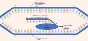
Two strands of DNA separated with an RNA polymerase attached to one of the strands and an RNA molecule coming out of the RNA polymerase
Illustrates the conversion of the template strand of DNA to the pre-mRNA molecule by RNA polymerase.

The enzyme RNA polymerase binds to the exposed template strand and reads from the gene in the 3' to 5' direction. Simultaneously, the RNA polymerase synthesizes a single strand of pre-mRNA in the 5'-to-3' direction by catalysing the formation of phosphodiester bonds between activated nucleotides (free in the nucleus) that are capable of complementary base pairing with the template strand. Behind the moving RNA polymerase the two strands of DNA rejoin, so only 12 base pairs of DNA are exposed at one time. RNA polymerase builds the pre-mRNA molecule at a rate of 20 nucleotides per second enabling the production of thousands of pre-mRNA molecules from the same gene in an hour. Despite the fast rate of synthesis, the RNA polymerase enzyme contains its own proofreading mechanism. The proofreading mechanisms allows the RNA polymerase to remove incorrect nucleotides (which are not complementary to the template strand of DNA) from the growing pre-mRNA molecule through an excision reaction. When RNA polymerases reaches a specific DNA sequence which terminates transcription, RNA polymerase detaches and pre-mRNA synthesis is complete.

The pre-mRNA molecule synthesized is complementary to the template DNA strand and shares the same nucleotide sequence as the coding DNA strand. However, there is one crucial difference in the nucleotide composition of DNA and mRNA molecules. DNA is composed of the bases - guanine, cytosine, adenine and thymine (G, C, A and T) - RNA is also composed of four bases - guanine, cytosine, adenine and uracil. In RNA molecules, the DNA base thymine is replaced by uracil which is able to base pair with adenine. Therefore, in the pre-mRNA molecule, all complementary bases which would be thymine in the coding DNA strand are replaced by uracil.

Post-transcriptional modifications

three strands of RNA at different stages of maturation, the first strand contains introns and exons only, the second strand has gained a 5' cap and 3' tail and contains still introns and exons, the third strand has the cap and tail but the introns have been removed
Outlines the process of post-transcriptionally modifying pre-mRNA through capping, polyadenylation and splicing to produce a mature mRNA molecule ready for export from the nucleus.

Once transcription is complete, the pre-mRNA molecule undergoes post-transcriptional modifications to produce a mature mRNA molecule.

There are 3 key steps within post-transcriptional modifications:

  1. Addition of a 5' cap to the 5' end of the pre-mRNA molecule
  2. Addition of a 3' poly(A) tail is added to the 3' end pre-mRNA molecule
  3. Removal of introns via RNA splicing

The 5' cap is added to the 5' end of the pre-mRNA molecule and is composed of a guanine nucleotide modified through methylation. The purpose of the 5' cap is to prevent break down of mature mRNA molecules before translation, the cap also aids binding of the ribosome to the mRNA to start translation and enables mRNA to be differentiated from other RNAs in the cell. In contrast, the 3' Poly(A) tail is added to the 3' end of the mRNA molecule and is composed of 100-200 adenine bases. These distinct mRNA modifications enable the cell to detect that the full mRNA message is intact if both the 5' cap and 3' tail are present.

This modified pre-mRNA molecule then undergoes the process of RNA splicing. Genes are composed of a series of introns and exons, introns are nucleotide sequences which do not encode a protein while, exons are nucleotide sequences that directly encode a protein. Introns and exons are present in both the underlying DNA sequence and the pre-mRNA molecule, therefore, to produce a mature mRNA molecule encoding a protein, splicing must occur. During splicing, the intervening introns are removed from the pre-mRNA molecule by a multi-protein complex known as a spliceosome (composed of over 150 proteins and RNA). This mature mRNA molecule is then exported into the cytoplasm through nuclear pores in the envelope of the nucleus.

Translation

Five strands of mRNA with all with a ribosome attached at different stages of translation. The first strand has a ribosome and one tRNA carrying its amino acid base pairing with the mRNA, the second strand has a ribosome and a second tRNA carrying an amino acid base pairing with the mRNA, the third strand has the ribosome catalysing a peptide bond between the two amino acids on the two tRNA's. The fourth strand has the first tRNA leaving the ribosome and a third tRNA with its amino acid arriving. The fifth strand has the ribosome catalysing a peptide bond between the amino acids on the second and third tRNA's with an arrowing indicating the cycle continues
Illustrates the translation process showing the cycle of tRNA codon-anti-codon pairing and amino acid incorporation into the growing polypeptide chain by the ribosome.
A ribosome on a strand of mRNA with tRNA's arriving, performing codon-anti-codon base pairing, delivering their amino acid to the growing polypeptide chain and leaving. Demonstrates the action of the ribosome as a biological machine which functions on a nanoscale to perform translation. The ribosome moves along the mature mRNA molecule incorporating tRNA and producing a polypeptide chain.

During translation, ribosomes synthesize polypeptide chains from mRNA template molecules. In eukaryotes, translation occurs in the cytoplasm of the cell, where the ribosomes are located either free floating or attached to the endoplasmic reticulum. In prokaryotes, which lack a nucleus, the processes of both transcription and translation occur in the cytoplasm.

Ribosomes are complex molecular machines, made of a mixture of protein and ribosomal RNA, arranged into two subunits (a large and a small subunit), which surround the mRNA molecule. The ribosome reads the mRNA molecule in a 5'-3' direction and uses it as a template to determine the order of amino acids in the polypeptide chain. To translate the mRNA molecule, the ribosome uses small molecules, known as transfer RNAs (tRNA), to deliver the correct amino acids to the ribosome. Each tRNA is composed of 70-80 nucleotides and adopts a characteristic cloverleaf structure due to the formation of hydrogen bonds between the nucleotides within the molecule. There are around 60 different types of tRNAs, each tRNA binds to a specific sequence of three nucleotides (known as a codon) within the mRNA molecule and delivers a specific amino acid.

The ribosome initially attaches to the mRNA at the start codon (AUG) and begins to translate the molecule. The mRNA nucleotide sequence is read in triplets - three adjacent nucleotides in the mRNA molecule correspond to a single codon. Each tRNA has an exposed sequence of three nucleotides, known as the anticodon, which are complementary in sequence to a specific codon that may be present in mRNA. For example, the first codon encountered is the start codon composed of the nucleotides AUG. The correct tRNA with the anticodon (complementary 3 nucleotide sequence UAC) binds to the mRNA using the ribosome. This tRNA delivers the correct amino acid corresponding to the mRNA codon, in the case of the start codon, this is the amino acid methionine. The next codon (adjacent to the start codon) is then bound by the correct tRNA with complementary anticodon, delivering the next amino acid to ribosome. The ribosome then uses its peptidyl transferase enzymatic activity to catalyze the formation of the covalent peptide bond between the two adjacent amino acids.

The ribosome then moves along the mRNA molecule to the third codon. The ribosome then releases the first tRNA molecule, as only two tRNA molecules can be brought together by a single ribosome at one time. The next complementary tRNA with the correct anticodon complementary to the third codon is selected, delivering the next amino acid to the ribosome which is covalently joined to the growing polypeptide chain. This process continues with the ribosome moving along the mRNA molecule adding up to 15 amino acids per second to the polypeptide chain. Behind the first ribosome, up to 50 additional ribosomes can bind to the mRNA molecule forming a polysome, this enables simultaneous synthesis of multiple identical polypeptide chains. Termination of the growing polypeptide chain occurs when the ribosome encounters a stop codon (UAA, UAG, or UGA) in the mRNA molecule. When this occurs, no tRNA can recognise it and a release factor induces the release of the complete polypeptide chain from the ribosome. Dr. Har Gobind Khorana, a scientist originating from India, decoded the RNA sequences for about 20 amino acids. He was awarded the Nobel prize in 1968, along with two other scientists, for his work.

Protein folding
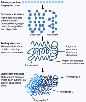
three individual polypeptide chains at different levels of folding and a cluster of chains
Shows the process of a polypeptide chain folding from its initial primary structure through to the quaternary structure.

Once synthesis of the polypeptide chain is complete, the polypeptide chain folds to adopt a specific structure which enables the protein to carry out its functions. The basic form of protein structure is known as the primary structure, which is simply the polypeptide chain i.e. a sequence of covalently bonded amino acids. The primary structure of a protein is encoded by a gene. Therefore, any changes to the sequence of the gene can alter the primary structure of the protein and all subsequent levels of protein structure, ultimately changing the overall structure and function.

The primary structure of a protein (the polypeptide chain) can then fold or coil to form the secondary structure of the protein. The most common types of secondary structure are known as an alpha helix or beta sheet, these are small structures produced by hydrogen bonds forming within the polypeptide chain. This secondary structure then folds to produce the tertiary structure of the protein. The tertiary structure is the proteins overall 3D structure which is made of different secondary structures folding together. In the tertiary structure, key protein features e.g. the active site, are folded and formed enabling the protein to function. Finally, some proteins may adopt a complex quaternary structure. Most proteins are made of a single polypeptide chain, however, some proteins are composed of multiple polypeptide chains (known as subunits) which fold and interact to form the quaternary structure. Hence, the overall protein is a multi-subunit complex composed of multiple folded, polypeptide chain subunits e.g. haemoglobin.

Post-translation events

There are events that follow protein biosynthesis such as proteolysis and protein-folding. Proteolysis refers to the cleavage of proteins by proteases and the breakdown of proteins into amino acids by the action of enzymes.

Post-translational modifications

When protein folding into the mature, functional 3D state is complete, it is not necessarily the end of the protein maturation pathway. A folded protein can still undergo further processing through post-translational modifications. There are over 200 known types of post-translational modification, these modifications can alter protein activity, the ability of the protein to interact with other proteins and where the protein is found within the cell e.g. in the cell nucleus or cytoplasm. Through post-translational modifications, the diversity of proteins encoded by the genome is expanded by 2 to 3 orders of magnitude.

There are four key classes of post-translational modification:

  1. Cleavage
  2. Addition of chemical groups
  3. Addition of complex molecules
  4. Formation of intramolecular bonds

Cleavage
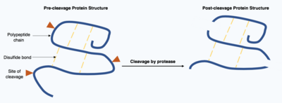
Two polypeptide chain, one chain is intact with three arrows indicating sites of protease cleavage on the chain and intermolecular disulphide bonds. The second chain is in three pieces connected by disulphide bonds.
Shows a post-translational modification of the protein by protease cleavage, illustrating that pre-existing bonds are retained even if when the polypeptide chain is cleaved.

Cleavage of proteins is an irreversible post-translational modification carried out by enzymes known as proteases. These proteases are often highly specific and cause hydrolysis of a limited number of peptide bonds within the target protein. The resulting shortened protein has an altered polypeptide chain with different amino acids at the start and end of the chain. This post-translational modification often alters the proteins function, the protein can be inactivated or activated by the cleavage and can display new biological activities.

Addition of chemical groups

Three polypeptide chains with one amino acid side chain showing, two have a lysine and one has a serine. Three arrows indicating different post-translational modifications with the new chemical group added to each side chain. The first is methylation then acetylation followed by phosphorylation.
Shows the post-translational modification of protein by methylation, acetylation and phosphorylation

Following translation, small chemical groups can be added onto amino acids within the mature protein structure. Examples of processes which add chemical groups to the target protein include methylation, acetylation and phosphorylation.

Methylation is the reversible addition of a methyl group onto an amino acid catalyzed by methyltransferase enzymes. Methylation occurs on at least 9 of the 20 common amino acids, however, it mainly occurs on the amino acids lysine and arginine. One example of a protein which is commonly methylated is a histone. Histones are proteins found in the nucleus of the cell. DNA is tightly wrapped round histones and held in place by other proteins and interactions between negative charges in the DNA and positive charges on the histone. A highly specific pattern of amino acid methylation on the histone proteins is used to determine which regions of DNA are tightly wound and unable to be transcribed and which regions are loosely wound and able to be transcribed.

Histone-based regulation of DNA transcription is also modified by acetylation. Acetylation is the reversible covalent addition of an acetyl group onto a lysine amino acid by the enzyme acetyltransferase. The acetyl group is removed from a donor molecule known as acetyl coenzyme A and transferred onto the target protein. Histones undergo acetylation on their lysine residues by enzymes known as histone acetyltransferase. The effect of acetylation is to weaken the charge interactions between the histone and DNA, thereby making more genes in the DNA accessible for transcription.

The final, prevalent post-translational chemical group modification is phosphorylation. Phosphorylation is the reversible, covalent addition of a phosphate group to specific amino acids (serine, threonine and tyrosine) within the protein. The phosphate group is removed from the donor molecule ATP by a protein kinase and transferred onto the hydroxyl group of the target amino acid, this produces adenosine diphosphate as a biproduct. This process can be reversed and the phosphate group removed by the enzyme protein phosphatase. Phosphorylation can create a binding site on the phosphorylated protein which enables it to interact with other proteins and generate large, multi-protein complexes. Alternatively, phosphorylation can change the level of protein activity by altering the ability of the protein to bind its substrate.

Addition of complex molecules

Two polypeptide chains, one with an asparagine side chain exposed and a polysaccharide attached to the nitrogen atom within asparagine. The other polypeptide has a serine side chain exposed and the core of a polysaccharide attached to the oxygen atom within serine.
Illustrates the difference in structure between N-linked and O-linked glycosylation on a polypeptide chain.

Post-translational modifications can incorporate more complex, large molecules into the folded protein structure. One common example of this is glycosylation, the addition of a polysaccharide molecule, which is widely considered to be most common post-translational modification.

In glycosylation, a polysaccharide molecule (known as a glycan) is covalently added to the target protein by glycosyltransferases enzymes and modified by glycosidases in the endoplasmic reticulum and Golgi apparatus. Glycosylation can have a critical role in determining the final, folded 3D structure of the target protein. In some cases glycosylation is necessary for correct folding. N-linked glycosylation promotes protein folding by increasing solubility and mediates the protein binding to protein chaperones. Chaperones are proteins responsible for folding and maintaining the structure of other proteins.

There are broadly two types of glycosylation, N-linked glycosylation and O-linked glycosylation. N-linked glycosylation starts in the endoplasmic reticulum with the addition of a precursor glycan. The precursor glycan is modified in the Golgi apparatus to produce complex glycan bound covalently to the nitrogen in an asparagine amino acid. In contrast, O-linked glycosylation is the sequential covalent addition of individual sugars onto the oxygen in the amino acids serine and threonine within the mature protein structure.

Formation of covalent bonds
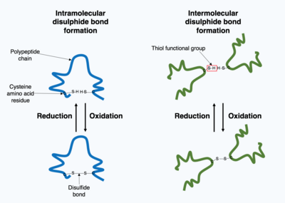
Formation of a disulfide bond between two cysteine amino acids within a single polypeptide chain and formation of a disulphide bond between two cysteine amino acids on different polypeptide chains, thereby joining the two chains.
Shows the formation of disulphide covalent bonds as a post-translational modification. Disulphide bonds can either form within a single polypeptide chain (left) or between polypeptide chains in a multi-subunit protein complex (right).

Many proteins produced within the cell are secreted outside the cell to function as extracellular proteins. Extracellular proteins are exposed to a wide variety of conditions. To stabilize the 3D protein structure, covalent bonds are formed either within the protein or between the different polypeptide chains in the quaternary structure. The most prevalent type is a disulfide bond (also known as a disulfide bridge). A disulfide bond is formed between two cysteine amino acids using their side chain chemical groups containing a Sulphur atom, these chemical groups are known as thiol functional groups. Disulfide bonds act to stabilize the pre-existing structure of the protein. Disulfide bonds are formed in an oxidation reaction between two thiol groups and therefore, need an oxidizing environment to react. As a result, disulfide bonds are typically formed in the oxidizing environment of the endoplasmic reticulum catalyzed by enzymes called protein disulfide isomerases. Disulfide bonds are rarely formed in the cytoplasm as it is a reducing environment.

Role of protein synthesis in disease

Many diseases are caused by mutations in genes, due to the direct connection between the DNA nucleotide sequence and the amino acid sequence of the encoded protein. Changes to the primary structure of the protein can result in the protein mis-folding or malfunctioning. Mutations within a single gene have been identified as a cause of multiple diseases, including sickle cell disease, known as single gene disorders.

Sickle cell disease

two blood curved vessels are shown, on the left one blood vessel contain normal red blood cells throughout the vessel. On the right, the red blood cells have a dish shape due to being sickled, a blockage composed of these distorted red blood cells is present at the curve in the blood vessel.
A comparison between an unaffected individual and an individual with sickle cell anaemia illustrating the different red blood cell shapes and differing blood flow within blood vessels.

Sickle cell disease is a group of diseases caused by a mutation in a subunit of hemoglobin, a protein found in red blood cells responsible for transporting oxygen. The most dangerous of the sickle cell diseases is known as sickle cell anemia. Sickle cell anemia is the most common homozygous recessive single gene disorder, meaning the affected individual must carry a mutation in both copies of the affected gene (one inherited from each parent) to experience the disease. Hemoglobin has a complex quaternary structure and is composed of four polypeptide subunits - two A subunits and two B subunits. Patients with sickle cell anemia have a missense or substitution mutation in the gene encoding the hemoglobin B subunit polypeptide chain. A missense mutation means the nucleotide mutation alters the overall codon triplet such that a different amino acid is paired with the new codon. In the case of sickle cell anemia, the most common missense mutation is a single nucleotide mutation from thymine to adenine in the hemoglobin B subunit gene. This changes codon 6 from encoding the amino acid glutamic acid to encoding valine.

This change in the primary structure of the hemoglobin B subunit polypeptide chain alters the functionality of the hemoglobin multi-subunit complex in low oxygen conditions. When red blood cells unload oxygen into the tissues of the body, the mutated haemoglobin protein starts to stick together to form a semi-solid structure within the red blood cell. This distorts the shape of the red blood cell, resulting in the characteristic "sickle" shape, and reduces cell flexibility. This rigid, distorted red blood cell can accumulate in blood vessels creating a blockage. The blockage prevents blood flow to tissues and can lead to tissue death which causes great pain to the individual.

Cancer
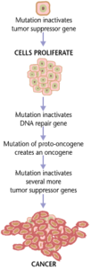
Formation of cancerous genes due to malfunction of suppressor genes.

Cancers form as a result of gene mutations as well as improper protein translation. In addition to cancer cells proliferating abnormally, they suppress the expression of anti-apoptotic or pro-apoptotic genes or proteins. Most cancer cells see a mutation in the signaling protein Ras, which functions as an on/off signal transductor in cells. In cancer cells, the RAS protein becomes persistently active, thus promoting the proliferation of the cell due to the absence of any regulation. Additionally, most cancer cells carry two mutant copies of the regulator gene p53, which acts as a gatekeeper for damaged genes and initiates apoptosis in malignant cells. In its absence, the cell cannot initiate apoptosis or signal for other cells to destroy it.

As the tumor cells proliferate, they either remain confined to one area and are called benign, or become malignant cells that migrate to other areas of the body. Oftentimes, these malignant cells secrete proteases that break apart the extracellular matrix of tissues. This then allows the cancer to enter its terminal stage called Metastasis, in which the cells enter the bloodstream or the lymphatic system to travel to a new part of the body.

Corticosterone

From Wikipedia, the free encyclopedia
 
Corticosterone
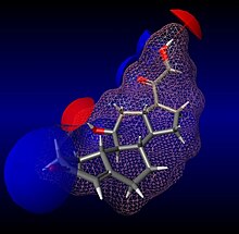
Corticosterone molecule
Names
IUPAC name
11β,21-Dihydroxypregn-4-ene-3,20-dione
Systematic IUPAC name
(1S,3aS,3bS,9aR,9bS,10S,11aS)-10-Hydroxy-1-(hydroxyacetyl)-9a,11a-dimethyl-1,2,3,3a,3b,4,5,8,9,9a,9b,10,11,11a-tetradecahydro-7H-cyclopropa[a]phenanthren-7-one
Identifiers
3D model (JSmol)
2339601
ChEBI
ChEMBL
ChemSpider
DrugBank
ECHA InfoCard 100.000.018 Edit this at Wikidata
EC Number
  • 200-019-6
KEGG
MeSH Corticosterone
UNII


Properties
C21H30O4
Molar mass 346.467 g·mol−1
Hazards
GHS labelling:
GHS07: Exclamation mark
Warning
H317
P261, P272, P280, P302+P352, P321, P333+P313, P363, P501
Except where otherwise noted, data are given for materials in their standard state (at 25 °C [77 °F], 100 kPa).

Corticosterone, also known as 17-deoxycortisol and 11β,21-dihydroxyprogesterone, is a 21-carbon steroid hormone of the corticosteroid type produced in the cortex of the adrenal glands. In the very rare case of congenital adrenal hyperplasia due to 17α-hydroxylase deficiency cortisol production is blocked.

Roles

In many species, including amphibians, reptiles, rodents and birds, corticosterone is a main glucocorticoid, involved in regulation of energy, immune reactions, and stress responses.

However, in humans, cortisol is the primary glucocorticoid that is produced primarily in the zona fasciculata of the adrenal cortex. Corticosterone has only weak glucocorticoid and mineralocorticoid potencies in humans and is important mainly as an intermediate in the steroidogenic pathway from pregnenolone to aldosterone. Corticosterone is converted to aldosterone by aldosterone synthase, found only in the mitochondria of glomerulosa cells. Glomerulosa cells are found in the zona glomerulosa, which is the most superficial region of endocrine cells in the adrenal cortex.

Corticosterone is the precursor molecule to the mineralocorticoid aldosterone, one of the major homeostatic modulators of sodium and potassium levels in vivo.

Release or generation mechanisms

One example of a release pathway relates to UV-B stimulation on the skins of certain amphibians such as the Rough-skinned Newt, Taricha granulosa; this trigger seems to cause the internal generation of corticosterone in that species.

Corticosterone in birds

A sizable amount of research has been done on the effects of corticosterone in birds. A brief survey of this research is below.

Corticosterone both inhibits protein synthesis and degrades proteins. Birds with increased levels of corticosterone will have slower feather growth during their molting period and an extended period of poor flight. As a result, many birds have reduced levels of corticosterone when they moult so as to prevent the degradation of their new feathers. Interestingly, higher levels of corticosterone are also associated with a wider range of exploration, despite beforementioned inhibited feather growth.

Corticosterone has further developmental effects on birds. Increased levels of corticosterone in chicks leads to increased begging for food and aggressiveness. In the short term this leads to higher chance of obtaining food, but in the long term, increased corticosterone in early life compromises the birds cognitive functioning (problem solving, association of visual cue with food, etc.).

Parental response to increased begging by chicks is an increased time foraging for food. This leaves the nest of chicks without protection for increased durations of time. To counter this, during extended periods of food shortage, chicks of some species may suppress corticosterone activity and thus reduce the negative effects elevated corticosterone induces.

Effect on memory

Corticosterone has multiple effects on memory. The main effects are seen through the impact of stress on emotional memories as well as long term memory (LTM).

With emotional memories, corticosterone is largely associated with fear memory recognition. Studies have shown that when fear memories are reactivated or consolidated, levels of corticosterone increased. The increase in corticosterone is linked to anxiety relief. This finding depends on the time at which the administration of corticosterone took place as compared to when the fear conditioning took place; corticosterone can either facilitate or interrupt conditioned fear.

Not only does corticosterone have effects on emotional memories but memory recognition and consolidation as well.

With respect to recognition and long term memories, corticosterone has variable effects. Studies show that the modification of certain chemical and brain processes that affect corticosterone levels can also impact stress effects on memory. In studies on rats, the fluctuations of corticosterone concentration are shown to prevent stress’ impairment of recognition memory in lower amounts. These lower levels seem to be linked to the rescue of stress-induced attenuation of CA1 long-term potentiation. When researchers looked at stress effects on LTM, they found many outcomes. In multiple studies, the formation of LTM (tested 24 h later) was found to be enhanced by corticosterone in some studies, while the persistence of LTM (tested at least 1 wk later) was only assisted by corticosterone in the late phase of memory consolidation and reconsolidation. Stress facilitates the consolidation but disrupts the reconsolidation of emotional memory. As mentioned previously, the persistence of LTM is selectively enhanced when stress and corticosterone are administered during the late phase after acquisition, but it is disrupted when stress and corticosterone are administered during the late phase after retrieval of memory. With regards to the persistence of LTM, there is a restricted time window between acquisition and retrieval where persistence is affected. These studies found that while persistence of LTM is selectively affected based on stage of memory, the formation of LTM is left intact after a certain length of time. Up to this point, studies have not agreed as to whether or not these processes are dependent on corticosterone or what even happens based on corticosterone in these processes and how memory is ultimately affected.

In the end, corticosterone affects many processes in terms of memory as well as different types of memories themselves.

Additional images

Center for Drug Evaluation and Research

FDA Building 51 is one of the main buildings in its White Oak campus that houses the Center for Drug Evaluation and Research.

The Center for Drug Evaluation and Research (CDER, pronounced "see'-der") is a division of the U.S. Food and Drug Administration (FDA) that monitors most drugs as defined in the Food, Drug, and Cosmetic Act. Some biological products are also legally considered drugs, but they are covered by the Center for Biologics Evaluation and Research. The center reviews applications for brand name, generic, and over the counter pharmaceuticals, manages US current Good Manufacturing Practice (cGMP) regulations for pharmaceutical manufacturing, determines which medications require a medical prescription, monitors advertising of approved medications, and collects and analyzes safety data about pharmaceuticals that are already on the market.

CDER receives considerable public scrutiny, and thus implements processes that tend toward objectivity and tend to isolate decisions from being attributed to specific individuals. The decisions on approval will often make or break a small company's stock price (e.g., Martha Stewart and Imclone), so the markets closely watch CDER's decisions.

The center has around 1,300 employees in "review teams" that evaluate and approve new drugs. Additionally, the CDER employs a "safety team" with 72 employees to determine whether new drugs are unsafe or present risks not disclosed in the product's labeling.

The FDA's budget for approving, labeling, and monitoring drugs is roughly $290 million per year. The safety team monitors the effects of more than 3,000 prescription drugs on 200 million people with a budget of about $15 million a year.

Patrizia Cavazzoni is the current director of CDER.

Responsibilities

CDER reviews New Drug Applications to ensure that the drugs are safe and effective. Its primary objective is to ensure that all prescription and over-the-counter (OTC) medications are safe and effective when used as directed.

The FDA requires a four-phased series of clinical trials for testing drugs. Phase I involves testing new drugs on healthy volunteers in small groups to determine the maximum safe dosage. Phase II trials involve patients with the condition the drug is intended to treat to test for safety and minimal efficacy in a somewhat larger group of people. Phase III trials involve one to five thousand patients to determine whether the drug is effective in treating the condition it is intended to be used for. After this stage, a new drug application is submitted. If the drug is approved, stage IV trials are conducted after marketing to ensure there are no adverse effects or long-term effects of the drug that were not previously discovered.

With the rapid advancement of biologically-derived treatments, the FDA has stated that it is working to modernize the process of approval for new drugs. In 2017, Commissioner Scott Gottlieb estimated that they have more than 600 active applications for gene and cell-based therapies.

Divisions

CDER is divided into 8 sections with different responsibilities:

  • Office of New Drugs
This office is responsible for oversight of clinical trials and other studies during drug development, and for the evaluation of new drug applications
The Office of New Drugs is divided into several departments based on the indication of the drug (the medical need for which it is being proposed)
  • Office of Generic Drugs
This office reviews generic drug applications to ensure generic drugs are equivalent to their branded forms
  • Office of Strategic Programs
This office is responsible for business programs, represents CDER in the FDA Bioinformatics Board, and communicates with other agencies
  • Office of Pharmaceutical Quality
This office is responsible for integrating assessment, inspection, surveillance, policy, and research activities to strengthen pharmaceutical quality on a global scale.
  • Office of Surveillance and Epidemiology
This office is responsible for post-marketing surveillance to identify adverse effects that may not have been apparent during clinical trials, using the MedWatch program
  • Office of Translational Sciences
This office promotes collaboration across offices in CDER by maintaining databases and biostatistical tools for evaluating drugs
  • Office of Medical and Regulatory Policy
This office develops and reviews guidelines pertinent to CDER's mission of ensuring the safety of drugs
  • Office of Compliance
This office ensures compliance with regulations relating to drug development and marketing

History

The FDA has had the responsibility of reviewing drugs since the passage of the 1906 Pure Food and Drugs Act. The 1938 Federal Food, Drug and Cosmetic Act required all new drugs to be tested before marketing by submitting the original form of the new drug application. Within the first year, the FDA's Drug Division, the predecessor to CDER, received over 1200 applications. The Drug Amendments of 1962 required manufacturers to prove to the FDA that the drug in question was both safe and effective. In 1966, the division was reorganized to create the Office of New Drugs, which was responsible for reviewing new drug applications and clinical testing of drugs.

In 1982, when the beginning of the biotechnology revolution blurred the line between a drug and a biologic, the Bureau of Drugs was merged with the FDA's Bureau of Biologics to form the National Center for Drugs and Biologics during an agency-wide reorganization under Commissioner Arthur Hayes. This reorganization similarly merged the bureaus responsible for medical devices and radiation control into the Center for Devices and Radiological Health.

In 1987, under Commissioner Frank Young, CDER and the Center for Biologics Evaluation and Research (CBER) were split into their present form. The two groups were charged with enforcing different laws and had significantly different philosophical and cultural differences. At that time, CDER was more cautious about approving therapeutics and had a more adversarial relationship with the industry. The growing crisis around HIV testing and treatment and an inter-agency dispute between officials from the former Bureau of Drugs and officials from the former Bureau of Biologics over whether to approve Genentech's Activase (tissue plasminogen activator) led to the split.

In its original form, CDER was composed of six offices: Management, Compliance, Drug Standards, Drug Evaluation I, Drug Evaluation II, Epidemiology and Biostatistics, and Research Resources. The Division of Antiviral Products was added in 1989 under Drug Evaluation II due to the large amount of drugs proposed for treating AIDS. The Office of Generic Drugs was also formed.

In 2002, the FDA transferred a number of biologically produced therapeutics to CDER. These include therapeutic monoclonal antibodies, proteins intended for therapeutic use, immunomodulators, and growth factors and other products designed to alter production of blood cells.

MDMA-assisted psychotherapy

From Wikipedia, the free encyclopedia
 
MDMA-assisted psychotherapy is the use of prescribed doses of MDMA as an adjunct to psychotherapy sessions. Research suggests that MDMA-assisted psychotherapy for post-traumatic stress disorder (PTSD), including Complex PTSD, might improve treatment effectiveness. In 2017, a Phase II clinical trial led to "breakthrough therapy" designation by the US Food and Drug Administration (FDA).

The research is controversial in part because recreational MDMA use has been associated with harmful effects among some users.

Therapeutic effects

Post-traumatic stress disorder (PTSD) is most commonly treated by cognitive behavioral therapy (particularly prolonged exposure and cognitive processing therapy), eye movement desensitization and reprocessing, and psychodynamic psychotherapy. However, over half of these patients continue to have PTSD after completing therapy, with results from military PTSD being especially poor.

PTSD is best treated when a patient is in the 'optimal arousal zone'. This is the zone in which emotions are engaged yet not overwhelming. In this zone, four symptom clusters of PTSD are sedated: These are:

  1. re-experiencing
  2. avoidance
  3. negative alterations in cognition/mood
  4. alterations in arousal and reactivity

Subjects with PTSD exhibit extreme emotional numbing or anxiety and struggle to remain in the optimal arousal zone during conservative therapies. Threatening interpretations of memories are reinforced when patients are in low emotional states. If traumatic memories are revisited in therapy when a patient is not within the optimal arousal state, therapy for PTSD can actually increase the patient's trauma.

When used in therapy, MDMA has been reported to increase empathy, closeness between patient and therapist, relaxation, motivation to engage with therapy and introspective thought, and to reduce depression and anxiety. MDMA makes it easier for a patient to stay in the optimal arousal zone by decreasing feelings of anxiety and defensiveness when revisiting traumatic memories. It also increases feelings of closeness and empathy, improving the patient's trust in the therapist and encouraging introspective thought to reassess memories and actions. Furthermore, research suggests that treatment may improve the quality of sleep of individuals affected by PTSD-related sleep disturbances. These factors may increase the success rate of psychotherapy.

Adverse effects, which can last from a few hours to several days, include diminished appetite, anxiety, headache, jaw tightness, tinnitus, nausea, asthenia, fatigue, acute sinusitis, nasopharyngitis, upper respiratory tract infection, disturbance in attention, tremor, tics, dysuria, erythema, and depression.

Research

With the approval of the FDA in 2017, MDMA has been cleared for use in assisting with psychotherapy. A phase 3 study indicated that MDMA-assisted therapy represents a potential breakthrough treatment for severe PTSD that merits expedited clinical evaluation. Based on this study, MDMA-assisted psychotherapy was granted breakthrough therapy designation by the FDA, a designation that indicates that there is preliminary evidence that an intervention might offer a substantial improvement over other options for a serious health condition. However, given the lack of blinding, several researchers have postulated that the results of the phase 3 trial might be heavily influenced by expectancy effects, and there are no trials comparing MDMA-assisted psychotherapy to already existing first-line psychological treatments for PTSD, which based on indirect evidence seems to attain similar or elevated symptom reduction compared with that due to MDMA-assisted psychotherapy.

Neuroscience & mechanism of action

Chemical compound of MDMA

PTSD inhibits a subject's ability to respond appropriately to trauma-related stimuli. The current model of PTSD proposes that it results from amplified and uncontrolled responses from the amygdala to trauma-specific cues. Oxytocin, which is increased by MDMA, has been found to increase trust and emotional awareness and reduce amygdala responses as well as reduce coupling of the amygdala to brainstem regions associated with autonomic and behavioral characteristics of fear. It has been proposed that these effects foster memory reconsolidation by allowing the patient to access the traumatic memory while feeling detached from the sense of imminent threat.

Legality

MDMA was first synthesized by the German pharmaceutical company Merck KGaA in 1912 as an intermediate in the synthesis of another compound. Its psychoactive effects were not noted until the early 1960s. In the 1960s and 1970s, the drug was used in psychotherapy, although it was not an approved drug and no clinical trials had been performed. The drug was studied in Switzerland for use in individual, couple, and group therapies until 1993 when the Swiss Ministry of Health withdrew permission to use MDMA and LSD by psychiatrists due to concerns about a lack of research methodology.

In 1986, MDMA was classed as a Schedule 1 drug by the United Nations according to its Convention on Psychotropic Substances of 1971 due to its high potential for abuse, and most research was stopped. Researchers interested in MDMA for use in psychotherapy founded and funded the Multidisciplinary Association for Psychedelic Studies (MAPS) in response. The US Food and Drug Administration (FDA) and Drug Enforcement Administration (DEA) granted approval for researching MDMA's efficacy as an adjunct to psychotherapy in 2004, and the first trial was carried out in 2011. In 2023, MAPS announced that it is compiling data from 18 different phase 2 and phase 3 studies with plans to file a New Drug Application with the FDA. MAPS hopes to receive FDA approval by the end of 2024.

Controversy & safety

MDMA's effects vary across people and settings and include adverse outcomes. The drug causes neurotransmitter activation across the main neural pathways (including serotonin and dopamine, noradrenaline) that can result in large mood swings. The memories that emerge under the influence of MDMA can evoke unwanted emotions. Side effects of MDMA use by recreational users include appetite fluctuations, food cravings, and disordered eating.

Once the effects of MDMA wear off, there is a "period of neurochemical depletion" that invokes anhedonia, lethargy, anger, depression, irritability, brooding, greater everyday stress, altered pain thresholds, changes in sleep, and bad dreams, especially in female participants. The symptoms are thought to be due to the depletion of serotonin, as a result of the large release of serotonin triggered by MDMA and have been called "neurotoxic in terms of causing serotonergic dysfunction."

There are also concerns surrounding "drug-dependent learning" — the theory that patients will return to the drug to access the state they were in when on the drug in therapy.

There were 92 MDMA-related deaths in England and Wales in 2018, up from 56 the year before, and 10,000 hospitalizations for MDMA-related illness/injury in 2011 in the US. However, as of 2021, there have been no such cases reported for clinical settings.

Media reports and statements of academic authors have often transmitted the view of MDMA as a possible medicine or treatment rather than as an adjunct to psychotherapy. This has been considered dangerous because it could lead people to believe that MDMA is an effective treatment alone, without concomitant psychotherapy.

Psychological stress and sleep

From Wikipedia, the free encyclopedia
Stature of distressed/ tired man
Tired man

Sleep is a naturally recurring state of mind and body, characterized by altered consciousness, relatively inhibited sensory activity, reduced muscle activity, and inhibition of nearly all voluntary muscles during rapid eye movement (REM) sleep, and reduced interactions with surroundings. An essential aspect of sleep is that it provides the human body with a period of reduced functioning that allows for the systems throughout the body to be repaired. This time allows for the body to recharge and return to a phase of optimal functioning. It is recommended that adults get 7 to 9 hours of sleep each night. Sleep is regulated by an internal process known as the circadian rhythm. This 24-hour cycle regulates periods of alertness and tiredness that an individual experiences. The correlation between psychological stress and sleep is complex and not fully understood. In fact, many studies have found a bidirectional relationship between stress and sleep. This means that sleep quality can affect stress levels, and stress levels can affect sleep quality. Sleep change depends on the type of stressor, sleep perception, related psychiatric conditions, environmental factors, and physiological limits.

Stress/sleep cycle

It is critical that we receive an adequate amount of sleep each night. According to the Centers for Disease Control and Prevention, people 18-60 years old need 7 or more hours of sleep per night. The majority of college students fall in this age range. While sleep is critical, many college students do not reach this threshold amount of sleep, and subsequently face detrimental effects. However, it is clear that stress and sleep in college students are interrelated, instead of one only affecting the other. "Stress and sleep affect each other. Poor sleep can increase stress, otherwise high-stress can also cause sleep disturbances". As stated in a different way, the way stress and sleep are related is bidirectional in nature.

Types of stressors

Stressors can be categorized into the Challenge/Hindrance stressor model. Challenge stressors, while unpleasant, allow for growth and achievement such as time pressure in a work context. Hindrance stressors are those that cause unnecessary burdens and do not contribute to achievement such as poor work supervision. Self-reported quality of sleep reduces in relation to more hindrance stressors but not to challenge stressors.

Sleep quality perception

Insomnia is a common condition affecting up to a fifth of the population in many countries across the world and is often complicated by several psychiatric conditions. Paradoxical insomnia is the phenomenon of a discrepancy between reported sleep duration and objective measurement of sleep. In some cases, however, the stress and anxiety produced do result in an objective reduction in the quality of sleep.

Stress factors that lead to a lack of sleep

The factors of stress that contribute to the lack of sleep include, but are not limited to, overthinking, excess caffeine consumption, and excess cortisol emission. Overthinking is a very common factor in keeping people awake because they are unable to calm their minds from their stressors. This rumination can impact the transitions between sleep stages since going to bed while overthinking can lead to disruptions in the stages in the middle of the night. Another cycle that also occurs with stress is that people under more stress tend to consume larger amounts of caffeine which can actually increase stress/anxiety levels. Furthermore, if people drink too much caffeine later in the day, they may have trouble falling asleep which will cause them to sleep fewer hours and have to continue the cycle of drinking more caffeine the next day to feel awake throughout the day. It is known that sleep deprivation can affect the efficiency at which one performs, such as at work for example. What is one way to counter this? More coffee. The cycle repeats itself. Cortisol, the stress hormone that is responsible for the fight-or-flight response, increases during times of stress and can often lead to a feeling of a jolt of energy. If people are feeling highly stressed right before bed, they may release higher amounts of cortisol which can cause unhealthy levels of sleep. It is also important to recognize that the stress one feels does not need to be chronic or trauma-induced. Everyday stressors in work, relationships, and other parts of life can impact a person's sleep cycle and continuous days of feeling stressed or overwhelmed can lead to multiple restless nights. The effects of stress on one's sleep quantity and quality can become frustrating, unhealthy, and harmful to a person's quality of life.

Increased prevalence of stress for teenagers

There are consistent stressors in life, some that are good, referred to as eustress, and some that are bad, which are referred to as distress. While these stressors have not necessarily increased over the years, the overall average stress levels have increased as data has shown that the typical high school student today has the same amount of anxiety as a psychiatric patient in the 1950s. The average stress level in the United States -- 5.6 out of 10 -- is far above past average levels and is the highest it has been in the last decade. There is also evidence showing that age impacts the correlation between lack of sleep and stress. Millennials (born between 1981 and 1996) and Gen Zers (born between 1997 and 2012) report the highest levels of stress out of all the generations, in which 44% of millennials and 48% of Gen Zers report being stressed all or most of the time. Correspondingly, younger Americans report getting fewer hours of sleep per night, in which a large portion only sleeps for about 6.5-7.5 hours per night on average. These correlating statistics reveal an epidemic that is being created with stress and an increased risk of chronic sleep deprivation. Stress often leads to difficulties falling asleep and staying asleep, however, a lack of sleep can also contribute to one's stress levels. This relationship leads to a never-ending cycle of being too stressed to sleep and then being unable to control one's anxieties because of the impacts of a lack of sleep. Unfortunately, this troubling cycle also causes an increased risk for the potential impacts of sleep deprivation and excessive stress, including many physical and mental health issues. These issues can have long-term consequences that may affect one's social life, academic capabilities, and relationships with others.

Neuropsychiatric mechanisms

Sleep can be broadly split into the lighter "rapid eye movement" (REM) and deeper "non-rapid eye movement" (NREM). Changes in sleep phase vary in animal models depending on the stressor but do not alter total sleep duration except for novelty which reduces both REM and NREM. Conditioned fear, for example, reduces REM sleep whereas auditory stimulation increases it.

In humans, models of stress have been closely linked to the context depression. Changes in sleep patterns in depression are very close to those seen in acutely stressed animals; these changes can be used as a predictor for developing depression. Once again, studies have shown a bidirectional nature between depressive symptoms and lack of sleep due to stress. Long-term/chronic psychosocial stress is known to cause depression symptoms but the effect of chronic stress on sleep can lead to a ripple effect of further damage including poor emotional stability, lowered attention span and self-control, and worse performance on cognitive tasks. Early life sleep disruption caused by stressors may affect neuroplasticity and synaptic connectivity potentially leading to the development of mood disorders. This poor sleep may become a stressor itself compounding the phenomenon.

Cholinergic neurons

In animal studies, psychologically stressed rats display an increase in total REM sleep and the average length of REM phase duration (but not the number of cycles). This change is mediated by cholinergic neurons as stressed animals' prolonged REM cycles can be reduced by using a cholinergic antagonist (atropine). One study found that auditory stimulation stressors act similarly by inhibiting the cholinergic reduction of REM sleep. Chronic mildly stressed rats display a reduction in inhibitory GABA receptors in the hypothalamus (increasing the release of stress hormones) and brain stem among others. Within the pedunculopontine tegmentum region, in the brainstem, reduced GABA imbibition of cholinergic neurons acts again in the same way in increasing REM sleep duration.

Hypothalamic-pituitary-adrenal axis

The neuroendocrine hypothalamic-pituitary-adrenal axis is a system of hormones that culminates in the release of cortisol from the adrenal glands in response to acute stress and is also seen to regulate sleep patterns. The reduction in GABA receptors in the hypothalamus seen in chronic stress reduces the inhibition of stress hormone release however does not appear to impact sleep patterns after exposure to a stressful social stimulus in animals.

Prenatal and childhood stress

Chronic maternal stress in pregnancy exposes the fetus to increased levels of glucocorticoid and inflammatory markers which in turn negatively affects the H-P-A axis and disrupts sleep regulation of the fetus. Up until the age of 2 years, children who have been exposed to prenatal stress have shortened and disorganized sleeping patterns. During early childhood development, the child's brain is particularly sensitive to adverse events such as family conflict, maternal postnatal depression, or abuse. It is thought that it is via sensitization of the H-P-A axis that an abnormal stress response is developed in response to these events/stressors which in turn causes emotional disorders and later life sleeping disorders.

Stress and substance abuse

Chronic stress leads to the malfunction of the HPA axis which can lead to sudden relapse in previously well-recovering alcoholics. Changes in HPA axis regulation lead to drastic over/underproduction of important stress response hormones because the system is usually kept under very tight regulation in order to quickly respond to external stimuli. Cortisol, the main stress hormone produced by the HPA axis is thought to be responsible for the vulnerability to alcohol abuse. Studies show that cortisol production correlates with heightened activity in the neural reward pathways of the brain, which could explain how stress leads to alcoholism.

Immune mediation

Observations have been made that there is an association between stress, sleep, and Interleukin-6 proposing a possible mechanism for sleep changes.

During both chronic and acute phase sleep deprivation, there are increases in the pro-inflammatory cytokine Interleukin-6 (IL-6). Not only is IL-6 influenced by the circadian rhythm but its effectiveness is increased by sleep itself as there is an increase in serum IL-6 receptors during sleep. After periods of long sleep deprivation, the first post-deprivation sleep shows a marked drop in IL-6 and an increase in slow wave sleep / "deep sleep". Similarly napping during the daytime has been shown to decrease IL-6 and reduce tiredness. When humans are injected with exogenous IL-6 they display an increase in fatigue and other sickness behaviour.

This IL-6 increase is also observed during times of increased psychological stress. In a laboratory setting, individuals exposed to psychological stressors have had raised IL-6 (an acute-phase protein CRP) measured especially in those who displayed anger or anxiety in response to the stressful stimulus. Just as the human body responds to inflammation-inducing illness with increased fatigue or reduced sleep quality, so too does it respond to psychological stress with a sickness behavior of tiredness and poor sleep quality. While sleep is important for recovery from stress, as, with an inflammatory illness, continuous and long-term increases of inflammatory markers with its associated behaviors may be considered maladaptive and be linked to long-term inefficient sleep. 

Military context

Since the American Civil War, there have been multiple "war syndromes" reported such as 'irritable heart', 'effort syndrome' and 'Gulf War Syndrome'. Thought to be discrete and different from post-traumatic stress disorder (PTSD), these war syndromes have a range of physical symptoms but commonly feature sleep disturbances, tiredness, poor concentration, and nightmares. The historic picture is unclear due to poor contemporary understanding of psychological illness and, in more modern conflicts, gathering data has been difficult due to operational priorities; no cause has been identified that is not connected to psychological stress.

PTSD

Sleep is often a core focus for both diagnosis and management of PTSD with 70% of PTSD patients reporting insomnia or sleep disturbances. When studied against controls, however, little difference was measured in the quality of sleep suggesting paradoxical insomnia along with physiological H-P-A axis involvement and "fight or flight responses". It is on this basis that CBT, a non-pharmacological therapy, is justified along with pharmacological intervention.

One month after the coronavirus outbreak, a study determined a frequency of PTSD (Post-traumatic stress disorder) symptoms among inhabitants of Wuhan and its surrounding villages. To better understand this phenomenon, a study was conducted in Canada after a two-month state of emergency was declared (2020). Many subjects began to have dreams highlighting deep-rooted trauma, like early childhood experiences, however not one subject described the pandemic as traumatic, simply stressful. The dream content was especially interesting because it had little to no relation to the pandemic; this study was consistent with others questioning the reemergence of PTSD when faced with a new stressor.

Stress and trauma can lead to vivid dreams. The classic "PTSD dream" can be used as a lens to understand the effect of waking stress on one's sleep. A PTSD dream occurs often and is a replay of a traumatic event. The person is woken up in sweats, shaking; fear, anxiety, anger, etc. are all induced.

Occupational context

One of the greatest factors affecting the stress and sleep of humans is their commitment to their jobs. In our society, one's employment schedule often dictates their sleep schedule. The bidirectional relationship between stress and sleep is also scientifically supported in terms of employment. When an employee does not get quality sleep, this often leads to poor performance at work and a greater chance of experiencing stress related to work. Similarly, when a person is experiencing occupational stress, their sleep is almost immediately negatively influenced. When individuals experience high levels of stress and insufficient amounts of sleep their mental and physical health is jeopardized.

Development of occupational stress

Many factors contribute to the development of occupational stress in one's life. Some of these factors include job scheduling, time commitment, lack of support, and conflicts within the workplace. Depending on the field in which people are employed, the demands of the job vary greatly. Some occupations demand at least 40 hours a week from an employee. This commitment, combined with other personal responsibilities, often leaves individuals little time for themselves, which elevates stress levels. When individuals have irregular work schedules, such as working at different times from week to week their regular day-to-day schedule is interrupted. This not only adds stress to their daily life but also influences an individual's circadian rhythm. The circadian rhythm is especially interrupted when individuals work night shifts. When faced with this challenge the body is not only stressed but must also adapt to new environmental factors. For instance, this would require the body to be more alert during late night hours as opposed to entering a relaxed state, as the average circadian rhythm would support.

Another main contributor to the development of occupational stress in employees is a feeling of a lack of support and recognition from superiors. When employees are not supported by their superiors, employees are more likely to experience stress related to their work responsibilities. Often employees dedicate extra time to perfect their work to achieve the desired validation they seek from superiors. When employees are not recognized and supported by their superiors, feelings of uncertainty begin to grow and overwhelm the individual. Additionally, when the responsibilities and expectations of an employee are not clearly understood this often leaves the employee questioning their place in the work environment.

Finally, conflicts within the workplace have been named as a major stressor when it comes to employment. When conflicts arise in the workplace the unsafe environment raises the employee's stress level and often produces an extreme emotional response. Furthermore, these anxiety-inducing conflicts lead employees to dread coming to work each day. These matters distract individuals and influence their overall productivity. It is important to note that these conflicts often revolve around a difference in power between the parties involved. Instances of sexual harassment within the work environment are the most prevalent and cause unnecessary amounts of stress.

Risk factors and interventions

Combining high levels of stress and a lack of sleep affects the day-to-day life of humans. For example, work performance often suffers under these conditions. Individuals suffering from stress and sleep deprivation also tend to have higher levels of absenteeism and take a greater number of sick days compared to their peers. When adequate amounts of sleep are not obtained, individuals are at a greater risk for developing heart disease, diabetes, hypertension, muscle pain, headaches, and a series of mental health problems. There is a strong association between lack of sleep and increased irritability, depression, and anxiety disorders. The working memory of individuals experiencing sleep deprivation is also affected.

There are various methods and practices that individuals can engage in to lower their levels of stress and optimize their sleep. Employers should address possible stressors within the work environment. Employers need to monitor the workloads and schedules of their employees to minimize burnout, but more importantly to prioritize the health of their workers. Providing training and clearly stating the expectations of an employee will help employees to better understand their role in the workplace. By building a positive work environment, employees will feel supported by their superiors and experience less stress in their daily life. An essential first step for employees is to acknowledge the stressor in their life. On an individual basis, employees experiencing high levels of stress and restlessness can engage in cognitive-behavioral interventions to manage their stress. Practicing simple self-care routines and setting aside time for the things that one enjoys will aid in minimizing the levels of stress that employees experience.

Performance and attention context

While stress and sleep greatly impact each other, their effect permeates into many more aspects of daily life. One specific concern is the harmful effects on cognitive performance and attention span. In addition, sleep deprivation can cause a change in perceptions as well. Being sleep-deprived for 24 hours leads to a dramatic decrease in cognitive performance tests similar to college exams, and causes individuals to have false perceptions about their performance. Hence, sleep-deprived individuals are cognitively performing worse but are not aware of it. In addition to cognitive performance, sleep deprivation can cause a decreased attention span on specific tasks at hand. This has further implications such as making more mistakes or not being as efficient. It can become a cycle of high stress, poor sleep, and lack of attention when these three things are intermixed.

Short-term physical health impacts of stress and sleep deprivation

Both excessive stress and sleep deprivation cause physical health impacts that may affect a person short-term or long-term. These impacts range in severity and it is important to be aware of the increased risk of health issues that may arise due to the stress-sleep cycle. Many of the physical impacts of stress overlap with the physical impacts of sleep deprivation, including short-term impacts like fatigue and headaches, and long-term impacts like high blood pressure, heart disease, diabetes, and obesity. Fatigue is a clear side effect of sleep deprivation, however, when combined with excessive stress, the feeling of fatigue can become overwhelming because one's body is having to work harder and is under more pressure which causes a person to feel further fatigued. Headaches are another short-term impact that occurs often for those who are feeling excessive levels of stress since stress often triggers a fight or flight response which can create tension headaches. When lack of sleep and excessive stress combine, the impacts of fatigue and headaches greatly increase. Although these impacts are both short-term, they can last for days, weeks, or even months if the stress continues to overwhelm a person and cause them to struggle to fall and stay asleep.

Long-term physical health impacts of stress and sleep deprivation

Cardiovascular impacts

Long-term effects can result from years of persistent feelings of excessive levels of stress and consequently getting a consistent lack of sleep. Excessive stress and sleep deprivation can cause cardiovascular issues, such as high blood pressure and heart disease. In a study focusing on the impacts of chronic stress on the heart, it was found that during times of chronic stress, the body hyperactivates the sympathetic nervous system which leads to changes in heart rate variability. Due to these changes in heart rate variability, which can harm the capabilities and strength of the heart, the risk of heart disease greatly increases due to elevated blood sugar and blood pressure levels. Prolongation of this stress on the body can cause plaque buildup in the artery walls which impedes blood flow and results in a much higher likelihood of major cardiovascular events, including heart attack and stroke. Sleep also adds to these cardiovascular impacts because blood pressure normally decreases during sleep. As a result, someone with a consistent lack of sleep has higher blood pressure levels for longer periods of time. In a study that was conducted to find the correlation between sleep deprivation and cardiovascular issues, it was found that one hour less of sleep each night increased the risk of calcium build-up in the arteries by 33%. Calcium buildup in the arteries is a major cause of plaque buildup, which was also mentioned as highly affected by increased stress levels. When combined, excessive stress and sleep deprivation cause a much larger increase in plaque buildup which can lead to an increased risk of heart disease, stroke, and high blood pressure. When someone is constantly feeling stressed throughout their day and then struggles to fall and stay asleep due to stress and anxiety, it creates a continued cycle of strain on the heart.

Increased chance of obesity

Another long-term effect that creates a combined risk is the effect of excessive stress and sleep deprivation on obesity. While short-term stress may cause people to lose their appetite, chronic stress causes the release of cortisol which increases a person's appetite. Many effects from stress cause people to overeat, including a lack of self-regulation during stressful times, a want for "comfort food" which is often high in sugar, fat, and calories, and cortisol's promotion of eating and fat deposition. These all cause stressful times to have many physiological and behavioral impacts on one's diet. Furthermore, sleep deprivation has been shown to decrease glucose tolerance and insulin sensitivity, and increase hunger and appetite, all of which impact one's diet and what foods they prefer. These hormonal changes often cause an increase in caloric intake and decrease the energy for many physical activities which combine to increase the likelihood of obesity. The many impacts of increasing stress levels and sleep deprivation show that there are many factors that can cause overall weight gain which may lead to obesity. It is important to note that many of these effects are more impactful after long-term chronic stress and sleep deprivation, however, since sleep deprivation and stress often coincide, it leads to a much greater chance of developing these harmful physical health impacts. Since both sleep deprivation and stress have similar impacts on one's body, the overall likelihood of serious health issues increases.

E-patient

From Wikipedia, the free encyclopedia https://en.wikipedi...