Search This Blog

Sunday, December 10, 2023

Nonsense mutation

From Wikipedia, the free encyclopedia

In genetics, a nonsense mutation is a point mutation in a sequence of DNA that results in a nonsense codon, or a premature stop codon in the transcribed mRNA, and leads to a truncated, incomplete, and possibly nonfunctional protein product. Nonsense mutation is not always harmful, the functional effect of a nonsense mutation depends on many aspects, such as the location of the stop codon within the coding DNA. For example, the effect of a nonsense mutation depends on the proximity of the nonsense mutation to the original stop codon, and the degree to which functional subdomains of the protein are affected. As nonsense mutations leads to premature termination of polypeptide chains; they are also called chain termination mutations.

Missense mutations differ from nonsense mutations since they are point mutations that exhibit a single nucleotide change to cause substitution of a different amino acid. A nonsense mutation also differs from a nonstop mutation, which is a point mutation that removes a stop codon. About 10% of patients facing genetic diseases have involvement with nonsense mutations. Some of the diseases that these mutations can cause are Duchenne muscular dystrophy (DMD), cystic fibrosis (CF), spinal muscular atrophy (SMA), cancers, metabolic diseases, and neurologic disorders. The rate of nonsense mutations is variable from gene-to-gene and tissue-to-tissue but gene silencing occurs in every patient with a nonsense mutation.

Simple example

    DNA: 5' - ATG ACT CAC CGA GCG CGA AGC TGA - 3'
         3' - TAC TGA GTG GCT CGC GCT TCG ACT - 5'
   mRNA: 5' - AUG ACU CAC CGA GCG CGA AGC UGA - 3'
Protein:      Met Thr His Arg Ala Arg Ser Stop

The example above begins with a 5' DNA sequence with eight nucleotides seen and its complementary strand shown below. The next row highlights the 5' mRNA strand, which is generated through transcription. Lastly, the final row showcases which the amino acids that are translated from each respective codon, with the eighth and final codon representing the stop codon. The codons corresponding to the fourth amino acid, Arginine, are highlighted because they will undergo a nonsense mutation in the following figure of this example.

    DNA: 5' - ATG ACT CAC TGA GCG CGA AGC TGA - 3'
         3' - TAC TGA GTG ACT CGC GCT TCG ACT - 5'
   mRNA: 5' - AUG ACU CAC UGA GCG CGU AGC UGA - 3'
Protein:      Met Thr His Stop

Now, suppose that a nonsense mutation was introduced at the fourth codon in the 5' DNA sequence (CGA) causing the cytosine to be replaced with thymine, yielding TGA in the 5' DNA sequence and ACT in the complementary strand. Because ACT is transcribed as UGA, it is translated as a stop codon. This leads the remaining codons of the mRNA to not be translated into protein because the stop codon is prematurely reached during translation. This can yield a truncated (i.e., abbreviated) protein product, which quite often lacks the functionality of the normal, non-mutant protein.

Possible outcomes

Deleterious

Deleterious outcomes represent the majority of nonsense mutations and are the most common outcome that is observed naturally. Deleterious nonsense mutations decreases the overall fitness and reproductive success of the organism. For example, a nonsense mutation occurring in a gene encoding a protein can cause structural or functional defects in the protein that disrupt cellular biology. Depending on the significance of the functions of this protein, this disruption now could be detrimental to the fitness and survival of that organism.

Neutral

When a nonsense mutation is neutral, it does not provide benefits or harm. These occur when the effects of the mutation are unnoticed. In other words, this means that the mutation does not positively or negatively affect the organism. As this effect is unnoticed, there is a lack of papers describing such mutations. An example of this type of nonsense mutation is one that occurs directly before the original stop codon for that given protein. Because this mutation occurred in such close proximity to the end of the protein chain, the impact of this change might not be as significant. This would suggest that this amino acid that was mutated did not have a large impact on the overall structure or function of the protein or the organism as a whole. This scenario is rare, but possible.

Beneficial

Beneficial nonsense mutations are considered as the rarest of possible nonsense mutation outcomes. Beneficial nonsense mutations increase the overall fitness and reproductive success of an organism, opposite of the effects of a deleterious mutation. Because a nonsense mutation introduces a premature stop codon within a sequence of DNA, it is extremely unlikely that this scenario can actually benefit the organism. An example of this would occur with a nonsense mutation that impacts a dysfunctional protein that releases toxins. The stop codon that this mutation brings would stop this dysfunctional protein from properly carrying out its function. Stopping this protein from performing at full strength causes less toxin to be released and the fitness of the organism to be improved. These types of situations with nonsense mutations occur a lot less frequently than the deleterious outcomes.

Suppressing nonsense mutations

Pictured on the left is a diagram of normal translation occurring without mutation. Blue circles are the peptides already translated while the grey circles are peptides going to be translated next. In the center is a diagram a nonsense mutation where the UUG codon is translated to the stop codon UAG. The stop codon recruits a release factor, terminating translation. On the right is a diagram of the tRNA suppression mechanism where the codon and the tRNA are both mutated, resulting in tRNA suppression. The mutated Tyr tRNA has the anticodon AUC which recognizes the UAG stop codon, continuing protein translation.

Nonsense-mediated mRNA decay

Despite an expected tendency for premature termination codons to yield shortened polypeptide products, in fact the formation of truncated proteins does not occur often in vivo. Many organisms—including humans and lower species, such as yeast—employ a nonsense-mediated mRNA decay pathway, which degrades mRNAs containing nonsense mutations before they are able to be translated into nonfunctional polypeptides.

tRNA Suppression

Because nonsense mutations result in altered mRNA with a premature stop codon, one way of suppressing the damage done to the final protein's function is to alter the tRNA that reads the mRNA. These tRNA’s are termed suppressor tRNA's. If the stop codon is UAG, any other amino acid tRNA could be altered from its original anticodon to AUC so it will recognize the UAG codon instead. This will result in the protein not being truncated, but it may still have an altered amino acid. These suppressor tRNA mutations are only possible if the cell has more than one tRNA that reads a particular codon, otherwise the mutation would kill the cell. The only stop codons are UAG, UAA, and UGA. UAG and UAA suppressors read their respective stop codons instead of their original codon, but UAA suppressors also read UAG due to wobble base pairing. UGA suppressors are very rare. Another hurdle to pass in this technique is the fact that stop codons are also recognized by release factors, so the tRNA still needs to compete with the release factors to keep the translation going. Because of this, suppression is usually only 10-40% successful. These suppressor tRNA mutations also target stop codons that are not mutations, causing some proteins to be much longer than they should be. Only bacteria and lower eukaryotes can survive with these mutations, mammal and insect cells die as a result of a suppressor mutation.

Common disease-associated nonsense mutations
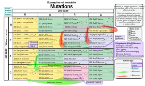
Selection of notable mutations, ordered in a standard table of the genetic code of amino acids. nonsense mutations are marked by red arrows.

Nonsense mutations comprise around 20% of single nucleotide substitutions within protein coding sequences that result in human disease. Nonsense mutation-mediated pathology is often attributed to reduced amounts of full-length protein, because only 5-25% of transcripts possessing nonsense mutations do not undergo nonsense-mediated decay (NMD). Translation of the remaining nonsense-bearing mRNA may generate abbreviated protein variants with toxic effects.

Twenty-three different single-point nucleotide substitutions are capable of converting a non-stop codon into a stop-codon, with the mutations CGATGA and CAGTAG being the most common disease-related substitutions characterized in the Human Gene Mutation Database (HGMD). As a result of different substitution frequencies for each nucleotide, the proportions of the three stop codons generated by disease-inducing nonsense mutations differs from stop codon distributions in non-diseased gene variants. Notably, the codon TAG is overrepresented, while the TGA and TAA codons are underrepresented in disease-related nonsense mutations.

Translation termination efficiency is influenced by the specific stop codon sequence on the mRNA, with the UAA sequence yielding the highest termination. Sequences surrounding the stop codon also impact termination efficiency. Consequently, the underlying pathology of diseases caused by nonsense mutations is ultimately dependent on the identity of the mutated gene, and specific location of the mutation.

Examples of diseases induced by nonsense mutations include:

Nonsense mutations in other genes may also drive dysfunction of several tissue or organ systems:

SMAD8

SMAD8 is the eighth homolog of the ENDOGLIN gene family and is involved in the signaling between TGF-b/BMP. It has been identified that novel nonsense mutations in SMAD8 are associated with pulmonary arterial hypertension. The pulmonary system relies on SMAD1, SMAD5, and SMAD 8 to regulate pulmonary vascular function. Downregulation and loss of signals that are normally operated by SMAD8 contributed to pathogenesis in pulmonary arterial hypertension. The ALK1 gene, a part of the TGF-B signaling family, was found to have been mutated while also down-regulating the SMAD8 gene in patients with pulmonary arterial hypertension. SMAD8 mutants were not phosphorylated by ALK1, disrupting interactions with SMAD4 that would normally allow for signaling in wild-type organisms.

LGR4

LGR4 binds R-spondins to activate the Wnt signaling pathway. Wnt signaling regulates bone mass and osteoblast differentiation and is important for the development of bone, heart, and muscle. An LGR4 nonsense mutation in a healthy population has been linked to low bone mass density and symptoms of osteoporosis. LGR4 mutant mice showed the observed low bone mass is not due to age-related bone loss. Mutations in LGR4 have been associated with family lineages with medical histories of rare bone disorders. Wild-type mice lacking LGR4 also displayed delayed osteoblast differentiation during development, showcasing the important role of LGR4 in bone mass regulation and development.

Therapeutics targeting nonsense mutation diseases

Therapeutics for diseases caused by nonsense mutations attempt to recapitulate wild-type function by decreasing the efficacy of NMD, facilitating readthrough of the premature stop codon during translation, or editing the genomic nonsense mutation.

Antisense oligonucleotides to suppress the expression of NMD and translation termination proteins are being explored in animal models of nonsense mutation-induced disease. Other RNA therapeutics under investigation include synthetic suppressor tRNAs that enable ribosomes to insert an amino acid, instead of initiating chain termination, upon encountering premature stop codons.

CRISPR-Cas9 based single nucleotide substitutions have been used to generate amino acid codons from stop codons, achieving an editing success rate of 10% in cell cultures.

Read-through has been achieved using small molecule drugs such as aminoglycosides and negamycin. An oxadiazole, Ataluren (previously PTC124), facilitates the selective read-through of aberrant stop codons, rendering it a potential therapeutic against nonsense mutation-induced disease. Ataluren, sold under the tradename Translarna, is currently an approved treatment for Duchenne muscular dystrophy in the European Economic area and Brazil. However, phase III trials of Ataluren as a cystic fibrosis therapeutic have failed to meet their primary endpoints.

Point mutation

From Wikipedia, the free encyclopedia
Point mutations of a codon, classified by their impact on protein sequence
Schematic of a single-stranded RNA molecule illustrating a series of three-base codons. Each three-nucleotide codon corresponds to an amino acid when translated to protein. When one of these codons is changed by a point mutation, the corresponding amino acid of the protein is changed.
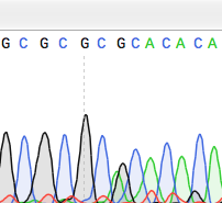
A to G point mutation detected with Sanger sequencing

A point mutation is a genetic mutation where a single nucleotide base is changed, inserted or deleted from a DNA or RNA sequence of an organism's genome. Point mutations have a variety of effects on the downstream protein product—consequences that are moderately predictable based upon the specifics of the mutation. These consequences can range from no effect (e.g. synonymous mutations) to deleterious effects (e.g. frameshift mutations), with regard to protein production, composition, and function.

Causes

Point mutations usually take place during DNA replication. DNA replication occurs when one double-stranded DNA molecule creates two single strands of DNA, each of which is a template for the creation of the complementary strand. A single point mutation can change the whole DNA sequence. Changing one purine or pyrimidine may change the amino acid that the nucleotides code for.

Point mutations may arise from spontaneous mutations that occur during DNA replication. The rate of mutation may be increased by mutagens. Mutagens can be physical, such as radiation from UV rays, X-rays or extreme heat, or chemical (molecules that misplace base pairs or disrupt the helical shape of DNA). Mutagens associated with cancers are often studied to learn about cancer and its prevention.

There are multiple ways for point mutations to occur. First, ultraviolet (UV) light and higher-frequency light are capable of ionizing electrons, which in turn can affect DNA. Reactive oxygen molecules with free radicals, which are a byproduct of cellular metabolism, can also be very harmful to DNA. These reactants can lead to both single-stranded DNA breaks and double-stranded DNA breaks. Third, bonds in DNA eventually degrade, which creates another problem to keep the integrity of DNA to a high standard. There can also be replication errors that lead to substitution, insertion, or deletion mutations.

Categorization

Transition/transversion categorization

Transitions (Alpha) and transversions (Beta).

In 1959 Ernst Freese coined the terms "transitions" or "transversions" to categorize different types of point mutations. Transitions are replacement of a purine base with another purine or replacement of a pyrimidine with another pyrimidine. Transversions are replacement of a purine with a pyrimidine or vice versa. There is a systematic difference in mutation rates for transitions (Alpha) and transversions (Beta). Transition mutations are about ten times more common than transversions.

Functional categorization

Nonsense mutations include stop-gain and start-loss. Stop-gain is a mutation that results in a premature termination codon (a stop was gained), which signals the end of translation. This interruption causes the protein to be abnormally shortened. The number of amino acids lost mediates the impact on the protein's functionality and whether it will function whatsoever. Stop-loss is a mutation in the original termination codon (a stop was lost), resulting in abnormal extension of a protein's carboxyl terminus. Start-gain creates an AUG start codon upstream of the original start site. If the new AUG is near the original start site, in-frame within the processed transcript and downstream to a ribosomal binding site, it can be used to initiate translation. The likely effect is additional amino acids added to the amino terminus of the original protein. Frame-shift mutations are also possible in start-gain mutations, but typically do not affect translation of the original protein. Start-loss is a point mutation in a transcript's AUG start codon, resulting in the reduction or elimination of protein production.

Missense mutations code for a different amino acid. A missense mutation changes a codon so that a different protein is created, a non-synonymous change. Conservative mutations result in an amino acid change. However, the properties of the amino acid remain the same (e.g., hydrophobic, hydrophilic, etc.). At times, a change to one amino acid in the protein is not detrimental to the organism as a whole. Most proteins can withstand one or two point mutations before their function changes. Non-conservative mutations result in an amino acid change that has different properties than the wild type. The protein may lose its function, which can result in a disease in the organism. For example, sickle-cell disease is caused by a single point mutation (a missense mutation) in the beta-hemoglobin gene that converts a GAG codon into GUG, which encodes the amino acid valine rather than glutamic acid. The protein may also exhibit a "gain of function" or become activated, such is the case with the mutation changing a valine to glutamic acid in the BRAF gene; this leads to an activation of the RAF protein which causes unlimited proliferative signalling in cancer cells. These are both examples of a non-conservative (missense) mutation.

Silent mutations code for the same amino acid (a "synonymous substitution"). A silent mutation does not affect the functioning of the protein. A single nucleotide can change, but the new codon specifies the same amino acid, resulting in an unmutated protein. This type of change is called synonymous change since the old and new codon code for the same amino acid. This is possible because 64 codons specify only 20 amino acids. Different codons can lead to differential protein expression levels, however.

Single base pair insertions and deletions

Sometimes the term point mutation is used to describe insertions or deletions of a single base pair (which has more of an adverse effect on the synthesized protein due to the nucleotides' still being read in triplets, but in different frames: a mutation called a frameshift mutation).

General consequences

Point mutations that occur in non-coding sequences are most often without consequences, although there are exceptions. If the mutated base pair is in the promoter sequence of a gene, then the expression of the gene may change. Also, if the mutation occurs in the splicing site of an intron, then this may interfere with correct splicing of the transcribed pre-mRNA.

By altering just one amino acid, the entire peptide may change, thereby changing the entire protein. The new protein is called a protein variant. If the original protein functions in cellular reproduction then this single point mutation can change the entire process of cellular reproduction for this organism.

Point germline mutations can lead to beneficial as well as harmful traits or diseases. This leads to adaptations based on the environment where the organism lives. An advantageous mutation can create an advantage for that organism and lead to the trait's being passed down from generation to generation, improving and benefiting the entire population. The scientific theory of evolution is greatly dependent on point mutations in cells. The theory explains the diversity and history of living organisms on Earth. In relation to point mutations, it states that beneficial mutations allow the organism to thrive and reproduce, thereby passing its positively affected mutated genes on to the next generation. On the other hand, harmful mutations cause the organism to die or be less likely to reproduce in a phenomenon known as natural selection.

There are different short-term and long-term effects that can arise from mutations. Smaller ones would be a halting of the cell cycle at numerous points. This means that a codon coding for the amino acid glycine may be changed to a stop codon, causing the proteins that should have been produced to be deformed and unable to complete their intended tasks. Because the mutations can affect the DNA and thus the chromatin, it can prohibit mitosis from occurring due to the lack of a complete chromosome. Problems can also arise during the processes of transcription and replication of DNA. These all prohibit the cell from reproduction and thus lead to the death of the cell. Long-term effects can be a permanent changing of a chromosome, which can lead to a mutation. These mutations can be either beneficial or detrimental. Cancer is an example of how they can be detrimental.

Other effects of point mutations, or single nucleotide polymorphisms in DNA, depend on the location of the mutation within the gene. For example, if the mutation occurs in the region of the gene responsible for coding, the amino acid sequence of the encoded protein may be altered, causing a change in the function, protein localization, stability of the protein or protein complex. Many methods have been proposed to predict the effects of missense mutations on proteins. Machine learning algorithms train their models to distinguish known disease-associated from neutral mutations whereas other methods do not explicitly train their models but almost all methods exploit the evolutionary conservation assuming that changes at conserved positions tend to be more deleterious. While majority of methods provide a binary classification of effects of mutations into damaging and benign, a new level of annotation is needed to offer an explanation of why and how these mutations damage proteins.

Moreover, if the mutation occurs in the region of the gene where transcriptional machinery binds to the protein, the mutation can affect the binding of the transcription factors because the short nucleotide sequences recognized by the transcription factors will be altered. Mutations in this region can affect rate of efficiency of gene transcription, which in turn can alter levels of mRNA and, thus, protein levels in general.

Point mutations can have several effects on the behavior and reproduction of a protein depending on where the mutation occurs in the amino acid sequence of the protein. If the mutation occurs in the region of the gene that is responsible for coding for the protein, the amino acid may be altered. This slight change in the sequence of amino acids can cause a change in the function, activation of the protein meaning how it binds with a given enzyme, where the protein will be located within the cell, or the amount of free energy stored within the protein.

If the mutation occurs in the region of the gene where transcriptional machinery binds to the protein, the mutation can affect the way in which transcription factors bind to the protein. The mechanisms of transcription bind to a protein through recognition of short nucleotide sequences. A mutation in this region may alter these sequences and, thus, change the way the transcription factors bind to the protein. Mutations in this region can affect the efficiency of gene transcription, which controls both the levels of mRNA and overall protein levels.

Specific diseases caused by point mutations

Cancer

Point mutations in multiple tumor suppressor proteins cause cancer. For instance, point mutations in Adenomatous Polyposis Coli promote tumorigenesis. A novel assay, Fast parallel proteolysis (FASTpp), might help swift screening of specific stability defects in individual cancer patients.

Neurofibromatosis

Neurofibromatosis is caused by point mutations in the Neurofibromin 1 or Neurofibromin 2 gene.

Sickle-cell anemia

Sickle-cell anemia is caused by a point mutation in the β-globin chain of hemoglobin, causing the hydrophilic amino acid glutamic acid to be replaced with the hydrophobic amino acid valine at the sixth position.

The β-globin gene is found on the short arm of chromosome 11. The association of two wild-type α-globin subunits with two mutant β-globin subunits forms hemoglobin S (HbS). Under low-oxygen conditions (being at high altitude, for example), the absence of a polar amino acid at position six of the β-globin chain promotes the non-covalent polymerisation (aggregation) of hemoglobin, which distorts red blood cells into a sickle shape and decreases their elasticity.

Hemoglobin is a protein found in red blood cells, and is responsible for the transportation of oxygen through the body. There are two subunits that make up the hemoglobin protein: beta-globins and alpha-globins. Beta-hemoglobin is created from the genetic information on the HBB, or "hemoglobin, beta" gene found on chromosome 11p15.5. A single point mutation in this polypeptide chain, which is 147 amino acids long, results in the disease known as Sickle Cell Anemia. Sickle-cell anemia is an autosomal recessive disorder that affects 1 in 500 African Americans, and is one of the most common blood disorders in the United States. The single replacement of the sixth amino acid in the beta-globin, glutamic acid, with valine results in deformed red blood cells. These sickle-shaped cells cannot carry nearly as much oxygen as normal red blood cells and they get caught more easily in the capillaries, cutting off blood supply to vital organs. The single nucleotide change in the beta-globin means that even the smallest of exertions on the part of the carrier results in severe pain and even heart attack. Below is a chart depicting the first thirteen amino acids in the normal and abnormal sickle cell polypeptide chain.

Sequence for normal hemoglobin
AUG GUG CAC CUG ACU CCU GAG GAG AAG UCU GCC GUU ACU
START Val His Leu Thr Pro Glu Glu Lys Ser Ala Val Thr

Sequence for sickle-cell hemoglobin
AUG GUG CAC CUG ACU CCU GUG GAG AAG UCU GCC GUU ACU
START Val His Leu Thr Pro Val Glu Lys Ser Ala Val Thr

Tay–Sachs disease

The cause of Tay–Sachs disease is a genetic defect that is passed from parent to child. This genetic defect is located in the HEXA gene, which is found on chromosome 15.

The HEXA gene makes part of an enzyme called beta-hexosaminidase A, which plays a critical role in the nervous system. This enzyme helps break down a fatty substance called GM2 ganglioside in nerve cells. Mutations in the HEXA gene disrupt the activity of beta-hexosaminidase A, preventing the breakdown of the fatty substances. As a result, the fatty substances accumulate to deadly levels in the brain and spinal cord. The buildup of GM2 ganglioside causes progressive damage to the nerve cells. This is the cause of the signs and symptoms of Tay-Sachs disease.

Repeat-induced point mutation

In molecular biology, repeat-induced point mutation or RIP is a process by which DNA accumulates G:C to A:T transition mutations. Genomic evidence indicates that RIP occurs or has occurred in a variety of fungi while experimental evidence indicates that RIP is active in Neurospora crassa, Podospora anserina, Magnaporthe grisea, Leptosphaeria maculans, Gibberella zeae and Nectria haematococca. In Neurospora crassa, sequences mutated by RIP are often methylated de novo.

RIP occurs during the sexual stage in haploid nuclei after fertilization but prior to meiotic DNA replication. In Neurospora crassa, repeat sequences of at least 400 base pairs in length are vulnerable to RIP. Repeats with as low as 80% nucleotide identity may also be subject to RIP. Though the exact mechanism of repeat recognition and mutagenesis are poorly understood, RIP results in repeated sequences undergoing multiple transition mutations.

The RIP mutations do not seem to be limited to repeated sequences. Indeed, for example, in the phytopathogenic fungus L. maculans, RIP mutations are found in single copy regions, adjacent to the repeated elements. These regions are either non-coding regions or genes encoding small secreted proteins including avirulence genes. The degree of RIP within these single copy regions was proportional to their proximity to repetitive elements.

Rep and Kistler have speculated that the presence of highly repetitive regions containing transposons, may promote mutation of resident effector genes. So the presence of effector genes within such regions is suggested to promote their adaptation and diversification when exposed to strong selection pressure.

As RIP mutation is traditionally observed to be restricted to repetitive regions and not single copy regions, Fudal et al. suggested that leakage of RIP mutation might occur within a relatively short distance of a RIP-affected repeat. Indeed, this has been reported in N. crassa whereby leakage of RIP was detected in single copy sequences at least 930 bp from the boundary of neighbouring duplicated sequences. To elucidate the mechanism of detection of repeated sequences leading to RIP may allow to understand how the flanking sequences may also be affected.

Mechanism

RIP causes G:C to A:T transition mutations within repeats, however, the mechanism that detects the repeated sequences is unknown. RID is the only known protein essential for RIP. It is a DNA methyltransferease-like protein, that when mutated or knocked out results in loss of RIP. Deletion of the rid homolog in Aspergillus nidulans, dmtA, results in loss of fertility while deletion of the rid homolog in Ascobolus immersens, masc1, results in fertility defects and loss of methylation induced premeiotically (MIP).

Consequences

RIP is believed to have evolved as a defense mechanism against transposable elements, which resemble parasites by invading and multiplying within the genome. RIP creates multiple missense and nonsense mutations in the coding sequence. This hypermutation of G-C to A-T in repetitive sequences eliminates functional gene products of the sequence (if there were any to begin with). In addition, many of the C-bearing nucleotides become methylated, thus decreasing transcription.

Use in molecular biology

Because RIP is so efficient at detecting and mutating repeats, fungal biologists often use it as a tool for mutagenesis. A second copy of a single-copy gene is first transformed into the genome. The fungus must then mate and go through its sexual cycle to activate the RIP machinery. Many different mutations within the duplicated gene are obtained from even a single fertilization event so that inactivated alleles, usually due to nonsense mutations, as well as alleles containing missense mutations can be obtained.

History

The cellular reproduction process of meiosis was discovered by Oscar Hertwig in 1876. Mitosis was discovered several years later in 1882 by Walther Flemming.

Hertwig studied sea urchins, and noticed that each egg contained one nucleus prior to fertilization and two nuclei after. This discovery proved that one spermatozoon could fertilize an egg, and therefore proved the process of meiosis. Hermann Fol continued Hertwig's research by testing the effects of injecting several spermatozoa into an egg, and found that the process did not work with more than one spermatozoon.

Flemming began his research of cell division starting in 1868. The study of cells was an increasingly popular topic in this time period. By 1873, Schneider had already begun to describe the steps of cell division. Flemming furthered this description in 1874 and 1875 as he explained the steps in more detail. He also argued with Schneider's findings that the nucleus separated into rod-like structures by suggesting that the nucleus actually separated into threads that in turn separated. Flemming concluded that cells replicate through cell division, to be more specific mitosis.

Matthew Meselson and Franklin Stahl are credited with the discovery of DNA replication. Watson and Crick acknowledged that the structure of DNA did indicate that there is some form of replicating process. However, there was not a lot of research done on this aspect of DNA until after Watson and Crick. People considered all possible methods of determining the replication process of DNA, but none were successful until Meselson and Stahl. Meselson and Stahl introduced a heavy isotope into some DNA and traced its distribution. Through this experiment, Meselson and Stahl were able to prove that DNA reproduces semi-conservatively.

Saturday, December 9, 2023

van der Waals force


From Wikipedia, the free encyclopedia
Rainwater flux from a canopy. Among the forces that govern drop formation: Van der Waals force, surface tension, cohesion, Plateau–Rayleigh instability.
Microfiber cloth makes use of van der Waals force to remove dirt without scratches.

In molecular physics and chemistry, the van der Waals force is a distance-dependent interaction between atoms or molecules. Unlike ionic or covalent bonds, these attractions do not result from a chemical electronic bond; they are comparatively weak and therefore more susceptible to disturbance. The van der Waals force quickly vanishes at longer distances between interacting molecules.

Named after Dutch physicist Johannes Diderik van der Waals, the van der Waals force plays a fundamental role in fields as diverse as supramolecular chemistry, structural biology, polymer science, nanotechnology, surface science, and condensed matter physics. It also underlies many properties of organic compounds and molecular solids, including their solubility in polar and non-polar media.

If no other force is present, the distance between atoms at which the force becomes repulsive rather than attractive as the atoms approach one another is called the van der Waals contact distance; this phenomenon results from the mutual repulsion between the atoms' electron clouds.

The van der Waals forces are usually described as a combination of the London dispersion forces between "instantaneously induced dipoles", Debye forces between permanent dipoles and induced dipoles, and the Keesom force between permanent molecular dipoles whose rotational orientations are dynamically averaged over time.

Definition

Van der Waals forces include attraction and repulsions between atoms, molecules, as well as other intermolecular forces. They differ from covalent and ionic bonding in that they are caused by correlations in the fluctuating polarizations of nearby particles (a consequence of quantum dynamics).

The force results from a transient shift in electron density. Specifically, the electron density may temporarily shift to be greater on one side of the nucleus. This shift generates a transient charge which a nearby atom can be attracted to or repelled by. The force is repulsive at very short distances, reaches zero at an equilibrium distance characteristic for each atom, or molecule, and becomes attractive for distances larger than the equilibrium distance. For individual atoms, the equilibrium distance is between 0.3 nm and 0.5 nm, depending on the atomic-specific diameter. When the interatomic distance is greater than 1.0 nm the force is not strong enough to be easily observed as it decreases as a function of distance r approximately with the 7th power (~r−7).

Van der Waals forces are often among the weakest chemical forces. For example, the pairwise attractive van der Waals interaction energy between H (Hydrogen) atoms in different H2 molecules equals 0.06 kJ/mol (0.6 meV) and the pairwise attractive interaction energy between O (Oxygen) atoms in different O2 molecules equals 0.44 kJ/mol (4.6 meV). The corresponding vaporization energies of H2 and O2 molecular liquids, which result as a sum of all van der Waals interactions per molecule in the molecular liquids, amount to 0.90 kJ/mol (9.3 meV) and 6.82 kJ/mol (70.7 meV), respectively, and thus approximately ~15 times the value of the individual pairwise interatomic interactions (excluding covalent bonds).

The strength of van-der-Waals bonds increases with higher polarizability of the participating atoms. For example, the pairwise van der Waals interaction energy for more polarizable atoms such as S (Sulfur) atoms in H2S and sulfides exceeds 1 kJ/mol (10 meV), and the pairwise interaction energy between even larger, more polarizable Xe (Xenon) atoms is 2.35 kJ/mol (24.3 meV). These van der Waals interactions are up to 40 times stronger than in H2, which has only one valence electron, and they are still not strong enough to achieve an aggregate state other than gas for Xe under standard conditions. The interactions between atoms in metals can also be effectively described as van-der-Waals interactions and account for the observed solid aggregate state with bonding strengths comparable to covalent and ionic interactions. The strength of pairwise van-der-Waals type interactions is on the order of 12 kJ/mol (120 meV) for low-melting Pb (Lead) and on the order of 32 kJ/mol (330 meV) for high-melting Pt (Platinum), which is about one order of magnitude stronger than in Xe due to the presence of a highly polarizable free electron gas. Accordingly, van der Waals forces can range from weak to strong interactions, and support integral structural loads when multitudes of such interactions are present.

More broadly, intermolecular forces have several possible contributions:

  1. A repulsive component resulting from the Pauli exclusion principle that prevents close contact of atoms, or the collapse of molecules.
  2. Attractive or repulsive electrostatic interactions between permanent charges (in the case of molecular ions), dipoles (in the case of molecules without inversion centre), quadrupoles (all molecules with symmetry lower than cubic), and in general between permanent multipoles. These interactions also include hydrogen bonds, cation-pi, and pi-stacking interactions. Orientation-averaged contributions from electrostatic interactions are sometimes called the Keesom interaction or Keesom force after Willem Hendrik Keesom.
  3. Induction (also known as polarization), which is the attractive interaction between a permanent multipole on one molecule with an induced multipole on another. This interaction is sometimes called Debye force after Peter J.W. Debye.
  4. Dispersion (usually named London dispersion interactions after Fritz London), which is the attractive interaction between any pair of molecules, including non-polar atoms, arising from the interactions of instantaneous multipoles.

Hereby, different texts may refer to a different spectrum of interactions using the term "van der Waals force". Typically, contributions (1) and (4) are considered as van-der-Waals forces, excluding effects from permanent multipoles as described in (2) and from permanent polarization in (3). However, some texts describe the van der Waals force as the totality of forces, including repulsion; others mean all the attractive forces (and then sometimes distinguish van der Waals–Keesom, van der Waals–Debye, and van der Waals–London).

All intermolecular/van der Waals forces are anisotropic (except those between two noble gas atoms), which means that they depend on the relative orientation of the molecules. The induction and dispersion interactions are always attractive, irrespective of orientation, but the electrostatic interaction changes sign upon rotation of the molecules. That is, the electrostatic force can be attractive or repulsive, depending on the mutual orientation of the molecules. When molecules are in thermal motion, as they are in the gas and liquid phase, the electrostatic force is averaged out to a large extent because the molecules thermally rotate and thus probe both repulsive and attractive parts of the electrostatic force. Random thermal motion can disrupt or overcome the electrostatic component of the van der Waals force but the averaging effect is much less pronounced for the attractive induction and dispersion forces.

The Lennard-Jones potential is often used as an approximate model for the isotropic part of a total (repulsion plus attraction) van der Waals force as a function of distance.

Van der Waals forces are responsible for certain cases of pressure broadening (van der Waals broadening) of spectral lines and the formation of van der Waals molecules. The London–van der Waals forces are related to the Casimir effect for dielectric media, the former being the microscopic description of the latter bulk property. The first detailed calculations of this were done in 1955 by E. M. Lifshitz. A more general theory of van der Waals forces has also been developed.

The main characteristics of van der Waals forces are:

  • They are weaker than normal covalent and ionic bonds.
  • The Van der Waals forces are additive in nature, consisting of several individual interactions, and cannot be saturated.
  • They have no directional characteristic.
  • They are all short-range forces and hence only interactions between the nearest particles need to be considered (instead of all the particles). Van der Waals attraction is greater if the molecules are closer.
  • Van der Waals forces are independent of temperature except for dipole-dipole interactions.

In low molecular weight alcohols, the hydrogen-bonding properties of their polar hydroxyl group dominate other weaker van der Waals interactions. In higher molecular weight alcohols, the properties of the nonpolar hydrocarbon chain(s) dominate and determine their solubility.

Van der Waals forces are also responsible for the weak hydrogen bond interactions between unpolarized dipoles particularly in acid-base aqueous solution and between biological molecules.

London dispersion force

London dispersion forces, named after the German-American physicist Fritz London, are weak intermolecular forces that arise from the interactive forces between instantaneous multipoles in molecules without permanent multipole moments. In and between organic molecules the multitude of contacts can lead to larger contribution of dispersive attraction, particularly in the presence of heteroatoms. London dispersion forces are also known as 'dispersion forces', 'London forces', or 'instantaneous dipole–induced dipole forces'. The strength of London dispersion forces is proportional to the polarizability of the molecule, which in turn depends on the total number of electrons and the area over which they are spread. Hydrocarbons display small dispersive contributions, the presence of heteroatoms lead to increased LD forces as function of their polarizability, e.g. in the sequence RI>RBr>RCl>RF. In absence of solvents weakly polarizable hydrocarbons form crystals due to dispersive forces; their sublimation heat is a measure of the dispersive interaction.

Van der Waals forces between macroscopic objects

For macroscopic bodies with known volumes and numbers of atoms or molecules per unit volume, the total van der Waals force is often computed based on the "microscopic theory" as the sum over all interacting pairs. It is necessary to integrate over the total volume of the object, which makes the calculation dependent on the objects' shapes. For example, the van der Waals interaction energy between spherical bodies of radii R1 and R2 and with smooth surfaces was approximated in 1937 by Hamaker (using London's famous 1937 equation for the dispersion interaction energy between atoms/molecules as the starting point) by:


(1)

where A is the Hamaker coefficient, which is a constant (~10−19 − 10−20 J) that depends on the material properties (it can be positive or negative in sign depending on the intervening medium), and z is the center-to-center distance; i.e., the sum of R1, R2, and r (the distance between the surfaces): .

The van der Waals force between two spheres of constant radii (R1 and R2 are treated as parameters) is then a function of separation since the force on an object is the negative of the derivative of the potential energy function,. This yields:


(2)

In the limit of close-approach, the spheres are sufficiently large compared to the distance between them; i.e., or , so that equation (1) for the potential energy function simplifies to:


(3)

with the force:


(4)

The van der Waals forces between objects with other geometries using the Hamaker model have been published in the literature.

From the expression above, it is seen that the van der Waals force decreases with decreasing size of bodies (R). Nevertheless, the strength of inertial forces, such as gravity and drag/lift, decrease to a greater extent. Consequently, the van der Waals forces become dominant for collections of very small particles such as very fine-grained dry powders (where there are no capillary forces present) even though the force of attraction is smaller in magnitude than it is for larger particles of the same substance. Such powders are said to be cohesive, meaning they are not as easily fluidized or pneumatically conveyed as their more coarse-grained counterparts. Generally, free-flow occurs with particles greater than about 250 μm.

The van der Waals force of adhesion is also dependent on the surface topography. If there are surface asperities, or protuberances, that result in a greater total area of contact between two particles or between a particle and a wall, this increases the van der Waals force of attraction as well as the tendency for mechanical interlocking.

The microscopic theory assumes pairwise additivity. It neglects many-body interactions and retardation. A more rigorous approach accounting for these effects, called the "macroscopic theory" was developed by Lifshitz in 1956. Langbein derived a much more cumbersome "exact" expression in 1970 for spherical bodies within the framework of the Lifshitz theory while a simpler macroscopic model approximation had been made by Derjaguin as early as 1934. Expressions for the van der Waals forces for many different geometries using the Lifshitz theory have likewise been published.

Use by geckos and arthropods

Gecko climbing a glass surface

The ability of geckos – which can hang on a glass surface using only one toe – to climb on sheer surfaces has been for many years mainly attributed to the van der Waals forces between these surfaces and the spatulae, or microscopic projections, which cover the hair-like setae found on their footpads.

There were efforts in 2008 to create a dry glue that exploits the effect, and success was achieved in 2011 to create an adhesive tape on similar grounds (i.e. based on van der Waals forces). In 2011, a paper was published relating the effect to both velcro-like hairs and the presence of lipids in gecko footprints.

A later study suggested that capillary adhesion might play a role, but that hypothesis has been rejected by more recent studies.

A 2014 study has shown that gecko adhesion to smooth Teflon and polydimethylsiloxane surfaces is mainly determined by electrostatic interaction (caused by contact electrification), not van der Waals or capillary forces.

Among the arthropods, some spiders have similar setae on their scopulae or scopula pads, enabling them to climb or hang upside-down from extremely smooth surfaces such as glass or porcelain.

Politics of Europe

From Wikipedia, the free encyclopedia ...