Search This Blog

Monday, January 15, 2024

Ontogeny

From Wikipedia, the free encyclopedia
The initial stages of human embryogenesis
Parts of a human embryo

Ontogeny (also ontogenesis) is the origination and development of an organism (both physical and psychological, e.g., moral development), usually from the time of fertilization of the egg to adult. The term can also be used to refer to the study of the entirety of an organism's lifespan.

Ontogeny is the developmental history of an organism within its own lifetime, as distinct from phylogeny, which refers to the evolutionary history of a species. Another way to think of ontogeny is that it is the process of an organism going through all of the developmental stages over its lifetime. The developmental history includes all the developmental events that occur during the existence of an organism, beginning with the changes in the egg at the time of fertilization and events from the time of birth or hatching and afterward (i.e., growth, remolding of body shape, development of secondary sexual characteristics, etc.). While developmental (i.e., ontogenetic) processes can influence subsequent evolutionary (e.g., phylogenetic) processes (see evolutionary developmental biology and recapitulation theory), individual organisms develop (ontogeny), while species evolve (phylogeny).

Ontogeny, embryology and developmental biology are closely related studies and those terms are sometimes used interchangeably. Aspects of ontogeny are morphogenesis, the development of form and shape of an organism; tissue growth; and cellular differentiation. The term ontogeny has also been used in cell biology to describe the development of various cell types within an organism. Ontogeny is a useful field of study in many disciplines, including developmental biology, cell biology, genetics, developmental psychology, developmental cognitive neuroscience, and developmental psychobiology. Ontogeny is used in anthropology as "the process through which each of us embodies the history of our own making".

Etymology

The word ontogeny comes from the Greek on meaning a being, individual; and existence, and from the suffix -geny from the Greek -geniea, meaning genesis, origin, and mode of production.

History

The term ontogeny was coined by Ernst Haeckel, a German zoologist and evolutionist in the 1860s. Haeckel, born in Germany on February 16, 1834, was also a strong supporter of Darwinism. Haeckel suggested that ontogeny briefly and sometimes incompletely recapitulated or repeated phylogeny in his 1866 book, Generelle Morphologie der Organismen ("General Morphology of Organisms"). Even though his book was widely read, the scientific community was not very convinced or interested in his ideas, so he turned to producing more publications to get more attention. In 1866, Haeckel and others imagined development as producing new structures after earlier additions to the developing organism have been established. He proposed that individual development followed developmental stages of previous generations and that the future generations would add something new to this process, and that there was a causal parallelism between an animal's ontogeny and phylogeny. In addition, Haeckel suggested a biogenetic law that ontogeny recapitulates phylogeny, based on the idea that the successive and progressive origin of new species was based on the same laws as the successive and progressive origin of new embryonic structures. According to Haeckel, development produced novelties, and natural selection would eliminate species that had become outdated or obsolete. Though his view of development and evolution wasn't justifiable, future embryologists tweaked and collaborated with Haeckel's proposals and showed how new morphological structures can occur by the hereditary modification of embryonic development. Marine biologist Walter Garstang reversed Haeckel's relationship between ontogeny and phylogeny, stating that ontogeny creates phylogeny, not recapitulates it.

A seminal 1963 paper by Nikolaas Tinbergen named ontogeny as one of the four primary questions of biology, along with Julian Huxley's three others: causation, survival value and evolution. Tinbergen emphasized that the change of behavioral machinery during development was distinct from the change in behavior during development. We can conclude that the thrush itself, i.e. its behavioral machinery, has changed only if the behavior change occurred while the environment was held constant...When we turn from description to causal analysis, and ask in what way the observed change in behavior machinery has been brought about, the natural first step is to try and distinguish between environmental influences and those within the animal...In ontogeny the conclusion that a certain change is internally controlled (is 'innate') is reached by elimination. Tinbergen was concerned that the elimination of environmental factors is difficult to establish, and the use of the word innate is often misleading.

Developmental stages

Development of an organism happens through fertilization, cleavage, blastulation, gastrulation, organogenesis, and metamorphosis into an adult. Each species of animal has a slightly different journey through these stages, since some stages might be shorter or longer when compared to other species, and where the offspring develops is different for each animal type (e.g., in a hard egg shell, uterus, soft egg shell, on a plant leaf, etc.).

Fertilization

In humans, the process of fetal development starts after sperm fertilizes an egg and they fuse together, kickstarting embryonic development. The fusion of egg and sperm into a zygote changes the surrounding membrane to not allow any more sperm to penetrate the egg, so multiple fertilizations can be prevented. Fusion of a zygote also activates the egg so it can begin undergoing cell division. Each animal species might not have specifically a sperm and an egg, but two gametes that contain half of the species' typical genetic material and the membranes of these gametes fuse to start creating an offspring.

Cleavage

Not long after successful fertilization by sperm, the zygote undergoes many mitotic divisions, which are also non-sexual cell divisions. Cleavage is the process of cell division, so the starting zygote becomes a collection of identical cells which is a morula and contains cells called blastomeres. Cleavage prepares the zygote to become an embryo, which is from 2 weeks to 8 weeks after conception (fertilization) in humans.
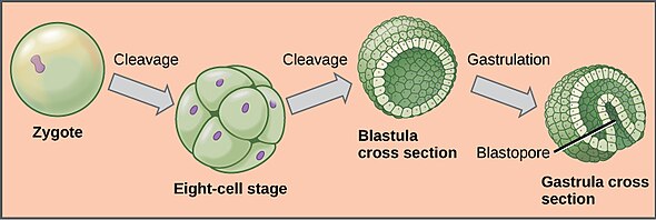
Process of zygote to gastrula in development

Blastulation

After the zygote has become an embryo, it continues dividing into a hollow sphere of cells, which is a blastula. These outer cells form a single epithelial layer, the blastoderm, that essentially encases the fluid-filled inside that is the blastocoel. The figure to the right shows the basic process that is modified in different species. Blastulation differs slightly in different species, but in mammals, the eight-cell stage embryo forms into a slightly different type of blastula, called a blastocyst. Other species such as sea stars, frogs, chicks, and mice have all the same structures in this stage, yet the orientation of these features differs, plus these species have additional types of cells in this stage.

Blastula to gastrula more detailed

Gastrulation

After blastulation, the single-layered blastula expands and reorganizes into multiple layers, a gastrula (seen in the figure to the right). Reptiles, birds and mammals are triploblastic organisms, meaning the gastrula comprises three germ layers; the endoderm (inner layer), mesoderm (middle layer), and ectoderm (outer layer). As seen in the figure below, each germ layer will become multi-potent stem cells that can become a specific tissue depending on the germ layer and is what happens in humans. This differentiation of germ layers differs slightly, because not all of the organs and tissues below are in all organisms, but corresponding body systems can be substituted in place of these.

Organogenesis

In the figure below, human germ cells are able to differentiate into the specific organs and tissues they become later on in life. Germ cells are able to migrate to their final locations to rearrange themselves and some organs are made of two germ layers; one for the outside, the other for the inside. The endoderm cells become the internal linings of organisms, such as the stomach, colon, small intestine, liver, and pancreas of the digestive system and the lungs. The mesoderm gives rise to other tissues not formed by the ectoderm, such as the heart, muscles, bones, blood, dermis of the skin, bone marrow, and the urogenital system. This germ layer is more specific for species, as it is the distinguishing layer of the three that can identify evolutionarily higher life-forms (e.g., bilateral organisms like humans) from lower-life forms (with radial symmetry). Lastly, the ectoderm is the outer layer of cells that become the epidermis and hair while being the precursor to the mammary glands, central nervous system, and the peripheral nervous systems.

Germ layers and what tissues they become in humans
Ernst Haeckel, Anthropogenie.

The figure above shows how the development of a pig, cow, rabbit, and human offspring are similar when compared to one another. This figure shows how the germ layers can become different organs and tissues in evolutionarily higher life-forms and how these species essentially develop very similarly. Additionally, it shows how multiple species develop in a parallel manner but branch off to develop more specific features for the organism such as hooves, a tail, or ears.

Primary neurulation detailed

Neurulation

In developing vertebrate offspring, a neural tube is formed through either primary or secondary neurulation. Some species develop their spine and nervous system using both primary and secondary neurulation, while others use only primary or secondary neurulation. In human fetal development, primary neurulation occurs during weeks 3 and 4 of gestation to develop the brain and spinal cord. Then during weeks 5 and 6 of gestation, secondary neurulation forms the lower sacral and coccygeal cord.

Primary Neurulation

The diagram to the right illustrates primary neurulation, which is the process of cells surrounding the neural plate interacting with neural plate cells to proliferate, converge, and pinch off to form a hollow tube above the notochord and mesoderm. This process is discontinuous and can start at different points along the cranial-caudal axis necessary for it to close. After the neural crest closes, the neural crest cells and ectoderm cells separate and the ectoderm becomes the epidermis surrounding this complex. The neural crest cells differentiate to become components of most of the peripheral nervous system in animals. Next, the notochord degenerates to become only the nucleus pulposus of the intervertebral discs and the mesoderm cells differentiate to become the somites and skeletal muscle later on. Also during this stage, the neural crest cells become the spinal ganglions, which function as the brain in organisms like earthworms and arthropods. In more advanced organisms like amphibians, birds and mammals; the spinal ganglions consists of a cluster of nerve bodies positioned along the spinal cord at the dorsal and ventral roots of a spinal nerve, which is a pair of nerves that correspond to a vertebra of the spine.

Secondary Neurulation

In secondary neurulation, caudal and sacral regions of the spine are formed after primary neurulation is finished. This process initiates once primary neurulation is finished and the posterior neuropore closes, so the tail bud can proliferate and condense, then create a cavity and fuse with the central canal of the neural tube. Secondary neurulation occurs in the small region starting at the spinal tail bud up to the posterior neuropore, which is the open neural folds near the tail region that don't close through primary neurulation. As canalization progresses over the next few weeks, neurons and ependymal cells (cells that create cerebral spinal fluid) differentiate to become the tail end of the spinal cord. Next, the closed neural tube contains neuroepithelial cells that immediately divide after closure and a second type of cell forms; the neuroblast. Neuroblast cells form the mantle layer, which later becomes the gray matter, which then gives rise to a marginal layer that becomes the white matter of the spinal cord. Secondary neurulation is seen in the neural tube of the lumbar and tail vertebrae of frogs and chicks and in both instances, this process is like a continuation of gastrulation.

Acraea zetes caterpillar to pupae to butterfly metamorphosis by Nick Hobgood

Larval and juvenile phases

In most species, the young organism that is just born or hatched is not sexually mature yet and in most animals, this young organism looks quite different than the adult form. This young organism is the larva and is the intermediate form before metamorphosing into an adult. A well known example of a larval form of an animal is the caterpillar of butterflies and moths. Caterpillars keep growing and feeding in order for enough energy during the pupal stage, when necessary body parts for metamorphosis are grown. The juvenile phase is different in plants and animals, but in plants juvenility is an early phase of plant growth in which plants can't flower. In animals, the juvenile stage is most commonly found in social mammals, such as wild dogs, monkeys, apes, lions, wolves, and more. In humans, puberty marks the end of this stage and adolescence follows. Some species begin puberty and reproduction before the juvenile stage is over, such as in female non-human primates.

Metamorphosis

The process of an organism's body undergoing structural and physical changes after birth or hatching to become suitable for its adult environment is metamorphosis. For example, amphibian tadpoles have a maturation of liver enzymes, hemoglobin, and eye pigments, in addition to their nervous, digestive, and reproductive systems being remodeled. In all species, molting and juvenile hormones appear to regulate these changes.

Adulthood

Adulthood is the stage of when physical and intellectual maturity have been achieved and this differs between species. In humans, adulthood is thought to be around 20 or 21 years old and is the longest stage of life, but in all species it ends with death. In dogs, small breeds (e.g., Yorkshire Terrier, Chihuahua, Cocker Spaniel, etc.) physically mature faster than large breeds (e.g., Saint Bernard, Great Dane, Golden Retriever, etc.), so adulthood is reached anywhere from 12 to 24 months or 1 to 2 years. In contrast, many insect species have long larval stages and the adult stage is only for reproduction. The silkworm moths don't have mouthparts and don't feed, so they have to consume enough food during the larval stage for energy to survive and mate.

Senescence

Senescence is when cells stop dividing but don't die, but these cells can build up and cause problems in the body. These cells can release substances that cause inflammation and can damage healthy nearby cells. Senescence can be induced by un-repaired DNA damage (e.g., from radiation, old age, etc.) or other cellular stress and also is the state of being old.

Ontogenetic allometry

Most organisms undergo allometric changes in shape as they grow and mature, while others engage in metamorphosis. Even reptiles (non-avian sauropsids, e.g., crocodilians, turtles, snakes, and lizards), in which the offspring are often viewed as miniature adults, show a variety of ontogenetic changes in morphology and physiology.

Metamorphosis

From Wikipedia, the free encyclopedia
A dragonfly in its final moult, undergoing metamorphosis, it begins transforming from its nymph form to an adult

Metamorphosis is a biological process by which an animal physically develops including birth transformation or hatching, involving a conspicuous and relatively abrupt change in the animal's body structure through cell growth and differentiation. Some insects, fish, amphibians, mollusks, crustaceans, cnidarians, echinoderms, and tunicates undergo metamorphosis, which is often accompanied by a change of nutrition source or behavior. Animals can be divided into species that undergo complete metamorphosis ("holometaboly"), incomplete metamorphosis ("hemimetaboly"), or no metamorphosis ("ametaboly").

Generally organisms with a larval stage undergo metamorphosis, and during metamorphosis the organism loses larval characteristics. 

Etymology

The word metamorphosis derives from Ancient Greek μεταμόρφωσις, "transformation, transforming", from μετα- (meta-), "after" and μορφή (morphe), "form".

Hormonal control

In insects, growth and metamorphosis are controlled by hormones synthesized by endocrine glands near the front of the body (anterior). Neurosecretory cells in an insect's brain secrete a hormone, the prothoracicotropic hormone (PTTH) that activates prothoracic glands, which secrete a second hormone, usually ecdysone (an ecdysteroid), that induces ecdysis (shedding of the exoskeleton). PTTH also stimulates the corpora allata, a retrocerebral organ, to produce juvenile hormone, which prevents the development of adult characteristics during ecdysis. In holometabolous insects, molts between larval instars have a high level of juvenile hormone, the moult to the pupal stage has a low level of juvenile hormone, and the final, or imaginal, molt has no juvenile hormone present at all. Experiments on firebugs have shown how juvenile hormone can affect the number of nymph instar stages in hemimetabolous insects.

In chordates, metamorphosis is iodothyronine-induced and an ancestral feature of all chordates.

Insects

Incomplete metamorphosis in the grasshopper with different instar nymphs. The largest specimen is adult.

All three categories of metamorphosis can be found in the diversity of insects, including no metamorphosis ("ametaboly"), incomplete or partial metamorphosis ("hemimetaboly"), and complete metamorphosis ("holometaboly"). While ametabolous insects show very little difference between larval and adult forms (also known as "direct development"), both hemimetabolous and holometabolous insects have significant morphological and behavioral differences between larval and adult forms, the most significant being the inclusion, in holometabolus organisms, of a pupal or resting stage between the larval and adult forms.

Development and terminology

Two types of metamorphosis are shown. In a complete (holometabolous) metamorphosis the insect passes through four distinct phases, which produce an adult that does not resemble the larva. In an incomplete (hemimetabolous) metamorphosis an insect does not go through a full transformation, but instead transitions from a nymph to an adult by molting its exoskeleton as it grows.

In hemimetabolous insects, immature stages are called nymphs. Development proceeds in repeated stages of growth and ecdysis (moulting); these stages are called instars. The juvenile forms closely resemble adults, but are smaller and lack adult features such as wings and genitalia. The size and morphological differences between nymphs in different instars are small, often just differences in body proportions and the number of segments; in later instars, external wing buds form. The period from one molt to the next is called a stadium.

In holometabolous insects, immature stages are called larvae and differ markedly from adults. Insects which undergo holometabolism pass through a larval stage, then enter an inactive state called pupa (called a "chrysalis" in butterfly species), and finally emerge as adults.

Evolution

The earliest insect forms showed direct development (ametabolism), and the evolution of metamorphosis in insects is thought to have fuelled their dramatic radiation (1,2). Some early ametabolous "true insects" are still present today, such as bristletails and silverfish. Hemimetabolous insects include cockroaches, grasshoppers, dragonflies, and true bugs. Phylogenetically, all insects in the Pterygota undergo a marked change in form, texture and physical appearance from immature stage to adult. These insects either have hemimetabolous development, and undergo an incomplete or partial metamorphosis, or holometabolous development, which undergo a complete metamorphosis, including a pupal or resting stage between the larval and adult forms.

A number of hypotheses have been proposed to explain the evolution of holometaboly from hemimetaboly, mostly centering on whether or not the intermediate stages of hemimetabolous forms are homologous in origin to the pupal stage of holometabolous forms.

Temperature-dependent metamorphosis

According to a 2009 study, temperature plays an important role in insect development as individual species are found to have specific thermal windows that allow them to progress through their developmental stages. These windows are not significantly affected by ecological traits, rather, the windows are phylogenetically adapted to the ecological circumstances insects are living in.

Recent research

According to research from 2008, adult Manduca sexta is able to retain behavior learned as a caterpillar. Another caterpillar, the ornate moth caterpillar, is able to carry toxins that it acquires from its diet through metamorphosis and into adulthood, where the toxins still serve for protection against predators.

Many observations published in 2002, and supported in 2013 indicate that programmed cell death plays a considerable role during physiological processes of multicellular organisms, particularly during embryogenesis, and metamorphosis. Additional research in 2019 found that both autophagy and apoptosis, the two ways programmed cell death occur, are processes undergone during insect metamorphosis. 

Below is the sequence of steps in the metamorphosis of the butterfly (illustrated):

Metamorphosis of butterfly (PSF)

1 – The larva of a butterfly
2 – The pupa is now spewing the thread to form chrysalis
3 – The chrysalis is fully formed
4 – Adult butterfly coming out of the chrysalis

Chordata

Amphioxus

In cephalochordata, metamorphosis is iodothyronine-induced and it could be an ancestral feature of all chordates.

Fish

Some fish, both bony fish (Osteichthyes) and jawless fish (Agnatha), undergo metamorphosis. Fish metamorphosis is typically under strong control by the thyroid hormone.

Examples among the non-bony fish include the lamprey. Among the bony fish, mechanisms are varied.

The salmon is diadromous, meaning that it changes from a freshwater to a saltwater lifestyle.

Many species of flatfish begin their life bilaterally symmetrical, with an eye on either side of the body; but one eye moves to join the other side of the fish – which becomes the upper side – in the adult form.

The European eel has a number of metamorphoses, from the larval stage to the leptocephalus stage, then a quick metamorphosis to glass eel at the edge of the continental shelf (eight days for the Japanese eel), two months at the border of fresh and salt water where the glass eel undergoes a quick metamorphosis into elver, then a long stage of growth followed by a more gradual metamorphosis to the migrating phase. In the pre-adult freshwater stage, the eel also has phenotypic plasticity because fish-eating eels develop very wide mandibles, making the head look blunt. Leptocephali are common, occurring in all Elopomorpha (tarpon- and eel-like fish).

Most other bony fish undergo metamorphosis initially from egg to immotile larvae known as sac fry (fry with a yolk sac), then to motile larvae (often known as fingerlings due to them roughly reaching the length of a human finger) that have to forage for themselves after the yolk sac resorbs, and then to the juvenile stage where the fish progressively start to resemble adult morphology and behaviors until finally reaching sexual maturity.

Amphibians

Just before metamorphosis, only 24 hours are needed to reach the stage in the next picture.
Almost functional common frog with some remains of the gill sac and a not fully developed jaw

In typical amphibian development, eggs are laid in water and larvae are adapted to an aquatic lifestyle. Frogs, toads, and newts all hatch from the eggs as larvae with external gills but it will take some time for the amphibians to interact outside with pulmonary respiration. Afterwards, newt larvae start a predatory lifestyle, while tadpoles mostly scrape food off surfaces with their horny tooth ridges.

Metamorphosis in amphibians is regulated by thyroxin concentration in the blood, which stimulates metamorphosis, and prolactin, which counteracts its effect. Specific events are dependent on threshold values for different tissues. Because most embryonic development is outside the parental body, development is subject to many adaptations due to specific ecological circumstances. For this reason tadpoles can have horny ridges for teeth, whiskers, and fins. They also make use of the lateral line organ. After metamorphosis, these organs become redundant and will be resorbed by controlled cell death, called apoptosis. The amount of adaptation to specific ecological circumstances is remarkable, with many discoveries still being made.

Frogs and toads

With frogs and toads, the external gills of the newly hatched tadpole are covered with a gill sac after a few days, and lungs are quickly formed. Front legs are formed under the gill sac, and hindlegs are visible a few days later. Following that there is usually a longer stage during which the tadpole lives off a vegetarian diet. Tadpoles use a relatively long, spiral‐shaped gut to digest that diet. Recent studies suggest tadpoles do not have a balanced homeostatic feedback control system until the beginning stages of metamorphosis. At this point, their long gut shortens and begins favoring the diet of insects.

Rapid changes in the body can then be observed as the lifestyle of the frog changes completely. The spiral‐shaped mouth with horny tooth ridges is resorbed together with the spiral gut. The animal develops a big jaw, and its gills disappear along with its gill sac. Eyes and legs grow quickly, a tongue is formed, and all this is accompanied by associated changes in the neural networks (development of stereoscopic vision, loss of the lateral line system, etc.) All this can happen in about a day, so it is truly a metamorphosis. It is not until a few days later that the tail is reabsorbed, due to the higher thyroxin concentrations required for tail resorption.

Salamanders

Salamander development is highly diverse; some species go through a dramatic reorganization when transitioning from aquatic larvae to terrestrial adults, while others, such as the axolotl, display pedomorphosis and never develop into terrestrial adults. Within the genus Ambystoma, species have evolved to be pedomorphic several times, and pedomorphosis and complete development can both occur in some species.

Newts

The large external gills of the crested newt

In newts, metamorphosis occurs due to the change in habitat, not a change in diet, because newt larvae already feed as predators and continue doing so as adults. Newts' gills are never covered by a gill sac and will be resorbed only just before the animal leaves the water. Adults can move faster on land than in water. Newts often have an aquatic phase in spring and summer, and a land phase in winter. For adaptation to a water phase, prolactin is the required hormone, and for adaptation to the land phase, thyroxin. External gills do not return in subsequent aquatic phases because these are completely absorbed upon leaving the water for the first time.

Caecilians

Basal caecilians such as Ichthyophis go through a metamorphosis in which aquatic larva transition into fossorial adults, which involves a loss of the lateral line. More recently diverged caecilians (the Teresomata) do not undergo an ontogenetic niche shift of this sort and are in general fossorial throughout their lives. Thus, most caecilians do not undergo an anuran-like metamorphosis.

Positive and negative predictive values

From Wikipedia, the free encyclopedia
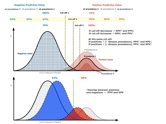
Positive and negative predictive values
Positive and negative predictive values - 2

The positive and negative predictive values (PPV and NPV respectively) are the proportions of positive and negative results in statistics and diagnostic tests that are true positive and true negative results, respectively. The PPV and NPV describe the performance of a diagnostic test or other statistical measure. A high result can be interpreted as indicating the accuracy of such a statistic. The PPV and NPV are not intrinsic to the test (as true positive rate and true negative rate are); they depend also on the prevalence. Both PPV and NPV can be derived using Bayes' theorem.

Although sometimes used synonymously, a positive predictive value generally refers to what is established by control groups, while a post-test probability refers to a probability for an individual. Still, if the individual's pre-test probability of the target condition is the same as the prevalence in the control group used to establish the positive predictive value, the two are numerically equal.

In information retrieval, the PPV statistic is often called the precision.

Definition

Positive predictive value (PPV)

The positive predictive value (PPV), or precision, is defined as

where a "true positive" is the event that the test makes a positive prediction, and the subject has a positive result under the gold standard, and a "false positive" is the event that the test makes a positive prediction, and the subject has a negative result under the gold standard. The ideal value of the PPV, with a perfect test, is 1 (100%), and the worst possible value would be zero.

The PPV can also be computed from sensitivity, specificity, and the prevalence of the condition:

cf. Bayes' theorem

The complement of the PPV is the false discovery rate (FDR):

Negative predictive value (NPV)

The negative predictive value is defined as:

where a "true negative" is the event that the test makes a negative prediction, and the subject has a negative result under the gold standard, and a "false negative" is the event that the test makes a negative prediction, and the subject has a positive result under the gold standard. With a perfect test, one which returns no false negatives, the value of the NPV is 1 (100%), and with a test which returns no true negatives the NPV value is zero.

The NPV can also be computed from sensitivity, specificity, and prevalence:

The complement of the NPV is the false omission rate (FOR):

Although sometimes used synonymously, a negative predictive value generally refers to what is established by control groups, while a negative post-test probability rather refers to a probability for an individual. Still, if the individual's pre-test probability of the target condition is the same as the prevalence in the control group used to establish the negative predictive value, then the two are numerically equal.

Relationship

The following diagram illustrates how the positive predictive value, negative predictive value, sensitivity, and specificity are related.


Predicted condition
Total population
= P + N
Predicted Positive (PP) Predicted Negative (PN) Informedness, bookmaker informedness (BM)
= TPR + TNR − 1
Prevalence threshold (PT)
=
Actual condition
Positive (P) True positive (TP),
hit
False negative (FN),
type II error, miss,
underestimation
True positive rate (TPR), recall, sensitivity (SEN), probability of detection, hit rate, power
= TP/P = 1 − FNR
False negative rate (FNR),
miss rate
= FN/P = 1 − TPR
Negative (N) False positive (FP),
type I error, false alarm,
overestimation
True negative (TN),
correct rejection
False positive rate (FPR),
probability of false alarm, fall-out
= FP/N = 1 − TNR
True negative rate (TNR),
specificity (SPC), selectivity
= TN/N = 1 − FPR

Prevalence
= P/P + N
Positive predictive value (PPV), precision
= TP/PP = 1 − FDR
False omission rate (FOR)
= FN/PN = 1 − NPV
Positive likelihood ratio (LR+)
= TPR/FPR
Negative likelihood ratio (LR−)
= FNR/TNR
Accuracy (ACC) = TP + TN/P + N False discovery rate (FDR)
= FP/PP = 1 − PPV
Negative predictive value (NPV) = TN/PN = 1 − FOR Markedness (MK), deltaP (Δp)
= PPV + NPV − 1
Diagnostic odds ratio (DOR) = LR+/LR−
Balanced accuracy (BA) = TPR + TNR/2 F1 score
= 2 PPV × TPR/PPV + TPR = 2 TP/2 TP + FP + FN
Fowlkes–Mallows index (FM) = Matthews correlation coefficient (MCC)
=
Threat score (TS), critical success index (CSI), Jaccard index = TP/TP + FN + FP

Note that the positive and negative predictive values can only be estimated using data from a cross-sectional study or other population-based study in which valid prevalence estimates may be obtained. In contrast, the sensitivity and specificity can be estimated from case-control studies.

Worked example

Suppose the fecal occult blood (FOB) screen test is used in 2030 people to look for bowel cancer:


Fecal occult blood screen test outcome

Total population
(pop.) = 2030
Test outcome positive Test outcome negative Accuracy (ACC)
= (TP + TN) / pop.
= (20 + 1820) / 2030
90.64%
F1 score
= 2 × precision × recall/precision + recall
0.174
Patients with
bowel cancer
(as confirmed
on endoscopy)
Actual condition
positive (AP)
= 30
(2030 × 1.48%)
True positive (TP)
= 20
(2030 × 1.48% × 67%)
False negative (FN)
= 10
(2030 × 1.48% × (100% − 67%))
True positive rate (TPR), recall, sensitivity
= TP / AP
= 20 / 30
66.7%
False negative rate (FNR), miss rate
= FN / AP
= 10 / 30
33.3%
Actual condition
negative (AN)
= 2000
(2030 × (100% − 1.48%))
False positive (FP)
= 180
(2030 × (100% − 1.48%) × (100% − 91%))
True negative (TN)
= 1820
(2030 × (100% − 1.48%) × 91%)
False positive rate (FPR), fall-out, probability of false alarm
= FP / AN
= 180 / 2000
= 9.0%
Specificity, selectivity, true negative rate (TNR)
= TN / AN
= 1820 / 2000
= 91%

Prevalence
= AP / pop.
= 30 / 2030
1.48%
Positive predictive value (PPV), precision
= TP / (TP + FP)
= 20 / (20 + 180)
= 10%
False omission rate (FOR)
= FN / (FN + TN)
= 10 / (10 + 1820)
0.55%
Positive likelihood ratio (LR+)
= TPR/FPR
= (20 / 30) / (180 / 2000)
7.41
Negative likelihood ratio (LR−)
= FNR/TNR
= (10 / 30) / (1820 / 2000)
0.366

False discovery rate (FDR)
= FP / (TP + FP)
= 180 / (20 + 180)
= 90.0%
Negative predictive value (NPV)
= TN / (FN + TN)
= 1820 / (10 + 1820)
99.45%
Diagnostic odds ratio (DOR)
= LR+/LR−
20.2

The small positive predictive value (PPV = 10%) indicates that many of the positive results from this testing procedure are false positives. Thus it will be necessary to follow up any positive result with a more reliable test to obtain a more accurate assessment as to whether cancer is present. Nevertheless, such a test may be useful if it is inexpensive and convenient. The strength of the FOB screen test is instead in its negative predictive value — which, if negative for an individual, gives us a high confidence that its negative result is true.

Problems

Other individual factors

Note that the PPV is not intrinsic to the test—it depends also on the prevalence. Due to the large effect of prevalence upon predictive values, a standardized approach has been proposed, where the PPV is normalized to a prevalence of 50%. PPV is directly proportional to the prevalence of the disease or condition. In the above example, if the group of people tested had included a higher proportion of people with bowel cancer, then the PPV would probably come out higher and the NPV lower. If everybody in the group had bowel cancer, the PPV would be 100% and the NPV 0%.

To overcome this problem, NPV and PPV should only be used if the ratio of the number of patients in the disease group and the number of patients in the healthy control group used to establish the NPV and PPV is equivalent to the prevalence of the diseases in the studied population, or, in case two disease groups are compared, if the ratio of the number of patients in disease group 1 and the number of patients in disease group 2 is equivalent to the ratio of the prevalences of the two diseases studied. Otherwise, positive and negative likelihood ratios are more accurate than NPV and PPV, because likelihood ratios do not depend on prevalence.

When an individual being tested has a different pre-test probability of having a condition than the control groups used to establish the PPV and NPV, the PPV and NPV are generally distinguished from the positive and negative post-test probabilities, with the PPV and NPV referring to the ones established by the control groups, and the post-test probabilities referring to the ones for the tested individual (as estimated, for example, by likelihood ratios). Preferably, in such cases, a large group of equivalent individuals should be studied, in order to establish separate positive and negative predictive values for use of the test in such individuals.

Bayesian updating

Bayes' theorem confers inherent limitations on the accuracy of screening tests as a function of disease prevalence or pre-test probability. It has been shown that a testing system can tolerate significant drops in prevalence, up to a certain well-defined point known as the prevalence threshold, below which the reliability of a positive screening test drops precipitously. That said, Balayla et al. showed that sequential testing overcomes the aforementioned Bayesian limitations and thus improves the reliability of screening tests. For a desired positive predictive value that approaches some constant , the number of positive test iterations needed is:

where

  • is the desired PPV
  • is the number of testing iterations necessary to achieve
  • is the sensitivity
  • is the specificity
  • is disease prevalence, and
  • is a constant.

Of note, the denominator of the above equation is the natural logarithm of the positive likelihood ratio (LR+).

Different target conditions

PPV is used to indicate the probability that in case of a positive test, that the patient really has the specified disease. However, there may be more than one cause for a disease and any single potential cause may not always result in the overt disease seen in a patient. There is potential to mix up related target conditions of PPV and NPV, such as interpreting the PPV or NPV of a test as having a disease, when that PPV or NPV value actually refers only to a predisposition of having that disease.

An example is the microbiological throat swab used in patients with a sore throat. Usually publications stating PPV of a throat swab are reporting on the probability that this bacterium is present in the throat, rather than that the patient is ill from the bacteria found. If presence of this bacterium always resulted in a sore throat, then the PPV would be very useful. However the bacteria may colonise individuals in a harmless way and never result in infection or disease. Sore throats occurring in these individuals are caused by other agents such as a virus. In this situation the gold standard used in the evaluation study represents only the presence of bacteria (that might be harmless) but not a causal bacterial sore throat illness. It can be proven that this problem will affect positive predictive value far more than negative predictive value. To evaluate diagnostic tests where the gold standard looks only at potential causes of disease, one may use an extension of the predictive value termed the Etiologic Predictive Value.

E-patient

From Wikipedia, the free encyclopedia https://en.wikipedi...