Search This Blog

Wednesday, March 27, 2024

Postural orthostatic tachycardia syndrome

From Wikipedia, the free encyclopedia
Postural orthostatic tachycardia syndrome


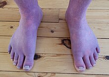
Acrocyanosis in a male Norwegian POTS patient. The patient's legs appear red and purple due to the condition.
Acrocyanosis in a male Norwegian POTS patient
SpecialtyCardiology, neurology
SymptomsMore often with standing: lightheadedness, trouble thinking, tachycardia, weakness, palpitations, heat intolerance, acrocyanosis
Usual onsetMost common (modal) age of onset is 14 years
TypesNeuropathic POTS, Hyperadrenergic POTS, Secondary POTS.
CausesAntibodies against the Alpha 1 adrenergic receptor and muscarinic acetylcholine M4 receptor
Risk factorsFamily history, Ehlers Danlos Syndrome
Diagnostic methodAn increase in heart rate by 30 beats/min with standing
Differential diagnosisDehydration, heart problems, adrenal insufficiency, epilepsy, Parkinson's disease, anemia
TreatmentAvoiding factors that bring on symptoms, increasing dietary salt and water, compression stockings, exercise, medications
MedicationOff label Medications: Beta blockers, Ivabradine, midodrine, and fludrocortisone.
Prognosisc. 90% improve with treatment, 25% of patients unable to work
Frequency~ 1,000,000 ~ 3,000,000 (US)

Postural orthostatic tachycardia syndrome (POTS) is a condition characterized by an abnormally large increase in heart rate upon sitting up or standing. POTS is a disorder of the autonomic nervous system that can lead the individual to experience a variety of symptoms. Symptoms may include lightheadedness, brain fog, blurred vision, weakness, fatigue, headaches, heart palpitations, exercise intolerance, nausea, diminished concentration, tremulousness (shaking), syncope (fainting), coldness or pain in the extremities, chest pain and shortness of breath. Other conditions associated with POTS include migraine headaches, Ehlers–Danlos syndrome, asthma, autoimmune disease, vasovagal syncope and mast cell activation syndrome. POTS symptoms may be treated with lifestyle changes such as increasing fluid and salt intake, wearing compression stockings, gentler and slow postural changes, avoiding prolonged bedrest, medication, increasing electrolyte intake, and physical therapy.

The causes of POTS are varied. POTS may develop after a viral infection, surgery, trauma, or pregnancy. It has been shown to emerge in previously healthy patients after COVID-19, or in rare cases after COVID-19 vaccination. POTS is more common among people who got infected with SARS-CoV-2 than among those who got vaccinated against COVID-19. Risk factors include a family history of the condition. POTS in adults is characterized by a heart rate increase of 30 beats per minute within ten minutes of standing up, accompanied by other symptoms. This increased heart rate should occur in the absence of orthostatic hypotension (>20 mm Hg drop in systolic blood pressure) to be considered POTS. A spinal fluid leak (called spontaneous intracranial hypotension) may have the same signs and symptoms as POTS and should be excluded. Prolonged bedrest may lead to multiple symptoms, including blood volume loss and postural tachycardia. Other conditions which can cause similar symptoms, such as dehydration, orthostatic hypotension, heart problems, adrenal insufficiency, epilepsy, and Parkinson's disease, must not be present. Treatment may include avoiding factors that bring on symptoms, increasing dietary salt and water, small and frequent meals, avoidance of immobilization, wearing compression stockings, and taking medications. Medications used may include beta blockers, pyridostigmine, midodrine or fludrocortisone. More than 50% of patients whose condition was triggered by a viral infection get better within five years. About 80% of patients have symptomatic improvement with treatment, while 25 percent of patients are so disabled they are unable to work. A retrospective study on patients with adolescent-onset has shown that five years after diagnosis, 19% of patients had a full resolution of symptoms.

It is estimated that 1–3 million people in the United States have POTS. The average age for POTS onset is 20 years, and it occurs about five times more frequently in females than in males.

Signs and symptoms

Person standing and measuring heart rate with a pulse oximeter which shows tachycardia of 108 bpm

In adults, the primary manifestation is an increase in heart rate of more than 30 beats per minute within ten minutes of standing up. The resulting heart rate is typically more than 120 beats per minute. For people between ages 12 and 19, the minimum increase for a POTS diagnosis is 40 beats per minute. POTS is often accompanied by common features of orthostatic intolerance—in which symptoms that develop while upright are relieved by reclining. These orthostatic symptoms include palpitations, light-headedness, chest discomfort, shortness of breath, nausea, weakness or "heaviness" in the lower legs, blurred vision, and cognitive difficulties. Symptoms may be exacerbated with prolonged sitting, prolonged standing, alcohol, heat, exercise, or eating a large meal.

Up to one-third of POTS patients experience fainting for many reasons, including but not limited to standing, physical exertion, or heat exposure. POTS patients may also experience orthostatic headaches. Some POTS patients may develop blood pooling in the extremities, characterized by a reddish-purple color of the legs and/or hands upon standing. 48% of people with POTS report chronic fatigue and 32% report sleep disturbances. Other POTS patients only exhibit the cardinal symptom of orthostatic tachycardia. Additional signs and symptoms are varied, and may include excessive sweating, lack of sweating, heat intolerance, digestive issues such as nausea, indigestion, constipation or diarrhea, post-exertional malaise, coat-hanger pain, brain fog, and syncope or presyncope.

Whereas POTS is primarily characterized by its profound impact on the autonomic and cardiovascular systems, it can lead to substantial functional impairment. This impairment, often manifesting as symptoms such as fatigue, cognitive dysfunction, and sleep disturbances, can significantly diminish the patient's quality of life.

Brain fog

One of the most disabling and prevalent symptoms in POTS is "brain fog", a term used by patients to describe the cognitive difficulties they experience. In one survey of 138 POTS patients, brain fog was defined as "forgetful" (91%), "difficulty thinking" (89%), and "difficulty focusing" (88%). Other common descriptions were "difficulty processing what others say" (80%), "confusion" (71%), "getting lost" (64%), and "thoughts moving too quickly" (40%). The same survey described the most common triggers of brain fog to be fatigue (91%), lack of sleep (90%), prolonged standing (87%), and dehydration (86%).

Neuropsychological testing has shown that a POTS patient has reduced attention (Ruff 2&7 speed and WAIS-III digits forward), short-term memory (WAIS-III digits back), cognitive processing speed (Symbol digits modalities test), and executive function (Stroop word color and trails B).

A potential cause for brain fog is a decrease in cerebral blood flow (CBF), especially in upright position.

There may be a loss of neurovascular coupling and reduced functional hyperemia in response to cognitive challenge under orthostatic stress – perhaps related to a loss of autoregulatory buffering of beat-by-beat fluctuations in arterial blood flow.

Causes

The symptoms of POTS can be caused by several distinct pathophysiological mechanisms. These mechanisms are poorly understood, and can overlap, with many patients showing features of multiple POTS types. Many people with POTS exhibit low blood volume (hypovolemia), which can decrease the rate of blood flow to the heart. To compensate for low blood volume, the heart increases its cardiac output by beating faster (reflex tachycardia), leading to the symptoms of presyncope.

In the 30% to 60% of cases classified as hyperadrenergic POTS, norepinephrine levels are elevated on standing, often due to hypovolemia or partial autonomic neuropathy. A smaller minority of people with POTS have (typically very high) standing norepinephrine levels that are elevated even in the absence of hypovolemia and autonomic neuropathy; this is classified as central hyperadrenergic POTS. The high norepinephrine levels contribute to symptoms of tachycardia. Another subtype, neuropathic POTS, is associated with denervation of sympathetic nerves in the lower limbs. In this subtype, it is thought that impaired constriction of the blood vessels causes blood to pool in the veins of the lower limbs. Heart rate increases to compensate for this blood pooling.

In up to 50% of cases, there was an onset of symptoms following a viral illness. It may also be linked to physical trauma, concussion, pregnancy, surgery or psychosocial stress. It is believed that these events could act as a trigger for an autoimmune response that result in POTS. It has been shown to emerge in previously healthy patients after COVID-19 or after COVID-19 vaccination. A 2023 review found that the chances of being diagnosed with POTS within 90 days after mRNA vaccination were 1.33 times higher compared to 90 days before vaccination, still, the results are inconclusive due to a small sample size; only 12 cases of newly diagnosed POTS after mRNA vaccination were reported, all these 12 patients having autoimmune antibodies. However, the risk of POTS-related diagnoses was 5.35 times higher after getting infected with SARS-CoV-2 compared to after mRNA vaccination. Possible mechanisms for COVID-induced POTS are hypovolemia, autoimmunity/inflammation from antibody production against autonomic nerve fibers, and direct toxic effects of COVID-19, or secondary sympathetic nervous system stimulation.

POTS is more common in females than males. POTS also has been linked to patients with a history of autoimmune diseases, Long Covid, irritable bowel syndrome, anemia, hyperthyroidism, fibromyalgia, diabetes, amyloidosis, sarcoidosis, systemic lupus erythematosus, and cancer. Genetics likely plays a role, with one study finding that one in eight POTS patients reported a history of orthostatic intolerance in their family.

Autoimmunity

There is an increasing number of studies indicating that POTS is an autoimmune disease. A high number of patients has elevated levels of autoantibodies against the adrenergic alpha 1 receptor and against the muscarinic acetylcholine M4 receptor.

Elevations of autoantibodies targeting adrenergic α1 receptor has been associated with symptoms severity in patients with POTS.

More recently, autoantibodies against other targets have been identified in small cohorts of POTS patients. Signs of innate immune system activation with elaboration of pro-inflammatory cytokines has also been reported in a cohort of POTS patients.

Secondary POTS

If POTS is caused by another condition, it may be classified as secondary POTS. Chronic diabetes mellitus is one common cause. POTS can also be secondary to gastrointestinal disorders that are associated with low fluid intake due to nausea or fluid loss through diarrhea, leading to hypovolemia. Systemic lupus erythematosus and other autoimmune diseases have also been linked to POTS.

There is a subset of patients who present with both POTS and mast cell activation syndrome (MCAS), and it is not yet clear whether MCAS is a secondary cause of POTS or simply comorbid, however, treating MCAS for these patients can significantly improve POTS symptoms.

POTS can also co-occur in all types of Ehlers–Danlos syndrome (EDS), a hereditary connective tissue disorder marked by loose hypermobile joints prone to subluxations and dislocations, skin that exhibits moderate or greater laxity, easy bruising, and many other symptoms. A trifecta of POTS, EDS, and mast cell activation syndrome (MCAS) is becoming increasingly more common, with a genetic marker common among all three conditions. POTS is also often accompanied by vasovagal syncope, with a 25% overlap being reported. There are some overlaps between POTS and chronic fatigue syndrome, with evidence of POTS in 10–20% of CFS cases. Fatigue and reduced exercise tolerance are prominent symptoms of both conditions, and dysautonomia may underlie both conditions.

POTS can sometimes be a paraneoplastic syndrome associated with cancer.

There are case reports of people developing POTS and other forms of dysautonomia post-COVID. There is no good large-scale empirical evidence yet to prove a connection, so for now the evidence is preliminary.

Diagnosis
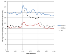
Results of a tilt table test positive for POTS

POTS is most commonly diagnosed by a cardiologist (41%), cardiac electrophysiologist (15%), or neurologist (19%). The average number of physicians seen before receiving diagnosis is seven, and the average delay before diagnosis is 4.7 years.

Diagnostic criteria

A POTS diagnosis requires the following characteristics:

  • For patients age 20 or older, a sustained increase in heart rate ≥30 bpm within ten minutes of upright posture (tilt table test or standing) from a supine position.
    • For patients age 12–19, heart rate increase must be >40 bpm.
  • Associated with frequent symptoms of lightheadedness, palpitations, tremulousness, generalized weakness, blurred vision, or fatigue that are worse with upright posture and that improve with recumbence.
  • An absence of orthostatic hypotension (i.e. no sustained systolic blood pressure drop of 20 mmHg or more).
  • Chronic symptoms that have lasted for longer than three months.
  • In the absence of other disorders, medications, or functional states that are known to predispose to orthostatic tachycardia.

Alternative tests to the tilt table test are also used, such as the NASA Lean Test and the adapted Autonomic Profile (aAP) which require less equipment to complete.

Orthostatic intolerance

An increase in heart rate upon moving to an upright posture is known as orthostatic (upright) tachycardia (fast heart rate). It occurs without any coinciding drop in blood pressure, as that would indicate orthostatic hypotension. Certain medications to treat POTS may cause orthostatic hypotension. It is accompanied by other features of orthostatic intolerance—symptoms that develop in an upright position and are relieved by reclining. These orthostatic symptoms include palpitations, light-headedness, chest discomfort, shortness of breath, nausea, weakness or "heaviness" in the lower legs, blurred vision, and cognitive difficulties.

Differential diagnoses

A variety of autonomic tests are employed to exclude autonomic disorders that could underlie symptoms, while endocrine testing is used to exclude hyperthyroidism and rarer endocrine conditions. Electrocardiography is normally performed on all patients to exclude other possible causes of tachycardia. In cases where a particular associated condition or complicating factor are suspected, other non-autonomic tests may be used: echocardiography to exclude mitral valve prolapse, and thermal threshold tests for small-fiber neuropathy.

Testing the cardiovascular response to prolonged head-up tilting, exercise, eating, and heat stress may help determine the best strategy for managing symptoms. POTS has also been divided into several types (see § Causes), which may benefit from distinct treatments. People with neuropathic POTS show a loss of sweating in the feet during sweat tests, as well as impaired norepinephrine release in the leg, but not arm. This is believed to reflect peripheral sympathetic denervation in the lower limbs. People with hyperadrenergic POTS show a marked increase of blood pressure and norepinephrine levels when standing, and are more likely to have from prominent palpitations, anxiety, and tachycardia. People with POTS can be misdiagnosed with inappropriate sinus tachycardia (IST) as they present similarly. One distinguishing feature is those with POTS rarely exhibit >100 bpm while in a supine position, while patients with IST often have a resting heart rate >100 bpm. Additionally patients with POTS display a more pronounced change in heart rate in response to postural change.

Treatment

POTS treatment involves using multiple methods in combination to counteract cardiovascular dysfunction, address symptoms, and simultaneously address any associated disorders. For most patients, water intake should be increased, especially after waking, in order to expand blood volume (reducing hypovolemia). Eight to ten cups of water daily are recommended. Increasing salt intake, by adding salt to food, taking salt tablets, or drinking sports drinks and other electrolyte solutions is an effective way to raise blood pressure by helping the body retain water. Different physicians recommend different amounts of sodium to their patients. Combining these techniques with gradual physical training enhances their effect. In some cases, when increasing oral fluids and salt intake is not enough, intravenous saline or the drug desmopressin is used to help increase fluid retention.

Large meals worsen symptoms for some people. These people may benefit from eating small meals frequently throughout the day instead. Alcohol and food high in carbohydrates can also exacerbate symptoms of orthostatic hypotension. Excessive consumption of caffeine beverages should be avoided, because they can promote urine production (leading to fluid loss) and consequently hypovolemia. Exposure to extreme heat may also aggravate symptoms.

Prolonged physical inactivity can worsen the symptoms of POTS. Techniques that increase a person's capacity for exercise, such as endurance training or graded exercise therapy, can relieve symptoms for some patients. Aerobic exercise performed for 20 minutes a day, three times a week, is sometimes recommended for patients who can tolerate it. Exercise may have the immediate effect of worsening tachycardia, especially after a meal or on a hot day. In these cases, it may be easier to exercise in a semi-reclined position, such as riding a recumbent bicycle, rowing, or swimming.

When changing to an upright posture, finishing a meal, or concluding exercise, a sustained hand grip can briefly raise the blood pressure, possibly reducing symptoms. Compression garments can also be of benefit by constricting blood pressures with external body pressure.

Aggravating factors include exertion (81%), continued standing (80%), heat (79%), and after meals (42%).

Medication

If nonpharmacological methods are ineffective, medication may be necessary. Medications used may include beta blockers, pyridostigmine, midodrine, or fludrocortisone. As of 2013, no medication has been approved by the U.S. Food and Drug Administration to treat POTS, but a variety are used off-label. Their efficacy has not yet been examined in long-term randomized controlled trials.

Fludrocortisone may be used to enhance sodium retention and blood volume, which may be beneficial not only by augmenting sympathetically mediated vasoconstriction, but also because a large subset of POTS patients appear to have low absolute blood volume. However, fludrocortisone may cause hypokalemia.

While people with POTS typically have normal or even elevated arterial blood pressure, the neuropathic form of POTS is presumed to constitute a selective sympathetic venous denervation. In these patients the selective Alpha-1 adrenergic receptor agonist midodrine may increase venous return, enhance stroke volume, and improve symptoms. Midodrine should only be taken during the daylight hours as it may promote supine hypertension.

Sinus node blocker Ivabradine can successfully restrain heart rate in POTS without affecting blood pressure, demonstrated in approximately 60% of people with POTS treated in an open-label trial of ivabradine experienced symptom improvement.

Pyridostigmine has been reported to restrain heart rate and improve chronic symptoms in approximately half of people. However, it may cause GI side effects that limit its use in around 20% of its patient population.

The selective alpha-1 agonist phenylephrine has been used successfully to enhance venous return and stroke volume in some people with POTS. However, this medication may be hampered by poor oral bioavailability.

Pharmacologic treatments for postural tachycardia syndrome
POTS subtypes Therapeutic action Goal Drug(s)
Neuropathic POTS Alpha-1 adrenergic receptor agonist Constrict the peripheral blood vessels aiding venous return. Midodrine
Splanchnic–mesenteric vasoconstriction Splanchnic vasoconstriction Octreotide
Hypovolemic POTS Synthetic mineralocorticoid Forces the body to retain salt. Increase blood volume Fludrocortisone (Florinef)
Vasopressin receptor agonist Helps retain water, Increase blood volume Desmopressin (DDAVP) 
Hyperadrenergic POTS Beta-blockers (non-selective) Decrease sympathetic tone and heart rate. Propranolol (Inderal)
Beta-blockers (selective) Metoprolol (Toprol), Bisoprolol
Selective sinus node blockade Directly reducing tachycardia. Ivabradine
Alpha-2 adrenergic receptor agonist Decreases blood pressure and sympathetic nerve traffic. Clonidine, Methyldopa
Anticholinesterase inhibitors Splanchnic vasoconstriction. Increase blood pressure. Pyridostigmine
Other (refractory POTS) Psychostimulant Improve cognitive symptoms (brain fog) Modafinil
Central nervous system stimulant Tighten blood vessels. Increases alertness and improves brain fog. Methylphenidate (Ritalin, Concerta)
Direct and indirect α1-adrenoreceptor agonist. Increased blood flows Ephedrine and Pseudoephedrine
Norepinephrine precursor Improve blood vessel contraction Droxidopa (Northera)
Alpha-2 adrenergic antagonist Increase blood pressure Yohimbine

Prognosis

POTS has a favorable prognosis when managed appropriately. Symptoms improve within five years of diagnosis for many patients, and 60% return to their original level of functioning. Approximately 90% of people with POTS respond to a combination of pharmacological and physical treatments. Those who develop POTS in their early to mid teens will likely respond well to a combination of physical methods as well as pharmacotherapy. Outcomes are more guarded for adults newly diagnosed with POTS. Some people do not recover, and a few even worsen with time. The hyperadrenergic type of POTS typically requires continuous therapy. If POTS is caused by another condition, outcomes depend on the prognosis of the underlying disorder.

Epidemiology

The prevalence of POTS is unknown. One study estimated a minimal rate of 170 POTS cases per 100,000 individuals, but the true prevalence is likely higher due to underdiagnosis. Another study estimated that there are at least 500,000 cases in the United States. POTS is more common in women than men, with a female-to-male ratio of 4:1. Most people with POTS are aged between 20 and 40, with an average onset of 21. Diagnoses of POTS beyond age 40 are rare, perhaps because symptoms improve with age.

As recently stated, up to one-third of POTS patients also present with Vasovagal Syncope (VVS).  This ratio is probably higher if pre-Syncope patients, patients that report the symptoms of Syncope without overt fainting, were included.  Given the difficulty with current autonomic measurements in quantitatively isolating and differentiating Parasympathetic (Vagal) activity from Sympathetic activity without assumption or approximation, the current direction of research and clinical assessment is understandable:  perpetuating uncertainty regarding underlying cause, prescribing beta-blockers and proper daily hydration as the only therapy, not addressing the orthostatic dysfunction as the underlying cause, and recommending acceptance and associated lifestyle changes to cope. 

Direct measures of Parasympathetic (Vagal) activity obviates the uncertainty and lack of true relief of POTS as well as VVS.  For example, the hypothesis that POTS is an auto-immune disorder may be an indication that a significant number of POTS cases are indeed co-morbid with VVS.  Remember the Parasympathetic Nervous System is the memory for, and controls and coordinates, the immune system.  If Parasympathetic (Vagal) over-, or prolonged-, activation is chronic then portions of the immune system may remain active beyond the limits of the infection.  Given that portions of the immune system are not of self, these portions remain active and continue to “feed.”  Once the only source of “feed” is self, the immune system begins to attack the host.  This is the definition of autoimmune.  This is a counter-hypothesis that may provide a simpler explanation with a more immediate plan for therapy and relief.  For it may be that relieving the Vagal over-activation, will retires the self-attacking portion of the immune system, thereby relieving the autoimmunity.

Another example may be “Hyperadrenergic POTS.”  A counter hypothesis and perhaps a simpler explanation that leads to more direct therapy and improved outcomes is again the fact that POTS and VVS may be co-morbid.  It is well known that Parasympathetic (Vagal) over-activation may cause secondary Sympathetic over-activation.  Without direct Parasympathetic (Vagal) measures, the resulting assumption is that the secondary Sympathetic over-activation (the definition of “hyperadrenergic”) is actually the primary autonomic dysfunction.  Simply treating the (secondary) Sympathetic over-activation may be just treating a symptom in these cases, which may work for a while but then the body compensates and more medication is needed or the patient become unresponsive and the permanent degraded lifestyles are considered the only option.  Again, this is unfortunate.  Given that cases of POTS with VVS involves different portions of the nervous system (Parasympathetic and Sympathetic), and that both branches may be treated simultaneously, albeit differently, true relief of both conditions, as needed, is quite possible, and the cases of these newer hypothesized causes may be relieved with current, less expensive, and shorter-term therapy modalities.

Co-morbidities

Conditions that are commonly reported with POTS include:

History

In 1871, physician Jacob Mendes Da Costa described a condition that resembled the modern concept of POTS. He named it irritable heart syndrome. Cardiologist Thomas Lewis expanded on the description, coining the term soldier's heart because it was often found among military personnel. The condition came to be known as Da Costa's syndrome, which is now recognized as several distinct disorders, including POTS. Postural tachycardia syndrome was coined in 1982 in a description of a patient who had postural tachycardia, but not orthostatic hypotension. Ronald Schondorf and Phillip A. Low of the Mayo Clinic first used the name postural orthostatic tachycardia syndrome, POTS, in 1993.

Notable cases

British politician Nicola Blackwood revealed in March 2015 that she had been diagnosed with Ehlers–Danlos syndrome in 2013 and that she had later been diagnosed with POTS. She was appointed Parliamentary Under-Secretary of State for Life Science by Prime Minister Theresa May in 2019 and given a life peerage that enabled her to take a seat in Parliament. As a junior minister, it is her responsibility to answer questions in parliament on the subjects of Health and departmental business. When answering these questions, it is customary for ministers to sit when listening to the question and then to rise to give an answer from the despatch box, thus standing up and sitting down numerous times in quick succession throughout a series of questions. On 17 June 2019, she fainted during one of these questioning sessions after standing up from a sitting position four times in the space of twelve minutes, and it was suggested that her POTS was a factor in her fainting. Asked about the incident, she stated: "I was frustrated and embarrassed my body gave up on me at work ... But I am grateful it gives me a chance to shine a light on a condition many others are also living with."

Trigeminal nerve

From Wikipedia, the free encyclopedia
https://en.wikipedia.org/wiki/Trigeminal_nerve

Trigeminal nerve
Schematic illustration of the trigeminal nerve and the organs (or structures) it supplies

In neuroanatomy, the trigeminal nerve (lit. triplet nerve), also known as the fifth cranial nerve, cranial nerve V, or simply CN V, is a cranial nerve responsible for sensation in the face and motor functions such as biting and chewing; it is the most complex of the cranial nerves. Its name (trigeminal, from Latin tri- 'three', and -geminus 'twin') derives from each of the two nerves (one on each side of the pons) having three major branches: the ophthalmic nerve (V1), the maxillary nerve (V2), and the mandibular nerve (V3). The ophthalmic and maxillary nerves are purely sensory, whereas the mandibular nerve supplies motor as well as sensory (or "cutaneous") functions. Adding to the complexity of this nerve is that autonomic nerve fibers as well as special sensory fibers (taste) are contained within it.

The motor division of the trigeminal nerve derives from the basal plate of the embryonic pons, and the sensory division originates in the cranial neural crest. Sensory information from the face and body is processed by parallel pathways in the central nervous system.

Structure

Origin

From the trigeminal ganglion, a single, large sensory root (radix sensoria s. portio major) enters the brainstem at the level of the pons. Immediately adjacent to the sensory root, a smaller motor root (radix motoria s. portio minor) emerges from the pons slightly rostrally and medially to the sensory root.

Motor fibers pass through the trigeminal ganglion without synapsing on their way to peripheral muscles, their cell bodies being located in the nucleus of the fifth nerve, deep within the pons.

Trigeminal ganglion

The three major branches of the trigeminal nerve—the ophthalmic nerve (V1), the maxillary nerve (V2) and the mandibular nerve (V3)—converge on the trigeminal ganglion (also called the semilunar ganglion or gasserian ganglion), located within Meckel's cave and containing the cell bodies of incoming sensory-nerve fibers. The trigeminal ganglion is analogous to the dorsal root ganglia of the spinal cord, which contain the cell bodies of incoming sensory fibers from the rest of the body.

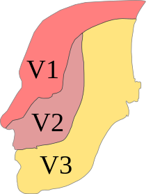
Drawing of the head, with areas served by specific nerves color-coded
Dermatome distribution of the trigeminal nerve

Sensory branches

Profile of the head, with the three sub-nerves color-coded
Dermatome distribution of the trigeminal nerve

The ophthalmic, maxillary and mandibular branches leave the skull through three separate foramina: the superior orbital fissure, the foramen rotundum and the foramen ovale, respectively. The ophthalmic nerve (V1) carries sensory information from the scalp and forehead, the upper eyelid, the conjunctiva and cornea of the eye, the nose (including the tip of the nose, except alae nasi), the nasal mucosa, the frontal sinuses and parts of the meninges (the dura and blood vessels). The maxillary nerve (V2) carries sensory information from the lower eyelid and cheek, the nares and upper lip, the upper teeth and gums, the nasal mucosa, the palate and roof of the pharynx, the maxillary, ethmoid and sphenoid sinuses and parts of the meninges. The mandibular nerve (V3) carries sensory information from the lower lip, the lower teeth and gums, the chin and jaw (except the angle of the jaw, which is supplied by C2-C3), parts of the external ear and parts of the meninges. The mandibular nerve carries touch-position and pain-temperature sensations from the mouth. Although it does not carry taste sensation (the chorda tympani is responsible for taste), one of its branches—the lingual nerve—carries sensation from the tongue.

The peripheral processes of mesencephalic nucleus of V neurons run in the motor root of the trigeminal nerve and terminate in the muscle spindles in the muscles of mastication. They are proprioceptive fibers, conveying information regarding the location of the masticatory muscles. The central processes of mesencephalic V neurons synapse in the motor nucleus V.

Dermatomes

The areas of cutaneous distribution (dermatomes) of the three sensory branches of the trigeminal nerve have sharp borders with relatively little overlap (unlike dermatomes in the rest of the body, which have considerable overlap). The injection of a local anesthetic, such as lidocaine, results in the complete loss of sensation from well-defined areas of the face and mouth. For example, teeth on one side of the jaw can be numbed by injecting the mandibular nerve. Occasionally, injury or disease processes may affect two (or all three) branches of the trigeminal nerve; in these cases, the involved branches may be termed:

  • V1/V2 distribution – Referring to the ophthalmic and maxillary branches
  • V2/V3 distribution – Referring to the maxillary and mandibular branches
  • V1-V3 distribution – Referring to all three branches

Nerves on the left side of the jaw slightly outnumber the nerves on the right side of the jaw.

Function

The sensory function of the trigeminal nerve is to provide tactile, proprioceptive, and nociceptive afference to the face and mouth. Its motor function activates the muscles of mastication, the tensor tympani, tensor veli palatini, mylohyoid and the anterior belly of the digastric.

The trigeminal nerve carries general somatic afferent fibers (GSA), which innervate the skin of the face via ophthalmic (V1), maxillary (V2) and mandibular (V3) divisions. The trigeminal nerve also carries special visceral efferent (SVE) axons, which innervate the muscles of mastication via the mandibular (V3) division.

Muscles

The motor component of the mandibular division (V3) of the trigeminal nerve controls the movement of eight muscles, including the four muscles of mastication: the masseter, the temporal muscle, and the medial and lateral pterygoids. The other four muscles are the tensor veli palatini, the mylohyoid, the anterior belly of the digastric and the tensor tympani.

With the exception of the tensor tympani, all these muscles are involved in biting, chewing and swallowing and all have bilateral cortical representation. A unilateral central lesion (for example, a stroke), no matter how large, is unlikely to produce an observable deficit. Injury to a peripheral nerve can cause paralysis of muscles on one side of the jaw, with the jaw deviating towards the paralyzed side when it opens. This direction of the mandible is due to the action of the functioning pterygoids on the opposite side.

Sensation

The two basic types of sensation are touch-position and pain-temperature. Touch-position input comes to attention immediately, but pain-temperature input reaches the level of consciousness after a delay; when a person steps on a pin, the awareness of stepping on something is immediate but the pain associated with it is delayed.

Touch-position information is generally carried by myelinated (fast-conducting) nerve fibers, and pain-temperature information by unmyelinated (slow-conducting) fibers. The primary sensory receptors for touch-position (Meissner's corpuscles, Merkel's receptors, Pacinian corpuscles, Ruffini's corpuscles, hair receptors, muscle spindle organs and Golgi tendon organs) are structurally more complex than those for pain-temperature, which are nerve endings.

Sensation in this context refers to the conscious perception of touch-position and pain-temperature information, rather than the special senses (smell, sight, taste, hearing and balance) processed by different cranial nerves and sent to the cerebral cortex through different pathways. The perception of magnetic fields, electrical fields, low-frequency vibrations and infrared radiation by some nonhuman vertebrates is processed by their equivalent of the fifth cranial nerve.

Touch in this context refers to the perception of detailed, localized tactile information, such as two-point discrimination (the difference between touching one point and two closely spaced points) or the difference between coarse, medium or fine sandpaper. People without touch-position perception can feel the surface of their bodies and perceive touch in a broad sense, but they lack perceptual detail.

Position, in this context, refers to conscious proprioception. Proprioceptors (muscle spindle and Golgi tendon organs) provide information about joint position and muscle movement. Although much of this information is processed at an unconscious level (primarily by the cerebellum and the vestibular nuclei), some is available at a conscious level.

Touch-position and pain-temperature sensations are processed by different pathways in the central nervous system. This hard-wired distinction is maintained up to the cerebral cortex. Within the cerebral cortex, sensations are linked with other cortical areas.

Sensory pathways

Sensory pathways from the periphery to the cortex are separate for touch-position and pain-temperature sensations. All sensory information is sent to specific nuclei in the thalamus. Thalamic nuclei, in turn, send information to specific areas in the cerebral cortex. Each pathway consists of three bundles of nerve fibers connected in series:

Flow chart from sensory receptors to the cerebral cortex

The secondary neurons in each pathway decussate (cross the spinal cord or brainstem), because the spinal cord develops in segments. Decussated fibers later reach and connect these segments with the higher centers. The optic chiasm is the primary cause of decussation; nasal fibers of the optic nerve cross (so each cerebral hemisphere receives contralateral—opposite—vision) to keep the interneuronal connections responsible for processing information short. All sensory and motor pathways converge and diverge to the contralateral hemisphere.

Although sensory pathways are often depicted as chains of individual neurons connected in series, this is an oversimplification. Sensory information is processed and modified at each level in the chain by interneurons and input from other areas of the nervous system. For example, cells in the main trigeminal nucleus (Main V in the diagram below) receive input from the reticular formation and cerebellar cortex. This information contributes to the final output of the cells in Main V to the thalamus.

Text-and-line diagram of sensory-nerve pathways
C = Cervical segment, S = Sacral segment, VPL = Ventral posterolateral nucleus, SI = Primary somatosensory cortex, VM = Ventromedial prefrontal cortex, MD = Medial dorsal thalamic nucleus, IL = Intralaminar nucleus, VPM = Ventral posteromedial nucleus, Main V = Main trigeminal nucleus, Spinal V = Spinal trigeminal nucleus

Touch-position information from the body is carried to the thalamus by the medial lemniscus, and from the face by the trigeminal lemniscus (both the anterior and posterior trigeminothalamic tracts). Pain-temperature information from the body is carried to the thalamus by the spinothalamic tract, and from the face by the anterior division of the trigeminal lemniscus (also called the anterior trigeminothalamic tract).

Pathways for touch-position and pain-temperature sensations from the face and body merge in the brainstem, and touch-position and pain-temperature sensory maps of the entire body are projected onto the thalamus. From the thalamus, touch-position and pain-temperature information is projected onto the cerebral cortex.

Summary

The complex processing of pain-temperature information in the thalamus and cerebral cortex (as opposed to the relatively simple, straightforward processing of touch-position information) reflects a phylogenetically older, more primitive sensory system. The detailed information received from peripheral touch-position receptors is superimposed on a background of awareness, memory and emotions partially set by peripheral pain-temperature receptors.

Although thresholds for touch-position perception are relatively easy to measure, those for pain-temperature perception are difficult to define and measure. "Touch" is an objective sensation, but "pain" is an individualized sensation which varies among different people and is conditioned by memory and emotion. Anatomical differences between the pathways for touch-position perception and pain-temperature sensation help explain why pain, especially chronic pain, is difficult to manage.

Trigeminal nuclei

Diagram of the brainstem
Brainstem nuclei: Red = Motor; Blue = Sensory; Dark blue = Trigeminal nucleus

All sensory information from the face, both touch-position and pain-temperature, is sent to the trigeminal nucleus. In classical anatomy most sensory information from the face is carried by the fifth nerve, but sensation from parts of the mouth, parts of the ear and parts of the meninges is carried by general somatic afferent fibers in cranial nerves VII (the facial nerve), IX (the glossopharyngeal nerve) and X (the vagus nerve).

All sensory fibers from these nerves terminate in the trigeminal nucleus. On entering the brainstem, sensory fibers from V, VII, IX and X are sorted and sent to the trigeminal nucleus (which contains a sensory map of the face and mouth). The spinal counterparts of the trigeminal nucleus (cells in the dorsal horn and dorsal column nuclei of the spinal cord) contain a sensory map of the rest of the body.

The trigeminal nucleus extends throughout the brainstem, from the midbrain to the medulla, continuing into the cervical cord (where it merges with the dorsal horn cells of the spinal cord). The nucleus is divided into three parts, visible in microscopic sections of the brainstem. From caudal to rostral (ascending from the medulla to the midbrain), they are the spinal trigeminal, the principal sensory and the mesencephalic nuclei. The parts of the trigeminal nucleus receive different types of sensory information; the spinal trigeminal nucleus receives pain-temperature fibers, the principal sensory nucleus receives touch-position fibers and the mesencephalic nucleus receives proprioceptor and mechanoreceptor fibers from the jaws and teeth.

Spinal trigeminal nucleus

The spinal trigeminal nucleus represents pain-temperature sensation from the face. Pain-temperature fibers from peripheral nociceptors are carried in cranial nerves V, VII, IX and X. On entering the brainstem, sensory fibers are grouped and sent to the spinal trigeminal nucleus. This bundle of incoming fibers can be identified in cross-sections of the pons and medulla as the spinal tract of the trigeminal nucleus, which parallels the spinal trigeminal nucleus. The spinal tract of V is analogous to, and continuous with, Lissauer's tract in the spinal cord.

The spinal trigeminal nucleus contains a pain-temperature sensory map of the face and mouth. From the spinal trigeminal nucleus, secondary fibers cross the midline and ascend in the trigeminothalamic (quintothalamic) tract to the contralateral thalamus. Pain-temperature fibers are sent to multiple thalamic nuclei. The central processing of pain-temperature information differs from the processing of touch-position information.

Somatotopic representation

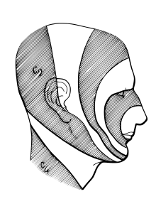
The head in profile, with trigeminal-nerve distribution illustrated
Onion-skin distribution of the trigeminal nerve

Exactly how pain-temperature fibers from the face are distributed to the spinal trigeminal nucleus is disputed. The present general understanding is that pain-temperature information from all areas of the human body is represented in the spinal cord and brainstem in an ascending, caudal-to-rostral fashion. Information from the lower extremities is represented in the lumbar cord, and that from the upper extremities in the thoracic cord. Information from the neck and the back of the head is represented in the cervical cord, and that from the face and mouth in the spinal trigeminal nucleus.

Within the spinal trigeminal nucleus, information is represented in a layered, or "onion-skin" fashion. The lowest levels of the nucleus (in the upper cervical cord and lower medulla) represent peripheral areas of the face (the scalp, ears and chin). Higher levels (in the upper medulla) represent central areas (nose, cheeks and lips). The highest levels (in the pons) represent the mouth, teeth and pharyngeal cavity.

The onion skin distribution differs from the dermatome distribution of the peripheral branches of the fifth nerve. Lesions which destroy lower areas of the spinal trigeminal nucleus (but spare higher areas) preserve pain-temperature sensation in the nose (V1), upper lip (V2) and mouth (V3) and remove pain-temperature sensation from the forehead (V1), cheeks (V2) and chin (V3). Although analgesia in this distribution is "nonphysiologic" in the traditional sense (because it crosses several dermatomes), this analgesia is found in humans after surgical sectioning of the spinal tract of the trigeminal nucleus.

The spinal trigeminal nucleus sends pain-temperature information to the thalamus and sends information to the mesencephalon and the reticular formation of the brainstem. The latter pathways are analogous to the spinomesencephalic and spinoreticular tracts of the spinal cord, which send pain-temperature information from the rest of the body to the same areas. The mesencephalon modulates painful input before it reaches the level of consciousness. The reticular formation is responsible for the automatic (unconscious) orientation of the body to painful stimuli. Incidentally, Sulfur-containing compounds found in plants in the onion family stimulate receptors found in trigeminal ganglia, bypassing the olfactory system.

Principal nucleus

The principal nucleus represents touch-pressure sensation from the face. It is located in the pons, near the entrance for the fifth nerve. Fibers carrying touch-position information from the face and mouth via cranial nerves V, VII, IX, and X are sent to this nucleus when they enter the brainstem.

The principal nucleus contains a touch-position sensory map of the face and mouth, just as the spinal trigeminal nucleus contains a complete pain-temperature map. This nucleus is analogous to the dorsal column nuclei (the gracile and cuneate nuclei) of the spinal cord, which contain a touch-position map of the rest of the body.

From the principal nucleus, secondary fibers cross the midline and ascend in the ventral trigeminothalamic tract to the contralateral thalamus. The ventral trigeminothalamic tract runs parallel to the medial lemniscus, which carries touch-position information from the rest of the body to the thalamus.

Some sensory information from the teeth and jaws is sent from the principal nucleus to the ipsilateral thalamus via the small dorsal trigeminal tract. Touch-position information from the teeth and jaws of one side of the face is represented bilaterally in the thalamus and cortex.

Mesencephalic nucleus

The mesencephalic nucleus is not a true nucleus; it is a sensory ganglion (like the trigeminal ganglion) embedded in the brainstem and the sole exception to the rule that sensory information passes through peripheral sensory ganglia before entering the central nervous system. It has been found in all vertebrates except lampreys and hagfishes. They are the only vertebrates without jaws and have specific cells in their brainstems. These "internal ganglion" cells were discovered in the late 19th century by medical student Sigmund Freud.

Two types of sensory fibers have cell bodies in the mesencephalic nucleus: proprioceptor fibers from the jaw and mechanoreceptor fibers from the teeth. Some of these incoming fibers go to the motor nucleus of the trigeminal nerve (V), bypassing the pathways for conscious perception. The jaw jerk reflex is an example; tapping the jaw elicits a reflex closure of the jaw in the same way that tapping the knee elicits a reflex kick of the lower leg. Other incoming fibers from the teeth and jaws go to the main nucleus of V. This information is projected bilaterally to the thalamus and available for conscious perception.

Activities such as biting, chewing and swallowing require symmetrical, simultaneous coordination of both sides of the body. They are automatic activities, requiring little conscious attention and involving a sensory component (feedback about touch-position) processed at the unconscious level in the mesencephalic nucleus.

Pathways to the thalamus and cortex

Sensation has been defined as the conscious perception of touch-position and pain-temperature information. With the exception of smell, all sensory input (touch-position, pain-temperature, sight, taste, hearing and balance) is sent to the thalamus and then the cortex. The thalamus is anatomically subdivided into nuclei.

Touch-position sensation

Diagram of functions controlled by the cerebral cortex
Cortical homunculus

Touch-position information from the body is sent to the ventral posterolateral nucleus (VPL) of the thalamus. Touch-position information from the face is sent to the ventral posteromedial nucleus (VPM) of the thalamus. From the VPL and VPM, information is projected to the primary somatosensory cortex (SI) in the parietal lobe.

The representation of sensory information in the postcentral gyrus is organized somatotopically. Adjacent areas of the body are represented by adjacent areas in the cortex. When body parts are drawn in proportion to the density of their innervation, the result is a "little man": the cortical homunculus.

Many textbooks have reproduced the outdated Penfield-Rasmussen diagram [ref?], with the toes and genitals on the mesial surface of the cortex when they are actually represented on the convexity. The classic diagram implies a single primary sensory map of the body, when there are multiple primary maps. At least four separate, anatomically distinct sensory homunculi have been identified in the postcentral gyrus. They represent combinations of input from surface and deep receptors and rapidly and slowly adapting peripheral receptors; smooth objects will activate certain cells, and rough objects will activate other cells.

Information from all four maps in SI is sent to the secondary sensory cortex (SII) in the parietal lobe. SII contains two more sensory homunculi. Information from one side of the body is generally represented on the opposite side in SI, but on both sides in SII. Functional MRI imaging of a defined stimulus (for example, stroking the skin with a toothbrush) "lights up" a single focus in SI and two foci in SII.

Pain-temperature sensation

Pain-temperature information is sent to the VPL (body) and VPM (face) of the thalamus (the same nuclei which receive touch-position information). From the thalamus, pain-temperature and touch-position information is projected onto SI.

Unlike touch-position information, however, pain-temperature information is also sent to other thalamic nuclei and projected onto additional areas of the cerebral cortex. Some pain-temperature fibers are sent to the medial dorsal thalamic nucleus (MD), which projects to the anterior cingulate cortex. Other fibers are sent to the ventromedial (VM) nucleus of the thalamus, which projects to the insular cortex. Finally, some fibers are sent to the intralaminar nucleus (IL) of the thalamus via the reticular formation. The IL projects diffusely to all parts of the cerebral cortex.

The insular and cingulate cortices are parts of the brain which represent touch-position and pain-temperature in the context of other simultaneous perceptions (sight, smell, taste, hearing and balance) in the context of memory and emotional state. Peripheral pain-temperature information is channeled directly to the brain at a deep level, without prior processing. Touch-position information is handled differently. Diffuse thalamic projections from the IL and other thalamic nuclei are responsible for a given level of consciousness, with the thalamus and reticular formation "activating" the brain; peripheral pain-temperature information also feeds directly into this system.

Clinical significance

Lateral medullary syndrome

Lateral medullary syndrome (Wallenberg syndrome) is a clinical demonstration of the anatomy of the trigeminal nerve, summarizing how it processes sensory information. A stroke usually affects only one side of the body; loss of sensation due to a stroke will be lateralized to the right or the left side of the body. The only exceptions to this rule are certain spinal-cord lesions and the medullary syndromes, of which Wallenberg syndrome is the best-known example. In this syndrome, a stroke causes a loss of pain-temperature sensation from one side of the face and the other side of the body.

This is explained by the anatomy of the brainstem. In the medulla, the ascending spinothalamic tract (which carries pain-temperature information from the opposite side of the body) is adjacent to the ascending spinal tract of the trigeminal nerve (which carries pain-temperature information from the same side of the face). A stroke which cuts off the blood supply to this area (for example, a clot in the posterior inferior cerebellar artery) destroys both tracts simultaneously. The result is a loss of pain-temperature (but not touch-position) sensation in a "checkerboard" pattern (ipsilateral face, contralateral body), facilitating diagnosis.

Sensory neuronopathy

Sensory neuronopathy (also known as sensory ganglionopathy) is a type of peripheral neuropathy in which sensory nerve cell bodies in the dorsal root ganglia, commonly including the trigeminal ganglion of the trigeminal nerve, are damaged due to a variety of mechanisms leading to sensory symptoms such as parasthesias, dysesthesias, or hyperalgesia in the affected nerve distribution including the distribution of the trigeminal nerve.

Copper in biology

From Wikipedia, the free encyclopedia https://en.wikipedia.org/wiki/Cop...