Search This Blog

Wednesday, December 26, 2018

Treatment-resistant depression

From Wikipedia, the free encyclopedia

Treatment-resistant depression
Classification and external resources
MeSHD061218

Treatment-resistant depression (TRD) or treatment-refractory depression is a term used in clinical psychiatry to describe a condition that affects people with major depressive disorder (MDD) who do not respond adequately to a course of appropriate antidepressant medication within a certain time. Typical definitions of TRD vary, and they do not include a resistance to psychological therapies. Inadequate response has traditionally been defined as no clinical response whatsoever (e.g. no improvement in depressive symptoms). However, many clinicians consider a response inadequate if the person does not achieve full remission of symptoms. People with treatment-resistant depression who do not adequately respond to antidepressant treatment are sometimes referred to as pseudoresistant. Some factors that contribute to inadequate treatment are: early discontinuation of treatment, insufficient dosage of medication, patient noncompliance, misdiagnosis, and concurrent psychiatric disorders. Cases of treatment-resistant depression may also be referred to by which medications people with TRD are resistant to (e.g.: SSRI-resistant).

Prevalence

Treatment-resistance is relatively common in people with MDD. Rates of total remission following antidepressant treatment are only 50.4%. In cases of depression treated by a primary care physician, 32% of people partially responded to treatment and 45% did not respond at all.

Predictors

Comorbid psychiatric disorders

Comorbid psychiatric disorders commonly go undetected in the treatment of depression. If left untreated, the symptoms of these disorders can interfere with both evaluation and treatment. Anxiety disorders are one of the most common disorder types associated with treatment-resistant depression. The two disorders commonly co-exist, and have some similar symptoms. Some studies have shown that patients with both MDD and panic disorder are the most likely to be nonresponsive to treatment. Substance abuse may also be a predictor of treatment-resistant depression. It may cause depressed patients to be noncompliant in their treatment, and the effects of certain substances can worsen the effects of depression. Other psychiatric disorders that may predict treatment-resistant depression include personality disorders, obsessive compulsive disorder, and eating disorders.

Comorbid medical disorders

Some people who are diagnosed with treatment-resistant depression may have an underlying undiagnosed health condition that is causing or contributing to their depression. Endocrine disorders like hypothyroidism, Cushing's disease, and Addison's disease are among the most commonly identified as contributing to depression. Others include diabetes, coronary artery disease, cancer, HIV, and Parkinson's disease. Another factor is that medications used to treat comorbid medical disorders may lessen the effectiveness of antidepressants or cause depression symptoms.

Features of depression

People with depression who also display psychotic symptoms such as delusions or hallucinations are more likely to be treatment resistant. Another depressive feature that has been associated with poor response to treatment is longer duration of depressive episodes. Finally, people with more severe depression and those who are suicidal are more likely to be nonresponsive to antidepressant treatment.

Drug treatment

There are three basic categories of drug treatment that can be used when a medication course is found to be ineffective. One option is to switch the patient to a different medication. Another option is to add a medication to the patient’s current treatment. This can include combination therapy: the combination of two different types of antidepressants, or augmentation therapy: the addition of a non-antidepressant medication that may increase the effectiveness of the antidepressant.

Dose increase

Increasing the dosage of an antidepressant is a common strategy to treat depression that does not respond after adequate treatment duration. Practitioners who use this strategy will usually increase the dose until the person reports intolerable side effects, symptoms are eliminated, or the dose is increased to the limit of what is considered safe.

Switching antidepressants

Studies have shown a wide variability in the effectiveness of switching antidepressants, with anywhere from 25-70% of people responding to a different antidepressant. There is support for the effectiveness of switching people to a different SSRI; 50% of people that were nonresponsive after taking one SSRI were responsive after taking a second type. Switching people with TRD to a different class of antidepressants may also be effective. People who are nonresponsive after taking an SSRI may respond to a Tricyclic antidepressant, bupropion or a MAOI.

Adding medication

Medications that have been shown to be effective in people with treatment-resistant depression include lithium, triiodothyronine, benzodiazepines, atypical antipsychotics, and stimulants. Adding lithium may be effective for people taking some types of antidepressants, it does not appear to be effective in patients taking SSRI’s. Triiodothyroxine (T3) is a type of thyroid hormone and has been associated with improvement in mood and depression symptoms. Benzodiazepines may improve treatment-resistant depression by decreasing the adverse side effects caused by some antidepressants and therefore increasing patient compliance. Since the entry of olanzapine into psychopharmacology, many psychiatrists have been adding low dose olanzapine to antidepressants and other atypical antipsychotics such as aripiprazole and quetiapine. Eli Lilly, the company that sells both olanzapine and fluoxetine individually, has also released a combo formulation which contains olanzapine and fluoxetine in a single capsule. 

These have shown promise in treating refractory depression but come with serious side effects. Stimulants such as amphetamines and methylphenidate have also been tested with positive results but have potential for abuse. However, stimulants have been shown to be effective for the unyielding depressed combined lacking addictive personality traits or heart problems.

Ketamine has been tested as a rapid-acting antidepressant for treatment-resistant depression in bipolar disorder, and major depressive disorder.

Other treatment options

Electroconvulsive therapy

Electroconvulsive therapy is generally only considered as a treatment option in severe cases of treatment-resistant depression. It is used when medication has repeatedly failed to improve symptoms, and usually when the patient’s symptoms are so severe that they have been hospitalized. Electroconvulsive therapy has been found to reduce thoughts of suicide and relieve depressive symptoms. It is associated with an increase in glial cell line derived neurotrophic factor.

Vagus nerve stimulation

Vagus nerve stimulation is a more invasive procedure than electroconvulsive therapy, but it has been shown to be well tolerated. During the procedure a stimulating electrode is surgically attached to the vagus nerve; this allows for continuous stimulation after implantation. Like electroconvulsive therapy, it is usually only used in severe cases of treatment-resistant depression that have been non-responsive to medication.

Psychological therapies

There is sparse evidence on the effectiveness of psychotherapy in cases of treatment-resistant depression. However, a review of the literature suggests that it may be an effective treatment option. Psychotherapy may be effective in people with TRD because it can help relieve stress that may contribute to depressive symptoms.

A Cochrane systematic review has shown that psychological therapies (including cognitive behavioural therapy, dialectal behavioural therapy, interpersonal therapy and intensive short term dynamic psychotherapy) added to usual care (with antidepressants) can be beneficial for depressive symptoms and for response and remission rates over the short term (up to 6 months) for patients with TRD. Medium‐ (7-12 months) and long‐term (longer than 12 months) effects seem similarly beneficial. Psychological therapies added to usual care (antidepressants) seem as acceptable as usual care alone.

rTMS

rTMS (Repetitive Transcranial Magnetic Stimulation) is gradually becoming recognised as a valuable therapeutic option in treatment-resistant depression. A number of randomised placebo-controlled trials have compared real versus sham rTMS. These trials have consistently demonstrated the efficacy of this treatment against major depression. There have also been a number of meta-analyses of RCTs  confirming the efficacy of rTMS in treatment-resistant major depression, as well as naturalistic studies showing its effectiveness in "real world" clinical settings. 

dTMS

dTMS (Deep Transcranial Magnetic Stimulation) is a continuation of the same idea as rTMS, but with the hope that deeper stimulation of subcortical areas of the brain leads to increased effect. A 2015 systematic review and health technology assessment found lacking evidence in order to recommend the method over either ECT or rTMS because so few studies had been published.

Outcomes

Treatment-resistant depression is associated with more instances of relapse than depression that is responsive to treatment. One study showed that as many as 80% of people with TRD who needed more than one course of treatment relapsed within a year. Treatment-resistant depression has also been associated with lower long term quality of life.

Reticular formation

From Wikipedia, the free encyclopedia

Reticular formation
Gray701.png
Axial section of the pons, at its upper part. (Formatio reticularis labeled at left.)
Gray694.png
Section of the medulla oblongata at about the middle of the olive. (Formatio reticularis grisea and formatio reticularis alba labeled at left.)
Details
Identifiers
Latinformatio reticularis
MeSHD012154
NeuroNames1223
NeuroLex IDnlx_143558
TAA14.1.00.021 A14.1.05.403 A14.1.06.327
FMA77719

The reticular formation is a set of interconnected nuclei that are located throughout the brainstem. The reticular formation is not anatomically well defined because it includes neurons located in diverse parts of the brain. The neurons of the reticular formation make up a complex set of networks in the core of the brainstem that stretch from the upper part of the midbrain to the lower part of the medulla oblongata. The reticular formation includes ascending pathways to the cortex in the ascending reticular activating system (ARAS) and descending pathways to the spinal cord via the reticulospinal tracts of the descending reticular formation.

Neurons of the reticular formation, particularly those of the ascending reticular activating system, play a crucial role in maintaining behavioral arousal and consciousness. The functions of the reticular formation are modulatory and premotor. The modulatory functions are primarily found in the rostral sector of the reticular formation and the premotor functions are localized in the neurons in more caudal regions.

The reticular formation is divided into three columns: raphe nuclei (median), gigantocellular reticular nuclei (medial zone), and parvocellular reticular nuclei (lateral zone). The raphe nuclei are the place of synthesis of the neurotransmitter serotonin, which plays an important role in mood regulation. The gigantocellular nuclei are involved in motor coordination. The parvocellular nuclei regulate exhalation.

The reticular formation is essential for governing some of the basic functions of higher organisms and is one of the phylogenetically oldest portions of the brain.

General structure

A cross section of the lower part of the pons showing the pontine reticular formation labeled as #9
 
The human reticular formation is composed of almost 100 brain nuclei and contains many projections into the forebrain, brainstem, and cerebellum, among other regions. It includes the reticular nuclei, reticulothalamic projection fibers, diffuse thalamo-cortical projections, ascending cholinergic projections, descending non-cholinergic projections, and descending reticulospinal projections. The reticular formation also contains two major neural subsystems, the ascending reticular activating system and descending reticulospinal tracts, which mediate distinct cognitive and physiological processes. It has been functionally cleaved both sagittally and coronally

Traditionally the reticular nuclei are divided into three columns:
  • In the median column – the raphe nuclei
  • In the medial column – gigantocellular nuclei (because of larger size of the cells)
  • In the lateral column – parvocellular nuclei (because of smaller size of the cells)
The original functional differentiation was a division of caudal and rostral. This was based upon the observation that the lesioning of the rostral reticular formation induces a hypersomnia in the cat brain. In contrast, lesioning of the more caudal portion of the reticular formation produces insomnia in cats. This study has led to the idea that the caudal portion inhibits the rostral portion of the reticular formation. 

Sagittal division reveals more morphological distinctions. The raphe nuclei form a ridge in the middle of the reticular formation, and, directly to its periphery, there is a division called the medial reticular formation. The medial RF is large and has long ascending and descending fibers, and is surrounded by the lateral reticular formation. The lateral RF is close to the motor nuclei of the cranial nerves, and mostly mediates their function.

Medial and lateral reticular formation

The medial reticular formation and lateral reticular formation are two columns of neuronal nuclei with ill-defined boundaries that send projections through the medulla and into the mesencephalon (midbrain). The nuclei can be differentiated by function, cell type, and projections of efferent or afferent nerves. Moving caudally from the rostral midbrain, at the site of the rostral pons and the midbrain, the medial RF becomes less prominent, and the lateral RF becomes more prominent.

Existing on the sides of the medial reticular formation is its lateral cousin, which is particularly pronounced in the rostral medulla and caudal pons. Out from this area spring the cranial nerves, including the very important vagus nerve. The Lateral RF is known for its ganglions and areas of interneurons around the cranial nerves, which serve to mediate their characteristic reflexes and functions.

General functions

The reticular formation consists of more than 100 small neural networks, with varied functions including the following:
  • Somatic motor control – Some motor neurons send their axons to the reticular formation nuclei, giving rise to the reticulospinal tracts of the spinal cord. These tracts function in maintaining tone, balance, and posture—especially during body movements. The reticular formation also relays eye and ear signals to the cerebellum so that the cerebellum can integrate visual, auditory, and vestibular stimuli in motor coordination. Other motor nuclei include gaze centers, which enable the eyes to track and fixate objects, and central pattern generators, which produce rhythmic signals of breathing with swallowing, and with defecation and urination.
  • Cardiovascular control – The reticular formation includes the cardiac and vasomotor centers of the medulla oblongata.
  • Pain modulation – The reticular formation is one means by which pain signals from the lower body reach the cerebral cortex. It is also the origin of the descending analgesic pathways. The nerve fibers in these pathways act in the spinal cord to block the transmission of some pain signals to the brain.
  • Sleep and consciousness – The reticular formation has projections to the thalamus and cerebral cortex that allow it to exert some control over which sensory signals reach the cerebrum and come to our conscious attention. It plays a central role in states of consciousness like alertness and sleep. Injury to the reticular formation can result in irreversible coma.
  • Habituation – This is a process in which the brain learns to ignore repetitive, meaningless stimuli while remaining sensitive to others. A good example of this is a person who can sleep through loud traffic in a large city, but is awakened promptly due to the sound of an alarm or crying baby. Reticular formation nuclei that modulate activity of the cerebral cortex are part of the ascending reticular activating system.

Major subsystems

Ascending reticular activating system

Ascending reticular activating system. Reticular formation labeled near center.

The ascending reticular activating system (ARAS), also known as the extrathalamic control modulatory system or simply the reticular activating system (RAS), is a set of connected nuclei in the brains of vertebrates that is responsible for regulating wakefulness and sleep-wake transitions. The ARAS is a part of the reticular formation and is mostly composed of various nuclei in the thalamus and a number of dopaminergic, noradrenergic, serotonergic, histaminergic, cholinergic, and glutamatergic brain nuclei.

Structure of the ARAS

The ARAS is composed of several neuronal circuits connecting the dorsal part of the posterior midbrain and anterior pons to the cerebral cortex via distinct pathways that project through the thalamus and hypothalamus. The ARAS is a collection of different nuclei – more than 20 on each side in the upper brainstem, the pons, medulla, and posterior hypothalamus. The neurotransmitters that these neurons release include dopamine, norepinephrine, serotonin, histamine, acetylcholine, and glutamate. They exert cortical influence through direct axonal projections and indirect projections through thalamic relays.

The thalamic pathway consists primarily of cholinergic neurons in the pontine tegmentum, whereas the hypothalamic pathway is composed primarily of neurons that release monoamine neurotransmitters, namely dopamine, norepinephrine, serotonin, and histamine. The glutamate-releasing neurons in the ARAS were identified much more recently relative to the monoaminergic and cholinergic nuclei; the glutamatergic component of the ARAS includes one glutamatergic nucleus in the hypothalamus and various glutamatergic brainstem nuclei. The orexin neurons of the lateral hypothalamus innervate every component of the ascending reticular activating system and coordinate activity within the entire system.

The key components of the ARAS are listed in the table below. 

Key components of the ascending reticular activating system
Nucleus type Corresponding nuclei that mediate arousal
Dopaminergic nuclei
Noradrenergic nuclei
Serotonergic nuclei
Histaminergic nuclei
Cholinergic nuclei
Glutamatergic nuclei
Thalamic nuclei

The ARAS consists of evolutionarily ancient areas of the brain, which are crucial to survival and protected during adverse periods. As a result, the ARAS still functions during inhibitory periods of hypnosis.
 
The ascending reticular activating system which sends neuromodulatory projections to the cortex - mainly connects to the prefrontal cortex. There is seen to be low connectivity to the motor areas of the cortex.

Functions of the ARAS

Consciousness
The ascending reticular activating system is an important enabling factor for the state of consciousness. The ascending system is seen to contribute to wakefulness as characterised by cortical and behavioural arousal.
Regulating sleep-wake transitions
The main function of the ARAS is to modify and potentiate thalamic and cortical function such that electroencephalogram (EEG) desynchronization ensues. There are distinct differences in the brain's electrical activity during periods of wakefulness and sleep: Low voltage fast burst brain waves (EEG desynchronization) are associated with wakefulness and REM sleep (which are electrophysiologically similar); high voltage slow waves are found during non-REM sleep. Generally speaking, when thalamic relay neurons are in burst mode the EEG is synchronized and when they are in tonic mode it is desynchronized. Stimulation of the ARAS produces EEG desynchronization by suppressing slow cortical waves (0.3–1 Hz), delta waves (1–4 Hz), and spindle wave oscillations (11–14 Hz) and by promoting gamma band (20 – 40 Hz) oscillations.

The physiological change from a state of deep sleep to wakefulness is reversible and mediated by the ARAS. Inhibitory influence from the brain is active at sleep onset, likely coming from the preoptic area (POA) of the hypothalamus. During sleep, neurons in the ARAS will have a much lower firing rate; conversely, they will have a higher activity level during the waking state. Therefore, low frequency inputs (during sleep) from the ARAS to the POA neurons result in an excitatory influence and higher activity levels (awake) will have inhibitory influence. In order that the brain may sleep, there must be a reduction in ascending afferent activity reaching the cortex by suppression of the ARAS.
Attention
The ARAS also helps mediate transitions from relaxed wakefulness to periods of high attention. There is increased regional blood flow (presumably indicating an increased measure of neuronal activity) in the midbrain reticular formation (MRF) and thalamic intralaminar nuclei during tasks requiring increased alertness and attention.

Clinical significance of the ARAS

Mass lesions in brainstem ARAS nuclei can cause severe alterations in level of consciousness (e.g., coma). Bilateral damage to the reticular formation of the midbrain may lead to coma or death.

Direct electrical stimulation of the ARAS produces pain responses in cats and educes verbal reports of pain in humans. Additionally, ascending reticular activation in cats can produce mydriasis, which can result from prolonged pain. These results suggest some relationship between ARAS circuits and physiological pain pathways.
Pathologies
Given the importance of the ARAS for modulating cortical changes, disorders of the ARAS should result in alterations of sleep-wake cycles and disturbances in arousal. Some pathologies of the ARAS may be attributed to age, as there appears to be a general decline in reactivity of the ARAS with advancing years. Changes in electrical coupling have been suggested to account for some changes in ARAS activity: If coupling were down-regulated, there would be a corresponding decrease in higher-frequency synchronization (gamma band). Conversely, up-regulated electrical coupling would increase synchronization of fast rhythms that could lead to increased arousal and REM sleep drive. Specifically, disruption of the ARAS has been implicated in the following disorders:
  • Narcolepsy: Lesions along the PPT/LDT nuclei are associated with narcolepsy. There is a significant down-regulation of PPN output and a loss of orexin peptides, promoting the excessive daytime sleepiness that is characteristic of this disorder.
  • Schizophrenia: Intractable schizophrenic patients have a significant increase (> 60%) in the number of PPN neurons and dysfunction of NO signaling involved in modulating cholinergic output of the ARAS.
  • Post-traumatic stress disorder, Parkinson's disease, REM behavior disorder: Patients with these syndromes exhibit a significant (>50%) decrease in the number of locus coeruleus (LC) neurons, resulting is increased disinhibition of the PPN.
  • Progressive supranuclear palsy (PSP): Dysfunction of NO signaling has been implicated in the development of PSP.
  • Depression, autism, Alzheimer's disease, attention deficit disorder: The exact role of the ARAS in each of these disorders has not yet been identified. However, it is expected that in any neurological or psychiatric disease that manifests disturbances in arousal and sleep-wake cycle regulation, there will be a corresponding dysregulation of some elements of the ARAS.
  • Parkinson's disease: REM sleep disturbances are common in Parkinson's. It is mainly a dopaminergic disease, but cholinergic nuclei are depleted as well. Degeneration in the ARAS begins early in the disease process.
Developmental influences
There are several potential factors that may adversely influence the development of the ascending reticular activating system:
  • Preterm birth: Regardless of birth weight or weeks of gestation, premature birth induces persistent deleterious effects on pre-attentional (arousal and sleep-wake abnormalities), attentional (reaction time and sensory gating), and cortical mechanisms throughout development.
  • Smoking during pregnancy: Prenatal exposure to cigarette smoke is known to produce lasting arousal, attentional and cognitive deficits in humans. This exposure can induce up-regulation of nicotinic receptors on α4b2 subunit on Pedunculopontine nucleus (PPN) cells, resulting in increased tonic activity, resting membrane potential, and hyperpolarization-activated cation current. These major disturbances of the intrinsic membrane properties of PPN neurons result in increased levels of arousal and sensory gating deficits (demonstrated by a diminished amount of habituation to repeated auditory stimuli). It is hypothesized that these physiological changes may intensify attentional dysregulation later in life.

Descending reticulospinal tracts

Spinal cord tracts - reticulospinal tract labeled in red, near-center at left in figure.

The reticulospinal tracts, also known as the descending or anterior reticulospinal tracts, are extrapyramidal motor tracts that descend from the reticular formation in two tracts to act on the motor neurons supplying the trunk and proximal limb flexors and extensors. The reticulospinal tracts are involved mainly in locomotion and postural control, although they do have other functions as well. The descending reticulospinal tracts are one of four major cortical pathways to the spinal cord for musculoskeletal activity. The reticulospinal tracts works with the other three pathways to give a coordinated control of movement, including delicate manipulations. The four pathways can be grouped into two main system pathways – a medial system and a lateral system. The medial system includes the reticulospinal pathway and the vestibulospinal pathway, and this system provides control of posture. The corticospinal and the rubrospinal tract pathways belong to the lateral system which provides fine control of movement.

Components of the reticulospinal tracts

The tract is divided into two parts, the medial (or pontine) and lateral (or medullary) reticulospinal tracts (MRST and LRST).
  • The MRST is responsible for exciting anti-gravity, extensor muscles. The fibers of this tract arise from the caudal pontine reticular nucleus and the oral pontine reticular nucleus and project to the lamina VII and lamina VIII of the spinal cord (BrainInfo)
  • The LRST is responsible for inhibiting excitatory axial extensor muscles of movement. It is also responsible for automatic breathing. The fibers of this tract arise from the medullary reticular formation, mostly from the gigantocellular nucleus, and descend the length of the spinal cord in the anterior part of the lateral column. The tract terminates in lamina VII mostly with some fibers terminating in lamina IX of the spinal cord.
The ascending sensory tract conveying information in the opposite direction is known as the spinoreticular tract.

Functions of the reticulospinal tracts

  • Integrates information from the motor systems to coordinate automatic movements of locomotion and posture
  • Facilitates and inhibits voluntary movement; influences muscle tone
  • Mediates autonomic functions
  • Modulates pain impulses
  • Influences blood flow to lateral geniculate nucleus of the thalamus.

Clinical significance of the reticulospinal tracts

The reticulospinal tracts are mostly inhibited by the corticospinal tract; if damage occurs at the level of or below the red nucleus (e.g. to the superior colliculus), it is called decerebration, and causes decerebrate rigidity: an unopposed extension of the head and limbs. The reticulospinal tracts also provide a pathway by which the hypothalamus can control sympathetic thoracolumbar outflow and parasympathetic sacral outflow.[citation needed]

History

The term "reticular formation" was coined in the late 19th century by Otto Deiters, coinciding with Ramon y Cajal’s neuron doctrine. Allan Hobson states in his book The Reticular Formation Revisited that the name is an etymological vestige from the fallen era of the aggregate field theory in the neural sciences. The term "reticulum" means "netlike structure", which is what the reticular formation resembles at first glance. It has been described as being either too complex to study or an undifferentiated part of the brain with no organization at all. Eric Kandel describes the reticular formation as being organized in a similar manner to the intermediate gray matter of the spinal cord. This chaotic, loose, and intricate form of organization is what has turned off many researchers from looking farther into this particular area of the brain. The cells lack clear ganglionic boundaries, but do have clear functional organizations and distinct cell types. The term "reticular formation" is seldom used anymore except to speak in generalities. Modern scientists usually refer to the individual nuclei that compose the reticular formation.

Moruzzi and Magoun first investigated the neural components regulating the brain's sleep-wake mechanisms in 1949. Physiologists had proposed that some structure deep within the brain controlled mental wakefulness and alertness. It had been thought that wakefulness depended only on the direct reception of afferent (sensory) stimuli at the cerebral cortex

The direct electrical stimulation of the brain could simulate electrocortical relays. Magoun used this principle to demonstrate, on two separate areas of the brainstem of a cat, how to produce wakefulness from sleep. First the ascending somatic and auditory paths; second, a series of "ascending relays from the reticular formation of the lower brain stem through the midbrain tegmentum, subthalamus and hypothalamus to the internal capsule." The latter was of particular interest, as this series of relays did not correspond to any known anatomical pathways for the wakefulness signal transduction and was coined the ascending reticular activating system (ARAS). 

Next, the significance of this newly identified relay system was evaluated by placing lesions in the medial and lateral portions of the front of the midbrain. Cats with mesancephalic interruptions to the ARAS entered into a deep sleep and displayed corresponding brain waves. In alternative fashion, cats with similarly placed interruptions to ascending auditory and somatic pathways exhibited normal sleeping and wakefulness, and could be awakened with somatic stimuli. Because these external stimuli would be blocked by the interruptions, this indicated that the ascending transmission must travel through the newly discovered ARAS.

Finally, Magoun recorded potentials within the medial portion of the brain stem and discovered that auditory stimuli directly fired portions of the reticular activating system. Furthermore, single-shock stimulation of the sciatic nerve also activated the medial reticular formation, hypothalamus, and thalamus. Excitation of the ARAS did not depend on further signal propagation through the cerebellar circuits, as the same results were obtained following decerebellation and decortication. The researchers proposed that a column of cells surrounding the midbrain reticular formation received input from all the ascending tracts of the brain stem and relayed these afferents to the cortex and therefore regulated wakefulness.

Ad Astra on VASIMIR propulsion system

Original link:  http://www.adastrarocket.com/aarc/research-and-development

The main focus of the Ad Astra's Research & Development efforts is the VX-200, which is a VASIMR® prototype designed to test flight-related hardware and technology in a space-like environment. The VX-200 technologically advanced components are the solid-state RF amplifiers, developed by Nautel, superconducting magnet, built by Scientific Magnetics, and the on-board computer control. The VX-200 serves as a technology demonstration and risk mitigation platform, in addition to serving as a means to explore fundamental plasma physics for academic purposes. The record performance numbers for VX-200 operating with argon propellant are:
  • RF Power: 200 kW;
  • thrust: 5.7 N;
  • exhaust speed: 50 km/s;
  • thruster efficiency: 72 % (jet power divided by coupled RF power).
In addition to our efforts towards the development of the VASIMR® engine, we also specialize functional testing of third-party hardware in extreme conditions such as microgravity, high magnetic field, high vacuum, and RFI environments. Our scientists have accumulated over 50 hours flight time with hardware in a microgravity environment on board NASA's ZERO-G aircraft. Ad Astra is willing to work with those who wish extreme limits of their technology or have a desire to fast-track their designs to flight readiness.

Ad Astra's control area where scientists are operating the VX-200 rocket. Credit: Kat's Photography
 
Ad Astra's scientists working around the 150 m3 vacuum chamber.  Credit: Ad Astra Rocket Company
 
Ad Astra's scientists using a laser alignment rig with the VX-200 to precisely line the rocket core with the magnetic field.  Credit: Ad Astra Rocket Company 

Zero-G Testing
Ad Astra scientist's testing hardware on board the NASA Zero-G aircraft

Locus coeruleus

From Wikipedia, the free encyclopedia

Locus coeruleus
Gray709.png
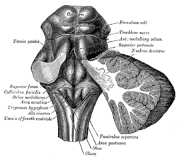
Rhomboid fossa. (Locus coeruleus not labeled, but is very near [just lateral to] the facial colliculus, which is labeled at center left.)
Locus ceruleus - high mag.jpg
Micrograph showing the locus coeruleus. HE-LFB stain.
Details
Identifiers
Latinlocus caeruleus ("blue place")
MeSHD008125
NeuroNames583
NeuroLex IDbirnlex_905
TAA14.1.05.436 A14.1.05.706
FMA72478

The locus coeruleus (\-si-ˈrü-lē-əs\, also spelled locus caeruleus or locus ceruleus) is a nucleus in the pons of the brainstem involved with physiological responses to stress and panic. It is a part of the reticular activating system.

The locus coeruleus is the principal site for brain synthesis of norepinephrine (noradrenaline). The locus coeruleus and the areas of the body affected by the norepinephrine it produces are described collectively as the locus coeruleus-noradrenergic system or LC-NA system. Norepinephrine may also be released directly into the blood from the adrenal medulla.

Anatomy

Micrograph showing the locus coeruleus (upper-right of image) in an axial section of the pons. The fourth ventricle (quasi-triangular white area) is in the upper-left of the image. The midline is seen on the left. The large white area in the upper-left corner is where the cerebellum would be. HE-LFB stain.
 
Locus coeruleus highlighted in green.

The locus coeruleus (LC) is located in the posterior area of the rostral pons in the lateral floor of the fourth ventricle. It is composed of mostly medium-size neurons. Melanin granules inside the neurons of the LC contribute to its blue colour. Thus, it is also known as the nucleus pigmentosus pontis, meaning "heavily pigmented nucleus of the pons." The neuromelanin is formed by the polymerization of noradrenaline and is analogous to the black dopamine-based neuromelanin in the substantia nigra

In adult humans (19-78) the locus coeruleus has 22,000 to 51,000 total pigmented neurons that range in size between 31,000 and 60,000 μm3.

Connections

The projections of this nucleus reach far and wide. For example, they innervate the spinal cord, the brain stem, cerebellum, hypothalamus, the thalamic relay nuclei, the amygdala, the basal telencephalon, and the cortex. The norepinephrine from the LC has an excitatory effect on most of the brain, mediating arousal and priming the brain’s neurons to be activated by stimuli.

As an important homeostatic control center of the body, the locus coeruleus receives afferents from the hypothalamus. The cingulate gyrus and the amygdala also innervate the LC, allowing emotional pain and stressors to trigger noradrenergic responses. The cerebellum and afferents from the raphe nuclei also project to the LC, in particular the pontine raphe nucleus and dorsal raphe nucleus.

Inputs

The locus coeruleus receives inputs from a number of other brain regions, primarily:

Outputs

The projections from the locus coeruleus consist of neurons that utilize norepinephrine as their primary neurotransmitter. These projections include the following connections:

Function

It is related to many functions via its widespread projections. The LC-NA system modulates cortical, subcortical, cerebellar, brainstem, and spinal cord circuits. Some of the most important functions influenced by this system are:
The locus coeruleus is a part of the reticular activating system, and is almost completely inactivated in rapid eye movement sleep.

Pathophysiology


Research continues to reveal that norepinephrine (NE) is a critical regulator of numerous activities from stress response, the formation of memory to attention and arousal. Many neuropsychiatric disorders precipitate from alterations to NE modulated neurocircuitry: disorders of affect, anxiety disorders, PTSD, ADHD and Alzheimer’s disease. Alterations in the locus coeruleus (LC) accompany dysregulation of NE function and likely play a key role in the pathophysiology of these neuropsychiatric disorders.

In stress

The locus coeruleus is responsible for mediating many of the sympathetic effects during stress. The locus coeruleus is activated by stress, and will respond by increasing norepinephrine secretion, which in turn will alter cognitive function (through the prefrontal cortex), increase motivation (through nucleus accumbens), activate the hypothalamic-pituitary-adrenal axis, and increase the sympathetic discharge/inhibit parasympathetic tone (through the brainstem). Specific to the activation of the hypothalamo-pituitary adrenal axis, norepinephrine will stimulate the secretion of corticotropin-releasing factor from the hypothalamus, that induces adrenocorticotropic hormone release from the anterior pituitary and subsequent cortisol synthesis in the adrenal glands. Norepinephrine released from locus coeruleus will feedback to inhibit its production, and corticotropin-releasing hormone will feedback to inhibit its production, while positively feeding to the locus coeruleus to increase norepinephrine production.

The LC's role in cognitive function in relation to stress is complex and multi-modal. Norepinephrine released from the LC can act on α2 receptors to increase working memory, or an excess of NE may decrease working memory by binding to the lower-affinity α1 receptors.

Psychiatric research has documented that enhanced noradrenergic postsynaptic responsiveness in the neuronal pathway (brain circuit) that originates in the locus coeruleus and ends in the basolateral nucleus of the amygdala is a major factor in the pathophysiology of most stress-induced fear-circuitry disorders and especially in posttraumatic stress disorder (PTSD). The LC neurons are probably the origin of the first or second “leg” of the "PTSD circuit." An important 2005 study of deceased American army veterans from World War II has shown combat-related PTSD to be associated with a postmortem-diminished number of neurons in the locus coeruleus (LC) on the right side of the brain.

In opiate withdrawal

Opioids inhibit the firing of neurons in the locus coeruleus. When opioid consumption is stopped, the increased activity of the locus coeruleus contributes to the symptoms of opiate withdrawal. The alpha2 adrenoceptor agonist clonidine is used to counteract this withdrawal effect by decreasing adrenergic neurotransmission from the locus coeruleus.

Rett syndrome

The genetic defect of the transcriptional regulator MECP2 is responsible for Rett syndrome. A MECP2 deficiency has been associated to catecholaminergic dysfunctions related to autonomic and sympathoadrenergic system in mouse models of Rett Syndrome (RTT). The Locus Coeruleus is the major source of noradrenergic innervation in the brain and sends widespread connections to rostral (cerebral cortex, hippocampus, hypothalamus) and caudal (cerebellum, brainstem nuclei) brain areas. Indeed, an alteration of this structure could contribute to several symptoms observed in MECP2-deficient mice. Changes in the electrophysiological properties of cells in the locus ceruleus were shown. These Locus Coeruleus cell changes include hyperexcitability and decreased functioning of its noradrenergic innervation. A reduction of the tyrosine hydroxylase (TH) mRNA level, the rate-limiting enzyme in catecholamine synthesis, was detected in the whole pons of MECP2-null male as well as in adult heterozygous female mice. Using immunoquantification techniques, a decrease of TH protein staining level, number of locus coeruleus TH-expressing neurons and density of dendritic arborization surrounding the structure was shown in symptomatic MECP2-deficient mice. However, locus coeruleus cells are not dying but are more likely losing their fully mature phenotype, since no apoptotic neurons in the pons were detected. Researchers have concluded that, "Because these neurons are a pivotal source of norepinephrine throughout the brainstem and forebrain and are involved in the regulation of diverse functions disrupted in Rett Syndrome, such as respiration and cognition, we hypothesize that the locus ceruleus is a critical site at which loss of MECP2 results in CNS dysfunction. Restoration of normal locus ceruleus function may therefore be of potential therapeutic value in the treatment of Rett Syndrome." This could explain why a norepinephrine reuptake inhibitor (desipramine, DMI), which enhances the extracellular NE levels at all noradrenergic synapses, ameliorated some Rett syndrome symptoms in a mouse model of Rett syndrome.

Neurodegenerative diseases

The locus ceruleus is affected in many forms of neurodegenerative diseases: genetic and idiopathic Parkinson's disease, progressive supranuclear palsy, Pick's disease or Alzheimer's disease. It is also affected in Down syndrome. For example, there is up to 80% loss of locus ceruleus neurons in Alzheimer's disease. Mouse models of Alzheimer's disease show accelerated progression after chemical destruction of the locus ceruleus The norepinephrine from locus ceruleus cells in addition to its neurotransmitter role locally diffuses from "varicosities". As such it provides an endogenous anti-inflammatory agent in the microenvironment around the neurons, glial cells, and blood vessels in the neocortex and hippocampus. It has been shown that norepinephrine stimulates mouse microglia to suppress -induced production of cytokines and promotes phagocytosis of Aβ. This suggests that degeneration of the locus ceruleus might be responsible for increased Aβ deposition in AD brains. Degeneration of pigmented neurons in this region in Alzheimer's and Parkinson's disease can be visualized in vivo with Neuromelanin MRI.

History

It was discovered in 1784 by Félix Vicq-d'Azyr, redescribed later by Johann Christian Reil in 1809 and named by Joseph Wenzel and Karl Wenzel brothers in 1812. High monoamine oxidase activity in the rodent LC was found in 1959, monoamines were found in 1964 and noradrenergic ubiquitous projections in the 1970s.

Etymology

Coeruleus or caeruleus

The 'English' name locus coeruleus is actually a Latin expression consisting of the noun, locus, place or spot and the adjective coeruleus, dark blue or sky-blue. This was aptly translated into English as blue place in 1907 in the English translation of the official Latin anatomic nomenclature of 1895, Nomina Anatomica. The name of the locus coeruleus is derived from its azure appearance in unstained brain tissue. The color is due to light scattering from neuromelanin in noradrenergic (producing or activated by norepinephrine) nerve cell bodies. The phenomenon is magnified by the Falck-Hillarp technique°, which combines freeze-dried tissue and formaldehyde to fluoresce the catecholamines and serotonin contained in the tissue.

The spelling coeruleus is actually considered incorrect with dictionaries of classical Latin preferring caeruleus instead. Caeruleus is derived from caelum, hence the spelling with -ae, like caeluleus → caeruleus. Caelum in classical Latin could refer to the sky, the heaven or the vault of heaven.

In mediaeval Latin, orthographic variants like coelum for classical Latin caelum and cerulans for classical Latin caerulans can be spotted. 

In English, the color adjective cerulean is derived from Latin caeruleus. In addition, ceiling is ultimately derived from Latin caelum as well.

Official Latin nomenclature

The official Latin nomenclature, Nomina Anatomica as ratified in Basel in 1895 and in Jena in 1935 contained the orthographic correct form locus caeruleus. The Nomina Anatomica published in 1955 inadvertently introduced the incorrect spelling locus coeruleus, without any further explanation. The subsequent edition monophthongized the diphthong, resulting in locus ceruleus, as they proclaimed that: "All diphthongs should be eliminated". This form was retained in the subsequent edition. The following two editions from 1977 and 1983 reverted the orthography back to the incorrect spelling locus coeruleus, while the subsequent edition from 1989 eventually returned to the correct spelling locus caeruleus. The current edition of the Nomina Anatomica, rebaptized as Terminologia Anatomica, dictates locus caeruleus in its list of Latin expressions and correspondingly mentions locus caeruleus in its list of English equivalents. This is in line with the statement made by the chairman of the Terminologia Anatomica that "the committee decided that Latin terms when used in English should be in correct Latin".

Geodesic

From Wikipedia, the free encyclopedia https://en.wikipedia.org/wiki/Geodesic   Klein quartic with 28 geodesics...