Search This Blog

Friday, July 19, 2019

Oncovirus

From Wikipedia, the free encyclopedia
An oncovirus is a virus that can cause cancer. This term originated from studies of acutely transforming retroviruses in the 1950–60s, often called oncornaviruses to denote their RNA virus origin. It now refers to any virus with a DNA or RNA genome causing cancer and is synonymous with "tumor virus" or "cancer virus". The vast majority of human and animal viruses do not cause cancer, probably because of longstanding co-evolution between the virus and its host. Oncoviruses have been important not only in epidemiology, but also in investigations of cell cycle control mechanisms such as the retinoblastoma protein.

The World Health Organization's International Agency for Research on Cancer estimated that in 2002, infection caused 17.8% of human cancers, with 11.9% caused by one of seven viruses. These cancers might be easily prevented through vaccination (e.g., papillomavirus vaccines), diagnosed with simple blood tests, and treated with less-toxic antiviral compounds.

Background

Generally, tumor viruses cause little or no disease after infection in their hosts, or cause non-neoplastic diseases such as acute hepatitis for hepatitis B virus or mononucleosis for Epstein–Barr virus. A minority of persons (or animals) will go on to develop cancers after infection. This has complicated efforts to determine whether or not a given virus causes cancer. The well-known Koch's postulates, 19th-century constructs developed by Robert Koch to establish the likelihood that Bacillus anthracis will cause anthrax disease, are not applicable to viral diseases. (Firstly, this is because viruses cannot truly be isolated in pure culture—even stringent isolation techniques cannot exclude undetected contaminating viruses with similar density characteristics, and viruses must be grown on cells. Secondly, asymptomatic virus infection and carriage is the norm for most tumor viruses, which violates Koch's third principle. Relman and Fredericks have described the difficulties in applying Koch's postulates to virus-induced cancers. Finally, the host restriction for human viruses makes it unethical to experimentally transmit a suspected cancer virus.) Other measures, such as A. B. Hill's criteria, are more relevant to cancer virology but also have some limitations in determining causality. 

Tumor viruses come in a variety of forms: Viruses with a DNA genome, such as adenovirus, and viruses with an RNA genome, like the Hepatitis C virus (HCV), can cause cancers, as can retroviruses having both DNA and RNA genomes (Human T-lymphotropic virus and hepatitis B virus, which normally replicates as a mixed double and single-stranded DNA virus but also has a retroviral replication component). In many cases, tumor viruses do not cause cancer in their native hosts but only in dead-end species. For example, adenoviruses do not cause cancer in humans but are instead responsible for colds, conjunctivitis and other acute illnesses. They only become tumorigenic when infected into certain rodent species, such as Syrian hamsters. Some viruses are tumorigenic when they infect a cell and persist as circular episomes or plasmids, replicating separately from host cell DNA (Epstein–Barr virus and Kaposi's sarcoma-associated herpesvirus). Other viruses are only carcinogenic when they integrate into the host cell genome as part of a biological accident, such as polyomaviruses and papillomaviruses. 

A direct oncogenic viral mechanism involves either insertion of additional viral oncogenic genes into the host cell or to enhance already existing oncogenic genes (proto-oncogenes) in the genome. Indirect viral oncogenicity involves chronic nonspecific inflammation occurring over decades of infection, as is the case for HCV-induced liver cancer. These two mechanisms differ in their biology and epidemiology: direct tumor viruses must have at least one virus copy in every tumor cell expressing at least one protein or RNA that is causing the cell to become cancerous. Because foreign virus antigens are expressed in these tumors, persons who are immunosuppressed such as AIDS or transplant patients are at higher risk for these types of cancers. Chronic indirect tumor viruses, on the other hand, can be lost (at least theoretically) from a mature tumor that has accumulated sufficient mutations and growth conditions (hyperplasia) from the chronic inflammation of viral infection. In this latter case, it is controversial but at least theoretically possible that an indirect tumor virus could undergo "hit-and-run" and so the virus would be lost from the clinically diagnosed tumor. In practical terms, this is an uncommon occurrence if it does occur.

Timeline of discovery

Non-human oncoviruses

  • 1908: Vilhelm Ellerman and Olaf Bang at the University of Copenhagen demonstrated that avian sarcoma leukosis virus could be transmitted between chickens after cell-free filtration and subsequently cause leukemia.
  • 1910: Peyton Rous at the Rockefeller University extended Bang and Ellerman's experiments to show cell-free transmission of a solid tumor sarcoma to chickens (now known as Rous sarcoma). The reasons why chickens are so receptive to such transmission may involve unusual characteristics of stability or instability as they relate to endogenous retroviruses.
  • 1933: Richard Edwin Shope discovered cottontail rabbit papillomavirus or Shope papillomavirus, the first mammalian tumor virus.
  • 1936: John J. Bittner identified the mouse mammary tumor virus, an "extrachromosomal factor" (i.e. virus) that could be transmitted between laboratory strains of mice by breast feeding. This was an extension of work on murine breast cancer caused by a transmissible agent as early as 1915, by A.F. Lathrop and L. Loeb.
  • 1953: Ludwik Gross, working at the Bronx VA Medical Center, isolated murine polyomavirus, which caused a variety of salivary gland and other tumors in specific strains of newborn mice, subsequently confirmed by Sarah Stewart and Bernice Eddy.
  • 1957: Charlotte Friend discovered the Friend virus, a strain of murine leukemia virus capable of causing cancers in immunocompetent mice. Though her findings received significant backlash, they were eventually accepted by the field and cemented the validity of viral oncogenesis.
  • 1961: Eddy discovered simian vacuolating virus 40 (SV40) at the NIH. Hillman and Sweet at Merck Laboratory also confirmed the existence of a rhesus macaque virus contaminating cells used to make Salk and Sabin polio vaccines. Several years later, it was shown to cause cancer in Syrian hamsters, raising concern about possible human health implications. Scientific consensus now strongly agrees that this is not likely to cause human cancer.

Human oncoviruses

  • 1964: Anthony Epstein, Bert Achong and Yvonne Barr identified the first human oncovirus from Burkitt's lymphoma cells. A herpesvirus, this virus is formally known as human herpesvirus 4 but more commonly called Epstein–Barr virus or EBV.
  • mid 1960s: Baruch Blumberg first physically isolated and characterized Hepatitis B while at NIH and later Fox Chase Laboratory, receiving the 1976 Nobel Prize in Medicine or Physiology. Although this agent was the clear cause of hepatitis and might contribute to liver cancer hepatocellular carcinoma, this link was not firmly established until epidemiologic studies were performed in the 1980s by R. Palmer Beasley and others.
  • 1980: Human T-lymphotropic virus 1 (HTLV I), the first human retrovirus, was discovered by Bernard Poiesz and Robert Gallo at NIH, and independently by Mitsuaki Yoshida and coworkers in Japan.
  • 1984–86: Harald zur Hausen and Lutz Gissman discovered HPV16 and HPV18, which together are responsible for approximately 70% of cervical cancers. For the discovery that human papillomaviruses (HPV) cause human cancer, zur Hausen shared the 2008 Nobel Prize in Medicine or Physiology.
  • 1987: Hepatitis C virus (HCV) was discovered by panning a cDNA library made from diseased tissues for foreign antigens recognized by patient sera. This work was performed by Michael Houghton at Chiron, a biotechnology company, and D. W. Bradley at the CDC. Controversy erupted when Chiron claimed all rights to the discovery although the work had been performed under contract with the CDC using Bradley's materials and ideas. Eventually, this was amicably settled. HCV was subsequently shown to be a major contributor to liver cancer (hepatocellular carcinoma) worldwide.
  • 1994: Patrick S. Moore and Yuan Chang (a husband and wife team then at Columbia University), working together with Frank Lee and Ethel Cesarman, isolated Kaposi's sarcoma-associated herpesvirus (KSHV or HHV8) using representational difference analysis. This search was prompted by work from V. Beral, T. Peterman and H. Jaffe, who inferred from the epidemic of Kaposi's sarcoma among patients with AIDS that this cancer must be caused by another infectious agent besides HIV, and that this was likely to be a second virus. Subsequent studies revealed that KSHV is the "KS agent" and is responsible for the epidemiologic patterns of KS and related cancers.
  • 2008: Chang and Moore, now at the University of Pittsburgh Cancer Institute, developed a new method to identify cancer viruses based on computer subtraction of human sequences from a tumor transcriptome, called digital transcriptome subtraction (DTS). DTS was used to isolate DNA fragments of Merkel cell polyomavirus from a Merkel cell carcinoma and it is now believed that this virus causes 70–80% of these cancers. This is the first polyomavirus to be well-established as the cause for a human cancer.

History

The theory that cancer could be caused by a virus began with the experiments of Oluf Bang and Vilhelm Ellerman in 1908 who first show that avian erythroblastosis (a form of chicken leukemia) could be transmitted by cell-free extracts. This was subsequently confirmed for solid tumors in chickens in 1910-1911 by Peyton Rous, and for liquid cancer in mice by Charlotte Friend.

By the early 1950s, it was known that viruses could remove and incorporate genes and genetic material in cells. It was suggested that such types of viruses could cause cancer by introducing new genes into the genome. Genetic analysis of mice infected with Friend virus confirmed that retroviral integration could disrupt tumor suppressor genes, causing cancer. Subsequently, many viral oncogenes were subsequently discovered and identified to cause cancer. 

The main viruses associated with human cancers are human papillomavirus, hepatitis B and hepatitis C virus, Epstein–Barr virus, human T-lymphotropic virus, Kaposi's sarcoma-associated herpesvirus (KSHV) and Merkel cell polyomavirus. Experimental and epidemiological data imply a causative role for viruses and they appear to be the second most important risk factor for cancer development in humans, exceeded only by tobacco usage. The mode of virally induced tumors can be divided into two, acutely transforming or slowly transforming. In acutely transforming viruses, the viral particles carry a gene that encodes for an overactive oncogene called viral-oncogene (v-onc), and the infected cell is transformed as soon as v-onc is expressed. In contrast, in slowly transforming viruses, the virus genome is inserted, especially as viral genome insertion is an obligatory part of retroviruses, near a proto-oncogene in the host genome. The viral promoter or other transcription regulation elements in turn cause overexpression of that proto-oncogene, which in turn induces uncontrolled cellular proliferation. Because viral genome insertion is not specific to proto-oncogenes and the chance of insertion near that proto-oncogene is low, slowly transforming viruses have very long tumor latency compared to acutely transforming viruses, which already carry the viral oncogene. 

Hepatitis viruses, including hepatitis B and hepatitis C, can induce a chronic viral infection that leads to liver cancer in 0.47% of hepatitis B patients per year (especially in Asia, less so in North America), and in 1.4% of hepatitis C carriers per year. Liver cirrhosis, whether from chronic viral hepatitis infection or alcoholism, is associated with the development of liver cancer, and the combination of cirrhosis and viral hepatitis presents the highest risk of liver cancer development. Worldwide, liver cancer is one of the most common, and most deadly, cancers due to a huge burden of viral hepatitis transmission and disease.

Through advances in cancer research, vaccines designed to prevent cancer have been created. The hepatitis B vaccine is the first vaccine that has been established to prevent cancer (hepatocellular carcinoma) by preventing infection with the causative virus. In 2006, the U.S. Food and Drug Administration approved a human papilloma virus vaccine, called Gardasil. The vaccine protects against four HPV types, which together cause 70% of cervical cancers and 90% of genital warts. In March 2007, the US Centers for Disease Control and Prevention (CDC) Advisory Committee on Immunization Practices (ACIP) officially recommended that females aged 11–12 receive the vaccine, and indicated that females as young as age 9 and as old as age 26 are also candidates for immunization.

DNA Oncoviruses

Introduction

DNA oncoviruses typically impair two families of tumor suppressor proteins: tumor proteins p53 and the retinoblastoma proteins (Rb). It is evolutionarily advantageous for viruses to inactivate p53 because p53 can trigger cell cycle arrest or apoptosis in infected cells when the virus attempts to replicate its DNA. Similarly, Rb proteins regulate many essential cell functions, including but not limited to a crucial cell cycle checkpoint, making them a target for viruses attempting to interrupt regular cell function.

While several DNA oncoviruses have been discovered, three have been studied extensively. Adenoviruses can lead to tumors in rodent models but do not cause cancer in humans; however, they have been exploited as delivery vehicles in gene therapy for diseases such as cystic fibrosis and cancer. Simian virus 40 (SV40), a polyomavirus, can cause tumors in rodent models but is not oncogenic in humans. This phenomenon has been one of the major controversies of oncogenesis in the 20th century because an estimated 100 million people were inadvertently exposed to SV40 through polio vaccines. The Human Papillomavirus-16 (HPV-16) has been shown to lead to cervical cancer and other cancers, including head and neck cancer. These three viruses have parallel mechanisms of action, forming an archetype for DNA oncoviruses. All three of these DNA oncoviruses are able to integrate their DNA into the host cell, and use this to transcribe it and transform cells by bypassing the G1/S checkpoint of the cell cycle.

Integration of viral DNA

DNA oncoviruses transform infected cells by integrating their DNA into the host cell’s genome. The DNA is believed to be inserted during transcription or replication, when the two annealed strands are separated. This event is relatively rare and generally unpredictable; there seems to be no deterministic predictor of the site of integration. After integration, the host’s cell cycle loses regulation from Rb and p53, and the cell begins cloning to form a tumor.

The G1/S Checkpoint

Rb and p53 regulate the transition between G1 and S phase, arresting the cell cycle before DNA replication until the appropriate checkpoint inputs, such as DNA damage repair, are completed. p53 regulates the p21 gene, which produces a protein which binds to the Cyclin D-Cdk4/6 complex. This prevents Rb phosphorylation and prevents the cell from entering S phase.[41] In mammals, when Rb is active (unphosphorylated), it inhibits the E2F family of transcription factors, which regulate the Cyclin E-Cdk2 complex, which inhibits Rb, forming a positive feedback loop, keeping the cell in G1 until the input crosses a threshold.[40] To drive the cell into S phase prematurely, the viruses must inactivate p53, which plays a central role in the G1/S checkpoint, as well as Rb, which, though downstream of it, is typically kept active by a positive feedback loop.

Inactivation of p53

Viruses employ various methods of inactivating p53. The adenovirus E1B protein (55K) prevents p53 from regulating genes by binding to the site on p53 which binds to the genome. In SV40, the large T antigen (LT) is an analogue; LT also binds to several other cellular proteins, such as p107 and p130, on the same residues. LT binds to p53’s binding domain on the DNA (rather than on the protein), again preventing p53 from appropriately regulating genes. HPV instead degrades p53: the HPV protein E6 binds to a cellular protein called the E6-associated protein (E6-AP, also known as UBE3A), forming a complex which causes the rapid and specific ubiquitination of p53.

Inactivation of Rb

Rb is inactivated (thereby allowing the G1/S transition to progress unimpeded) by different but analogous viral oncoproteins. The adenovirus early region 1A (E1A) is an oncoprotein which binds to Rb and can stimulate transcription and transform cells. SV40 uses the same protein for inactivating Rb, LT, to inactivate p53. HPV contains a protein, E7, which can bind to Rb in much the same way. Rb can be inactivated by phosphorylation, or by being bound to a viral oncoprotein, or by mutations—mutations which prevent oncoprotein binding are also associated with cancer.

Variations

DNA oncoviruses typically cause cancer by inactivating p53 and Rb, thereby allowing unregulated cell division and creating tumors. There may be many different mechanisms which have evolved separately; in addition to those described above, for example, the Hepatitis B virus (an RNA virus) inactivates p53 by sequestering it in the cytoplasm.

SV40 has been well studied and does not cause cancer in humans, but a recently discovered analogue called Merkel cell polyomavirus has been associated with Merkel cell carcinoma, a form of skin cancer. The Rb binding feature is believed to be the same between the two viruses.

RNA Oncoviruses

Brief history

In the 1960s, the replication process of RNA virus was believed to be similar to other single-stranded RNA. Single-stranded RNA replication involves RNA-dependent RNA synthesis which meant that virus-coding enzymes would make partial double-stranded RNA. This belief was proven to be incorrect because there were no double-stranded RNA found in the retrovirus cell. In 1964, Howard Temin proposed a provirus hypothesis, but shortly after reverse transcription in the retrovirus genome was discovered.

Description of virus

All retroviruses have three major coding domains; gag, pol and env. In the gag region of the virus, the synthesis of the internal virion proteins are maintained which make up the matrix, capsid and nucleocapsid proteins. In pol, the information for the reverse transcription and integration enzymes are stored. In env, it is derived from the surface and transmembrane for the viral envelope protein. There is a fourth coding domain which is smaller, but exists in all retroviruses. Pol is the domain that encodes the virion protease.

Retrovirus enters host cell

The retrovirus begins the journey into a host cell by attaching a surface glycoprotein to the cell's plasma membrane receptor. Once inside the cell, the retrovirus goes through reverse transcription in the cytoplasm and generates a double-stranded DNA copy of the RNA genome. Reverse transcription also produces identical structures known as long terminal repeats (LTRs). Long terminal repeats are at the ends of the DNA strands and regulates viral gene expression. The viral DNA is then translocated into the nucleus where one strand of the retroviral genome is put into the chromosomal DNA by the help of the virion intergrase. At this point the retrovirus is referred to as provirus. Once in the chromosomal DNA, the provirus is transcribed by the cellular RNA polymerase II. The transcription leads to the splicing and full-length mRNAs and full-length progeny virion RNA. The virion protein and progeny RNA assemble in the cytoplasm and leave the cell, whereas the other copies send translated viral messages in the cytoplasm.

Classification

DNA viruses

RNA viruses

Not all oncoviruses are DNA viruses. Some RNA viruses have also been associated such as the hepatitis C virus as well as certain retroviruses, e.g., human T-lymphotropic virus (HTLV-1) and Rous sarcoma virus (RSV). 

Overview table

Virus Percent of cancers Associated cancer types
Hepatitis B (HBV)
Hepatocarcinoma
Hepatitis C (HCV)
HCV is a known carcinogen, causing hepatocarcinoma
Human T-lymphotropic virus (HTLV) 0.03 Adult T-cell leukemia
Human papillomaviruses (HPV) 5.2 The types 16 and 18 are associated with cancers of cervix, anus, penis, vulva/vagina, and oropharyngeal cancer.
Kaposi's sarcoma-associated herpesvirus (HHV-8) 0.9 Kaposi’s sarcoma, multicentric Castleman's disease and primary effusion lymphoma
Merkel cell polyomavirus (MCV) NA Merkel cell carcinoma
Epstein–Barr virus (EBV) NA Burkitt's lymphoma, Hodgkin’s lymphoma, Post-transplant lymphoproliferative disease and Nasopharyngeal carcinoma.

Estimated percent of new cancers attributable to the virus worldwide in 2002. NA indicates not available. The association of other viruses with human cancer is continually under research.
 

Thursday, July 18, 2019

Hepatitis B

From Wikipedia, the free encyclopedia
 
Hepatitis B
Hepatitis-B virions.jpg
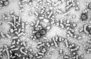
Electron micrograph of hepatitis B virus
SpecialtyInfectious disease, gastroenterology
SymptomsNone, yellowish skin, tiredness, dark urine, abdominal pain
ComplicationsCirrhosis, liver cancer
Usual onsetSymptoms may take up to 6 months to appear
DurationShort or long term
CausesHepatitis B virus spread by some body fluids
Risk factorsIntravenous drug use, sexual intercourse, dialysis, living with an infected person
Diagnostic methodBlood tests
PreventionHepatitis B vaccine
TreatmentAntiviral medication (tenofovir, interferon), liver transplantation
Frequency356 million (2015)
Deaths65,400 direct (2015), >750,000 (total)

Hepatitis B is an infectious disease caused by the hepatitis B virus (HBV) that affects the liver. It can cause both acute and chronic infections. Many people have no symptoms during the initial infection. Some develop a rapid onset of sickness with vomiting, yellowish skin, tiredness, dark urine and abdominal pain. Often these symptoms last a few weeks and rarely does the initial infection result in death. It may take 30 to 180 days for symptoms to begin. In those who get infected around the time of birth 90% develop chronic hepatitis B while less than 10% of those infected after the age of five do. Most of those with chronic disease have no symptoms; however, cirrhosis and liver cancer may eventually develop. Cirrhosis or liver cancer occur in about 25% of those with chronic disease.

The virus is transmitted by exposure to infectious blood or body fluids. Infection around the time of birth or from contact with other people's blood during childhood is the most frequent method by which hepatitis B is acquired in areas where the disease is common. In areas where the disease is rare, intravenous drug use and sexual intercourse are the most frequent routes of infection.[1] Other risk factors include working in healthcare, blood transfusions, dialysis, living with an infected person, travel in countries where the infection rate is high, and living in an institution. Tattooing and acupuncture led to a significant number of cases in the 1980s; however, this has become less common with improved sterility. The hepatitis B viruses cannot be spread by holding hands, sharing eating utensils, kissing, hugging, coughing, sneezing, or breastfeeding. The infection can be diagnosed 30 to 60 days after exposure. The diagnosis is usually confirmed by testing the blood for parts of the virus and for antibodies against the virus. It is one of five main hepatitis viruses: A, B, C, D, and E.

The infection has been preventable by vaccination since 1982. Vaccination is recommended by the World Health Organization in the first day of life if possible. Two or three more doses are required at a later time for full effect. This vaccine works about 95% of the time. About 180 countries gave the vaccine as part of national programs as of 2006. It is also recommended that all blood be tested for hepatitis B before transfusion, and that condoms be used to prevent infection. During an initial infection, care is based on the symptoms that a person has. In those who develop chronic disease, antiviral medication such as tenofovir or interferon may be useful; however, these drugs are expensive. Liver transplantation is sometimes used for cirrhosis.

About a third of the world population has been infected at one point in their lives, including 343 million who have chronic infections. Another 129 million new infections occurred in 2013. Over 750,000 people die of hepatitis B each year. About 300,000 of these are due to liver cancer. The disease is now only common in East Asia and sub-Saharan Africa where between 5 and 10% of adults are chronically infected. Rates in Europe and North America are less than 1%. It was originally known as "serum hepatitis". Research is looking to create foods that contain HBV vaccine. The disease may affect other great apes as well.

Signs and symptoms

Acute infection with hepatitis B virus is associated with acute viral hepatitis, an illness that begins with general ill-health, loss of appetite, nausea, vomiting, body aches, mild fever, and dark urine, and then progresses to development of jaundice. It has been noted that itchy skin has been an indication as a possible symptom of all hepatitis virus types. The illness lasts for a few weeks and then gradually improves in most affected people. A few people may have a more severe form of liver disease known as fulminant hepatic failure and may die as a result. The infection may be entirely asymptomatic and may go unrecognized.

Chronic infection with hepatitis B virus either may be asymptomatic or may be associated with a chronic inflammation of the liver (chronic hepatitis), leading to cirrhosis over a period of several years. This type of infection dramatically increases the incidence of hepatocellular carcinoma (HCC; liver cancer). Across Europe, hepatitis B and C cause approximately 50% of hepatocellular carcinomas. Chronic carriers are encouraged to avoid consuming alcohol as it increases their risk for cirrhosis and liver cancer. Hepatitis B virus has been linked to the development of membranous glomerulonephritis (MGN).

Symptoms outside of the liver are present in 1–10% of HBV-infected people and include serum-sickness–like syndrome, acute necrotizing vasculitis (polyarteritis nodosa), membranous glomerulonephritis, and papular acrodermatitis of childhood (Gianotti–Crosti syndrome). The serum-sickness–like syndrome occurs in the setting of acute hepatitis B, often preceding the onset of jaundice. The clinical features are fever, skin rash, and polyarteritis. The symptoms often subside shortly after the onset of jaundice but can persist throughout the duration of acute hepatitis B. About 30–50% of people with acute necrotizing vasculitis (polyarteritis nodosa) are HBV carriers. HBV-associated nephropathy has been described in adults but is more common in children. Membranous glomerulonephritis is the most common form. Other immune-mediated hematological disorders, such as essential mixed cryoglobulinemia and aplastic anemia have been described as part of the extrahepatic manifestations of HBV infection, but their association is not as well-defined; therefore, they probably should not be considered etiologically linked to HBV.

Cause

Transmission

Transmission of hepatitis B virus results from exposure to infectious blood or body fluids containing blood. It is 50 to 100 times more infectious than human immunodeficiency virus (HIV). Possible forms of transmission include sexual contact, blood transfusions and transfusion with other human blood products, re-use of contaminated needles and syringes, and vertical transmission from mother to child (MTCT) during childbirth. Without intervention, a mother who is positive for HBsAg has a 20% risk of passing the infection to her offspring at the time of birth. This risk is as high as 90% if the mother is also positive for HBeAg. HBV can be transmitted between family members within households, possibly by contact of nonintact skin or mucous membrane with secretions or saliva containing HBV. However, at least 30% of reported hepatitis B among adults cannot be associated with an identifiable risk factor. Breastfeeding after proper immunoprophylaxis does not appear to contribute to mother-to-child-transmission (MTCT) of HBV. The virus may be detected within 30 to 60 days after infection and can persist and develop into chronic hepatitis B. The incubation period of the hepatitis B virus is 75 days on average but can vary from 30 to 180 days.

Virology

Structure

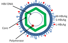
The structure of hepatitis B virus
 
Hepatitis B virus (HBV) is a member of the hepadnavirus family. The virus particle (virion) consists of an outer lipid envelope and an icosahedral nucleocapsid core composed of protein. These virions are 30–42 nm in diameter. The nucleocapsid encloses the viral DNA and a DNA polymerase that has reverse transcriptase activity. The outer envelope contains embedded proteins that are involved in viral binding of, and entry into, susceptible cells. The virus is one of the smallest enveloped animal viruses. The 42 nm virions, which are capable of infecting liver cells known as hepatocytes, are referred to as "Dane particles". In addition to the Dane particles, filamentous and spherical bodies lacking a core can be found in the serum of infected individuals. These particles are not infectious and are composed of the lipid and protein that forms part of the surface of the virion, which is called the surface antigens (HBsAg), and is produced in excess during the life cycle of the virus.

Genome

The genome organisation of HBV. The genes overlap.
 
The genome of HBV is made of circular DNA, but it is unusual because the DNA is not fully double-stranded. One end of the full length strand is linked to the viral DNA polymerase. The genome is 3020–3320 nucleotides long (for the full-length strand) and 1700–2800 nucleotides long (for the short length-strand). The negative-sense (non-coding) is complementary to the viral mRNA. The viral DNA is found in the nucleus soon after infection of the cell. The partially double-stranded DNA is rendered fully double-stranded by completion of the (+) sense strand and removal of a protein molecule from the (−) sense strand and a short sequence of RNA from the (+) sense strand. Non-coding bases are removed from the ends of the (−) sense strand and the ends are rejoined. There are four known genes encoded by the genome, called C, X, P, and S. The core protein is coded for by gene C (HBcAg), and its start codon is preceded by an upstream in-frame AUG start codon from which the pre-core protein is produced. HBeAg is produced by proteolytic processing of the pre-core protein. In some rare strains of the virus known as Hepatitis B virus precore mutants, no HBeAg is present. The DNA polymerase is encoded by gene P. Gene S is the gene that codes for the surface antigen (HBsAg). The HBsAg gene is one long open reading frame but contains three in frame "start" (ATG) codons that divide the gene into three sections, pre-S1, pre-S2, and S. Because of the multiple start codons, polypeptides of three different sizes called large (the order from surface to the inside: pre-S1, pre-S2, and S ), middle (pre-S2, S), and small (S)  are produced. The function of the protein coded for by gene X is not fully understood but it is associated with the development of liver cancer. It stimulates genes that promote cell growth and inactivates growth regulating molecules.

Pathogenesis

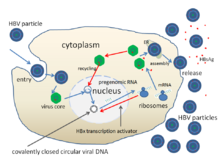
Hepatitis B virus replication
 
The life cycle of hepatitis B virus is complex. Hepatitis B is one of a few known pararetroviruses: non-retroviruses that still use reverse transcription in their replication process. The virus gains entry into the cell by binding to NTCP  on the surface and being endocytosed. Because the virus multiplies via RNA made by a host enzyme, the viral genomic DNA has to be transferred to the cell nucleus by host proteins called chaperones. The partially double-stranded viral DNA is then made fully double stranded by a viral polymerase and transformed into covalently closed circular DNA (cccDNA). This cccDNA serves as a template for transcription of four viral mRNAs by host RNA polymerase. The largest mRNA, (which is longer than the viral genome), is used to make the new copies of the genome and to make the capsid core protein and the viral DNA polymerase. These four viral transcripts undergo additional processing and go on to form progeny virions that are released from the cell or returned to the nucleus and re-cycled to produce even more copies. The long mRNA is then transported back to the cytoplasm where the virion P protein (the DNA polymerase) synthesizes DNA via its reverse transcriptase activity.

Serotypes and genotypes

The virus is divided into four major serotypes (adr, adw, ayr, ayw) based on antigenic epitopes presented on its envelope proteins, and into eight major genotypes (A–H). The genotypes have a distinct geographical distribution and are used in tracing the evolution and transmission of the virus. Differences between genotypes affect the disease severity, course and likelihood of complications, and response to treatment and possibly vaccination. There are two other genotypes I and J but they are not universally accepted as of 2015.

Genotypes differ by at least 8% of their sequence and were first reported in 1988 when six were initially described (A–F). Two further types have since been described (G and H). Most genotypes are now divided into subgenotypes with distinct properties.

Mechanisms

Hepatitis B virus primarily interferes with the functions of the liver by replicating in hepatocytes. A functional receptor is NTCP. There is evidence that the receptor in the closely related duck hepatitis B virus is carboxypeptidase D. The virions bind to the host cell via the preS domain of the viral surface antigen and are subsequently internalized by endocytosis. HBV-preS-specific receptors are expressed primarily on hepatocytes; however, viral DNA and proteins have also been detected in extrahepatic sites, suggesting that cellular receptors for HBV may also exist on extrahepatic cells.

During HBV infection, the host immune response causes both hepatocellular damage and viral clearance. Although the innate immune response does not play a significant role in these processes, the adaptive immune response, in particular virus-specific cytotoxic T lymphocytes(CTLs), contributes to most of the liver injury associated with HBV infection. CTLs eliminate HBV infection by killing infected cells and producing antiviral cytokines, which are then used to purge HBV from viable hepatocytes. Although liver damage is initiated and mediated by the CTLs, antigen-nonspecific inflammatory cells can worsen CTL-induced immunopathology, and platelets activated at the site of infection may facilitate the accumulation of CTLs in the liver.

Diagnosis

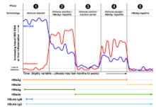
Hepatitis B viral antigens and antibodies detectable in the blood following acute infection
 
Hepatitis B viral antigens and antibodies detectable in the blood of a chronically infected person
 
The tests, called assays, for detection of hepatitis B virus infection involve serum or blood tests that detect either viral antigens (proteins produced by the virus) or antibodies produced by the host. Interpretation of these assays is complex.

The hepatitis B surface antigen (HBsAg) is most frequently used to screen for the presence of this infection. It is the first detectable viral antigen to appear during infection. However, early in an infection, this antigen may not be present and it may be undetectable later in the infection as it is being cleared by the host. The infectious virion contains an inner "core particle" enclosing viral genome. The icosahedral core particle is made of 180 or 240 copies of the core protein, alternatively known as hepatitis B core antigen, or HBcAg. During this 'window' in which the host remains infected but is successfully clearing the virus, IgM antibodies specific to the hepatitis B core antigen (anti-HBc IgM) may be the only serological evidence of disease. Therefore, most hepatitis B diagnostic panels contain HBsAg and total anti-HBc (both IgM and IgG).

Shortly after the appearance of the HBsAg, another antigen called hepatitis B e antigen (HBeAg) will appear. Traditionally, the presence of HBeAg in a host's serum is associated with much higher rates of viral replication and enhanced infectivity; however, variants of the hepatitis B virus do not produce the 'e' antigen, so this rule does not always hold true. During the natural course of an infection, the HBeAg may be cleared, and antibodies to the 'e' antigen (anti-HBe) will arise immediately afterwards. This conversion is usually associated with a dramatic decline in viral replication. 

Ground glass hepatocytes as seen in a chronic hepatitis B liver biopsy. H&E stain
 
If the host is able to clear the infection, eventually the HBsAg will become undetectable and will be followed by IgG antibodies to the hepatitis B surface antigen and core antigen (anti-HBs and anti HBc IgG). The time between the removal of the HBsAg and the appearance of anti-HBs is called the window period. A person negative for HBsAg but positive for anti-HBs either has cleared an infection or has been vaccinated previously. 

Individuals who remain HBsAg positive for at least six months are considered to be hepatitis B carriers. Carriers of the virus may have chronic hepatitis B, which would be reflected by elevated serum alanine aminotransferase (ALT) levels and inflammation of the liver, if they are in the immune clearance phase of chronic infection. Carriers who have seroconverted to HBeAg negative status, in particular those who acquired the infection as adults, have very little viral multiplication and hence may be at little risk of long-term complications or of transmitting infection to others. However, it is possible for individuals to enter an "immune escape" with HBeAg-negative hepatitis. 

The five phases of chronic hepatitis B infection as defined by European Association for the Study of the Liver
 
PCR tests have been developed to detect and measure the amount of HBV DNA, called the viral load, in clinical specimens. These tests are used to assess a person's infection status and to monitor treatment. Individuals with high viral loads, characteristically have ground glass hepatocytes on biopsy.

Prevention

Vaccine

Vaccines for the prevention of hepatitis B have been routinely recommended for babies since 1991 in the United States. The first dose is generally recommended within a day of birth.

Most vaccines are given in three doses over a course of months. A protective response to the vaccine is defined as an anti-HBs antibody concentration of at least 10 mIU/ml in the recipient's serum. The vaccine is more effective in children and 95 percent of those vaccinated have protective levels of antibody. This drops to around 90% at 40 years of age and to around 75 percent in those over 60 years. The protection afforded by vaccination is long lasting even after antibody levels fall below 10 mIU/ml. For newborns of HBsAg-positive mothers: hepatitis B vaccine alone, hepatitis B immunoglobulin alone, or the combination of vaccine plus hepatitis B immunoglobulin, all prevent hepatitis B occurrence. Furthermore, the combination of vaccine plus hepatitis B immunoglobulin is superior to vaccine alone. This combination prevents HBV transmission around the time of birth in 86% to 99% of cases.

Tenofovir given in the second or third trimester can reduce the risk of mother to child transmission by 77% when combined with hepatitis B immunoglobulin and the hepatitis B vaccine, especially for pregnant women with high hepatitis B virus DNA levels. However, there is no sufficient evidence that the administration of hepatitis B immunoglobulin alone during pregnancy, might reduce transmission rates to the newborn infant. No randomized control trial has been conducted to assess the effects of hepatitis B vaccine during pregnancy for preventing infant infection.

All those with a risk of exposure to body fluids such as blood should be vaccinated, if not already. Testing to verify effective immunization is recommended and further doses of vaccine are given to those who are not sufficiently immunized.

In 10- to 22-year follow-up studies there were no cases of hepatitis B among those with a normal immune system who were vaccinated. Only rare chronic infections have been documented. Vaccination is particularly recommended for high risk groups including: health workers, people with chronic renal failure, and men who have sex with men.

Both types of the hepatitis B vaccine, the plasma-derived vaccine (PDV) and recombinant vaccine (RV) are of similar effectiveness in preventing the infection in both healthcare workers and chronic renal failure groups. With one difference noticed among health worker group, that the RV intramuscular route is significantly more effective compared with RV intradermal route of administration.

Other

In assisted reproductive technology, sperm washing is not necessary for males with hepatitis B to prevent transmission, unless the female partner has not been effectively vaccinated. In females with hepatitis B, the risk of transmission from mother to child with IVF is no different from the risk in spontaneous conception.

Those at high risk of infection should be tested as there is effective treatment for those who have the disease. Groups that screening is recommended for include those who have not been vaccinated and one of the following: people from areas of the world where hepatitis B occurs in more than 2%, those with HIV, intravenous drug users, men who have sex with men, and those who live with someone with hepatitis B.

Treatment

Acute hepatitis B infection does not usually require treatment and most adults clear the infection spontaneously. Early antiviral treatment may be required in fewer than 1% of people, whose infection takes a very aggressive course (fulminant hepatitis) or who are immunocompromised. On the other hand, treatment of chronic infection may be necessary to reduce the risk of cirrhosis and liver cancer. Chronically infected individuals with persistently elevated serum alanine aminotransferase, a marker of liver damage, and HBV DNA levels are candidates for therapy. Treatment lasts from six months to a year, depending on medication and genotype. Treatment duration when medication is taken by mouth, however, is more variable and usually longer than one year.

Although none of the available medications can clear the infection, they can stop the virus from replicating, thus minimizing liver damage. As of 2018, there are eight medications licensed for the treatment of hepatitis B infection in the United States. These include antiviral medications lamivudine, adefovir, tenofovir disoproxil, tenofovir alafenamide, telbivudine, and entecavir, and the two immune system modulators interferon alpha-2a and PEGylated interferon alpha-2a. In 2015 the World Health Organization recommended tenofovir or entecavir as first-line agents. Those with current cirrhosis are in most need of treatment.

The use of interferon, which requires injections daily or thrice weekly, has been supplanted by long-acting PEGylated interferon, which is injected only once weekly. However, some individuals are much more likely to respond than others, and this might be because of the genotype of the infecting virus or the person's heredity. The treatment reduces viral replication in the liver, thereby reducing the viral load (the amount of virus particles as measured in the blood). Response to treatment differs between the genotypes. Interferon treatment may produce an e antigen seroconversion rate of 37% in genotype A but only a 6% seroconversion in type D. Genotype B has similar seroconversion rates to type A while type C seroconverts only in 15% of cases. Sustained e antigen loss after treatment is ~45% in types A and B but only 25–30% in types C and D.

Prognosis

Estimate of disability-adjusted life year for hepatitis B per 100,000 inhabitants as of 2004
 

Hepatitis B virus infection may be either acute (self-limiting) or chronic (long-standing). Persons with self-limiting infection clear the infection spontaneously within weeks to months. 

Children are less likely than adults to clear the infection. More than 95% of people who become infected as adults or older children will stage a full recovery and develop protective immunity to the virus. However, this drops to 30% for younger children, and only 5% of newborns that acquire the infection from their mother at birth will clear the infection. This population has a 40% lifetime risk of death from cirrhosis or hepatocellular carcinoma. Of those infected between the age of one to six, 70% will clear the infection.

Hepatitis D (HDV) can occur only with a concomitant hepatitis B infection, because HDV uses the HBV surface antigen to form a capsid. Co-infection with hepatitis D increases the risk of liver cirrhosis and liver cancer. Polyarteritis nodosa is more common in people with hepatitis B infection.

Cirrhosis

A number of different tests are available to determine the degree of cirrhosis present. Transient elastography (FibroScan) is the test of choice, but it is expensive. Aspartate aminotransferase to platelet ratio index may be used when cost is an issue.

Reactivation

Hepatitis B virus DNA persists in the body after infection, and in some people the disease recurs. Although rare, reactivation is seen most often following alcohol or drug use, or in people with impaired immunity. HBV goes through cycles of replication and non-replication. Approximately 50% of overt carriers experience acute reactivation. Males with baseline ALT of 200 UL/L are three times more likely to develop a reactivation than people with lower levels. Although reactivation can occur spontaneously, people who undergo chemotherapy have a higher risk. Immunosuppressive drugs favor increased HBV replication while inhibiting cytotoxic T cell function in the liver. The risk of reactivation varies depending on the serological profile; those with detectable HBsAg in their blood are at the greatest risk, but those with only antibodies to the core antigen are also at risk. The presence of antibodies to the surface antigen, which are considered to be a marker of immunity, does not preclude reactivation. Treatment with prophylactic antiviral drugs can prevent the serious morbidity associated with HBV disease reactivation.

Epidemiology

Prevalence of hepatitis B virus as of 2005
 
In 2004, an estimated 350 million individuals were infected worldwide. National and regional prevalences range from over 10% in Asia to under 0.5% in the United States and Northern Europe. 

Routes of infection include vertical transmission (such as through childbirth), early life horizontal transmission (bites, lesions, and sanitary habits), and adult horizontal transmission (sexual contact, intravenous drug use).

The primary method of transmission reflects the prevalence of chronic HBV infection in a given area. In low prevalence areas such as the continental United States and Western Europe, injection drug abuse and unprotected sex are the primary methods, although other factors may also be important. In moderate prevalence areas, which include Eastern Europe, Russia, and Japan, where 2–7% of the population is chronically infected, the disease is predominantly spread among children. In high-prevalence areas such as China and South East Asia, transmission during childbirth is most common, although in other areas of high endemicity such as Africa, transmission during childhood is a significant factor. The prevalence of chronic HBV infection in areas of high endemicity is at least 8% with 10–15% prevalence in Africa/Far East. As of 2010, China has 120 million infected people, followed by India and Indonesia with 40 million and 12 million, respectively. According to World Health Organization (WHO), an estimated 600,000 people die every year related to the infection.

In the United States about 19,000 new cases occurred in 2011 down nearly 90% from 1990.

History

The hepatitis B virus has infected humans since at least the Bronze Age. The evidence was obtained from 4,500-year-old human remains. According to the 2018 study, the viral genomes obtained by shotgun sequencing became the oldest ever recovered from vertebrate samples. It was also found that some ancient hepatitis viral strains still infect humans, while other became extinct. This disproved the belief that hepatitis B originated in the New World and spread to Europe around 16th century. Another 2018 study of the remains of a mummified child found in the Basilica of San Domenico Maggiore in Naples concluded that the child, who had lived in the 16th century, had a form of HBV, and that the virus was closely related to modern variants.

The earliest record of an epidemic caused by hepatitis B virus was made by Lurman in 1885. An outbreak of smallpox occurred in Bremen in 1883 and 1,289 shipyard employees were vaccinated with lymph from other people. After several weeks, and up to eight months later, 191 of the vaccinated workers became ill with jaundice and were diagnosed as suffering from serum hepatitis. Other employees who had been inoculated with different batches of lymph remained healthy. Lurman's paper, now regarded as a classical example of an epidemiological study, proved that contaminated lymph was the source of the outbreak. Later, numerous similar outbreaks were reported following the introduction, in 1909, of hypodermic needles that were used, and, more importantly, reused, for administering Salvarsan for the treatment of syphilis. The virus was not discovered until 1966 when Baruch Blumberg, then working at the National Institutes of Health (NIH), discovered the Australia antigen (later known to be hepatitis B surface antigen, or HBsAg) in the blood of Aboriginal Australian people. Although a virus had been suspected since the research published by Frederick MacCallum in 1947, David Dane and others discovered the virus particle in 1970 by electron microscopy. By the early 1980s the genome of the virus had been sequenced, and the first vaccines were being tested.

Society and culture

World Hepatitis Day, observed July 28, aims to raise global awareness of hepatitis B and hepatitis C and encourage prevention, diagnosis, and treatment. It has been led by the World Hepatitis Alliance since 2007 and in May 2010, it received global endorsement from the World Health Organization.

Inequality (mathematics)

From Wikipedia, the free encyclopedia https://en.wikipedia.org/wiki/Inequality...