Search This Blog

Wednesday, December 11, 2019

Tau protein

From Wikipedia, the free encyclopedia
 
MAPT
PDB 1i8h EBI.jpg
Available structures
PDBOrtholog search: PDBe RCSB
Identifiers
AliasesMAPT, DDPAC, FTDP-17, MAPTL, MSTD, MTBT1, MTBT2, PPND, PPP1R103, TAU, microtubule associated protein tau, Tau proteins
External IDsOMIM: 157140 MGI: 97180 HomoloGene: 74962 GeneCards: MAPT
Gene location (Human)
Chromosome 17 (human)
Chr.Chromosome 17 (human)
Chromosome 17 (human)
Genomic location for MAPT
Band17q21.31Start45,894,382 bp
End46,028,334 bp
RNA expression pattern
PBB GE MAPT 203928 x at fs.png

PBB GE MAPT 203929 s at fs.png

PBB GE MAPT 203930 s at fs.png
More reference expression data
Orthologs
SpeciesHumanMouse
Entrez


Ensembl


UniProt


RefSeq (mRNA)
NM_001038609
NM_010838
NM_001285454
NM_001285455
NM_001285456
RefSeq (protein)
NP_001033698
NP_001272383
NP_001272384
NP_001272385
NP_034968
Location (UCSC)Chr 17: 45.89 – 46.03 MbChr 11: 104.23 – 104.33 Mb

Tau proteins (or τ proteins, after the Greek letter with that name) are proteins that stabilize microtubules. They are abundant in neurons of the central nervous system and are less common elsewhere, but are also expressed at very low levels in CNS astrocytes and oligodendrocytes. Pathologies and dementias of the nervous system such as Alzheimer's disease and Parkinson's disease are associated with tau proteins that have become defective and no longer stabilize microtubules properly.

The tau proteins are the product of alternative splicing from a single gene that in humans is designated MAPT (microtubule-associated protein tau) and is located on chromosome 17.

The tau proteins were identified in 1975 as heat-stable proteins essential for microtubule assembly  and since then, they have been characterized as intrinsically disordered proteins.

Neurons were grown in tissue culture and stained with antibody to MAP2 protein in green and MAP tau in red using the immunofluorescence technique. MAP2 is found only in dendrites and perikarya, while tau is found not only in the dendrites and perikarya but also in axons. As a result, axons appear red while the dendrites and perikarya appear yellow, due to superimposition of the red and green signals. DNA is shown in blue using the DAPI stain which highlights the nuclei.

Function

Tau protein is a highly soluble microtubule-associated protein tau (MAPT). In humans, these proteins are found mostly in neurons compared to non-neuronal cells. One of tau's main functions is to modulate the stability of axonal microtubules. Other nervous system MAPs may perform similar functions, as suggested by tau knock out mice that did not show abnormalities in brain development - possibly because of compensation in tau deficiency by other MAPs. Tau is not present in dendrites and is active primarily in the distal portions of axons where it provides microtubule stabilization but also flexibility as needed. This contrasts with MAP6 (STOP) proteins in the proximal portions of axons, which, in essence, lock down the microtubules and MAP2 that stabilizes microtubules in dendrites. In addition to their microtubule stabilizing functions, MAPTs have also been found to recruit signaling proteins and regulation of microtubule-mediated transport.

Tau proteins interact with tubulin to stabilize microtubules and promote tubulin assembly into microtubules. Tau has two ways of controlling microtubule stability: isoforms and phosphorylation.

Tau regulates cytoskeletal stability and translation (negatively) in Drosophila as well, but also facilitates habituation (a form of non-associative learning) and negatively regulates long-term memory, two higher and more integrated neural functions.

Genetics

In humans, the MAPT gene for encoding tau protein is located on chromosome 17q21, containing 16 exons. The major tau protein in the human brain is encoded by 11 exons. Exons 2, 3 and 10 are alternatively spliced that lead to formation of six tau isoforms. In human brain, tau proteins constitute a family of six isoforms with a range of 352-441 amino acids. Tau isoforms are different in either zero, one, or two inserts of 29 amino acids at the N-terminal part (exon 2 and 3), and three or four repeat-regions at the C-terminal part (exon 10). Thus, the longest isoform in the CNS has four repeats (R1, R2, R3 and R4) and two inserts (441 amino acids total), while the shortest isoform has three repeats (R1, R3 and R4) and no insert (352 amino acids total). 

The MAPT gene has two haplogroups, H1 and H2, in which the gene appears in inverted orientations. Haplogroup H2 is common only in Europe and in people with European ancestry. Haplogroup H1 appears to be associated with increased probability of certain dementias, such as Alzheimer's disease. The presence of both haplogroups in Europe means that recombination between inverted haplotypes can result in the lack of one of the functioning copies of the gene, resulting in congenital defects.

Structure

Six tau isoforms exist in human brain tissue, and they are distinguished by their number of binding domains. Three isoforms have three binding domains and the other three have four binding domains. The binding domains are located in the carboxy-terminus of the protein and are positively charged (allowing it to bind to the negatively charged microtubule). The isoforms with four binding domains are better at stabilizing microtubules than those with three binding domains. The isoforms are a result of alternative splicing in exons 2, 3, and 10 of the tau gene. Tau is a phosphoprotein with 79 potential Serine (Ser) and Threonine (Thr) phosphorylation sites on the longest tau isoform. Phosphorylation has been reported on approximately 30 of these sites in normal tau proteins.

Phosphorylation of tau is regulated by a host of kinases, including PKN, a serine/threonine kinase. When PKN is activated, it phosphorylates tau, resulting in disruption of microtubule organization. Phosphorylation of tau is also developmentally regulated. For example, fetal tau is more highly phosphorylated in the embryonic CNS than adult tau. The degree of phosphorylation in all six isoforms decreases with age due to the activation of phosphatases. Like kinases, phosphatases too play a role in regulating the phosphorylation of tau. For example, PP2A and PP2B are both present in human brain tissue and have the ability to dephosphorylate Ser396. The binding of these phosphatases to tau affects tau's association with MTs. 

Mechanism

The accumulation of hyperphosphorylated tau in neurons leads to the neurofibrillary degeneration. The actual mechanism of how tau propagates from one cell to another is not well identified. Also, other mechanisms, including tau release and toxicity, are unclear. As tau aggregates, it replaces tubulin, which in turn enhances fibrilization of tau. Several propagation methods have been proposed which occur by synaptic contact such as synaptic cell adhesion proteins and neuronal activity and other synaptic and non-synaptic mechanisms. The mechanism of tau aggregation is still not completely elucidated, but several factors favor this process, including tau phosphorylation and zinc ions.

Release

Tau involves in uptake and release process, which is known as seeding. Uptake of tau protein mechanism requires the presence of heparan sulfate proteoglycans at the cell surface, which happen by macropinocytosis. On the other hand, tau release depends on neuronal activity. Many factors influence tau release, for example, type of isoforms or MAPT mutations that change the extracellular level of tau. According to Asai and his colleagues, spreading of tau protein occurs from entorhinal cortex to the hippocampal region in the early stages of the disease. They also suggested that microglia were also involved in the transport process and their actual role is still unknown.

Toxicity

Tau causes toxic effects through its accumulation inside cells. Many enzymes are involved in toxicity mechanism such as PAR-1 kinase. This enzyme stimulates phosphorylation of serine 262 and 356, which in turn leads to activate other kinases (GSK-3 and Cdk5) that cause disease-associated phosphoepitopes. The degree of toxicity is affected by different factors, such as the degree of microtubule binding. Toxicity could also happen by neurofibrillary tangles (NFTs), which leads to cell death and cognitive decline.

Clinical significance

Hyperphosphorylation of the tau protein (tau inclusions, pTau) can result in the self-assembly of tangles of paired helical filaments and straight filaments, which are involved in the pathogenesis of Alzheimer's disease, frontotemporal dementia, and other tauopathies.

All of the six tau isoforms are present in an often hyperphosphorylated state in paired helical filaments from Alzheimer's disease brain. In other neurodegenerative diseases, the deposition of aggregates enriched in certain tau isoforms has been reported. When misfolded, this otherwise very soluble protein can form extremely insoluble aggregates that contribute to a number of neurodegenerative diseases. 

Tau protein has a direct effect on the breakdown of a living cell caused by tangles that form and block nerve synapses. Tangles are clumps of tau protein that stick together and block essential nutrients that need to be distributed to cells in the brain, causing the cells to die.

Gender-specific tau gene expression across different regions of the human brain has recently been implicated in gender differences in the manifestations and risk for tauopathies.

Some aspects of how the disease functions also suggest that it has some similarities to prion proteins.

Traumatic brain injury

Repetitive mild traumatic brain injury (TBI) is now recognized as a central component of brain injury in contact sports, especially American football, and the concussive force of military blasts. It can lead to chronic traumatic encephalopathy (CTE) that is characterized by fibrillar tangles of hyperphosphorylated tau.

High levels of tau protein in fluid bathing the brain are linked to poor recovery after head trauma.

Tau hypothesis of Alzheimer's disease

The tau hypothesis states that excessive or abnormal phosphorylation of tau results in the transformation of normal adult tau into paired helical filament (PHF) tau and neurofibrillary tangles (NFTs). The stage of the disease determines NFTs' phosphorylation. In AD, at least 19 amino acids are phosphorylated, such as pre-NFT phosphorylation occurs at serine 119, 202, and 409. While intra-NFT phosphorylation happens at serine 396 and threonine 231. Tau protein is a highly soluble microtubule-associated protein tau (MAPT). Through its isoforms and phosphorylation, tau protein interacts with tubulin to stabilize microtubule assembly. All of the six tau isoforms are present in an often hyperphosphorylated state in paired helical filaments (PHFs) from AD.

Tau mutations have many consequences such as changing the expression level of tau isoforms or lead to MTs dysfunction. Mutations that alter function and isoform expression of tau lead to hyperphosphorylation. The process of tau aggregation in the absence of mutations is not known, but might result from increased phosphorylation, protease action, or exposure to polyanions, such as glycosaminoglycans. Hyperphosphorylated tau disassembles microtubules and sequesters normal tau, MAPT 1 (microtubule associated protein tau 1), MAPT 2, and ubiquitin into tangles of PHFs. This insoluble structure damages cytoplasmic functions and interferes with axonal transport, which can lead to cell death. Hyperphosphorylated forms of tau protein are the main component of PHFs of NFTs in the brain of AD patients. It has been well demonstrated that regions of tau six-residue segments, namely PHF6 (VQIVYK) and PHF6 (VQIINK), can form tau PHF aggregation in AD. Apart from the PHF6, some other residue sites like Ser285, Ser289, Ser293, Ser305, and Tyr310, located near the C-terminal of the PHF6 sequences, play key roles in the phosphorylation of tau.

A68 Protein

The A68 protein is a hyperphosphorylated Tau protein differing in its sensitivity and its Kinase as well as Alkaline phosphatase and is along with beta-amyloid a component of pathologic lesions in Alzheimer disease, and is found in the brains of individuals with Alzheimer's disease.

Interactions

Tau protein has been shown to interact with proto-oncogene tyrosine-protein kinase:

Alcohol-related dementia

From Wikipedia, the free encyclopedia
https://en.wikipedia.org/wiki/Alcohol-related_dementia

Alcohol-related dementia (ARD) is a form of dementia caused by long-term, excessive consumption of alcoholic beverages, resulting in neurological damage and impaired cognitive function.

Terminology

Alcohol-related dementia is a broad term currently preferred among medical professionals. Many experts use the terms alcohol (or alcoholic) dementia to describe a specific form of ARD, characterized by impaired executive function (planning, thinking, and judgment). Another form of ARD is known as wet brain (Wernicke-Korsakoff syndrome), characterized by short term memory loss and thiamine (vitamin B1) deficiency. ARD patients often have symptoms of both forms, i.e. impaired ability to plan, apathy, and memory loss. ARD may occur with other forms of dementia (mixed dementia). The diagnosis of ARD is widely recognized but rarely applied, due to a lack of specific diagnostic criteria.

On many non-medical websites, the terms wet brain and alcohol-related dementia are often used interchangeably, creating significant confusion. Additionally, the term alcohol-induced persistent dementia is another nonspecific name that is sometimes used.

Signs and symptoms

Alcohol-related dementia presents as a global deterioration in intellectual function with memory not being specifically affected, but it may occur with other forms of dementia, resulting in a wide range of symptoms. Certain individuals with alcohol-related dementia present with damage to the frontal lobes of their brain causing disinhibition, loss of planning and executive functions, and a disregard for the consequences of their behavior. Other types of alcohol-related dementia such as Korsakoff's Syndrome cause the destruction of certain areas of the brain, where changes in memory, primarily a loss of short-term memory, are the main symptom. Most presentations of alcohol dementia are somewhere along the spectrum between a global dementia and Korsakoff's psychosis, and may include symptoms of both.

Individuals affected by alcohol-related dementia may develop memory problems, language impairment, and an inability to perform complex motor tasks such as getting dressed. Heavy alcohol abuse also damages the nerves in arms and legs, i.e. peripheral neuropathy, as well as the cerebellum that controls coordination thereby leading to the development of cerebellar ataxia. These patients frequently have problems with sensation in their extremities and may demonstrate unsteadiness on their feet.

Alcohol-related dementia can produce a variety of psychiatric problems including psychosis (disconnection from reality), depression, anxiety, and personality changes. Patients with alcoholic dementia often develop apathy, related to frontal lobe damage, that may mimic depression. People with alcoholism are more likely to become depressed than people without alcoholism, and it may be difficult to differentiate between depression and alcohol dementia. 

Pathophysiology

Alcohol has a direct effect on brain cells in the front part of the brain, resulting in poor judgment, difficulty making decisions, and lack of insight. Long-term alcohol abuse can often lead to poor nutrition problems causing parts of the brain to be damaged by vitamin deficiencies. These problems could also cause personality changes in some people.

Diagnosis

The signs and symptoms of alcohol-related dementia are essentially the same as the symptoms present in other types of dementia, making alcohol-related dementia difficult to diagnose. There are very few qualitative differences between alcohol dementia and Alzheimer's disease and it is therefore difficult to distinguish between the two. Some of these warning signs may include memory loss, difficulty performing familiar tasks, poor or impaired judgment and problems with language. However the biggest indicator is friends or family members reporting changes in personality.

A simple test for intellectual function, like the Folstein Mini-Mental Status Examination, is the minimum screen for dementia. The test requires 15–20 minutes to administer and is available in mental health centers.

Diagnosing alcohol-related dementia can be difficult due to the wide range of symptoms and a lack of specific brain pathology. The Diagnostic and Statistical Manual of Mental Disorders (DSM-IV) is a guide to aid doctors in diagnosing a range of psychiatric disorders, and may be helpful in diagnosing dementia.

Diagnostic criteria

The existence of alcohol-related dementia is widely acknowledged but not often used as a diagnosis, due to a lack of widely accepted, non-subjective diagnostic criteria; more research is needed. Criteria for alcohol-induced persistent dementia in the Diagnostic and Statistical Manual of Mental Disorders (DSM-IV) include the following:
A. The development of multiple cognitive deficits manifested by both:
  1. Memory impairment (impaired ability to learn new information or to recall previously learned information)
  2. One (or more) of the following cognitive disturbances:
  • (a) Aphasia (language disturbance)
  • (b) Apraxia (impaired ability to carry out motor activities despite intact motor function)
  • (c) Agnosia (failure to recognize or identify objects despite intact sensory function)
  • (d) Disturbance in executive functioning (i.e. planning, organizing, sequencing, abstracting)
B. The cognitive deficits in criteria A1 and A2 each cause significant impairment in social or occupational functioning and represent a significant decline from a previous level of functioning.
C. The deficits do not occur exclusively during the course of a delirium and persist beyond the usual duration of substance intoxication or withdrawal.
D. There is evidence from the history, physical examination, or laboratory findings that deficits are etiologically related to the persisting effects of substance use (e.g. drug of abuse; medication).
There are problems with DSM diagnostic criteria, however. Firstly, they are vague and subjective. Furthermore, the criteria for diagnosis of dementia were inspired by the clinical presentation of Alzheimer's disease and are poorly adapted to the diagnosis of other dementias. This has led to efforts to develop better diagnostic models.

Oslin (Int J Geriatr Psychiatry 1998) proposed alternative clinical diagnostic criteria which were validated. The criteria include a clinical diagnosis of dementia at least 60 days after last exposure to alcohol, significant alcohol use (i.e. minimum 35 standard drinks/week for males and 28 for women) for more than five years, and significant alcohol use occurring within three years of the initial onset of cognitive deficits. Oslin proposed the new and refined diagnostic criteria for alcohol-related dementia because he hoped that the redefined classification system would bring more awareness and clarity to the relationship between alcohol use and dementia.


Oslin's proposed classification of ARD:
  • Definite alcohol-related dementia
At the current time there are no acceptable criteria to definitively define alcohol-related dementia.
  • Probable alcohol-related dementia
A. The criteria for the clinical diagnosis of probable alcohol-related dementia include the following:
  1. A clinical diagnosis of dementia at least 60 days after the last exposure to alcohol.
  2. Significant alcohol use as defined by a minimum average of 35 standard drinks per week for men (28 for women) for greater than a period of five years. The period of significant alcohol use must occur within three years of the initial onset of dementia.
B. The diagnosis of alcohol-related dementia is supported by the presence of any of the following
  1. Alcohol related hepatic, pancreatic, gastrointestinal, cardiovascular, or renal disease i.e. other end-organ damage.
  2. Ataxia or peripheral sensory polyneuropathy (not attributed to other causes).
  3. Beyond 60 days of abstinence, the cognitive impairment stabilizes or improves.
  4. After 60 days of abstinence, any neuroimaging evidence of ventricular or sulcal dilatation improves.
  5. Neuroimaging evidence of cerebellar atrophy, especially in the vermis.
C. The following clinical features cast doubt on the diagnosis of alcohol-related dementia
  1. The presence of language impairment, especially dysnomia or anomia.
  2. the presence of focal neurologic signs or symptoms (except ataxia or peripheral sensory polyneuropathy).
  3. Neuroimaging evidence for cortical or subcortical infarction, subdural hematoma, or other focal brain pathology.
  4. Elevated Hachinski Ischemia Scale score.
D. Clinical features that are neither supportive nor cast doubt on the diagnosis of alcohol-related dementia included:
  1. Neuroimaging evidence of cortical atrophy.
  2. The presence of periventricular or deep white matter lesions on neuroimaging in the absence of focal infarct(s).
  3. The presence of the Apolipoprotein c4 allele.

Treatment

If the symptoms of alcohol dementia are caught early enough, the effects may be reversed. The person must stop drinking and start on a healthy diet, replacing the lost vitamins, including, but not limited to, thiamine. Recovery is more easily achievable for women than men, but in all cases it is necessary that they have the support of family and friends and abstain from alcohol.

Epidemiology

The onset of alcohol dementia can occur as early as age 30, although it is far more common that the dementia will reveal itself anywhere from age 50 to 70. The onset and the severity of this type of dementia is directly correlated to the amount of alcohol that a person consumes over their lifetime.

Epidemiological studies show an association between long-term alcohol intoxication and dementia. Alcohol can damage the brain directly as a neurotoxin, or it can damage it indirectly by causing malnutrition, primarily a loss of thiamine (vitamin B1). Alcohol abuse is common in older persons, and alcohol-related dementia is under-diagnosed. A discredited French study, looking at other studies of thousands of subjects, found that moderate alcohol consumption (up to four glasses of wine per week) protected against dementia, whereas higher rates of consumption were found to increase the chances of getting it.

Notable sufferers

According to her family, the socialite Leonore Lemmon spent the last few years of her life with alcohol dementia, before dying in 1989. The Australian entertainer and "King of Comedy" Graham Kennedy was suffering from alcohol-related dementia at time of his death in 2005.

Frontotemporal dementia

From Wikipedia, the free encyclopedia
 
Frontotemporal dementia
SpecialtyPsychiatry, neurology
Causesfrontotemporal lobar degeneration

The frontotemporal dementias (FTD) encompass six types of dementia involving the frontal or temporal lobes. They are: behavioral variant of FTD, semantic variant primary progressive aphasia, nonfluent agrammatic variant primary progressive aphasia, corticobasal syndrome, progressive supranuclear palsy, and FTD associated with motor neuron disease.

One variant is the clinical presentation of frontotemporal lobar degeneration, which is characterized by progressive neuronal loss predominantly involving the frontal or temporal lobes, and typical loss of over 70% of spindle neurons, while other neuron types remain intact.

It was first described by Arnold Pick in 1892 and was originally called "Pick's disease", a term now reserved for Pick disease, one specific type of frontotemporal dementia. Second only to Alzheimer's disease (AD) in prevalence, FTD accounts for 20% of young-onset dementia cases. Signs and symptoms typically manifest in late adulthood, more commonly between the ages of 45 and 65, approximately equally affecting men and women.

Common signs and symptoms include significant changes in social and personal behavior, apathy, blunting of emotions, and deficits in both expressive and receptive language. Currently, there is no cure for FTD, but there are treatments that help alleviate symptoms.

Signs and symptoms

Frontotemporal dementia (FTD) classically affects adults in their fifth to sixth decade of life.These patients usually describe a gradual onset and progression of changes in behavior or language deficits for several years prior to presentation to a neurologist.

FTD is traditionally difficult to diagnose due to the heterogeneity of the associated symptoms. Signs and symptoms are classified into three groups based on the functions of the frontal and temporal lobes:
  • Behavioural variant frontotemporal dementia (BvFTD) is characterized by changes in social behavior and conduct, with loss of social awareness and poor impulse control.
  • Semantic dementia (SD) is characterized by the loss of semantic understanding, resulting in impaired word comprehension, although speech remains fluent and grammatically faultless.
  • Progressive nonfluent aphasia (PNFA) is characterized by progressive difficulties in speech production.
However, the following abilities in the person with FTD are preserved:
In later stages of FTD, the clinical phenotypes may overlap. FTD patients tend to struggle with binge eating and compulsive behaviors. These binge eating habits are often associated with abnormal eating behavior including overeating, stuffing oneself with food, changes in food preferences (cravings for more sweets, carbohydrates), eating inedible objects and snatching food from others. Recent findings from structural MRI research have indicated that eating changes in FTD are associated with atrophy (wasting) in the right ventral insula, striatum, and orbitofrontal cortex.

Patients with FTD show marked deficiencies in executive functioning and working memory. Most FTD patients become unable to perform skills that require complex planning or sequencing. In addition to the characteristic cognitive dysfunction, a number of primitive reflexes known as frontal release signs are often able to be elicited. Usually the first of these frontal release signs to appear is the palmomental reflex which appears relatively early in the disease course whereas the palmar grasp reflex and rooting reflex appear late in the disease course.

In rare cases, FTD can occur in patients with motor neuron disease (MND) (typically amyotrophic lateral sclerosis). The prognosis for people with MND is worse when combined with FTD, shortening survival by about a year.

Genetics

A higher proportion of FTD cases seem to have a familial component than more common neurodegenerative diseases like Alzheimer's disease. More and more mutations and genetic variants are being identified all the time, so the lists of genetic influences require consistent updating.
  • Tau-positive frontotemporal dementia with parkinsonism (FTDP-17) is caused by mutations in the MAPT gene on chromosome 17 that encodes the Tau protein It has been determined that there is a direct relationship between the type of tau mutation and the neuropathology of gene mutations. The mutations at the splice junction of exon 10 of tau lead to the selective deposition of the repetitive tau in neurons and glia. The pathological phenotype associated with mutations elsewhere in tau is less predictable with both typical neurofibrillary tangles (consisting of both 3 repeat and 4 repeat tau) and Pick bodies (consisting of 3 repeat tau) having been described. The presence of tau deposits within glia is also variable in families with mutations outside of exon 10. This disease is now informally designated FTDP-17T. FTD shows a linkage to the region of the tau locus on chromosome 17, but it is believed that there are two loci leading to FTD within megabases of each other on chromosome 17.
  • FTD caused by FTLD-TDP43 has numerous genetic causes. Some cases are due to mutations in the GRN gene, also located on chromosome 17. Others are caused by VCP mutations, although these patients present with a complex picture of multisystem proteinopathy that can include amyotrophic lateral sclerosis, inclusion body myopathy, Paget's disease of bone, and FTD. The most recent addition to the list is a hexanucleotide repeat expansion in intron 1 of C9ORF72. Only one or two cases have been reported describing TARDBP (the TDP-43 gene) mutations in a clinically pure FTD (FTD without MND).
  • No genetic causes of FUS pathology in FTD have yet been reported.

Pathology

There are three main histological subtypes found at post-mortem: FTLD-tau, FTLD-TDP, and FTLD-FUS. Dementia lacking distinctive histology (DLDH) is a rare and controversial entity. New analyses has allowed many cases previously described as DLDH to be reclassified into one of the positively defined subgroups. In rare cases, patients with clinical FTD were found to have changes consistent with Alzheimer's disease on autopsy.[13] The most severe brain atrophy appears to be associated with Pick's disease, corticobasal degeneration, and TDP pathology associated with behavioral-variant FTD.

With regard to the genetic defects that have been found, repeat expansion in the C9orf72 gene is considered a major contribution to frontotemporal lobar degeneration, although defects in the GRN and MAPT genes are also associated with it.

Diagnosis

Structural MRI scans often reveal frontal lobe and/or anterior temporal lobe atrophy but in early cases the scan may seem normal. Atrophy can be either bilateral or asymmetric. Registration of images at different points of time (e.g., one year apart) can show evidence of atrophy that otherwise (at individual time points) may be reported as normal. Many research groups have begun using techniques such as magnetic resonance spectroscopy, functional imaging and cortical thickness measurements in an attempt to offer an earlier diagnosis to the FTD patient. Fluorine-18-fluorodeoxyglucose positron emission tomography (FDG-PET) scans classically show frontal and/or anterior temporal hypometabolism, which helps differentiate the disease from Alzheimer's disease. The PET scan in Alzheimer's disease classically shows biparietal hypometabolism. Meta-analyses based on imaging methods have shown that frontotemporal dementia mainly affects a frontomedial network discussed in the context of social cognition or 'theory of mind'. This is entirely in keeping with the notion that on the basis of cognitive neuropsychological evidence, the ventromedial prefrontal cortex is a major locus of dysfunction early on in the course of the behavioural variant of frontotemporal degeneration. The language subtypes of frontotemporal lobar degeneration (semantic dementia and progressive nonfluent aphasia) can be regionally dissociated by imaging approaches in vivo.

The confusion between Alzheimer's and FTD is justifiable due to the similarities between their initial symptoms. Patients do not have difficulty with movement and other motor tasks. As FTD symptoms appear, it is difficult to differentiate between a diagnosis of Alzheimer's disease and FTD. There are distinct differences in the behavioral and emotional symptoms of the two dementias, notably, the blunting of emotions seen in FTD patients. In the early stages of FTD, anxiety and depression are common, which may result in an ambiguous diagnosis. However, over time, these ambiguities fade away as this dementia progresses and defining symptoms of apathy, unique to FTD, start to appear.

Recent studies over several years have developed new criteria for the diagnosis of behavioral variant frontotemporal dementia (bvFTD). Six distinct clinical features have been identified as symptoms of bvFTD.
  1. Disinhibition
  2. Apathy/Inertia
  3. Loss of Sympathy/Empathy
  4. Perseverative/compulsive behaviors
  5. Hyperorality
  6. Dysexecutive neuropsychological profile
Of the six features, three must be present in a patient to diagnose one with possible bvFTD. Similar to standard FTD, the primary diagnosis stems from clinical trials that identify the associated symptoms, instead of imaging studies. The above criteria are used to distinguish bvFTD from disorders such as Alzheimer's and other causes of dementia. In addition, the new criteria allow for a diagnostic hierarchy distinguished possible, probable, and definite bvFTD based on the number of symptoms present. 

Neuropsychological tests

The progression of the degeneration caused by bvFTD may follow a predictable course. The degeneration begins in the orbitofrontal cortex and medial aspects such as ventromedial cortex. In later stages, it gradually expands its area to the dorsolateral cortex and the temporal lobe. Thus, the detection of dysfunction of the orbitofrontal cortex and ventromedial cortex is important in the detection of early stage bvFTD. As stated above, a behavioural change may occur before the appearance of any atrophy in the brain in the course of the disease. Because of that, image scanning such as MRI can be insensitive to the early degeneration and it is difficult to detect early-stage bvFTD.

In neuropsychology, there is an increasing interest in using neuropsychological tests such as the Iowa gambling task or Faux Pas Recognition test as an alternative to imaging for the diagnosis of bvFTD. Both the Iowa gambling task and the Faux Pas test are known to be sensitive to dysfunction of the orbitofrontal cortex.

Faux Pas Recognition test is intended to measure one’s ability to detect faux pas types of social blunders (accidentally make a statement or an action that offends others). It is suggested that people with orbitofrontal cortex dysfunction show a tendency to make social blunders due to a deficit in self-monitoring. Self-monitoring is the ability of individuals to evaluate their behaviour to make sure that their behaviour is appropriate in particular situations. The impairment in self-monitoring leads to a lack of social emotion signals. The social emotions such as embarrassment are important in the way that they signal the individual to adapt social behaviour in an appropriate manner to maintain relationships with others. Though patients with damage to the OFC retain intact knowledge of social norms, they fail to apply it to actual behaviour because they fail to generate social emotions that promote adaptive social behaviour.

The other test, the Iowa gambling task, is a psychological test intended to simulate real-life decision making. The underlying concept of this test is the somatic marker hypothesis. This hypothesis argues that when people have to make complex uncertain decisions, they employ both cognitive and emotional processes to assess the values of the choices available to them. Each time a person makes a decision, both physiological signals and evoked emotion (somatic marker) are associated with their outcomes and it accumulates as experience. People tend to choose the choice which might produce the outcome reinforced with positive stimuli, thus it biases decision-making towards certain behaviours while avoiding others. It is thought that somatic marker is processed in orbitofrontal cortex. 

The symptoms observed in bvFTD are caused by dysfunction of the orbitofrontal cortex, thus these two neuropsychological tests might be useful in detecting the early stage bvFTD. However, as self-monitoring and somatic marker processes are so complex, it likely involves other brain regions. Therefore, neuropsychological tests are sensitive to the dysfunction of orbitofrontal cortex, yet not specific to it. The weakness of these tests is that they do not necessarily show dysfunction of the orbitofrontal cortex.

In order to solve this problem, some researchers combined neuropsychological tests which detect the dysfunction of orbitofrontal cortex into one so that it increases its specificity to the degeneration of the frontal lobe in order to detect the early-stage bvFTD. They invented the Executive and Social Cognition Battery which comprises five neuropsychological tests.
The result has shown that this combined test is more sensitive in detecting the deficits in early bvFTD.

Management

Currently, there is no cure for FTD. Treatments are available to manage the behavioral symptoms. Disinhibition and compulsive behaviors can be controlled by selective serotonin reuptake inhibitors (SSRIs). Although Alzheimer's and FTD share certain symptoms, they cannot be treated with the same pharmacological agents because the cholinergic systems are not affected in FTD.

Because FTD often occurs in younger people (i.e. in their 40s or 50s), it can severely affect families. Patients often still have children living in the home. Financially, it can be devastating as the disease strikes at the time of life that often includes the top wage-earning years.

Prognosis

Symptoms of frontotemporal dementia progress at a rapid, steady rate. Patients suffering from the disease can survive between 2–20 years. Eventually patients will need 24-hour care for daily function.

CSF leaks are a known cause of reversible frontotemporal dementia.

History

Frontotemporal dementia was first described by Pick in 1892. In 1989, Snowden suggested the term “semantic dementia” to describe the patient with predominant left temporal atrophy and aphasia that Pick described. The first research criteria for FTD “Clinical and neuropathological criteria for frontotemporal dementia. The Lund and Manchester Groups,” was developed in 1994.The clinical diagnostic criteria were revised in the late 1990s, when the FTD spectrum was divided into a behavioral variant, a nonfluent aphasia variant and a semantic dementia variant. The most recent revision of the clinical research criteria was by International Behavioural Variant FTD Criteria Consortium (FTDC) in 2011.

Chronic traumatic encephalopathy

 
Chronic traumatic encephalopathy
Other namesTraumatic encephalopathy syndrome, dementia pugilistica, punch drunk syndrome
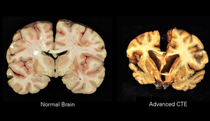
Chronic Traumatic Encephalopathy.png
A normal brain (left) and one with CTE (right)
SpecialtyNeurology, psychiatry, sports medicine
SymptomsBehavioral problems, mood problems, problems with thinking
ComplicationsDementia, aggression, depression, suicidal thoughts
Usual onsetYears after initial injuries
CausesRepeated head injuries
Risk factorsContact sports, military, domestic abuse, repeated banging of the head
Diagnostic methodAutopsy
Differential diagnosisAlzheimer's disease, Parkinson's disease
TreatmentSupportive care
FrequencyUncertain

Chronic traumatic encephalopathy (CTE) is a neurodegenerative disease caused by repeated head injuries. Symptoms may include behavioral problems, mood problems, and problems with thinking. Symptoms typically do not begin until years after the injuries. CTE often gets worse over time and can result in dementia. It is unclear if the risk of suicide is altered.

Most documented cases have occurred in athletes involved in contact sports such as boxing, American football, professional wrestling, ice hockey, rugby and soccer. Other risk factors include being in the military, prior domestic violence, and repeated banging of the head. The exact amount of trauma required for the condition to occur is unknown. Definitive diagnosis can only occur at autopsy. Chronic traumatic encephalopathy is a form of tauopathy.

There is no specific treatment. Rates of disease have been found to be about 30% among those with a history of multiple head injuries. Population rates, however, are unclear. Research in brain damage as a result of repeated head injuries began in the 1920s, at which time the condition was known as dementia pugilistica or "punch drunk syndrome". Changing the rules in some sports has been discussed as a means of prevention.

Signs and symptoms

Symptoms of CTE, which occur in four stages, generally appear eight to ten years after an individual experiences repetitive mild traumatic brain injuries.

First-stage symptoms include attention deficit hyperactivity disorder as well as confusion, disorientation, dizziness, and headaches. Second-stage symptoms include memory loss, social instability, impulsive behavior, and poor judgment. Third and fourth stages include progressive dementia, movement disorders, hypomimia, speech impediments, sensory processing disorder, tremors, vertigo, deafness, depression and suicidality.

Additional symptoms include dysarthria, dysphagia, cognitive disorders such as amnesia, and ocular abnormalities, such as ptosis.

The condition manifests as dementia, or declining mental ability, problems with memory, dizzy spells or lack of balance to the point of not being able to walk under one's own power for a short time and/or Parkinsonism, or tremors and lack of coordination. It can also cause speech problems and an unsteady gait. Patients with CTE may be prone to inappropriate or explosive behavior and may display pathological jealousy or paranoia.

Causes

Most documented cases have occurred in athletes with mild repetitive brain trauma (RBT) over an extended period of time. Specifically contact sports such as boxing, American football, wrestling, ice hockey, rugby, and football (soccer). In soccer whether this is just associated with prolific headers or other injuries is unclear as of 2017. Other potential risk factors include military personnel (repeated exposure to concussions charges or large caliber ordnance), domestic violence, and repeated banging of the head. The exact amount of trauma required for the condition to occur is unknown.

Pathology

The neuropathological appearance of CTE is distinguished from other tauopathies, such as Alzheimer's disease. The four clinical stages of observable CTE disability have been correlated with tau pathology in brain tissue, ranging in severity from focal perivascular epicenters of neurofibrillary tangles in the frontal neocortex to severe tauopathy affecting widespread brain regions.

The primary physical manifestations of CTE include a reduction in brain weight, associated with atrophy of the frontal and temporal cortices and medial temporal lobe. The lateral ventricles and the third ventricle are often enlarged, with rare instances of dilation of the fourth ventricle.[10] Other physical manifestations of CTE include anterior cavum septi pellucidi and posterior fenestrations, pallor of the substantia nigra and locus ceruleus, and atrophy of the olfactory bulbs, thalamus, mammillary bodies, brainstem and cerebellum. As CTE progresses, there may be marked atrophy of the hippocampus, entorhinal cortex, and amygdala.

On a microscopic scale, the pathology includes neuronal loss, tau deposition, TAR DNA-binding Protein 43 (TDP 43) deposition, white matter changes, and other abnormalities. The tau deposition occurs as dense neurofibrillary tangles (NFT), neurites, and glial tangles, which are made up of astrocytes and other glial cells Beta-amyloid deposition is a relatively uncommon feature of CTE.

A small group of individuals with CTE have chronic traumatic encephalomyopathy (CTEM), which is characterized by symptoms of motor-neuron disease and which mimics amyotrophic lateral sclerosis (ALS). Progressive muscle weakness and balance and gait problems (problems with walking) seem to be early signs of CTEM.

Exosome vesicles created by the brain are potential biomarkers of TBI, including CTE. A subtype of CTE is dementia pugilistica or boxer's dementia (from Latin pugilator - boxer) as it was initially found in those with a history of boxing, also called "punch-drunk syndrome". 

Loss of neurons, scarring of brain tissue, collection of proteinaceous, senile plaques, hydrocephalus, attenuation of the corpus callosum, diffuse axonal injury, neurofibrillary tangles, and damage to the cerebellum are implicated in the syndrome. The condition may be etiologically related to Alzheimer's disease. Neurofibrillary tangles have been found in the brains of dementia pugilistica patients, but not in the same distribution as is usually found in people with Alzheimer's. One group examined slices of brain from patients having had multiple mild traumatic brain injuries and found changes in the cells' cytoskeletons, which they suggested might be due to damage to cerebral blood vessels.

Increased exposure to concussions and sub-concussive blows is regarded as the most important risk factor, which can depend on the total number of fights, number of knockout losses, the duration of career, fight frequency, age of retirement, and boxing style.

Diagnosis

Diagnosis of CTE cannot be made in living individuals. A clear diagnosis is possible during an autopsy. Though there are signs and symptoms some researchers associate with CTE, there is no definitive test to prove the existence in a living person. Signs are also very similar to that of other neurological conditions such as Alzheimer's.

The lack of distinct biomarkers is the reason CTE cannot typically be diagnosed while a person is alive. Concussions are non-structural injuries and do not result in brain bleeding, which is why most concussions cannot be seen on routine neuroimaging tests such as CT or MRI. Acute concussion symptoms (those that occur shortly after an injury) should not be confused with CTE. Differentiating between prolonged post-concussion syndrome (PCS, where symptoms begin shortly after a concussion and last for weeks, months, and sometimes even years) and CTE symptoms can be difficult. Research studies are currently examining whether neuroimaging can detect subtle changes in axonal integrity and structural lesions that can occur in CTE. Recently, more progress in in-vivo diagnostic techniques for CTE has been made, using DTI, fMRI, MRI, and MRS imaging; however, more research needs to be done before any such techniques can be validated.

PET tracers that bind specifically to tau protein are desired to aid diagnosis of CTE in living individuals. One candidate is the tracer [18F]FDDNP, which is retained in the brain in individuals with a number of dementing disorders such as Alzheimer's disease, Down syndrome, progressive supranuclear palsy, corticobasal degeneration, familial frontotemporal dementia, and Creutzfeldt–Jakob disease. In a small study of 5 retired NFL players with cognitive and mood symptoms, the PET scans revealed accumulation of the tracer in their brains. However, [18F]FDDNP binds to beta-amyloid and other proteins as well. Moreover, the sites in the brain where the tracer was retained were not consistent with the known neuropathology of CTE. A more promising candidate is the tracer [18F]-T807, which binds only to tau. It is being tested in several clinical trials.

A putative biomarker for CTE is the presence in serum of autoantibodies against the brain. The autoantibodies were detected in football players who experienced a large number of head hits but no concussions, suggesting that even sub-concussive episodes may be damaging to the brain. The autoantibodies may enter the brain by means of a disrupted blood-brain barrier, and attack neuronal cells which are normally protected from an immune onslaught. Given the large numbers of neurons present in the brain (86 billion), and considering the poor penetration of antibodies across a normal blood-brain barrier, there is an extended period of time between the initial events (head hits) and the development of any signs or symptoms. Nevertheless, autoimmune changes in blood of players may consist the earliest measurable event predicting CTE.

Imaging

Although the diagnosis of CTE cannot be determined by imagining, the effects of head trauma may be seen with the use of structural imaging. Imaging techniques include the use of magnetic resonance imaging, nuclear magnetic resonance spectroscopy, CT scan, single-photon emission computed tomography, Diffusion MRI, and Positron Emission Tomography (PET). One specific use of imaging is the use of a PET scan is to evaluate for tau deposition, most commonly conducted on retired NFL players 

Prevention

Prevention of CTE in sport is not an idealistic goal because repetitive concussions increase the risk for this condition. Prevention techniques are also difficult because diagnosis of the condition can only be during a postmortem autopsy. The initial onset of this condition can not yet be determined, and therefore creating techniques for prevention pose a struggle.

Some common preventative methods have been the utilization of helmets and mouth-guards, though neither have significant research to support their use, they have shown prevention in direct head trauma. Although there is no significant research to support the use of helmets to reduce the risk of concussions, there is evidence to support that helmet use lowers the impactive forces. Mouth guards have been shown to decrease dental injuries, but again have not shown significant evidence to reduce concussions. A growing area of practice is the improved recognition and treatment for concussions and other head trauma, because repeated impacts are thought to increase the likelihood of CTE development, removal from sport during these traumatic incidences is essential. Proper return to play protocol during brain injuries is also important to decrease the significance of future impacts.

Another factor that has been implemented and continues to be an area of debate is to change the rules of many contact sports to make the effectively safer. Examples of these rules are the evolution of tackle technique rules in American football, such as the banning of helmet-first tackles, and addition of rules to protect defenseless players. Likewise, another growing area of debate is the better implementation of current rules that have previously been put in place to protect athletes.

Because of the concern that boxing may cause CTE, there is a movement among medical professionals to ban the sport. Medical professionals have called for such a ban since as early as the 1950s.

Management

No cure currently exists for CTE. Treatment is supportive as with other forms of dementia. Those with CTE-related symptoms may receive medication and non medication related treatments.

Epidemiology

Rates of disease have been found to be about 30% among those with a history of multiple head injuries. Population rates, however, are unclear.

Professional level athletes are the largest group with CTE, due to frequent concussions and sub-concussive impacts from play in contact sport. These contact-sports include American football, ice hockey, rugby, boxing, mixed martial arts, association football, wrestling, and war veterans. In association football, only prolific headers are known to have developed CTE.

Other individuals diagnosed with CTE were those involved in military service, had a previous history of chronic seizures, were domestically abused, or were involved in activities resulting in repetitive head collisions.

History

CTE was originally studied in boxers in the 1920s as dementia pugilistica. DP was first described in 1928 by a forensic pathologist, Dr. Harrison Stanford Martland, who was the chief medical examiner of Essex County in Newark, New Jersey in a Journal of the American Medical Association article, in which he noted the tremors, slowed movement, confusion, and speech problems typical of the condition. The initial diagnosis of dementia pugilistica was derived from the Latin word for boxer pugil (akin to pugnus ‘fist’, pugnāre ‘to fight’).

Other terms for the condition have included chronic boxer's encephalopathy, traumatic boxer's encephalopathy, boxer's dementia, pugilistic dementia, chronic traumatic brain injury associated with boxing (CTBI-B), and punch-drunk syndrome.

The seminal work on the disease came from British neurologist Macdonald Critchley, who in 1949 wrote a paper titled "Punch-drunk syndromes: the chronic traumatic encephalopathy of boxers." CTE was first recognized as affecting individuals who took considerable blows to the head, but was believed to be confined to boxers and not other athletes. As evidence pertaining to the clinical and neuropathological consequences of repeated mild head trauma grew, it became clear that this pattern of neurodegeneration was not restricted to boxers, and the term chronic traumatic encephalopathy became most widely used. In the early 2000s, Nigerian-American neuropathologist Bennet Omalu worked on the case of American football player Mike Webster, who died following unusual and unexplained behavior. In 2005 Omalu, along with colleagues in the Department of Pathology at the University of Pittsburgh, published his findings in the journal Neurosurgery in a paper which he titled "Chronic Traumatic Encephalopathy in a National Football League Player." This was followed by a paper on a second case in 2006 describing similar pathology. 

In 2008, the Sports Legacy Institute joined with the Boston University School of Medicine (BUSM) to form the Center for the Study of Traumatic Encephalopathy (CSTE). Brain Injury Research Institute (BIRI) also studies the impact of concussions.

Research

In 2005 forensic pathologist Bennet Omalu, along with colleagues in the Department of Pathology at the University of Pittsburgh, published a paper, "Chronic Traumatic Encephalopathy in a National Football League Player", in the journal Neurosurgery, based on analysis of the brain of deceased former NFL center Mike Webster. This was then followed by a paper on a second case in 2006 describing similar pathology, based on findings in the brain of former NFL player Terry Long

In 2008, the CSTE at Boston University at the BU School of Medicine started the CSTE brain bank at the Bedford VA Hospital to analyze the effects of CTE and other neurodegenerative diseases on the brain and spinal cord of athletes, military veterans, and civilians To date, the CSTE Brain Bank is the largest CTE tissue repository in the world. On December 21, 2009, the National Football League Players Association announced that it would collaborate with the CSTE at the Boston University School of Medicine to support the Center's study of repetitive brain trauma in athletes. Additionally, in 2010 the National Football League gave the CSTE a $1 million gift with no strings attached. In 2008, twelve living athletes (active and retired), including hockey players Pat LaFontaine and Noah Welch as well as former NFL star Ted Johnson, committed to donate their brains to CSTE after their deaths. In 2009, NFL Pro Bowlers Matt Birk, Lofa Tatupu, and Sean Morey pledged to donate their brains to the CSTE. In 2010, 20 more NFL players and former players pledged to join the CSTE Brain Donation Registry, including Chicago Bears linebacker Hunter Hillenmeyer, Hall of Famer Mike Haynes, Pro Bowlers Zach Thomas, Kyle Turley, and Conrad Dobler, Super Bowl Champion Don Hasselbeck and former pro players Lew Carpenter, and Todd Hendricks. In 2010, Professional Wrestlers Mick Foley, Booker T and Matt Morgan also agreed to donate their brains upon their deaths. Also in 2010, MLS player Taylor Twellman, who had to retire from the New England Revolution because of post-concussion symptoms, agreed to donate his brain upon his death. As of 2010, the CSTE Brain Donation Registry consists of over 250 current and former athletes. In 2011, former North Queensland Cowboys player Shaun Valentine became the first rugby league player to agree to donate his brain upon his death, in response to recent concerns about the effects of concussions on Rugby League players, who do not use helmets. Also in 2011, boxer Micky Ward, whose career inspired the film The Fighter, agreed to donate his brain upon his death.

In related research, the Center for the Study of Retired Athletes, which is part of the Department of Exercise and Sport Science at the University of North Carolina at Chapel Hill, is conducting research funded by National Football League Charities to "study former football players, a population with a high prevalence of exposure to prior Mild Traumatic Brain Injury (MTBI) and sub-concussive impacts, in order to investigate the association between increased football exposure and recurrent MTBI and neurodegenerative disorders such as cognitive impairment and Alzheimer's disease (AD)".

In February 2011, Dave Duerson committed suicide, leaving text messages to loved ones asking that his brain be donated to research for CTE. The family got in touch with representatives of the Boston University center studying the condition, said Robert Stern, the co-director of the research group. Stern said Duerson's gift was the first time of which he was aware that such a request had been made by someone who had committed suicide that was potentially linked to CTE. Stern and his colleagues found high levels of the protein tau in Duerson's brain. These elevated levels, which were abnormally clumped and pooled along the brain sulci, are indicative of CTE.

In July 2010, NHL enforcer Bob Probert died of heart failure. Before his death, he asked his wife to donate his brain to CTE research because it was noticed that Probert experienced a mental decline in his 40s. In March 2011, researchers at Boston University concluded that Probert had CTE upon analysis of the brain tissue he donated. He is the second NHL player from the program at the Center for the Study of Traumatic Encephalopathy to be diagnosed with CTE postmortem.

BUSM has also found indications of links between amyotrophic lateral sclerosis (ALS) and CTE in athletes who have participated in contact sports. Tissue for the study was donated by twelve athletes and their families to the CSTE Brain Bank at the Bedford, Massachusetts VA Medical Center.

In 2013, President Barack Obama announced the creation of the Chronic Effects of Neurotrauma Consortium or CENC, a federally funded research project devised to address the long-term effects of mild traumatic brain injury in military service personnel (SM's) and Veterans. The CENC is a multi-center collaboration linking premiere basic science, translational, and clinical neuroscience researchers from the DoD, VA, academic universities, and private research institutes to effectively address the scientific, diagnostic, and therapeutic ramifications of mild TBI and its long-term effects. Nearly 20% of the more than 2.5 million U.S. Service Members (SMs) deployed since 2003 to Operation Enduring Freedom (OEF) and Operation Iraqi Freedom (OIF) have sustained at least one traumatic brain injury (TBI), predominantly mild TBI (mTBI), and almost 8% of all OEF/OIF Veterans demonstrate persistent post-TBI symptoms more than six months post-injury. Unlike those head injuries incurred in most sporting events, recent military head injuries are most often the result of blast wave exposure. After a competitive application process, a consortium led by Virginia Commonwealth University was awarded funding. The project principal investigator for the CENC is David Cifu, Chairman and Herman J. Flax professor of the Department of Physical Medicine and Rehabilitation (PM&R) at Virginia Commonwealth University (VCU) in Richmond, Virginia, with co-principal investigators Ramon Diaz-Arrastia, Professor of Neurology, Uniformed Services University of the Health Sciences, and Rick L. Williams, statistician at RTI International.

Archetype

From Wikipedia, the free encyclopedia https://en.wikipedia.org/wiki/Archetype The concept of an archetyp...