Search This Blog

Saturday, February 22, 2020

Lumbar puncture

From Wikipedia, the free encyclopedia
https://en.wikipedia.org/wiki/Lumbar_puncture 
 
Lumbar puncture
Wikipedian getting a lumbar puncture (2006).jpg
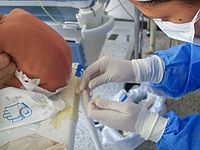
Lumbar puncture in a sitting position. The reddish-brown swirls on the patient's back are tincture of iodine (an antiseptic).
Other namesSpinal tap
ICD-9-CM03.31
MeSHD013129
eMedicine80773

Lumbar puncture (LP), also known as a spinal tap, is a medical procedure in which a needle is inserted into the spinal canal, most commonly to collect cerebrospinal fluid (CSF) for diagnostic testing. The main reason for a lumbar puncture is to help diagnose diseases of the central nervous system, including the brain and spine. Examples of these conditions include meningitis and subarachnoid hemorrhage. It may also be used therapeutically in some conditions. Increased intracranial pressure (pressure in the skull) is a contraindication, due to risk of brain matter being compressed and pushed toward the spine. Sometimes, lumbar puncture cannot be performed safely (for example due to a severe bleeding tendency). It is regarded as a safe procedure, but post-dural-puncture headache is a common side effect.

The procedure is typically performed under local anesthesia using a sterile technique. A hypodermic needle is used to access the subarachnoid space and fluid collected. Fluid may be sent for biochemical, microbiological, and cytological analysis. Using ultrasound to landmark may increase success.

Lumbar puncture was first introduced in 1891 by the German physician Heinrich Quincke.

Medical uses

The reason for a lumbar puncture may be to make a diagnosis or to treat a disease.

Diagnosis

The chief diagnostic indications of lumbar puncture are for collection of cerebrospinal fluid (CSF). Analysis of CSF may exclude infectious, inflammatory, and neoplastic diseases affecting the central nervous system. The most common purpose is in suspected meningitis, since there is no other reliable tool with which meningitis, a life-threatening but highly treatable condition, can be excluded. A lumbar puncture can also be used to detect whether someone has 'Stage 1' or 'Stage 2' Trypanosoma brucei. Young infants commonly require lumbar puncture as a part of the routine workup for fever without a source. This is due to higher rates of meningitis than in older persons. Infants also do not reliably show classic symptoms of meningeal irritation (meningismus) like neck stiffness and headache the way adults do. In any age group, subarachnoid hemorrhage, hydrocephalus, benign intracranial hypertension, and many other diagnoses may be supported or excluded with this test. It may also be used to detect the presence of malignant cells in the CSF, as in carcinomatous meningitis or medulloblastoma. CSF containing less than 10 red blood cells (RBCs)/mm³ constitutes a "negative" tap in the context of a workup for subarachnoid hemorrhage, for example. Taps that are "positive" have an RBC count of 100/mm³ or more.

Treatment

Lumbar punctures may also be done to inject medications into the cerebrospinal fluid ("intrathecally"), particularly for spinal anesthesia or chemotherapy

Serial lumbar punctures may be useful in temporary treatment of idiopathic intracranial hypertension (IIH). This disease is characterized by increased pressure of CSF which may cause headache and permanent loss of vision. While mainstays of treatment are medication, in some cases lumbar puncture performed multiple times may improve symptoms. It is not recommended as a staple of treatment due to discomfort and risk of the procedure, and the short duration of its efficacy.

Additionally, some people with normal pressure hydrocephalus (characterized by urinary incontinence, a changed ability to walk properly, and dementia) receive some relief of symptoms after removal of CSF.

Contraindications

Lumbar puncture should not be performed in the following situations:
  • Idiopathic (unidentified cause) increased intracranial pressure (ICP)
    • Rationale: lumbar puncture in the presence of raised ICP may cause uncal herniation
    • Exception: therapeutic use of lumbar puncture to reduce ICP, but only if obstruction (for example in the third ventricle of the brain) has been ruled out
    • Precaution
      • CT brain, especially in the following situations
        • Age >65
        • Reduced GCS
        • Recent history of seizure
        • Focal neurological signs
        • Abnormal respiratory pattern
        • Hypertension with bradycardia and deteriorating consciousness
      • Ophthalmoscopy for papilledema
  • Bleeding diathesis (relative)
/L)
  • Infections
    • Skin infection at puncture site
  • Vertebral deformities (scoliosis or kyphosis), in hands of an inexperienced physician.[11][12]
  • Adverse effects

    Headache

    Post spinal headache with nausea is the most common complication; it often responds to pain medications and infusion of fluids. It was long taught that this complication can be prevented by strict maintenance of a supine posture for two hours after the successful puncture; this has not been borne out in modern studies involving large numbers of people. Doing the procedure with the person on their side might decrease the risk.[13] Intravenous caffeine injection is often quite effective in aborting these spinal headaches. A headache that is persistent despite a long period of bedrest and occurs only when sitting up may be indicative of a CSF leak from the lumbar puncture site. It can be treated by more bedrest, or by an epidural blood patch, where the person's own blood is injected back into the site of leakage to cause a clot to form and seal off the leak.

    The risk of headache and need for analgesia and blood patch is much reduced if "atraumatic" needles are used. This does not affect the success rate of the procedure in other ways.

    Other

    Contact between the side of the lumbar puncture needle and a spinal nerve root can result in anomalous sensations (paresthesia) in a leg during the procedure; this is harmless and people can be warned about it in advance to minimize their anxiety if it should occur.

    Serious complications of a properly performed lumbar puncture are extremely rare. They include spinal or epidural bleeding, adhesive arachnoiditis and trauma to the spinal cord or spinal nerve roots resulting in weakness or loss of sensation, or even paraplegia. The latter is exceedingly rare, since the level at which the spinal cord ends (normally the inferior border of L1, although it is slightly lower in infants) is several vertebral spaces above the proper location for a lumbar puncture (L3/L4). There are case reports of lumbar puncture resulting in perforation of abnormal dural arterio-venous malformations, resulting in catastrophic epidural hemorrhage; this is exceedingly rare.

    The procedure is not recommended when epidural infection is present or suspected, when topical infections or dermatological conditions pose a risk of infection at the puncture site or in patients with severe psychosis or neurosis with back pain. Some authorities believe that withdrawal of fluid when initial pressures are abnormal could result in spinal cord compression or cerebral herniation; others believe that such events are merely coincidental in time, occurring independently as a result of the same pathology that the lumbar puncture was performed to diagnose. In any case, computed tomography of the brain is often performed prior to lumbar puncture if an intracranial mass is suspected.

    Technique

    Mechanism

    The brain and spinal cord are enveloped by a layer of cerebrospinal fluid, 125-150 ml in total (in adults) which acts as a shock absorber and provides a medium for the transfer of nutrients and waste products. The majority is produced by the choroid plexus in the brain and circulates from there to other areas, before being reabsorbed into the circulation (predominantly by the arachnoid granulations).

    The cerebrospinal fluid can be accessed most safely in the lumbar cistern. Below the first or second lumbar vertebrae (L1 or L2) the spinal cord terminates (conus medullaris). Nerves continue down the spine below this, but in a loose bundle of nerve fibers called the cauda equina. There is lower risk with inserting a needle into the spine at the level of the cauda equina because these loose fibers move out of the way of the needle without being damaged. The lumbar cistern extends into the sacrum.

    Procedure

    Illustration depicting lumbar puncture (spinal tap)
     
    Spinal needles used in lumbar puncture.
     
    Illustration depicting common positions for lumbar puncture procedure.

    The person is usually placed on their side (left more commonly than right). The patient bends the neck so the chin is close to the chest, hunches the back, and brings knees toward the chest. This approximates a fetal position as much as possible. Patients may also sit on a stool and bend their head and shoulders forward. The area around the lower back is prepared using aseptic technique. Once the appropriate location is palpated, local anaesthetic is infiltrated under the skin and then injected along the intended path of the spinal needle. A spinal needle is inserted between the lumbar vertebrae L3/L4, L4/L5 or L5/S1 and pushed in until there is a "give" as it enters the lumbar cistern wherein the ligamentum flavum is housed. The needle is again pushed until there is a second 'give' that indicates the needle is now past the dura mater. The arachnoid membrane and the dura mater exist in flush contact with one another in the living person's spine due to fluid pressure from CSF in the subarachnoid space pushing the arachnoid membrane out towards the dura. Therefore, once the needle has pierced the dura mater it has also traversed the thinner arachnoid membrane. The needle is then in the subarachnoid space. The stylet from the spinal needle is then withdrawn and drops of cerebrospinal fluid are collected. The opening pressure of the cerebrospinal fluid may be taken during this collection by using a simple column manometer. The procedure is ended by withdrawing the needle while placing pressure on the puncture site. The spinal level is so selected to avoid spinal injuries. In the past, the patient would lie on their back for at least six hours and be monitored for signs of neurological problems. There is no scientific evidence that this provides any benefit. The technique described is almost identical to that used in spinal anesthesia, except that spinal anesthesia is more often done with the patient in a seated position.

    The upright seated position is advantageous in that there is less distortion of spinal anatomy which allows for easier withdrawal of fluid. Some practitioners prefer it for lumbar puncture in obese patients, where lying on their side would cause a scoliosis and unreliable anatomical landmarks. However, opening pressures are notoriously unreliable when measured in the seated position. Therefore, patients will ideally lie on their side if practitioners need to measure opening pressure. 

    Reinsertion of the stylet may decrease the rate of post lumbar puncture headaches.

    Although not available in all clinical settings, use of ultrasound is helpful for visualizing the interspinous space and assessing the depth of the spine from the skin. Use of ultrasound reduces the number of needle insertions and redirections, and results in higher rates of successful lumbar puncture. If the procedure is difficult, such as in people with spinal deformities such as scoliosis, it can also be performed under fluoroscopy (under continuous X-ray imaging).

    Children

    In children, a sitting flexed position was as successful as lying on the side with respect to obtaining non-traumatic CSF, CSF for culture, and cell count. There was a higher success rate in obtaining CSF in the first attempt in infants younger than 12 months in the sitting flexed position.

    The spine of an infant at the time of birth differs from the adult spine. The conus medullaris (bottom of the spinal cord) terminates at the level of L1 in adults, but may range in term neonates (newly born babies) from L1-L3 levels. It is important to insert the spinal needle below the conus medullaris at the L3/L4 or L4/L5 interspinous levels. With growth of the spine, the conus typically reaches the adult level (L1) by 2 years of age.

    The ligamentum flavum and dura mater are not as thick in infants and children as they are in adults. Therefore, it is difficult to assess when the needle passes through them into the subarachnoid space because the characteristic "pop" or "give" may be subtle or nonexistent in the pediatric lumbar puncture. To decrease the chances of inserting the spinal needle too far, some clinicians use the "Cincinnati" method. This method involves removing the stylet of the spinal needle once the needle has advanced through the dermis. After removal of the stylet, the needle is inserted until CSF starts to come out of the needle. Once all of the CSF is collected, the stylet is then reinserted before removal of the needle.

    Interpretation

    Analysis of the cerebrospinal fluid generally includes a cell count and determination of the glucose and protein concentrations. The other analytical studies of cerebrospinal fluid are conducted according to the diagnostic suspicion.

    Pressure determination

    Lumbar puncture in a child suspected of having meningitis.

    Increased CSF pressure can indicate congestive heart failure, cerebral edema, subarachnoid hemorrhage, hypo-osmolality resulting from hemodialysis, meningeal inflammation, purulent meningitis or tuberculous meningitis, hydrocephalus, or pseudotumor cerebri. In the setting of raised pressure (or normal pressure hydrocephalus, where the pressure is normal but there is excessive CSF), lumbar puncture may be therapeutic.

    Decreased CSF pressure can indicate complete subarachnoid blockage, leakage of spinal fluid, severe dehydration, hyperosmolality, or circulatory collapse. Significant changes in pressure during the procedure can indicate tumors or spinal blockage resulting in a large pool of CSF, or hydrocephalus associated with large volumes of CSF.

    Cell count

    The presence of white blood cells in cerebrospinal fluid is called pleocytosis. A small number of monocytes can be normal; the presence of granulocytes is always an abnormal finding. A large number of granulocytes often heralds bacterial meningitis. White cells can also indicate reaction to repeated lumbar punctures, reactions to prior injections of medicines or dyes, central nervous system hemorrhage, leukemia, recent epileptic seizure, or a metastatic tumor. When peripheral blood contaminates the withdrawn CSF, a common procedural complication, white blood cells will be present along with erythrocytes, and their ratio will be the same as that in the peripheral blood. 

    The finding of erythrophagocytosis, where phagocytosed erythrocytes are observed, signifies haemorrhage into the CSF that preceded the lumbar puncture. Therefore, when erythrocytes are detected in the CSF sample, erythrophagocytosis suggests causes other than a traumatic tap, such as intracranial haemorrhage and haemorrhagic herpetic encephalitis. In which case, further investigations are warranted, including imaging and viral culture.

    Microbiology

    CSF can be sent to the microbiology lab for various types of smears and cultures to diagnose infections.
    • Gram staining may demonstrate gram positive bacteria in bacterial meningitis.
    • Microbiological culture is the gold standard for detecting bacterial meningitis. Bacteria, fungi, and viruses can all be cultured by using different techniques.
    • Polymerase chain reaction (PCR) has been a great advance in the diagnosis of some types of meningitis, such as meningitis from herpesvirus and enterovirus. It has high sensitivity and specificity for many infections of the CNS, is fast, and can be done with small volumes of CSF. Even though testing is expensive, cost analyses of PCR testing in neonatal patients demonstrated savings via reduced cost of hospitalization.
    • Numerous antibody-mediated tests for CSF are available in some countries: these include rapid tests for antigens of common bacterial pathogens, treponemal titers for the diagnosis of neurosyphilis and Lyme disease, Coccidioides antibody, and others.
    • The India ink test is still used for detection of meningitis caused by Cryptococcus neoformans, but the cryptococcal antigen (CrAg) test has a higher sensitivity.

    Chemistry

    Several substances found in cerebrospinal fluid are available for diagnostic measurement.
    • Glucose is present in the CSF; the level is usually about 60% that in the peripheral circulation. A fingerstick or venipuncture at the time of lumbar puncture may therefore be performed to assess peripheral glucose levels and determine a predicted CSF glucose value. Decreased glucose levels can indicate fungal, tuberculous or pyogenic infections; lymphomas; leukemia spreading to the meninges; meningoencephalitic mumps; or hypoglycemia. A glucose level of less than one third of blood glucose levels in association with low CSF lactate levels is typical in hereditary CSF glucose transporter deficiency also known as De Vivo disease.
    • Increased glucose levels in the fluid can indicate diabetes, although the 60% rule still applies.
    • Increased levels of glutamine are often involved with hepatic encephalopathies, Reye's syndrome, hepatic coma, cirrhosis, hypercapnia and depression.
    • Increased levels of lactate can occur the presence of cancer of the CNS, multiple sclerosis, heritable mitochondrial disease, low blood pressure, low serum phosphorus, respiratory alkalosis, idiopathic seizures, traumatic brain injury, cerebral ischemia, brain abscess, hydrocephalus, hypocapnia or bacterial meningitis.
    • The enzyme lactate dehydrogenase can be measured to help distinguish meningitides of bacterial origin, which are often associated with high levels of the enzyme, from those of viral origin in which the enzyme is low or absent.
    • Changes in total protein content of cerebrospinal fluid can result from pathologically increased permeability of the blood-cerebrospinal fluid barrier, obstructions of CSF circulation, meningitis, neurosyphilis, brain abscesses, subarachnoid hemorrhage, polio, collagen disease or Guillain–Barré syndrome, leakage of CSF, increases in intracranial pressure, or hyperthyroidism. Very high levels of protein may indicate tuberculous meningitis or spinal block.
    • IgG synthetic rate is calculated from measured IgG and total protein levels; it is elevated in immune disorders such as multiple sclerosis, transverse myelitis, and neuromyelitis optica of Devic. Oligoclonal bands may be detected in CSF but not in serum, suggesting intrathecal antibody production.
    Infection Appearance WBCs / mm3 Protein (g/l) Glucose
    Normal Clear <5 font=""> 0.15 to 0.45 > 2/3 of blood glucose
    Bacterial Yellowish, turbid > 1,000 (mostly PMNs) > 1 Low
    Viral Clear < 200 (mostly lymphocytes) Mild increase Normal or mildly low
    Tuberculosis Yellowish and viscous Modest increase Markedly Increased Decreased
    Fungal Yellowish and viscous < 50 (mostly lymphocytes) Initially normal than increased

    Normal or mildly low

    History

    Lumbar puncture, early 20th century.

    The first technique for accessing the dural space was described by the London physician Walter Essex Wynter. In 1889 he developed a crude cut down with cannulation in four patients with tuberculous meningitis. The main purpose was the treatment of raised intracranial pressure rather than for diagnosis. The technique for needle lumbar puncture was then introduced by the German physician Heinrich Quincke, who credits Wynter with the earlier discovery; he first reported his experiences at an internal medicine conference in Wiesbaden, Germany, in 1891. He subsequently published a book on the subject.

    The lumbar puncture procedure was taken to the United States by Arthur H. Wentworth an assistant professor at the Harvard Medical School, based at Children's Hospital. In 1893 he published a long paper on diagnosing cerebrospinal meningitis by examining spinal fluid. However, he was criticized by antivivisectionists for having obtained spinal fluid from children. He was acquitted, but, nevertheless, he was uninvited from the then forming Johns Hopkins School of Medicine, where he would have been the first professor of pediatrics.

    Historically lumbar punctures were also employed in the process of performing a pneumoencephalography, a nowadays obsolete X-ray imaging study of the brain that was performed extensively from the 1920s until the advent of modern non-invasive neuroimaging techniques such as MRI and CT in the 1970s. During this quite painful procedure, CSF was replaced with air or some other gas via the lumbar puncture in order to enhance the appearance of certain areas of the brain on plain radiographs.

    African trypanosomiasis

    From Wikipedia, the free encyclopedia
    African trypanosomiasis
    Other namesSleeping sickness, African sleeping sickness
    Trypanosoma sp. PHIL 613 lores.jpg
    Trypanosoma forms in a blood smear
    SpecialtyInfectious disease
    SymptomsStage 1: Fevers, headaches, itchiness, joint pains
    Stage 2: Trouble sleeping, confusion, poor coordination
    Usual onset1–3 weeks post exposure
    CausesTrypanosoma brucei spread by tsetse flies
    Diagnostic methodBlood smear, lumbar puncture
    MedicationPentamidine, suramin, eflornithine
    PrognosisFatal without treatment
    Frequency11,000
    Deaths3,500

    African trypanosomiasis, also known as African sleeping sickness or simply sleeping sickness, is an insect-borne parasitic disease of humans and other animals. It is caused by protozoa of the species Trypanosoma brucei. Humans are infected by two types, Trypanosoma brucei gambiense (TbG) and Trypanosoma brucei rhodesiense (TbR). TbG causes over 98% of reported cases. Both are usually transmitted by the bite of an infected tsetse fly and are most common in rural areas.

    Initially, the first stage of the disease is characterized by fevers, headaches, itchiness, and joint pains, beginning one to three weeks after the bite. Weeks to months later the second stage begins with confusion, poor coordination, numbness, and trouble sleeping. Diagnosis is by finding the parasite in a blood smear or in the fluid of a lymph node. A lumbar puncture is often needed to tell the difference between first and second stage disease.

    Prevention of severe disease involves screening the population at risk with blood tests for TbG. Treatment is easier when the disease is detected early and before neurological symptoms occur. Treatment of the first stage is with the medications pentamidine or suramin. Treatment of the second stage involves eflornithine or a combination of nifurtimox and eflornithine for TbG. While melarsoprol works for both stages, it is typically only used for TbR, due to serious side effects. Without treatment sleeping sickness typically results in death.

    The disease occurs regularly in some regions of sub-Saharan Africa with the population at risk being about 70 million in 36 countries. An estimated 11,000 people are currently infected with 2,800 new infections in 2015. In 2015 it caused around 3,500 deaths, down from 34,000 in 1990. More than 80% of these cases are in the Democratic Republic of the Congo. Three major outbreaks have occurred in recent history: one from 1896 to 1906 primarily in Uganda and the Congo Basin and two in 1920 and 1970 in several African countries. It is classified as a neglected tropical disease. Other animals, such as cows, may carry the disease and become infected in which case it is known as animal trypanosomiasis.

    Signs and symptoms

    African trypanosomiasis symptoms occur in two stages. The first stage, known as the hemolymphatic phase, is characterized by fever, headaches, joint pains, and itching. Fever is intermittent, with attacks lasting from a day to a week, separated by intervals of a few days to a month or longer. Invasion of the circulatory and lymphatic systems by the parasites is associated with severe swelling of lymph nodes, often to tremendous sizes. Winterbottom's sign, the tell-tale swollen lymph nodes along the back of the neck, may appear. Occasionally, a chancre (red sore) will develop at the location of the tsetse fly bite. If left untreated, the disease overcomes the host's defenses and can cause more extensive damage, broadening symptoms to include anemia, endocrine, cardiac, and kidney dysfunctions.

    The second phase of the disease, the neurological phase, begins when the parasite invades the central nervous system by passing through the blood–brain barrier. Disruption of the sleep cycle is a leading symptom of this stage and is the one that gave the disease the name 'sleeping sickness.' Infected individuals experience a disorganized and fragmented 24-hour rhythm of the sleep-wake cycle, resulting in daytime sleep episodes and nighttime periods of wakefulness.

    Other neurological symptoms include confusion, tremor, general muscle weakness, hemiparesis, and paralysis of a limb. Parkinson-like movements might arise due to non-specific movement disorders and speech disorders. Individuals may also exhibit psychiatric symptoms such as irritability, psychotic reactions, aggressive behaviour, or apathy which can sometimes dominate the clinical diagnosis. Without treatment, the disease is invariably fatal, with progressive mental deterioration leading to coma, systemic organ failure, and death. An untreated infection with T. b. rhodesiense will cause death within months whereas an untreated infection with T. b. gambiense will cause death after several years. Damage caused in the neurological phase is irreversible.

    Cause

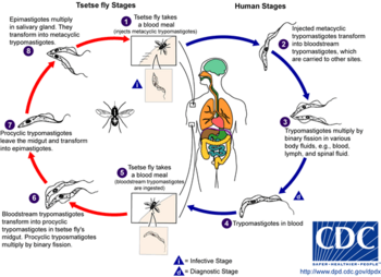
    The life cycle of the Trypanosoma brucei parasites.

    Trypanosoma brucei gambiense accounts for the majority of African trypanosomiasis cases, with humans as the main reservoir needed for the transmission, while Trypanosoma brucei rhodesiense is mainly zoonotic, with the occasional human infection. African Trypanosomiasis is dependent on the interaction of the parasite (trypanosome) with the tsetse flies (vector), as well as the host (human for Trypanosoma brucei gambiense, and animals for Trypanosoma brucei rhodesiense). The risk of contracting African Trypanosomiasis is dependent on coming in contact with an infected tsetse fly.

    Trypanosoma brucei

    There are two subspecies of the parasite that are responsible for starting the disease in humans. Trypanosoma brucei gambiense causes the diseases in west and central Africa, whereas Trypanosoma brucei rhodesiense has a limited geographical range and is responsible for causing the disease in east and southern Africa. In addition, a third subspecies of the parasite known as Trypanosoma brucei brucei is responsible for affecting animals but not humans.

    Humans are the main reservoir for T. b. gambiense but this species can also be found in pigs and other animals. Wild game animals and cattle are the main reservoir of T. b. rhodesiense. These parasites primarily infect individuals in sub-Saharan Africa because that is where the vector (tsetse fly) is located. The two human forms of the disease also vary greatly in intensity. T. b. gambiense causes a chronic condition that can remain in a passive phase for months or years before symptoms emerge and the infection can last about 3 years before death occurs.

    T. b. rhodesiense is the acute form of the disease, and death can occur within months since the symptoms emerge within weeks and it is more virulent and faster developing than T. b. gambiense. Furthermore, trypanosomes are surrounded by a coat that is composed of variant surface glycoproteins (VSG). These proteins act to protect the parasite from any lytic factors that are present in human plasma. The host's immune system recognizes the glycoproteins present on the coat of the parasite leading to the production of different antibodies (IgM and IgG).

    These antibodies will then act to destroy the parasites that circulate around the blood. However, from the several parasites present in the plasma, a small number of them will experience changes in their surface coats resulting in the formation of new VSGs. Thus, the antibodies produced by the immune system will no longer recognize the parasite leading to proliferation until new antibodies are created to combat the novel VSGs. Eventually the immune system will no longer be able to fight off the parasite due to the constant changes in VSGs and infection will arise.

    Vector

    The tsetse fly (genus Glossina) is a large, brown, biting fly that serves as both a host and vector for the trypanosome parasites. While taking blood from a mammalian host, an infected tsetse fly injects metacyclic trypomastigotes into skin tissue. From the bite, parasites first enter the lymphatic system and then pass into the bloodstream. Inside the mammalian host, they transform into bloodstream trypomastigotes, and are carried to other sites throughout the body, reach other body fluids (e.g., lymph, spinal fluid), and continue to replicate by binary fission.

    The entire life cycle of African trypanosomes is represented by extracellular stages. A tsetse fly becomes infected with bloodstream trypomastigotes when taking a blood meal on an infected mammalian host. In the fly's midgut, the parasites transform into procyclic trypomastigotes, multiply by binary fission, leave the midgut, and transform into epimastigotes. The epimastigotes reach the fly's salivary glands and continue multiplication by binary fission.

    The entire life cycle of the fly takes about three weeks. In addition to the bite of the tsetse fly, the disease can be transmitted by:
    • Mother-to-child infection: the trypanosome can sometimes cross the placenta and infect the fetus.
    • Laboratories: accidental infections, for example, through the handling of blood of an infected person and organ transplantation, although this is uncommon.
    • Blood transfusion
    • Sexual contact (This may be possible)
    Horse-flies (Tabanidae) and stable flies (Muscidae) possibly play a role in transmission of nagana (the animal form of sleeping sickness) and the human disease form.

    Sleeping sickness Trypanosoma Distribution Vector
    Chronic form T. brucei gambiense Western Africa G. palpalis G. tachinoides
    G. fuscipes
    G. morsitans
    Acute form T. brucei rhodesiense Eastern Africa G. morsitans G. swynnertoni
    G. pallidipes
    G. fuscipes

    Mechanism

    Tryptophol is a chemical compound that induces sleep in humans. It is produced by the trypanosomal parasite in sleeping sickness.

    Diagnosis

    Two areas from a blood smear from a person with African trypanosomiasis, thin blood smear stained with Giemsa: Typical trypomastigote stages (the only stages found in people), with a posterior kinetoplast, a centrally located nucleus, an undulating membrane, and an anterior flagellum. The two Trypanosoma brucei subspecies that cause human trypanosomiasis, T. b. gambiense and T. b. rhodesiense, are indistinguishable morphologically. The trypanosomes' length range is 14 to 33 µm, Source: CDC

    The gold standard for diagnosis is identification of trypanosomes in a sample by microscopic examination. Samples that can be used for diagnosis include chancre fluid, lymph node aspirates, blood, bone marrow, and, during the neurological stage, cerebrospinal fluid. Detection of trypanosome-specific antibodies can be used for diagnosis, but the sensitivity and specificity of these methods are too variable to be used alone for clinical diagnosis. Further, seroconversion occurs after the onset of clinical symptoms during a T. b. rhodesiense infection, so is of limited diagnostic use.

    Trypanosomes can be detected from samples using two different preparations. A wet preparation can be used to look for the motile trypanosomes. Alternatively, a fixed (dried) smear can be stained using Giemsa's or Field's technique and examined under a microscope. Often, the parasite is in relatively low abundance in the sample, so techniques to concentrate the parasites can be used prior to microscopic examination. For blood samples, these include centrifugation followed by examination of the buffy coat; mini anion-exchange/centrifugation; and the quantitative buffy coat (QBC) technique. For other samples, such as spinal fluid, concentration techniques include centrifugation followed by examination of the sediment.

    Three serological tests are also available for detection of the parasite: the micro-CATT, wb-CATT, and wb-LATEX. The first uses dried blood, while the other two use whole blood samples. A 2002 study found the wb-CATT to be the most efficient for diagnosis, while the wb-LATEX is a better exam for situations where greater sensitivity is required.

    Prevention

    Currently there are few medically related prevention options for African Trypanosomiasis (i.e. no vaccine exists for immunity). Although the risk of infection from a tsetse fly bite is minor (estimated at less than 0.1%), the use of insect repellants, wearing long-sleeved clothing, avoiding tsetse-dense areas, implementing bush clearance methods and wild game culling are the best options to avoid infection available for local residents of affected areas.

    In July 2000, a resolution was passed to form the Pan African Tsetse and Trypanosomiasis Eradication Campaign (PATTEC). The campaign works to eradicate the tsetse vector population levels and subsequently the protozoan disease, by use of insecticide-impregnated targets, fly traps, insecticide-treated cattle, ultra-low dose aerial/ground spraying (SAT) of tsetse resting sites and the sterile insect technique (SIT). The use of SIT in Zanzibar proved effective in eliminating the entire population of tsetse flies but was expensive and is relatively impractical to use in many of the endemic countries afflicted with African trypanosomiasis.

    A pilot program in Senegal has reduced the tsetse fly population by as much as 99% by introducing male flies which have been sterilized by exposure to gamma rays.

    Regular active surveillance, involving detection and prompt treatment of new infections, and tsetse fly control is the backbone of the strategy used to control sleeping sickness. Systematic screening of at-risk communities is the best approach, because case-by-case screening is not practical in endemic regions. Systematic screening may be in the form of mobile clinics or fixed screening centres where teams travel daily to areas of high infection rates. Such screening efforts are important because early symptoms are not evident or serious enough to warrant people with gambiense disease to seek medical attention, particularly in very remote areas. Also, diagnosis of the disease is difficult and health workers may not associate such general symptoms with trypanosomiasis. Systematic screening allows early-stage disease to be detected and treated before the disease progresses, and removes the potential human reservoir. A single case of sexual transmission of West African sleeping sickness has been reported.

    Treatment

    First stage

    The current treatment for first-stage disease is intravenous or intramuscular pentamidine for T. b. gambiense or intravenous suramin for T. b. rhodesiense.

    Fexinidazole has been approved in the Democratic Republic Congo for T.b. gambiense sleeping sickness. Benefits include that it is taken by mouth and treats both stages of the disease.

    Second stage

    For T. b. gambiense a regimen involving the combination of nifurtimox and eflornithine, nifurtimox-eflornithine combination treatment (NECT), or eflornithine alone appear to be more effective and result in fewer side effects. These treatments may replace melarsoprol when available with the combination being first line. NECT has the benefit of requiring less injections of eflornithine.

    Intravenous melarsoprol was previously the standard treatment for second-stage (neurological phase) disease and is effective for both types. Melarsoprol is the only treatment for second stage T. b. rhodesiense; however, it causes death in 5% of people who take it. Resistance to melarsoprol can occur.

    Epidemiology

    Deaths per 100,000 population due to African trypanosomiasis by country in 2002.

    In 2010, it caused around 9,000 deaths, down from 34,000 in 1990. As of 2000, the disability-adjusted life-years (9 to 10 years) lost due to sleeping sickness are 2.0 million. From 2010-2014, there was an estimated 55 million people at risk for gambiense African Trypanosomiasis and over 6 million people at risk for rhodesiense African Trypanosomiasis. In 2014, the World Health Organization reported 3,797 cases of Human African Trypanosomiasis when the predicted number of cases were to be 5,000. The number of total reported cases in 2014 is an 86% reduction to the total number of cases reported in 2000.

    The disease has been recorded as occurring in 37 countries, all in sub-Saharan Africa. It occurs regularly in southeast Uganda and western Kenya, and killed more than 48,000 Africans in 2008. The Democratic Republic of the Congo is the most affected country in the world, accounting for 75% of the Trypanosoma brucei gambiense cases. The population at risk being about 69 million with one third of this number being at a 'very high' to 'moderate' risk and the remaining two thirds at a 'low' to 'very low' risk. The number of people being affected by the disease has declined. At this rate, sleeping sickness elimination is a possibility. The World Health Organization plans to eradicate sleeping sickness by the year 2020.

    Prognosis

    If untreated, T. b. gambiense almost always results in death, with only a few individuals shown in a long-term 15 year follow-up to have survived after refusing treatment. T. b. rhodesiense, being a more acute and severe form of the disease, is consistently fatal if not treated. Disease progression greatly varies depending on disease form. For individuals which are infected by T. b. gambiense, which accounts for 98% of all of the reported cases, a person can be infected for months or even years without signs or symptoms until the advanced disease stage, where it is too late to be treated successfully. For individuals affected by T. b. rhodesiense, which accounts for 2% of all reported cases, symptoms appear within weeks or months of the infection. Disease progression is rapid and invades the central nervous system, causing death within a short amount of time.

    History

    In 1903, David Bruce recognized the tsetse fly as the arthropod vector.

    The condition has been present in Africa for thousands of years. Because of a lack of travel between indigenous people, sleeping sickness in humans had been limited to isolated pockets. This changed after Arab slave traders entered central Africa from the east, following the Congo River, bringing parasites along. Gambian sleeping sickness travelled up the Congo River, and then further east.

    An Arab writer of the 14th century left the following description in the case of a sultan of the Mali Kingdom: "His end was to be overtaken by the sleeping sickness (illat an-nawm) which is a disease that frequently befalls the inhabitants of these countries especially their chieftains. Sleep overtakes one of them in such a manner that it is hardly possible to awake him."

    The British naval surgeon John Atkins described the disease on his return from West Africa in 1734:
    "The Sleepy Distemper (common among the Negroes) gives no other previous Notice, than a want of Appetite 2 or 3 days before; their sleeps are sound, and Sense and Feeling very little; for pulling, drubbing or whipping will scarce stir up Sense and Power enough to move; and the Moment you cease beating the smart is forgot, and down they fall again into a state of Insensibility, drivling constantly from the Mouth as in deep salivation; breathe slowly, but not unequally nor snort. Young people are more subject to it than the old; and the Judgement generally pronounced is Death, the Prognostik seldom failing. If now and then one of them recovers, he certainly loses the little Reason he had, and turns Ideot..."
    In 1901, a devastating epidemic erupted in Uganda, killing more than 250,000 people, including about two-thirds of the population in the affected lakeshore areas. According to The Cambridge History of Africa, "It has been estimated that up to half the people died of sleeping-sickness and smallpox in the lands on either bank of the lower river Congo."

    The causative agent and vector were identified in 1903 by David Bruce, and the subspecies of the protozoa were differentiated in 1910. Bruce had earlier shown that T. brucei was the cause of a similar disease in horses and cattle that was transmitted by the tse-tse fly (Glossina morsitans).

    The first effective treatment, atoxyl, an arsenic-based drug developed by Paul Ehrlich and Kiyoshi Shiga, was introduced in 1910, but blindness was a serious side effect.

    Suramin was first synthesized by Oskar Dressel and Richard Kothe in 1916 for Bayer. It was introduced in 1920 to treat the first stage of the disease. By 1922, Suramin was generally combined with tryparsamide (another pentavalent organoarsenic drug), the first drug to enter the nervous system and be useful in the treatment of the second stage of the gambiense form. Tryparsamide was announced in the Journal of Experimental Medicine in 1919 and tested in the Belgian Congo by Louise Pearce of the Rockefeller Institute in 1920. It was used during the grand epidemic in West and Central Africa on millions of people and was the mainstay of therapy until the 1960s. American medical missionary Arthur Lewis Piper was active in using tryparsamide to treat sleeping sickness in the Belgian Congo in 1925.

    Pentamidine, a highly effective drug for the first stage of the disease, has been used since 1937. During the 1950s, it was widely used as a prophylactic agent in western Africa, leading to a sharp decline in infection rates. At the time, eradication of the disease was thought to be at hand.

    The organoarsenical melarsoprol (Arsobal) developed in the 1940s is effective for people with second-stage sleeping sickness. However, 3–10% of those injected have reactive encephalopathy (convulsions, progressive coma, or psychotic reactions), and 10–70% of such cases result in death; it can cause brain damage in those who survive the encephalopathy. However, due to its effectiveness, melarsoprol is still used today. Resistance to melarsoprol is increasing, and combination therapy with nifurtimox is currently under research.

    Eflornithine (difluoromethylornithine or DFMO), the most modern treatment, was developed in the 1970s by Albert Sjoerdsma and underwent clinical trials in the 1980s. The drug was approved by the United States Food and Drug Administration in 1990. Aventis, the company responsible for its manufacture, halted production in 1999. In 2001, Aventis, in association with Médecins Sans Frontières and the World Health Organization, signed a long-term agreement to manufacture and donate the drug.

    In addition to sleeping sickness, previous names have included negro lethargy, maladie du sommeil (Fr), Schlafkrankheit (Ger), African lethargy, and Congo trypanosomiasis.

    Research

    The genome of the parasite has been sequenced and several proteins have been identified as potential targets for drug treatment. Analysis of the genome also revealed the reason why generating a vaccine for this disease has been so difficult. T. brucei has over 800 genes that make proteins the parasite "mixes and matches" to evade immune system detection.

    Using a genetically modified form of a bacterium that occurs naturally in the gut of the vectors is being studied as a method of controlling the disease.

    Recent findings indicate that the parasite is unable to survive in the bloodstream without its flagellum. This insight gives researchers a new angle with which to attack the parasite.

    Trypanosomiasis vaccines are undergoing research.

    Additionally, the Drugs for Neglected Disease Initiative has contributed to the African sleeping sickness research by developing a compound called fexinidazole. This project was originally started in April 2007 and enrolled 749 people in the DRC and Central African Republic. The results showed efficacy and safety in both stages of the disease, both in adults and children ≥ 6 years old and weighing ≥ 20 kg. The European Medicines Agency approved it for first and second stage disease outside of Europe in November 2018. The treatment was approved in the DRC in December 2018.

    Funding

    For current funding statistics, human African trypanosomiasis is grouped with kinetoplastid infections. Kinetoplastids refer to a group of flagellate protozoa. Kinetoplastid infections include African sleeping sickness, Chagas' disease, and Leishmaniasis. All together, these three diseases accounted for 4.4 million disability adjusted life years (DALYs) and an additional 70,075 recorded deaths yearly. For kinetoplastid infections, the total global research and development funding was approximately $136.3 million in 2012. Each of the three diseases, African sleeping sickness, Chagas' disease, and Leishmaniasis each received approximately a third of the funding, which was about $36.8 million US dollars, $38.7 million US dollars, and $31.7 million US dollars, respectively.

    For sleeping sickness, funding was split into basic research, drug discovery, vaccines, and diagnostics. The greatest amount of funding was directed towards basic research of the disease; approximately $21.6 million US dollars was directed towards that effort. As for therapeutic development, approximately $10.9 million was invested.

    The top funder for kinetoplastid infection research and development are public sources. About 62% of the funding comes from high-income countries while 9% comes from low- and middle-income countries. High-income countries' public funding is the largest contributor to the neglected disease research effort. However, in recent years, funding from high-income countries has been steadily decreasing; in 2007, high-income countries provided 67.5% of the total funding whereas, in 2012, high-income countries public funds only provided 60% of the total funding for kinetoplastid infections. This downwards trend leaves a gap for other funders, such as philanthropic foundations and private pharmaceutical companies to fill.

    Much of the progress that has been made in African sleeping sickness and neglected disease research as a whole is a result of the other non-public funders. One of these major sources of funding has come from foundations, which have increasingly become more committed to neglected disease drug discovery in the 21st century. In 2012, philanthropic sources provided 15.9% of the total funding. The Bill and Melinda Gates Foundation has been a leader in providing funding for neglected disease drug development. They have provided $444.1 million US dollars towards neglected disease research in 2012. To date, they have donated over $1.02 billion US dollars towards the neglected disease discovery efforts.

    For kinetoplastid infections specifically, they have donated an average of $28.15 million US dollars annually between the years 2007 to 2011. They have labeled human African trypanosomiasis a high-opportunity target meaning it is a disease that presents the greatest opportunity for control, elimination, and eradication, through the development of new drugs, vaccines, public-health programs, and diagnostics. They are the second highest funding source for neglected diseases, immediately behind the US National Institutes of Health. At a time where public funding is decreasing and government grants for scientific research are harder to obtain, the philanthropic world has stepped in to push the research forward. 

    Another important component of increased interest and funding has come from industry. In 2012, they contributed 13.1% total to the kinetoplastid research and development effort, and have additionally played an important role by contributing to public-private partnerships (PPP) as well as product-development partnerships (PDP). A public-private partnership is an arrangement between one or more public entities and one or more private entities that exists to achieve a specific health outcome or to produce a health product. The partnership can exist in numerous ways; they may share and exchange funds, property, equipment, human resources, and intellectual property. These public-private partnerships and product-development partnerships have been established to address challenges in pharmaceutical industry, especially related to neglected disease research. These partnerships can help increase the scale of the effort towards therapeutic development by using different knowledge, skills, and expertise from different sources. These types of partnerships have been shown to be more effective than industry or public groups working independently.

    Operator (computer programming)

    From Wikipedia, the free encyclopedia https://en.wikipedia.org/wiki/Operator_(computer_programmin...