Search This Blog

Saturday, April 18, 2020

Shenandoah National Park

From Wikipedia, the free encyclopedia
 
Shenandoah National Park
IUCN category II (national park)
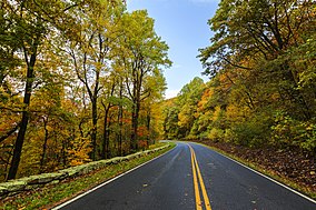
Skyline Drive in the Fall (21852619608).jpg
Skyline Drive
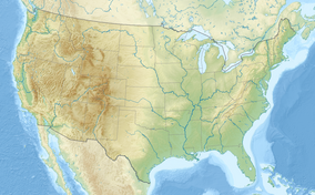
Map showing the location of Shenandoah National Park
Location in the United States
LocationVirginia, United States
Nearest cityFront Royal
Coordinates38°32′N 78°21′WCoordinates: 38°32′N 78°21′W
Area199,173 acres (311.208 sq mi; 806.02 km2)
EstablishedDecember 26, 1935
Visitors1,264,880 (in 2018)
Governing bodyNational Park Service
WebsiteOfficial website

Shenandoah National Park /ˈʃɛnənˌdə/ (often /ˈʃænənˌdə/) is an American national park that encompasses part of the Blue Ridge Mountains in the state of Virginia. The park is long and narrow, with the Shenandoah River and its broad valley to the west, and the rolling hills of the Virginia Piedmont to the east. Skyline Drive is the main park road, generally traversing near the ridgeline of the mountains. Almost 40% of the land area—79,579 acres (124.3 sq mi; 322.0 km2)—has been designated as wilderness and is protected as part of the National Wilderness Preservation System. The highest peak is Hawksbill Mountain at 4,051 feet (1,235 m).

Geography

Park map (click on map to enlarge)

The park encompasses parts of eight counties. On the west side of Skyline Drive they are, from northeast to southwest, Warren, Page, Rockingham, and Augusta counties. On the east side of Skyline Drive they are Rappahannock, Madison, Greene, and Albemarle counties. The park stretches for 105 miles (169 km) along Skyline Drive from near the town of Front Royal in the northeast to near the city of Waynesboro in the southwest. The park headquarters are located in Luray.

Geology

Shenandoah National Park lies along the Blue Ridge Mountains in north-central Virginia. These mountains form a distinct highland rising to elevations above 4,000 feet (1,200 m). Local topographic relief between the Blue Ridge Mountains and Shenandoah Valley exceeds 3,000 feet (910 m) at some locations. The crest of the range divides the Shenandoah River drainage basin, part of the Potomac River drainage, on the west side, from the James and Rappahannock River drainage basins on the east side.

Some of the rocks exposed in the park date to over one billion years in age, making them among the oldest in Virginia. Bedrock in the park includes Grenville-age granitic basement rocks (1.2–1.0 billion years old) and a cover sequence of metamorphosed Neoproterozoic (570–550 million years old) sedimentary and volcanic rocks of the Swift Run and Catoctin formations. Columns of Catoctin Formation metamorphosed basalt can be seen at Compton Peak. Clastic rocks of the Chilhowee Group are of early Cambrian age (542–520 million years old). Quaternary surficial deposits are common and cover much of the bedrock throughout the park.

The park is located along the western part of the Blue Ridge anticlinorium, a regional-scale Paleozoic structure at the eastern margin of the Appalachian fold and thrust belt. Rocks within the park were folded, faulted, distorted, and metamorphosed during the late Paleozoic Alleghanian orogeny (325 to 260 million years ago). The rugged topography of Blue Ridge Mountains is a result of differential erosion during the Cenozoic, although some post-Paleozoic tectonic activity occurred in the region.

History

Satellite view of Shenandoah in autumn, the leaf peeping season

Creation of the park

Legislation to create a national park in the Appalachian mountains was first introduced by freshman Virginia congressman Henry D. Flood in 1901, but despite the support of President Theodore Roosevelt, failed to pass. The first national park was Yellowstone, in Wyoming, Montana, and Idaho. It was signed into law in 1872. Yosemite National Park was created in 1890. When Congress created the National Park Service (NPS) in 1916, additional parks had maintained the western pattern (Crater Lake in 1902, Wind Cave in 1903, Mesa Verde in 1906, then Denali in 1917). Grand Canyon, Zion and Acadia were all created in 1919 during the administration of Virginia-born president Woodrow Wilson. Acadia finally broke the western mold, becoming the first eastern national park. It was also based on donations from wealthy private landowners. Stephen Mather, the first NPS director, saw a need for a national park in the southern states, and solicited proposals in his 1923 year-end report. In May 1925, Congress and President Calvin Coolidge authorized the NPS to acquire a minimum of 250,000 acres (390.6 sq mi; 1,011.7 km2) and a maximum of 521,000 acres (814.1 sq mi; 2,108.4 km2) to form Shenandoah National Park, and also authorized creation of Great Smoky Mountains National Park. However, the legislation also required that no federal funds would be used to acquire the land. Thus, Virginia needed to raise private funds, and could also authorize state funds and use its eminent domain (condemnation) power to acquire the land to create Shenandoah National Park. 

Virginia's Democratic gubernatorial candidate (and the late Congressman Flood's nephew), Harry F. Byrd supported creation of Shenandoah National Park, as did his friend William E. Carson, a businessman who had become Virginia's first chairman of the Commission on Conservation and Development. Development of the western national parks had assisted tourism, which produced jobs, which Byrd and local politicians supported. The land that became Shenandoah park was scenic, mountainous, and had also lost about half of its trees to the Chestnut blight (which was incurable and affected trees as they reached maturity). However, it had been held as private property for over a century, so many farms and orchards existed. After Byrd became governor and convinced the legislature to appropriate $1 million for land acquisition and other work, Carson and his teams (including surveyors and his brother Kit who was Byrd's law partner) tried to figure out who owned the land. They found that it consisted of more than 5,000 parcels, some of them inhabited by tenant farmers or squatters (who were ineligible to receive compensation). Some landowners, including wealthy resort owner George Freeman Pollock and Luray Realtor and developer L. Ferdinand Zerkel, had long wanted the park created and had formed the Northern Virginia Park Association to win over the national park selection committee. However, many local families who had lived in the area for generations (especially people over 60 years old) did not want to sell their land, and some refused to sell at any price. Carson promised that if they sold to the state, they could still live on their homesteads for the rest of their lives. Carson also lobbied the new president, Herbert Hoover, who bought land to establish a vacation fishing camp near the headwaters of the Rapidan River (and would ultimately donate it to the park as he left office; it remains as Rapidan Camp).

A small family cemetery along Skyline Drive
 
The commonwealth of Virginia slowly acquired the land through eminent domain, and then gave it to the U.S. federal government to establish the national park. Carson's brother suggested that Virginia's legislature authorize condemnation by counties (followed by arbitration for individual parcels) rather than condemn each parcel. Some families accepted the payments because they needed the money and wanted to escape the subsistence lifestyle. Nearly 90 percent of the inhabitants worked the land for a living: selling timber, charcoal or crops. They had previously been able to earn money to buy supplies by harvesting the now-rare chestnuts, by working during the apple and peach harvest season (but the drought of 1930 devastated those crops and killed many fruit trees), by selling handmade textiles and crafts (displaced by factories) and moonshine (illegal after Prohibition started).

However, Carson and the politicians did not seek citizen input early in the process, nor convince residents that they could live better in a tourist economy. Instead they started with an advertising campaign to raise the funds, and courthouse property evaluations and surveys. Upon Mather's death in 1929, the new NPS director, Horace M. Albright also decided that the federal agency would only accept vacant land, so even elderly residents would be forced to leave. Thus, many families and entire communities were forced to vacate portions of the Blue Ridge Mountains in eight Virginia counties. Although the Skyline Drive right-of-way was purchased from owners without condemnation, the costs of the acreage purchased trebled over initial estimates and the acreage decreased to what Carson called a "fish-bone" shape and others a "shoestring". Although Byrd and Carson convinced Congress to reduce the minimum size of Shenandoah Park to just over 160,000 acres (250.0 sq mi; 647.5 km2) to eliminate some high-priced lands, in 1933 newly elected President Franklin D. Roosevelt decided to also create the Blue Ridge Parkway to connect to then-under-construction Skyline Drive on the Shenandoah National Park ridgeline, which required additional condemnations.

When many families continued to refuse to sell their land in 1932 and 1933, proponents changed tactics. Freeman hired social worker Miriam Sizer to teach at a summer school he had set up near one of his workers' communities, and asked her to write a report about the conditions in which they lived. Although later discredited, the report depicted the local population as very poor and inbred, and was soon used to support forcible evictions and burning of former cabins so residents would not sneak back. University of Chicago sociologists Fay-Cooper Cole and Mandel Sherman described how the small valley communities or hollows had existed "without contact with law or government" for centuries, which some analogized to a popular comic strip Li'l Abner and his fictional community, Dogpatch. In 1933, Sherman and journalist Thomas Henry published Hollow Folk drawing pitying eyes to local conditions and "hillbillies." As in many rural areas of the time, most remote homesteads in the Shenandoah lacked electricity and often running water, as well as access to schools and health facilities during many months. However, Hoover had hired experienced rural teacher Christine Vest to teach near his summer home (and who believed the other reports exaggerated, as did Episcopal missionary teachers in other Blue Ridge areas).

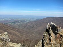
View from the summit of Hawksbill Mountain
 
Carson had had ambitions to become governor in 1929 and 1933, but Byrd instead selected George C. Peery of the state's southwest corner to succeed easterner Pollard. After winning election, Peery and Carson's successor would establish Virginia's state park system, although plans to relocate reluctant residents kept changing and basically failed. Carson had hoped to head that new state agency, but was not selected because of his growing differences with Byrd, over fees owed his brother and especially over the evictions that began in late 1933 against his advice but pursuant to new federal policies and that garnered much negative publicity. 

Most of the reluctant families came from the park's central counties (Madison, Page, and Rappahannock), not the northern counties nearest Byrd's and Carson's bases, or from the southern end where residents could see tourism's benefits at Thomas Jefferson's Monticello since the 1920s, as well as the jobs available in the Shenandoah and new Blue Ridge projects. In 1931 and 1932, residents were allowed to petition the state agency to stay another year to gather crops, etc. However, some refused to cooperate to any extent, others wanted to continue to use resources now protected (including timber or homes and gardens vacated by others), and many found the permit process arbitrary. Businessman Robert H. Via filed suit against the condemnations in 1934 but did not prevail (and ended up moving to Pennsylvania and never cashed his condemnation check).

Carson announced his resignation from his unpaid job effective in December 1934. As one of his final acts, Carson wrote the new NPS director, Arno B. Cammerer, urging that 60 people over 60 years of age whose plots were not visible from the new Skyline Drive not be evicted. When evictions kept creating negative publicity in 1935, photographer Arthur Rothstein coordinated with the Hollow Folk authors and then went to document the conditions they claimed.

View from Skyline Drive

Creation of the park had immediate benefits to some Virginians. During the Great Depression, many young men received training and jobs through the Civilian Conservation Corps (CCC). The first CCC camp in Virginia was established in the George Washington National Forest near Luray, and Governor Pollard quickly filled his initial quota of 5,000 workers. About 1,000 men and boys worked on Skyline Drive, and about 100,000 worked in Virginia during the agency's existence. In Shenandoah Park, CCC crews removed many of the dead chestnut trees whose skeletons marred views in the new park, as well as constructed trails and facilities. Tourism revenues also skyrocketed. On the other hand, CCC crews were assigned to burn and destroy some cabins in the park, to prevent residents from coming back. Also, U.S. Secretary of the Interior Harold Ickes who had jurisdiction over the NPS and partial jurisdiction over the CCC, tried to use his authority to force Byrd to cooperate on other New Deal projects. 

Shenandoah National Park was finally established on December 26, 1935, and soon construction began on the Blue Ridge Parkway that Byrd wanted. President Franklin Delano Roosevelt formally opened Shenandoah National Park on July 3, 1936. Eventually, about 40 people (on the "Ickes list") were allowed to live out their lives on land that became the park. One of them was George Freeman Pollock, whose residence Killahevlin was later listed on the National Register, and whose Skyland Resort reopened under a concessionaire in 1937. Carson also donated significant land; a mountain in the park is now named in his honor and signs acknowledge his contributions. The last grandmothered resident was Annie Lee Bradley Shenk. NPS employees had watched and cared for her since 1950; she died in 1979 at age 92. Most others left quietly. 85-year-old Hezekiah Lam explained, "I ain't so crazy about leavin' these hills but I never believed in bein' ag'in (against) the Government. I signed everythin' they asked me."

Segregation and desegregation

Mount Marshall and Hogback Mountain covered in clouds in winter
 
In the early 1930s, the National Park Service began planning the park facilities and envisioned separate provisions for blacks and whites. At that time, in Jim Crow Virginia, racial segregation was the order of the day. In its transfer of the parkland to the federal government, Virginia initially attempted to ban African Americans entirely from the park, but settled for enforcing its segregation laws in the park's facilities.

By the 1930s, there were several concessions operated by private firms within the area that would become the park, some going back to the late 19th century. These early private facilities at Skyland Resort, Panorama Resort, and Swift Run Gap were operated only for whites. By 1937, the Park Service accepted a bid from Virginia Sky-Line Company to take over the existing facilities and add new lodges, cabins, and other amenities, including Big Meadows Lodge. Under their plan, all the sites in the parks, save one, were for "whites only". Their plan included a separate facility for African Americans at Lewis Mountain—a picnic ground, a smaller lodge, cabins and a campground. The site opened in 1939, and it was substantially inferior to the other park facilities. By then, however, the Interior Department was increasingly anxious to eliminate segregation from all parks. Pinnacles picnic ground was selected to be the initial integrated site in the Shenandoah, but Virginia Sky-Line Company continued to balk, and distributed maps showing Lewis Mountain as the only site for African Americans. During World War II, concessions closed and park usage plunged. But once the War ended, in December 1945, the NPS mandated that all concessions in all national parks were to be desegregated. In October 1947 the dining rooms of Lewis Mountain and Panorama were integrated and by early 1950, the mandate was fully accomplished.

Social history

Particularly after the 1960s, park operations broadened from nature-focused to include social history. The Potomac Appalachian Trail Club had restored some cabins beginning in the 1940s, and made them available to overnight hikers. Some displaced residents (and their descendants) created the Children of the Shenandoah to lobby for more balanced presentations. 

In the 1990s, the park hired cultural resource specialists and conducted an archeological inventory of existing structures, the Survey of Rural Mountain Settlement. Eventually, the park's new focus on cultural resources coincided with agitation from a descendant's organization known as the Children of Shenandoah, which resulted in the removal of questionable interpretive displays. Hikes and tours that explained the social history of the displaced mountain people began.

Attractions

Skyline Drive

View from Skyline Drive's Pinnacles Overlook
 
The park is best known for Skyline Drive, a 105-mile (169 km) road that runs the length of the park along the ridge of the mountains. 101 miles (163 km) of the Appalachian Trail are also in the park. In total, there are over 500 miles (800 km) of trails within the park. There is also horseback riding, camping, bicycling, and a number of waterfalls. The Skyline Drive is the first National Park Service road east of the Mississippi River listed as a National Historic Landmark on the National Register of Historic Places. It is also designated as a National Scenic Byway.

Backcountry camping

Shenandoah National Park offers 196,000 acres (306.2 sq mi; 793.2 km2) of backcountry and wilderness camping. While in the backcountry, campers must use a "Leave No Trace" policy that includes burying excrement and not building campfires.

Backcountry campers must also be careful of wildlife such as bears and venomous snakes. Campers must suspend their food from trees while not in use in "bear bags" or park-approved bear canisters to prevent unintentionally feeding the bears, who then become habituated to humans and their food and therefore dangerous. All animals are protected by federal law.

Lodging

Campgrounds and cabins

Most of the campgrounds are open from April to October–November. There are five major campgrounds:
  • Mathews Arm Campground
  • Big Meadows Campground
  • Lewis Mountain Campground
  • Loft Mountain Campground
  • Dundo Group Campground

Lodges

There are three lodges/cabins:
Massanutten Lodge at Skyland Resort
 
Lodges are located at Skyland and Big Meadows. The park's Harry F. Byrd Visitor Center is also located at Big Meadows. Another visitor center is located at Dickey Ridge. Campgrounds are located at Mathews Arm, Big Meadows, Lewis Mountain, and Loft Mountain.

Rapidan Camp, the restored presidential fishing retreat Herbert Hoover built on the Rapidan River in 1929, is accessed by a 4.1-mile (6.6 km) round-trip hike on Mill Prong Trail, which begins on the Skyline Drive at Milam Gap (Mile 52.8). The NPS also offers guided van trips that leave from the Byrd Center at Big Meadows.

Shenandoah National Park is one of the most dog-friendly in the national park system. The campgrounds all allow dogs, and dogs are allowed on almost all of the trails including the Appalachian Trail, if kept on leash (6 feet or shorter). Dogs are not allowed on ten trails: Fox Hollow Trail, Stony Man Trail, Limberlost Trail, Post Office Junction to Old Rag Shelter, Old Rag Ridge Trail, Old Rag Saddle Trail, Dark Hollow Falls Trail, Story of the Forest Trail, Bearfence Mountain Trail, Frazier Discovery Trail. These ten trails fall short of a total of 20 miles of the 500 miles of trails of the Shenandoah National Park.

Streams and rivers in the park are very popular with fly fisherman for native brook trout.

Waterfalls

Many waterfalls are located within the park boundaries. Below is a list of significant falls.
Falls Height Location Description
Overall Run 93 ft (28 m) Mile 21.1, parking lot just south of Hogback Overlook The tallest waterfall in the park. 6.5 mile (10 km) round trip hike. Go before June as this waterfall tends to dry up.
Whiteoak Canyon 86 ft (26 m) Mile 42.6, Whiteoak Canyon parking area Whiteoak Canyon has a series of six waterfalls, the first (and tallest) is 86 feet (28 m). Not all the falls are easily accessible from the trail. Start at the lowest and work your way up to the tallest waterfall.
Cedar Run 34 ft (10 m) Mile 45.6, Hawksbill Gap parking area Difficult 3.4 mile (5 km) round trip hike. Sights along the way include waterfalls, swimming holes, and natural rock slides of varying lengths.
Rose River 67 ft (20 m) Mile 49.4, parking at Fishers Gap Overlook A 2.6 mile (4 km) round trip hike. Can also be done as a longer loop hike.
Dark Hollow Falls 70 ft (21 m) Mile 50.7, Dark Hollow Falls parking area 1.4 mile (2 km) round trip hike. The closest waterfall to Skyline Drive and the most popular. No pets allowed on this trail.
Lewis Falls 81 ft (25 m) Mile 51.4, parking lot just south of Big Meadows, next to a service road 2 mile (3 km) round trip hike.
South River Falls 83 ft (25 m) Mile 62.8, park at South River picnic area 3.3 mile (5 km) loop hike to an overlook above the falls. There is also a rocky, 1 mile (2 km) round trip spur trail that goes to the base of the falls. The "shortcut" is before the overlook but watch out for water snakes as they're very common in this area.
Doyles River Falls 28 and 63 ft (9 and 19 m) Mile 81.1, Doyles River parking area A 3-mile (4.8 km) round trip hike to see both the upper and lower falls. Be sure to go a little past the lower falls viewing spot for a better view. Can also be turned into a 7.8-mile (12.6 km) loop trail that also goes by Jones Run Falls
Jones Run Falls 42 ft (13 m) Mile 84.1, Jones Run parking area A 3.6-mile (5.8 km) round trip hike. Can also be turned into a longer loop hike that goes by Doyles River upper and lower falls

Hiking trails

Dark Hollow Falls Trail

Dark Hollow Falls
 
Beginning at mile 50.7 of the Skyline Drive near the Byrd Visitor Center, Dark Hollow Falls Trail leads downhill beside Hogcamp Branch to Dark Hollow Falls, a 70-foot cascade. The distance from the trailhead to the base of the falls is 0.7 mile, although the trail continues beyond that point, crossing the creek and connecting with the Rose River fire road. Various fauna can be viewed along the trail, including occasional sightings of black bears and timber rattlesnakes. While the trail is relatively short, parts of it are steep and may prove challenging to some visitors. There is no view from the brink of the falls, and slippery rocks make it inadvisable to leave the trail.

Climate

According to the Köppen climate classification system, Shenandoah National Park has a humid continental climate with warm summers and no dry season (Dfb). According to the United States Department of Agriculture, the plant hardiness zone at Big Meadows Visitor Center (3514 ft / 1071 m) is 6a with an average annual extreme minimum temperature of -7.1 °F (-21.7 °C).

Ecology

Deer at Tanner Ridge Overlook

The climate of the park and its flora and fauna are typical for mountainous regions of the eastern Mid-Atlantic woodland, while a large portion of common species are also typical of ecosystems at lower altitudes. A. W. Kuchler's potential natural vegetation type for the park is Appalachian oak (104) within an eastern hardwood forest vegetation form (25), also known as a temperate broadleaf and mixed forest.

Pines predominate on the southwestern faces of some of the southernmost hillsides, where an occasional prickly pear cactus may also grow naturally. In contrast, some of the northeastern aspects are most likely to have small but dense stands of moisture loving hemlocks and mosses in abundance. Other commonly found plants include oak, hickory, chestnut, maple, tulip poplar, mountain laurel, milkweed, daisies, and many species of ferns. The once predominant American chestnut tree was effectively brought to extinction by a fungus known as the chestnut blight during the 1930s; though the tree continues to grow in the park, it does not reach maturity and dies back before it can reproduce. Various species of oaks superseded the chestnuts and became the dominant tree species. Gypsy moth infestations beginning in the early 1990s began to erode the dominance of the oak forests as the moths would primarily consume the leaves of oak trees. Though the gypsy moths seem to have abated, they continue to affect the forest and have destroyed almost ten percent of the oak groves.

Wildlife

Mammals include black bear, coyote, striped skunk, spotted skunk, raccoon, beaver, river otter, opossum, woodchuck, two species of foxes, white-tailed deer, and eastern cottontail rabbit. Though unsubstantiated, there have been some reported sightings of cougar in remote areas of the park. Over 200 species of birds make their home in the park for at least part of the year. About thirty live in the park year round, including the barred owl, Carolina chickadee, red-tailed hawk, and wild turkey. The peregrine falcon was reintroduced into the park in the mid-1990s and by the end of the 20th century there were numerous nesting pairs in the park. Thirty-two species of fish have been documented in the park, including brook trout, longnose and eastern blacknose dace, and the bluehead chub.

Ranger programs

Park rangers organize several programs from spring to fall. These include ranger-led hikes, as well as discussions of the history, flora, and fauna. Shenandoah Live is an online series where listeners may chat live with rangers and learn about some of the park's features. Rangers discuss a wide range of topics while answering questions and talking with experts from the field.

Artist-in-Residence Program

In 2014, under the leadership of Superintendent Jim Northup, Shenandoah National Park established an Artist-in-Residence Program that is administered by the Shenandoah National Park Trust, the park's philanthropic partner. Photographer Sandy Long was selected as the park's first artist-in-residence. The results of Long's residency were featured in the photography exhibit "Wild Beauty: The Artful Nature of Shenandoah National Park" held at the Looking Glass Art Gallery in the historic Hawley Silk Mill, in Hawley, Pennsylvania.
Cardiac action potential

From Wikipedia, the free encyclopedia
 
The cardiac action potential is a brief change in voltage (membrane potential) across the cell membrane of heart cells. This is caused by the movement of charged atoms (called ions) between the inside and outside of the cell, through proteins called ion channels. The cardiac action potential differs from action potentials found in other types of electrically excitable cells, such as nerves. Action potentials also vary within the heart; this is due to the presence of different ion channels in different cells (see below).

Unlike the action potential in skeletal muscle cells, the cardiac action potential is not initiated by nervous activity. Instead, it arises from a group of specialized cells, that have automatic action potential generation capability. In healthy hearts, these cells are found in the right atrium and are called the sinoatrial node (SAN; see below for more details). They produce roughly 60-100 action potentials every minute. This action potential passes along the cell membrane causing the cell to contract, therefore the activity of the SAN results in a resting heart rate of roughly 60-100 beats per minute. All cardiac muscle cells are electrically linked to one another, by structures known as gap junctions (see below) which allow the action potential to pass from one cell to the next. This means that all atrial cells can contract together, and then all ventricular cells.

Rate dependence of the action potential is a fundamental property of cardiac cells and alterations can lead to severe cardiac diseases including cardiac arrhythmia and sometimes sudden death. Action potential activity within the heart can be recorded to produce an electrocardiogram (ECG). This is a series of upward and downward spikes (labelled P, Q, R, S and T) that represent the depolarization (voltage becoming more positive) and repolarization (voltage becoming more negative) of the action potential in the atria and ventricles.

Overview

Figure 1: Intra- and extracellular ion concentrations (mmol/L)
Element Ion Extracellular Intracellular Ratio
Sodium Na+ 135 - 145 10 14:1
Potassium K+ 3.5 - 5.0 155 1:30
Chloride Cl 95 - 110 10 - 20 4:1
Calcium Ca2+ 2 10−4 2 x 10⁴:1
Although intracellular Ca2+ content is about 2 mM, most of this is bound or sequestered in intracellular organelles (mitochondria and sarcoplasmic reticulum).

Similar to skeletal muscle, the resting membrane potential (voltage when the cell is not electrically excited) of ventricular cells, is around -90 millivolts (mV; 1 mV = 0.001 V) i.e. the inside of the membrane is more negative than the outside. The main ions found outside the cell at rest are sodium (Na+), and chloride (Cl), whereas inside the cell it is mainly potassium (K+).

The action potential begins with the voltage becoming more positive; this is known as depolarization and is mainly due to the opening of sodium channels that allow Na+ to flow into the cell. After a delay (known as the absolute refractory period; see below), termination of the action potential then occurs, as potassium channels open, allowing K+ to leave the cell and causing the membrane potential to return to negative, this is known as repolarization. Another important ion is calcium (Ca2+), which can be found outside of the cell as well as inside the cell, in a calcium store known as the sarcoplasmic reticulum (SR). Release of Ca2+ from the SR, via a process called calcium-induced calcium release, is vital for the plateau phase of the action potential (see phase 2, below) and is a fundamental step in cardiac excitation-contraction coupling.

There are important physiological differences between the cells that spontaneously generate the action potential (pacemaker cells; e.g. SAN) and those that simply conduct it (non-pacemaker cells; e.g. ventricular myocytes). The specific differences in the types of ion channels expressed and mechanisms by which they are activated results in differences in the configuration of the action potential waveform, as shown in figure 2.

Phases of the cardiac action potential

Action potentials recorded from sheep atrial and ventricular cardiomyocytes with phases shown. Ion currents approximate to ventricular action potential.
 
The standard model used to understand the cardiac action potential is that of the ventricular myocyte. Outlined below are the five phases of the ventricular myocyte action potential, with reference also to the SAN action potential. 

Figure 2a: Ventricular action potential (left) and sinoatrial node action potential (right) waveforms. The main ionic currents responsible for the phases are below (upwards deflections represent ions flowing out of cell, downwards deflection represents inward current).

Phase 4

In the ventricular myocyte, phase 4 occurs when the cell is at rest, in a period known as diastole. In the standard non-pacemaker cell the voltage during this phase is more or less constant, at roughly -90 mV. The resting membrane potential results from the flux of ions having flowed into the cell (e.g. sodium and calcium) and the ions having flowed out of the cell (e.g. potassium, chloride and bicarbonate) being perfectly balanced.

The leakage of these ions across the membrane is maintained by the activity of pumps which serve to keep the intracellular concentration more or less constant, so for example, the sodium (Na+) and potassium (K+) ions are maintained by the sodium-potassium pump which uses energy (in the form of adenosine triphosphate (ATP)) to move three Na+ out of the cell and two K+ into the cell. Another example is the sodium-calcium exchanger which removes one Ca2+ from the cell for three Na+ into the cell.

During this phase the membrane is most permeable to K+, which can travel into or out of cell through leak channels, including the inwardly rectifying potassium channel. Therefore, the resting membrane potential is mainly determined by K+ equilibrium potential and can be calculated using the Goldman-Hodgkin-Katz voltage equation

However, pacemaker cells are never at rest. In these cells, phase 4 is also known as the pacemaker potential. During this phase, the membrane potential slowly becomes more positive, until it reaches a set value (around -40 mV; known as the threshold potential) or until it is depolarized by another action potential, coming from a neighboring cell. 

The pacemaker potential is thought to be due to a group of channels, referred to as HCN channels (Hyperpolarization-activated cyclic nucleotide-gated). These channels open at very negative voltages (i.e. immediately after phase 3 of the previous action potential; see below) and allow the passage of both K+ and Na+ into the cell. Due to their unusual property of being activated by very negative membrane potentials, the movement of ions through the HCN channels is referred to as the funny current (see below).

Another hypothesis regarding the pacemaker potential is the ‘calcium clock’. Here, calcium is released from the sarcoplasmic reticulum, within the cell. This calcium then increases activation of the sodium-calcium exchanger resulting in the increase in membrane potential (as a +3 charge is being brought into the cell (by the 3Na+) but only a +2 charge is leaving the cell (by the Ca2+) therefore there is a net charge of +1 entering the cell). This calcium is then pumped back into the cell and back into the SR via calcium pumps (including the SERCA).

Phase 0

This phase consists of a rapid, positive change in voltage across the cell membrane (depolarization) lasting less than 2 ms, in ventricular cells and 10/20 ms in SAN cells.[13] This occurs due to a net flow of positive charge into the cell.

In non-pacemaker cells (i.e. ventricular cells), this is produced predominantly by the activation of Na+ channels, which increases the membrane conductance (flow) of Na+ (gNa). These channels are activated when an action potential arrives from a neighbouring cell, through gap junctions. When this happens, the voltage within the cell increases slightly. If this increased voltage reaches a certain value (threshold potential; ~-70 mV) it causes the Na+ channels to open. This produces a larger influx of sodium into the cell, rapidly increasing the voltage further (to ~ +50 mV; i.e. towards the Na+ equilibrium potential). However, if the initial stimulus is not strong enough, and the threshold potential is not reached, the rapid sodium channels will not be activated and an action potential will not be produced; this is known as the all-or-none law. The influx of calcium ions (Ca2+) through L-type calcium channels also constitutes a minor part of the depolarisation effect. The slope of phase 0 on the action potential waveform (see figure 2) represents the maximum rate of voltage change, of the cardiac action potential and is known as dV/dtmax.

In pacemaker cells (e.g. sinoatrial node cells), however, the increase in membrane voltage is mainly due to activation of L-type calcium channels. These channels are also activated by an increase in voltage, however this time it is either due to the pacemaker potential (phase 4) or an oncoming action potential. The L-type calcium channels activate towards the end of the pacemaker potential (and therefore contribute to the latter stages of the pacemaker potential). The L-type calcium channels are activated more slowly than the sodium channels, in the ventricular cell, therefore, the depolarization slope in the pacemaker action potential waveform is less steep than that in the non-pacemaker action potential waveform.

Phase 1

This phase begins with the rapid inactivation of the Na+ channels by the inner gate (inactivation gate), reducing the movement of sodium into the cell. At the same time potassium channels (called Ito1) open and close rapidly, allowing for a brief flow of potassium ions out of the cell, making the membrane potential slightly more negative. This is referred to as a ‘notch’ on the action potential waveform.

There is no obvious phase 1 present in pacemaker cells.

Phase 2

This phase is also known as the "plateau" phase due to the membrane potential remaining almost constant, as the membrane slowly begins to repolarize. This is due to the near balance of charge moving into and out of the cell. During this phase delayed rectifier potassium channels allow potassium to leave the cell while L-type calcium channels (activated by the flow of sodium during phase 0), allow the movement of calcium ions into the cell. These calcium ions bind to and open more calcium channels (called ryanodine receptors) located on the sarcoplasmic reticulum within the cell, allowing the flow of calcium out of the SR. These calcium ions are responsible for the contraction of the heart. Calcium also activates chloride channels called Ito2, which allow Cl to enter the cell. Together the movement of both Ca2+ and Cl oppose the voltage change caused by K+ . As well as this the increased calcium concentration increases the activity of the sodium-calcium exchanger, and the increase in sodium entering the cell increases activity of the sodium-potassium pump. The movement of all of these ions results in the membrane potential remaining relatively constant. This phase is responsible for the large duration of the action potential and is important in preventing irregular heartbeat (cardiac arrhythmia). 

There is no plateau phase present in pacemaker action potentials.

Phase 3

During phase 3 (the "rapid repolarization" phase) of the action potential, the L-type Ca2+ channels close, while the slow delayed rectifier (IKs) K+ channels remain open as more potassium leak channels open. This ensures a net outward positive current, corresponding to negative change in membrane potential, thus allowing more types of K+ channels to open. These are primarily the rapid delayed rectifier K+ channels (IKr) and the inwardly rectifying K+ current, IK1. This net outward, positive current (equal to loss of positive charge from the cell) causes the cell to repolarize. The delayed rectifier K+ channels close when the membrane potential is restored to about -85 to -90 mV, while IK1 remains conducting throughout phase 4, which helps to set the resting membrane potential.

Ionic pumps as discussed above, like the sodium-calcium exchanger and the sodium-potassium pump restore ion concentrations back to balanced states pre-action potential. This means that the intracellular calcium is pumped out, which was responsible for cardiac myocyte contraction. Once this is lost the contraction stops and myocytic cells relax, which in turn relaxes the heart muscle. 

During this phase, the action potential fatefully commits to repolarisation. This begins with the closing of the L-type Ca2+channels, while the K+ channels (from phase 2) remain open. The main potassium channels involved in repolarization are the delayed rectifiers (IKr) and (IKs) as well as the inward rectifier (IK1). Overall there is a net outward positive current, that produces negative change in membrane potential. The delayed rectifier channels close when the membrane potential is restored to resting potential, whereas the inward rectifier channels and the ion pumps remain active throughout phase 4, resetting the resting ion concentrations. This means that the calcium used for muscle contraction, is pumped out of the cell, resulting in muscle relaxation.

In the sinoatrial node, this phase is also due to the closure of the L-type calcium channels, preventing inward flux of Ca2+ and the opening of the rapid delayed rectifier potassium channels (IKr).

Refractory period

Cardiac cells have two refractory periods, the first from the beginning of phase 0 until part way through phase 3; this is known as the absolute refractory period during which it is impossible for the cell to produce another action potential. This is immediately followed, until the end of phase 3, by a relative refractory period, during which a stronger-than-usual stimulus is required to produce another action potential.

These two refractory periods are caused by changes in the states of sodium and potassium channels. The rapid depolarization of the cell, during phase 0, causes the membrane potential to approach sodium's equilibrium potential (i.e. the membrane potential at which sodium is no longer drawn into or out of the cell). As the membrane potential becomes more positive, the sodium channels then close and lock, this is known as the "inactivated" state. During this state the channels cannot be opened regardless of the strength of the excitatory stimulus—this gives rise to the absolute refractory period. The relative refractory period is due to the leaking of potassium ions, which makes the membrane potential more negative (i.e. it is hyperpolarised), this resets the sodium channels; opening the inactivation gate, but still leaving the channel closed. This means that it is possible to initiate an action potential, but a stronger stimulus than normal is required.

Gap junctions

Gap junctions allow the action potential to be transferred from one cell to the next (they are said to electrically couple neighbouring cardiac cells). They are made from the connexin family of proteins, that form a pore through which ions (including Na+, Ca2+ and K+) can pass. As potassium is highest within the cell, it is mainly potassium that passes through. This increased potassium in the neighbour cell causes the membrane potential to increase slightly, activating the sodium channels and initiating an action potential in this cell. (A brief chemical gradient driven efflux of Na+ through the connexon at peak depolarization causes the conduction of cell to cell depolarization, not potassium.) These connections allow for the rapid conduction of the action potential throughout the heart and are responsible for allowing all of the cells in the atria to contract together as well as all of the cells in the ventricles. Uncoordinated contraction of heart muscles is the basis for arrhythmia and heart failure.

Channels

Figure 3: Major currents during the cardiac ventricular action potential
Current (I) α subunit protein α subunit gene Phase / role
Na+ INa NaV1.5 SCN5A 0
Ca2+ ICa(L) CaV1.2 CACNA1C 0-2
K+ Ito1 KV4.2/4.3 KCND2/KCND3 1, notch
K+ IKs KV7.1 KCNQ1 2,3
K+ IKr KV11.1 (hERG) KCNH2 3
K+ IK1 Kir2.1/2.2/2.3 KCNJ2/KCNJ12/KCNJ4 3,4
Na+, Ca2+ INaCa 3Na+-1Ca2+-exchanger NCX1 (SLC8A1) ion homeostasis
Na+, K+ INaK 3Na+-2K+-ATPase ATP1A ion homeostasis
Ca2+ IpCa Ca2+-transporting ATPase ATP1B ion homeostasis

Ion channels are proteins, that change shape in response to different stimuli to either allow or prevent the movement of specific ions across a membrane (they are said to be selectively permeable). Stimuli, which can either come from outside the cell or from within the cell, can include the binding of a specific molecule to a receptor on the channel (also known as ligand-gated ion channels) or a change in membrane potential around the channel, detected by a sensor (also known as voltage-gated ion channels) and can act to open or close the channel. The pore formed by an ion channel is aqueous (water filled) and allows the ion to rapidly travel across the membrane. Ion channels can be selective for specific ions, so there are Na+, K+, Ca2+, and Cl specific channels. They can also be specific for a certain charge of ions (i.e. positive or negative).

Each channel is coded by a set of DNA instructions that tell the cell how to make it. These instructions are known as a gene. Figure 3 shows the important ion channels involved in the cardiac action potential, the current (ions) that flows through the channels, their main protein subunits (building blocks of the channel), some of their controlling genes that code for their structure and the phases they are active during the cardiac action potential. Some of the most important ion channels involved in the cardiac action potential are described briefly below.

Hyperpolarisation activated cyclic nucleotide gated (HCN) channels

Located mainly in pacemaker cells, these channels become active at very negative membrane potentials and allow for the passage of both Na+ and K+ into the cell (this movement is known as a funny current, If). These poorly selective, cation (positively charged ions) channels conduct more current as the membrane potential becomes more negative (hyperpolarised). The activity of these channels in the SAN cells causes the membrane potential to depolarise slowly and so they are thought to be responsible for the pacemaker potential. Sympathetic nerves directly affect these channels, resulting in an increased heart rate (see below).

The fast Na+ channel

These sodium channels are voltage-dependent, opening rapidly due to depolarization of the membrane, which usually occurs from neighboring cells, through gap junctions. They allow for a rapid flow of sodium into the cell, depolarizing the membrane completely and initiating an action potential. As the membrane potential increases, these channels then close and lock (become inactive). Due to the rapid influx sodium ions (steep phase 0 in action potential waveform) activation and inactivation of these channels happens almost at exactly the same time. During the inactivation state, Na+ cannot pass through (absolute refractory period). However they begin to recover from inactivation as the membrane potential becomes more negative (relative refractory period).

Potassium channels

The two main types of potassium channels in cardiac cells are inward rectifiers and voltage-gated potassium channels. 

Inwardly rectifying potassium channels (Kir) favour the flow of K+ into the cell. This influx of potassium, however, is larger when the membrane potential is more negative than the equilibrium potential for K+ (~-90 mV). As the membrane potential becomes more positive (i.e. during cell stimulation from a neighbouring cell), the flow of potassium into the cell via the Kir decreases. Therefore, Kir is responsible for maintaining the resting membrane potential and initiating the depolarization phase. However, as the membrane potential continues to become more positive, the channel begins to allow the passage of K+ out of the cell. This outward flow of potassium ions at the more positive membrane potentials means that the Kir can also aid the final stages of repolarisation.

The voltage-gated potassium channels (Kv) are activated by depolarization. The currents produced by these channels include the transient out potassium current Ito1. This current has two components. Both components activate rapidly, but Ito,fast inactivates more rapidly than Ito, slow. These currents contribute to the early repolarization phase (phase 1) of the action potential.

Another form of voltage-gated potassium channels are the delayed rectifier potassium channels. These channels carry potassium currents which are responsible for the plateau phase of the action potential, and are named based on the speed at which they activate: slowly activating IKs, rapidly activating IKr and ultra-rapidly activating IKur.

Calcium channels

There are two voltage-gated calcium channels within cardiac muscle: L-type calcium channels ('L' for Long-lasting) and T-type calcium channels ('T' for Transient, i.e. short). L-type channels are more common and are most densely populated within the t-tubule membrane of ventricular cells, whereas the T-type channels are found mainly within atrial and pacemaker cells, but still to a lesser degree than L-type channels. 

These channels respond to voltage changes across the membrane differently: L-type channels are activated by more positive membrane potentials, take longer to open and remain open longer than T-type channels. This means that the T-type channels contribute more to depolarization (phase 0) whereas L-type channels contribute to the plateau (phase 2).

Autorhythmicity

Figure 4: The electrical conduction system of the heart
 
Electrical activity that originates from the sinoatrial node is propagated via the His-Purkinje network, the fastest conduction pathway within the heart. The electrical signal travels from the sinoatrial node (SAN), which stimulates the atria to contract, to the atrioventricular node (AVN) which slows down conduction of the action potential, from the atria to the ventricles. This delay allows the ventricles to fully fill with blood before contraction. The signal then passes down through a bundle of fibres called the bundle of His, located between the ventricles, and then to the purkinje fibers at the bottom (apex) of the heart, causing ventricular contraction. This is known as the electrical conduction system of the heart, see figure 4. 

Other than the SAN, the AVN and purkinje fibres also have pacemaker activity and can therefore spontaneously generate an action potential. However, these cells usually do not depolarize spontaneously, simply because, action potential production in the SAN is faster. This means that before the AVN or purkinje fibres reach the threshold potential for an action potential, they are depolarized by the oncoming impulse from the SAN. This is called "overdrive suppression". Pacemaker activity of these cells is vital, as it means that if the SAN were to fail, then the heart could continue to beat, albeit at a lower rate (AVN= 40-60 beats per minute, purkinje fibres = 20-40 beats per minute). These pacemakers will keep a patient alive until the emergency team arrives.

An example of premature ventricular contraction, is the classic athletic heart syndrome. Sustained training of athletes causes a cardiac adaptation where the resting SAN rate is lower (sometimes around 40 beats per minute). This can lead to atrioventricular block, where the signal from the SAN is impaired in its path to the ventricles. This leads to uncoordinated contractions between the atria and ventricles, without the correct delay in between and in severe cases can result in sudden death.

Regulation by the autonomic nervous system

The speed of action potential production in pacemaker cells is affected, but not controlled by the autonomic nervous system

The sympathetic nervous system (nerves dominant during the bodies fight or flight response) increase heart rate (positive chronotropy), by decreasing the time to produce an action potential in the SAN. Nerves from the spinal cord release a molecule called noradrenaline, which binds to and activates receptors on the pacemaker cell membrane called β1 adrenoceptors. This activates a protein, called a Gs-protein (s for stimulatory). Activation of this G-protein leads to increased levels of cAMP in the cell (via the cAMP pathway). cAMP binds to the HCN channels (see above), increasing the funny current and therefore increasing the rate of depolarization, during the pacemaker potential. The increased cAMP also increases the opening time of L -type calcium channels, increasing the Ca2+ current through the channel, speeding up phase 0.

The parasympathetic nervous system (nerves dominant while the body is resting and digesting) decreases heart rate (negative chronotropy), by increasing the time taken to produce an action potential in the SAN. A nerve called the vagus nerve, that begins in the brain and travels to the sinoatrial node, releases a molecule called acetylcholine (ACh) which binds to a receptor located on the outside of the pacemaker cell, called an M2 muscarinic receptor. This activates a Gi-protein (I for inhibitory), which is made up of 3 subunits (α, β and γ) which, when activated, separate from the receptor. The β and γ subunits activate a special set of potassium channels, increasing potassium flow out of the cell and decreasing membrane potential, meaning that the pacemaker cells take longer to reach their threshold value. The Gi-protein also inhibits the cAMP pathway therefore reducing the sympathetic effects caused by the spinal nerves.

Cardiac pacemaker

From Wikipedia, the free encyclopedia
Image showing the cardiac pacemaker or SA node, the normal pacemaker within the electrical conduction system of the heart.
 
The contraction of cardiac muscle (heart muscle) in all animals is initiated by electrical impulses known as action potentials. The rate at which these impulses fire, controls the rate of cardiac contraction, that is, the heart rate. The cells that create these rhythmic impulses, setting the pace for blood pumping, are called pacemaker cells, and they directly control the heart rate. They make up the cardiac pacemaker, that is, the natural pacemaker of the heart. In most humans, the concentration of pacemaker cells in the sinoatrial (SA) node is the natural pacemaker, and the resultant rhythm is a sinus rhythm

Sometimes an ectopic pacemaker sets the pace, if the SA node is damaged or if the electrical conduction system of the heart has problems. Cardiac arrhythmias can cause heart block, in which the contractions lose any useful rhythm. In humans, and occasionally in animals, a mechanical device called an artificial pacemaker (or simply "pacemaker") may be used after damage to the body's intrinsic conduction system to produce these impulses synthetically.

Control

Schematic representation of the sinoatrial node and the atrioventricular bundle of His. The location of the SA node is shown in blue. The bundle, represented in red, originates near the orifice of the coronary sinus, undergoes slight enlargement to form the AV node. The AV node tapers down into the bundle of HIS, which passes into the ventricular septum and divides into two bundle branches, the left and right bundles. The ultimate distribution cannot be completely shown in this diagram.

Primary (SA node)

One percent of the cardiomyocytes in the myocardium possess the ability to generate electrical impulses (or action potentials) spontaneously.

A specialized portion of the heart, called the sinoatrial node (SA node), is responsible for atrial propagation of this potential.

The sinoatrial node (SA node) is a group of cells positioned on the wall of the right atrium, near the entrance of the superior vena cava. These cells are modified cardiomyocytes. They possess rudimentary contractile filaments, but contract relatively weakly compared to the cardiac contractile cells.

The pacemaker cells are connected to neighboring contractile cells via gap junctions, which enable them to locally depolarize adjacent cells. Gap junctions allow the passage of positive cations from the depolarization of the pacemaker cell to adjacent contractile cells. This starts the depolarization and eventual action potential in contractile cells. Having cardiomyocytes connected via gap junctions allow all contractile cells of the heart to act in a coordinated fashion and contract as a unit. All the while being in sync with the pacemaker cells; this is the property that allows the pacemaker cells to control contraction in all other cardiomyocytes. 

Cells in the SA node spontaneously depolarize, ultimately resulting in contraction, approximately 100 times per minute. This native rate is constantly modified by the activity of sympathetic and parasympathetic nerve fibers via the autonomic nervous system, so that the average resting cardiac rate in adult humans is about 70 beats per minute. Because the sinoatrial node is responsible for the rest of the heart's electrical activity, it is sometimes called the primary pacemaker.

Secondary (AV junction and Bundle of His)

If the SA node does not function properly and is unable to control the heart rate, a group of cells further down the heart will become the ectopic pacemaker of the heart. These cells form the Atrioventricular node (or AV node), which is an area between the left atrium and the right ventricle within the atrial septum, will take over the pacemaker responsibility.

The cells of the AV node normally discharge at about 40-60 beats per minute, and are called the secondary pacemaker

Further down the electrical conducting system of the heart is the Bundle of His. The left and right branches of this bundle, and the Purkinje fibers, will also produce a spontaneous action potential at a rate of 30-40 beats per minute, so if the SA and AV node both fail to function, these cells can become pacemakers. It is important to realize that these cells will be initiating action potentials and contraction at a much lower rate than the primary or secondary pacemaker cells. 

The SA node controls the rate of contraction for the entire heart muscle because its cells have the quickest rate of spontaneous depolarization, thus they initiate action potentials the quickest. The action potential generated by the SA node passes down the electrical conduction system of the heart, and depolarizes the other potential pacemaker cells (AV node) to initiate action potentials before these other cells have had a chance to generate their own spontaneous action potential, thus they contract and propagate electrical impulses to the pace set by the cells of the SA node. This is the normal conduction of electrical activity in the heart.

Generation of action potentials

There are 3 main stages in the generation of an action potential in a pacemaker cell. Since the stages are analogous to contraction of cardiac muscle cells, they have the same naming system. This can lead to some confusion. There is no phase 1 or 2, just phases 0, 3, and 4.

Phase 4 - Pacemaker potential

The key to the rhythmic firing of pacemaker cells is that, unlike other neurons in the body, these cells will slowly depolarize by themselves and do not need any outside innervation from the autonomic nervous system to fire action potentials. 

As in all other cells, the resting potential of a pacemaker cell (-60mV to -70mV) is caused by a continuous outflow or "leak" of potassium ions through ion channel proteins in the membrane that surrounds the cells. However, in pacemaker cells, this potassium permeability (efflux) decreases as time goes on, causing a slow depolarization. In addition, there is a slow, continuous inward flow of sodium, called the "funny" or pacemaker current. These two relative ion concentration changes slowly depolarize (make more positive) the inside membrane potential (voltage) of the cell, giving these cells their pacemaker potential. When the membrane potential gets depolarized to about -40mV it has reached threshold (cells enter phase 0), allowing an action potential to be generated.

Phase 0 - Upstroke

Though much faster than the depolarization of phase 4, the upstroke in a pacemaker cell is slow compared to that in an axon.

The SA and AV node do not have fast sodium channels like neurons, and the depolarization is mainly caused by a slow influx of calcium ions. (The funny current also increases). Calcium enters the cell via voltage-sensitive calcium channels that open when the threshold is reached. This calcium influx produces the rising phase of the action potential, which results in the reversal of membrane potential to a peak of about +10mV. It is important to note that intracellular calcium causes muscular contraction in contractile cells, and is the effector ion. In heart pacemaker cells, phase 0 depends on the activation of L-type calcium channels instead of the activation of voltage-gated fast sodium channels, which are responsible for initiating action potentials in contractile (non-pacemaker) cells. For this reason, the pacemaker action potential rising phase slope is more gradual than that of the contractile cell (image 2).

Phase 3 - Repolarization

The reversal of membrane potential triggers the opening of potassium leak channels, resulting in the rapid loss of potassium ions from the inside of the cell, causing repolarization (Vm gets more negative). The calcium channels are also inactivated soon after they open. In addition, as sodium channels become inactivated, sodium permeability into the cell is decreased. These ion concentration changes slowly repolarize the cell to resting membrane potential (-60mV). Another important note at this phase is that ionic pumps restore ion concentrations to pre-action potential status. The sodium-calcium exchanger ionic pump works to pump calcium out of the intracellular space, thus effectively relaxing the cell. The sodium/potassium pump restores ion concentrations of sodium and potassium ions by pumping sodium out of the cell and pumping (exchanging) potassium into the cell. Restoring these ion concentrations is vital because it enables the cell to reset itself and enables it to repeat the process of spontaneous depolarization leading to activation of an action potential.

Clinical significance

Damage to the SA node

If the SA node does not function, or the impulse generated in the SA node is blocked before it travels down the electrical conduction system, a group of cells further down the heart will become its pacemaker. This center is typically represented by cells inside the atrioventricular node (AV node), which is an area between the atria and ventricles, within the atrial septum. If the AV node also fails, Purkinje fibers are occasionally capable of acting as the default or "escape" pacemaker. The reason Purkinje cells do not normally control the heart rate is that they generate action potentials at a lower frequency than the AV or SA nodes.

Lie point symmetry

From Wikipedia, the free encyclopedia https://en.wikipedia.org/wiki/Lie_point_symmetry     ...