Search This Blog

Friday, May 8, 2020

Cytogenetics

From Wikipedia, the free encyclopedia
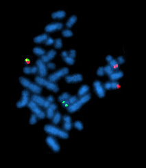
A metaphase cell positive for the BCR/ABL rearrangement using FISH

Cytogenetics is essentially a branch of genetics, but is also a part of cell biology/cytology (a subdivision of human anatomy), that is concerned with how the chromosomes relate to cell behaviour, particularly to their behaviour during mitosis and meiosis. Techniques used include karyotyping, analysis of G-banded chromosomes, other cytogenetic banding techniques, as well as molecular cytogenetics such as fluorescent in situ hybridization (FISH) and comparative genomic hybridization (CGH).

History

During early time

Chromosomes were first observed in plant cells by Karl Wilhelm von Nägeli in 1842. Their behavior in animal (salamander) cells was described by Walther Flemming, the discoverer of mitosis, in 1882. The name was coined by another German anatomist, von Waldeyer in 1888.

The next stage took place after the development of genetics in the early 20th century, when it was appreciated that the set of chromosomes (the karyotype) was the carrier of the genes. Levitsky seems to have been the first to define the karyotype as the phenotypic appearance of the somatic chromosomes, in contrast to their genic contents. Investigation into the human karyotype took many years to settle the most basic question: how many chromosomes does a normal diploid human cell contain? In 1912, Hans von Winiwarter reported 47 chromosomes in spermatogonia and 48 in oogonia, concluding an XX/XO sex determination mechanism. Painter in 1922 was not certain whether the diploid number of humans was 46 or 48, at first favoring 46. He revised his opinion later from 46 to 48, and he correctly insisted on humans having an XX/XY system of sex-determination. Considering their techniques, these results were quite remarkable. In science books, the number of human chromosomes remained at 48 for over thirty years. New techniques were needed to correct this error. Joe Hin Tjio working in Albert Levan's lab was responsible for finding the approach: 

Using cells in culture
  1. Pre-treating cells in a hypotonic solution, which swells them and spreads the chromosomes
  2. Arresting mitosis in metaphase by a solution of colchicine 
  3. Squashing the preparation on the slide forcing the chromosomes into a single plane
  4. Cutting up a photomicrograph and arranging the result into an indisputable karyogram.
It took until 1956 for it to be generally accepted that the karyotype of man included only 46 chromosomes. The great apes have 48 chromosomes. Human chromosome 2 was formed by a merger of ancestral chromosomes, reducing the number.

Applications in biology

McClintock's work on maize

Barbara McClintock began her career as a maize cytogeneticist. In 1931, McClintock and Harriet Creighton demonstrated that cytological recombination of marked chromosomes correlated with recombination of genetic traits (genes). McClintock, while at the Carnegie Institution, continued previous studies on the mechanisms of chromosome breakage and fusion flare in maize. She identified a particular chromosome breakage event that always occurred at the same locus on maize chromosome 9, which she named the "Ds" or "dissociation" locus. McClintock continued her career in cytogenetics studying the mechanics and inheritance of broken and ring (circular) chromosomes of maize. During her cytogenetic work, McClintock discovered transposons, a find which eventually led to her Nobel Prize in 1983.

Natural populations of Drosophila

In the 1930s, Dobzhansky and his coworkers collected Drosophila pseudoobscura and D. persimilis from wild populations in California and neighboring states. Using Painter's technique they studied the polytene chromosomes and discovered that the wild populations were polymorphic for chromosomal inversions. All the flies look alike whatever inversions they carry: this is an example of a cryptic polymorphism.

Evidence rapidly accumulated to show that natural selection was responsible. Using a method invented by L'Héritier and Teissier, Dobzhansky bred populations in population cages, which enabled feeding, breeding and sampling whilst preventing escape. This had the benefit of eliminating migration as a possible explanation of the results. Stocks containing inversions at a known initial frequency can be maintained in controlled conditions. It was found that the various chromosome types do not fluctuate at random, as they would if selectively neutral, but adjust to certain frequencies at which they become stabilised. By the time Dobzhansky published the third edition of his book in 1951 he was persuaded that the chromosome morphs were being maintained in the population by the selective advantage of the heterozygotes, as with most polymorphisms.

Lily and mouse

The lily is a favored organism for the cytological examination of meiosis since the chromosomes are large and each morphological stage of meiosis can be easily identified microscopically. Hotta et al. presented evidence for a common pattern of DNA nicking and repair synthesis in male meiotic cells of lilies and rodents during the zygotene–pachytene stages of meiosis when crossing over was presumed to occur. The presence of a common pattern between organisms as phylogenetically distant as lily and mouse led the authors to conclude that the organization for meiotic crossing-over in at least higher eukaryotes is probably universal in distribution.

Human abnormalities and medical applications

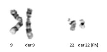
Philadelphia translocation t(9;22)(q34;q11.2) seen in chronic myelogenous leukemia.

Following the advent of procedures which allowed easy enumeration of chromosomes, discoveries were quickly made related to aberrant chromosomes or chromosome number. In some congenital disorders, such as Down syndrome, cytogenetics revealed the nature of the chromosomal defect: a "simple" trisomy. Abnormalities arising from nondisjunction events can cause cells with aneuploidy (additions or deletions of entire chromosomes) in one of the parents or in the fetus. In 1959, Lejeune discovered patients with Down syndrome had an extra copy of chromosome 21. Down syndrome is also referred to as trisomy 21.

Other numerical abnormalities discovered include sex chromosome abnormalities. A female with only one X chromosome has Turner syndrome, whereas an additional X chromosome in a male, resulting in 47 total chromosomes, has Klinefelter syndrome. Many other sex chromosome combinations are compatible with live birth including XXX, XYY, and XXXX. The ability for mammals to tolerate aneuploidies in the sex chromosomes arises from the ability to inactivate them, which is required in normal females to compensate for having two copies of the chromosome. Not all genes on the X chromosome are inactivated, which is why there is a phenotypic effect seen in individuals with extra X chromosomes.

Trisomy 13 was associated with Patau syndrome and trisomy 18 with Edwards syndrome.

In 1960, Peter Nowell and David Hungerford discovered a small chromosome in the white blood cells of patients with Chronic myelogenous leukemia (CML). This abnormal chromosome was dubbed the Philadelphia chromosome - as both scientists were doing their research in Philadelphia, Pennsylvania. Thirteen years later, with the development of more advanced techniques, the abnormal chromosome was shown by Janet Rowley to be the result of a translocation of chromosomes 9 and 22. Identification of the Philadelphia chromosome by cytogenetics is diagnostic for CML.

Advent of banding techniques

Human male karyotype.

In the late 1960s, Torbjörn Caspersson developed a quinacrine fluorescent staining technique (Q-banding) which revealed unique banding patterns for each chromosome pair. This allowed chromosome pairs of otherwise equal size to be differentiated by distinct horizontal banding patterns. Banding patterns are now used to elucidate the breakpoints and constituent chromosomes involved in chromosome translocations. Deletions and inversions within an individual chromosome can also be identified and described more precisely using standardized banding nomenclature. G-banding (utilizing trypsin and Giemsa/ Wright stain) was concurrently developed in the early 1970s and allows visualization of banding patterns using a bright field microscope. 

Diagrams identifying the chromosomes based on the banding patterns are known as idiograms. These maps became the basis for both prenatal and oncological fields to quickly move cytogenetics into the clinical lab where karyotyping allowed scientists to look for chromosomal alterations. Techniques were expanded to allow for culture of free amniocytes recovered from amniotic fluid, and elongation techniques for all culture types that allow for higher-resolution banding.

Beginnings of molecular cytogenetics

In the 1980s, advances were made in molecular cytogenetics. While radioisotope-labeled probes had been hybridized with DNA since 1969, movement was now made in using fluorescent labeled probes. Hybridizing them to chromosomal preparations using existing techniques came to be known as fluorescence in situ hybridization (FISH). This change significantly increased the usage of probing techniques as fluorescent labeled probes are safer. Further advances in micromanipulation and examination of chromosomes led to the technique of chromosome microdissection whereby aberrations in chromosomal structure could be isolated, cloned and studied in ever greater detail.

Techniques

Karyotyping

The routine chromosome analysis (Karyotyping) refers to analysis of metaphase chromosomes which have been banded using trypsin followed by Giemsa, Leishmanns, or a mixture of the two. This creates unique banding patterns on the chromosomes. The molecular mechanism and reason for these patterns is unknown, although it likely related to replication timing and chromatin packing.

Several chromosome-banding techniques are used in cytogenetics laboratories. Quinacrine banding (Q-banding) was the first staining method used to produce specific banding patterns. This method requires a fluorescence microscope and is no longer as widely used as Giemsa banding (G-banding). Reverse banding, or R-banding, requires heat treatment and reverses the usual black-and-white pattern that is seen in G-bands and Q-bands. This method is particularly helpful for staining the distal ends of chromosomes. Other staining techniques include C-banding and nucleolar organizing region stains (NOR stains). These latter methods specifically stain certain portions of the chromosome. C-banding stains the constitutive heterochromatin, which usually lies near the centromere, and NOR staining highlights the satellites and stalks of acrocentric chromosomes. High-resolution banding involves the staining of chromosomes during prophase or early metaphase (prometaphase), before they reach maximal condensation. Because prophase and prometaphase chromosomes are more extended than metaphase chromosomes, the number of bands observable for all chromosomes increases from about 300 to 450 to as many as 800. This allows the detection of less obvious abnormalities usually not seen with conventional banding.

Slide preparation

Cells from bone marrow, blood, amniotic fluid, cord blood, tumor, and tissues (including skin, umbilical cord, chorionic villi, liver, and many other organs) can be cultured using standard cell culture techniques in order to increase their number. A mitotic inhibitor (colchicine, colcemid) is then added to the culture. This stops cell division at mitosis which allows an increased yield of mitotic cells for analysis. The cells are then centrifuged and media and mitotic inhibitor are removed, and replaced with a hypotonic solution. This causes the white blood cells or fibroblasts to swell so that the chromosomes will spread when added to a slide as well as lyses the red blood cells. After the cells have been allowed to sit in hypotonic solution, Carnoy's fixative (3:1 methanol to glacial acetic acid) is added. This kills the cells and hardens the nuclei of the remaining white blood cells. The cells are generally fixed repeatedly to remove any debris or remaining red blood cells. The cell suspension is then dropped onto specimen slides. After aging the slides in an oven or waiting a few days they are ready for banding and analysis.

Analysis

Analysis of banded chromosomes is done at a microscope by a clinical laboratory specialist in cytogenetics (CLSp(CG)). Generally 20 cells are analyzed which is enough to rule out mosaicism to an acceptable level. The results are summarized and given to a board-certified cytogeneticist for review, and to write an interpretation taking into account the patient's previous history and other clinical findings. The results are then given out reported in an International System for Human Cytogenetic Nomenclature 2009 (ISCN2009).

Fluorescent in situ hybridization

Interphase cells positive for a t(9;22) rearrangement

Fluorescent in situ hybridization (FISH) refers to using fluorescently labeled probe to hybridize to cytogenetic cell preparations. 

In addition to standard preparations FISH can also be performed on:

Slide preparation

This section refers to preparation of standard cytogenetic preparations
 
The slide is aged using a salt solution usually consisting of 2X SSC (salt, sodium citrate). The slides are then dehydrated in ethanol, and the probe mixture is added. The sample DNA and the probe DNA are then co-denatured using a heated plate and allowed to re-anneal for at least 4 hours. The slides are then washed to remove excess unbound probe, and counterstained with 4',6-Diamidino-2-phenylindole (DAPI) or propidium iodide.

Analysis

Analysis of FISH specimens is done by fluorescence microscopy by a clinical laboratory specialist in cytogenetics. For oncology generally a large number of interphase cells are scored in order to rule out low-level residual disease, generally between 200 and 1,000 cells are counted and scored. For congenital problems usually 20 metaphase cells are scored.

Future of cytogenetics

Advances now focus on molecular cytogenetics including automated systems for counting the results of standard FISH preparations and techniques for virtual karyotyping, such as comparative genomic hybridization arrays, CGH and Single nucleotide polymorphism arrays.
Spindle apparatus

From Wikipedia, the free encyclopedia
Micrograph showing condensed chromosomes in blue, kinetochores in pink, and microtubules in green during metaphase of mitosis
 
In cell biology, the spindle apparatus (or mitotic spindle) refers to the cytoskeletal structure of eukaryotic cells that forms during cell division to separate sister chromatids between daughter cells. It is referred to as the mitotic spindle during mitosis, a process that produces genetically identical daughter cells, or the meiotic spindle during meiosis, a process that produces gametes with half the number of chromosomes of the parent cell.

Besides chromosomes, the spindle apparatus is composed of hundreds of proteins. Microtubules comprise the most abundant components of the machinery.

Spindle structure

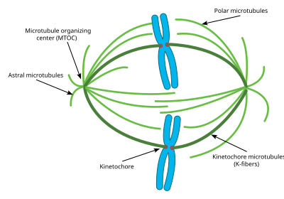
This diagram depicts the organization of a typical mitotic spindle found in animal cells. Chromosomes are attached to kinetochore microtubules via a multiprotein complex called the kinetochore. Polar microtubules interdigitate at the spindle midzone and push the spindle poles apart via motor proteins. Astral microtubules anchor the spindle poles to the cell membrane. Microtubule polymerization is nucleated at the microtubule organizing center.
 
Attachment of microtubules to chromosomes is mediated by kinetochores, which actively monitor spindle formation and prevent premature anaphase onset. Microtubule polymerization and depolymerization dynamic drive chromosome congression. Depolymerization of microtubules generates tension at kinetochores; bipolar attachment of sister kinetochores to microtubules emanating from opposite cell poles couples opposing tension forces, aligning chromosomes at the cell equator and poising them for segregation to daughter cells. Once every chromosome is bi-oriented, anaphase commences and cohesin, which couples sister chromatids, is severed, permitting the transit of the sister chromatids to opposite poles.

The cellular spindle apparatus includes the spindle microtubules, associated proteins, which include kinesin and dynein molecular motors, condensed chromosomes, and any centrosomes or asters that may be present at the spindle poles depending on the cell type. The spindle apparatus is vaguely ellipsoid in cross section and tapers at the ends. In the wide middle portion, known as the spindle midzone, antiparallel microtubules are bundled by kinesins. At the pointed ends, known as spindle poles, microtubules are nucleated by the centrosomes in most animal cells. Acentrosomal or anastral spindles lack centrosomes or asters at the spindle poles, respectively, and occur for example during female meiosis in most animals. In this instance, a Ran GTP gradient is the main regulator of spindle microtubule organization and assembly. In fungi, spindles form between spindle pole bodies embedded in the nuclear envelope, which does not break down during mitosis.

Microtubule-associated proteins and spindle dynamics

The dynamic lengthening and shortening of spindle microtubules, through a process known as dynamic instability determines to a large extent the shape of the mitotic spindle and promotes the proper alignment of chromosomes at the spindle midzone. Microtubule-associated proteins (MAPs) associate with microtubules at the midzone and the spindle poles to regulate their dynamics. γ-tubulin is a specialized tubulin variant that assembles into a ring complex called γ-TuRC which nucleates polymerization of α/β tubulin heterodimers into microtubules. Recruitment of γ-TuRC to the pericentrosomal region stabilizes microtubule minus-ends and anchors them near the microtubule-organizing center. The microtubule-associated protein Augmin acts in conjunction with γ-TURC to nucleate new microtubules off of existing microtubules.

The growing ends of microtubules are protected against catastrophe by the action of plus-end microtubule tracking proteins (+TIPs) to promote their association with kinetochores at the midzone. CLIP170 was shown to localize near microtubule plus-ends in HeLa cells  and to accumulate in kinetochores during prometaphase. Although how CLIP170 recognizes plus-ends remains unclear, it has been shown that its homologues protect against catastrophe and promote rescue, suggesting a role for CLIP170 in stabilizing plus-ends and possibly mediating their direct attachment to kinetochores. CLIP-associated proteins like CLASP1 in humans have also been shown to localize to plus-ends and the outer kinetochore as well as to modulate the dynamics of kinetochore microtubules (Maiato 2003). CLASP homologues in Drosophila, Xenopus, and yeast are required for proper spindle assembly; in mammals, CLASP1 and CLASP2 both contribute to proper spindle assembly and microtubule dynamics in anaphase. Plus-end polymerization may be further moderated by the EB1 protein, which directly binds the growing ends of microtubules and coordinates the binding of other +TIPs.

Opposing the action of these microtubule-stabilizing proteins are a number of microtubule-depolymerizing factors which permit the dynamic remodeling of the mitotic spindle to promote chromosome congression and attainment of bipolarity. The kinesin-13 superfamily of MAPs contains a class of plus-end-directed motor proteins with associated microtubule depolymerization activity including the well-studied mammalian MCAK and Xenopus XKCM1. MCAK localizes to the growing tips of microtubules at kinetochores where it can trigger catastrophe in direct competition with stabilizing +TIP activity. These proteins harness the energy of ATP hydrolysis to induce destabilizing conformational changes in protofilament structure that cause kinesin release and microtubule depolymerization. Loss of their activity results in numerous mitotic defects. Additional microtubule destabilizing proteins include Op18/stathmin and katanin which have roles in remodeling the mitotic spindle as well as promoting chromosome segregation during anaphase.

The activities of these MAPs are carefully regulated to maintain proper microtubule dynamics during spindle assembly, with many of these proteins serving as Aurora and Polo-like kinase substrates.

Organizing the spindle apparatus

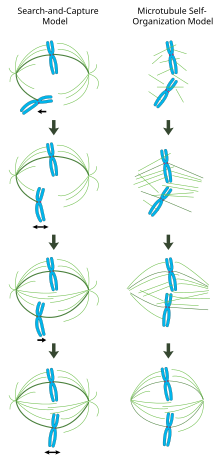
In the centrosome-mediated “search and capture” model (left), microtubules nucleated from centrosomes contact chromosomes by chance and become stabilized at kinetochores to form the spindle. In the chromatin-mediated “self-organization” model (right), microtubules are nucleated around the vicinity of mitotic chromatin and organized into a bipolar array by motor proteins.
 
In a properly formed mitotic spindle, bi-oriented chromosomes are aligned along the equator of the cell with spindle microtubules oriented roughly perpendicular to the chromosomes, their plus-ends embedded in kinetochores and their minus-ends anchored at the cell poles. The precise orientation of this complex is required to ensure accurate chromosome segregation and to specify the cell division plane. However, it remains unclear how the spindle becomes organized. Two models predominate the field, which are synergistic and not mutually exclusive. In the search-and-capture model, the spindle is predominantly organized by the poleward separation of centrosomal microtubule organizing centers (MTOCs). Spindle microtubules emanate from centrosomes and 'seek' out kinetochores; when they bind a kinetochore they become stabilized and exert tension on the chromosomes. In an alternative self assembly model, microtubules undergo acentrosomal nucleation among the condensed chromosomes. Constrained by cellular dimensions, lateral associations with antiparallel microtubules via motor proteins, and end-on attachments to kinetochores, microtubules naturally adopt a spindle-like structure with chromosomes aligned along the cell equator.

Centrosome-mediated "search-and-capture" model

In this model, microtubules are nucleated at microtubule organizing centers and undergo rapid growth and catastrophe to 'search' the cytoplasm for kinetochores. Once they bind a kinetochore, they are stabilized and their dynamics are reduced. The newly mono-oriented chromosome oscillates in space near the pole to which it is attached until a microtubule from the opposite pole binds the sister kinetochore. This second attachment further stabilizes kinetochore attachment to the mitotic spindle. Gradually, the bi-oriented chromosome is pulled towards the center of the cell until microtubule tension is balanced on both sides of the centromere; the congressed chromosome then oscillates at the metaphase plate until anaphase onset releases cohesion of the sister chromatids.

In this model, microtubule organizing centers are localized to the cell poles, their separation driven by microtubule polymerization and 'sliding' of antiparallel spindle microtubules with respect to one another at the spindle midzone mediated by bipolar, plus-end-directed kinesins.[19][20] Such sliding forces may account not only for spindle pole separation early in mitosis, but also spindle elongation during late anaphase.

Chromatin-mediated self-organization of the mitotic spindle

In contrast to the search-and-capture mechanism in which centrosomes largely dictate the organization of the mitotic spindle, this model proposes that microtubules are nucleated acentrosomally near chromosomes and spontaneously assemble into anti-parallel bundles and adopt a spindle-like structure. Classic experiments by Heald and Karsenti show that functional mitotic spindles and nuclei form around DNA-coated beads incubated in Xenopus egg extracts and that bipolar arrays of microtubules are formed in the absence of centrosomes and kinetochores. Indeed, it has also been shown that laser ablation of centrosomes in vertebrate cells inhibits neither spindle assembly nor chromosome segregation. Under this scheme, the shape and size of the mitotic spindle are a function of the biophysical properties of the cross-linking motor proteins.

Chromatin-mediated microtubule nucleation by the Ran GTP gradient

The guanine nucleotide exchange factor for the small GTPase Ran (Regulator of chromosome condensation 1 or RCC1) is attached to nucleosomes via core histones H2A and H2B. Thus, a gradient of GTP-bound Ran is generated around the vicinity of mitotic chromatin. Glass beads coated with RCC1 induce microtubule nucleation and bipolar spindle formation in Xenopus egg extracts, revealing that the Ran GTP gradient alone is sufficient for spindle assembly. The gradient triggers release of spindle assembly factors (SAFs) from inhibitory interactions via the transport proteins importin β/α. The unbound SAFs then promote microtubule nucleation and stabilization around mitotic chromatin, and spindle bipolarity is organized by microtubule motor proteins.

Regulation of spindle assembly

Spindle assembly is largely regulated by phosphorylation events catalyzed by mitotic kinases. Cyclin dependent kinase complexes (CDKs) are activated by mitotic cyclins, whose translation increases during mitosis. CDK1 (also called CDC2) is considered the main mitotic kinase in mammalian cells and is activated by Cyclin B1. Aurora kinases are required for proper spindle assembly and separation. Aurora A associates with centrosomes and is believed to regulate mitotic entry. Aurora B is a member of the chromosomal passenger complex and mediates chromosome-microtubule attachment and sister chromatid cohesion. Polo-like kinase, also known as PLK, especially PLK1 has important roles in the spindle maintenance by regulating microtubule dynamics.

Mitotic chromosome structure

By the end of DNA replication, sister chromatids are bound together in an amorphous mass of tangled DNA and protein that would be virtually impossible to partition into each daughter cell. To avoid this problem, mitotic entry triggers a dramatic reorganization of the duplicated genome. Sister chromatids are disentangled and resolved from one another. Chromosomes also shorten in length, up to 10,000 fold in animal cells, in a process called condensation. Condensation begins in prophase and chromosomes are maximally compacted into rod-shaped structures by the time they are aligned in the middle of the spindle at metaphase. This gives mitotic chromosomes the classic “X” shape seen in karyotypes, with each condensed sister chromatid linked along their lengths by cohesin proteins and joined, often near the center, at the centromere.

While these dynamic rearrangements are vitally important to ensure accurate and high-fidelity segregation of the genome, our understanding of mitotic chromosome structure remains largely incomplete. A few specific molecular players have been identified, however: Topoisomerase II uses ATP hydrolysis to catalyze decatenation of DNA entanglements, promoting sister chromatid resolution. Condensins are 5-subunit complexes that also use ATP-hydrolysis to promote chromosome condensation. Experiments in Xenopus egg extracts have also implicated linker Histone H1 as an important regulator of mitotic chromosome compaction.

Mitotic spindle assembly checkpoint

The completion of spindle formation is a crucial transition point in the cell cycle called the spindle assembly checkpoint. If chromosomes are not properly attached to the mitotic spindle by the time of this checkpoint, the onset of anaphase will be delayed. Failure of this spindle assembly checkpoint can result in aneuploidy and may be involved in aging and the formation of cancer.

Spindle apparatus orientation

Cartoon of the dividing epithelium cell surrounded by epithelium tissue. Spindle apparatus rotates inside the cell. The rotation is a result of astral microtubules pulling towards tri-cellular-junctions (TCJ), signaling centers localized at the regions where three cells meet.
 
Cell division orientation is of major importance for tissue architecture, cell fates and morphogenesis. Cells tend to divide along their long axis according to the so-called Hertwig rule. The axis of cell division is determined by the orientation of the spindle apparatus. Cells divide along the line connecting two centrosomes of the spindle apparatus. After formation, the spindle apparatus undergoes rotation inside the cell. The astral microtubules originating from centrosomes reach the cell membrane where they are pulled towards specific cortical clues. In vitro, the distribution of cortical clues is set up by the adhesive pattern. In vivo polarity cues are determined by localization of Tricellular junctions localized at cell vertices. The spatial distribution of cortical clues leads to the force field that determine final spindle apparatus orientation and the subsequent orientation of cell division.

UNESCO-CEPES

From Wikipedia, the free encyclopedia
 
UNESCO-CEPES (Centre Européen pour l’Enseignement Supérieur – CEPES) was established in 1972 at Bucharest, Romania, as a de-centralized office for the European Centre for Higher Education. The Centre was closed in 2011 due to lack of funding. The centre promoted international cooperation in the sphere of higher education among UNESCO’s Member States in Central, Eastern and South-East Europe and also served Canada, the United States and Israel. Higher Education in Europe, a scholarly publication focusing on major problems and trends in higher education, was the official journal of UNESCO-CEPES. The CEPES headquarters was in the Kretzulescu Palace in Bucharest.

The CEPES member countries

Central Europe Eastern Europe South-Eastern Other regions
Austria Estonia Albania Belgium
Czech Republic Latvia Bosnia and Herzegovina Denmark
Croatia Lithuania Bulgaria Finland
France Republic of Moldova Cyprus Holy See
Germany Ukraine Republic of Macedonia Ireland
Hungary
Republic of Montenegro Malta
Italy
Turkey Netherlands
Liechtenstein

Norway
Poland

Portugal
Romania

Spain
Serbia

Sweden
Slovakia

UK
Slovenia

Canada
Switzerland

USA


Israel

History

On 21 September 1972, as the only intergovernmental Centre for Higher Education in Europe region, North America and Israel, UNESCO European Centre for Higher Education (Centre Européen pour l’Enseignement Supérieur – CEPES) was established in Bucharest. The early mission of the CEPES was to encourage cooperation, to disseminate information, and to research modern trends in higher education within the Europe Region. In the early 1990s, with the fall of the communist regimes in Central and Eastern Europe, the role of UNESCO-CEPES extended its round has been broader.

on this endeavour, the UNITWIN/UNESCO Chairs Programme constituted "a major breakthrough with regard to the reinforcement of inter-university co-operation at the sub-regional, regional and interregional levels as a means to improve the quality in higher education as well as to strengthen national capabilities for higher level training and research in the developing countries." 

In April 1997, the joint Council of Europe/UNESCO Convention on the Recognition of Qualifications Concerning Higher Education in the European Region was adopted, and UNESCO-CEPES assumed a Co-Secretariat function to the Convention. From the late 1990s, the Centre gradually more co-worked on European Union projects aimed at the reform of higher education in Eastern and Central Europe and reinforced its cooperation with international organisations such as World Bank, OECD, and others.

In September 2003, UNESCO-CEPES was nominated a consultative member of the Follow-up Group of the Bologna Process (BFUG), charged with the accomplishment of Bologna Process goals, and the actualisation of the European Higher Education Area (EHEA).

On 25 September 2009, according as a Memorandum of Understanding (MoU) was signed between UNESCO and the Romanian Government on transitional arrangements for UNESCO-CEPES, The MoU realigns the Centre's mandate with the new education landscape in Europe and provides that during the 2010-2011 period. CEPES will focus on addressing the needs of higher education of UNESCO's Member States in Central, Eastern and South-East Europe.

On 31 December 2011, the Centre was closed as funding was not ensured by the Government of Romania or other countries in the region, which is a requirement for all UNESCO Regional Centres.

Mission

The UNESCO European Centre for Higher Education/Centre européen pour l'enseignement supérieur (CEPES) promotes co-operation and provides technical support in the field of higher education among UNESCO's Member States in Central, Eastern and South-East Europe.

Specifically UNESCO-CEPES:
  • Undertakes projects relevant to the development and reform of higher education, specifically in view of the follow-up to the 2009 UNESCO World Conference on Higher Education, and the Bologna Process aiming at the creation of the European Higher Education Area;
  • Promotes policy development and research on higher education and serves as a forum for the discussion of important topics in higher education;
  • Gathers and disseminates a wide range of information on higher education;
  • Coordinates, within the UNITWIN/UNESCO Chairs Programme, relations with a designated number of UNESCO Chairs relevant to its activities;
  • Provides consultancy services;
  • Participates in the activities of other governmental and non-governmental organizations;
  • Serves as a link between UNESCO Headquarters and Romania.

Recent Events

Date Events
9 March 2010 Reception for Presentation of the Book Collection "Patrimoniul Umanităţii din România".
4–5 March 2010 Romanian Research Assessment Exercise, organized by UEFISCSU
24 February 2010 Programme Planning Meeting, organized by UNICEF
18 February 2010 Mutual Learning Workshop, organized by UEFISCSU
3 December 2009 Stakeholders Engagement Day, organized by Accountability and Aston Eco Management Company
20 November 2009 UN Convention Day for the Right of the Child, organized by ONG Romanian Federation for Children
26–30 October 2009 The Art Exhibition organized by UNESCO-CEPES on the occasion of UN DAY 2009
23 October 2009 Opening Ceremony of the Art exposition "Our World; Our Climate"
27 September 2009 First Forum on Energy Efficiency in Romania
24–25 September 2009 Systems Thinking for Foresight: The case of Romanian Higher Education System

UNESCO Institutes and Centres for Education around the world

Including UNESCO-CEPES, there are many institutes and centres on the world that UNESCO established. following lists work as part of UNESCO's Education Programme to assist countries to tackle challenges in education.

Global

Regional

Africa
  • International Institute for Capacity Building in Africa (IICBA), Addis Ababa, Ethiopia. - Strengthening Africa's educational institutions.
Europe and North America
  • European Centre for Higher Education (CEPES), Bucharest, Romania. - Promoting cooperation and reform in higher education in Central and Eastern Europe.
Latin America and Caribbean
  • International Institute for Higher Education in Latin America and the Caribbean (IESALC), (website in Spanish), Caracas, Venezuela. - Developing and transforming higher education in the region.
Centres under the auspices of UNESCO
The five centres under the auspices of UNESCO (category 2) complement and expand UNESCO's education programme.
  • Asia-Pacific Centre of Education for International Understanding (APCEIU), Icheon, Republic of Korea.
  • International Centre for Girls and Women's Education in Africa (CIEFFA), Ouagadougou, Burkina Faso.
  • Guidance, Counselling and Youth Development Centre for Africa (GCYDCA), Lilongwe, Malawi.
  • International Research and Training Center for Rural Education (INRULED), Beijing, China.
  • Regional Centre for Educational Planning (RCEP), Sharjah, United Arab Emirates.
New centres and institutes to be established
  • The South-East Asian Centre for Lifelong Learning for Sustainable Development (SEACLLSD), Manila, Philippines
  • The Regional Centre for Early Childhood Care and Education in the Arab States Damascus, Syrian Arab Republic

Partner and Support Organisations

Partner International Governmental and Non-governmental Organizations Operating in the Field of (Higher) Education Reform and Policy Development
  • Center for Higher Education Development (CHE)
  • Council of Europe (CE)
  • German Academic Exchange Service (DAAD)
  • Education International (EI)
  • Elias Foundation
  • European Association for Quality Assurance in Higher Education (ENQA)
  • European Quality Assurance Register for Higher Education (EQAR)
  • European Centre for Strategic Management of Universities (ESMU)
  • European Students' Union (ESU)
  • European University Association (EUA)
  • European Union (EU)
  • European Commission - Eurydice
  • International Association of Universities (IAU)
  • Observatory of the Magna Charta Universitatum
  • UNESCO International Bureau of Education (IBE)
  • UNESCO International Institute for Educational Planning (IIEP)
  • The Quality Assurance Agency for Higher Education (QAA)
  • Organization for Economic Co-operation and Development (OECD)
  • United Nations Development Programme (UNDP)
  • United Nations Educational, Scientific and Cultural Organization (UNESCO)
  • UNESCO National Commissions
  • University of Cambridge
  • World University Service (WUS)
Support Organizations of UNESCO – CEPES Activities
  • British Council Romania
  • Calouste Gulbenkian Foundation
  • Deutsche Telekom
  • Hertie Foundation
  • Microsoft Romania
  • National Bank of Romania
  • Okian Publishing Romania
  • RAO Publishing
  • Samsung Romania

Intangible cultural heritage

From Wikipedia, the free encyclopedia
 
Logo of Convention for the Safeguarding of the Intangible Cultural Heritage
 
An intangible cultural heritage (ICH) is a practice, representation, expression, knowledge, or skill considered by UNESCO to be part of a place's cultural heritage. Buildings, historic places, monuments, and artifacts are physical intellectual wealth. Intangible heritage is comprised of nonphysical intellectual wealth, such as folklore, customs, beliefs, traditions, knowledge, and language. Intangible cultural heritage is considered by member states of UNESCO in relation to the tangible World Heritage focusing on intangible aspects of culture. In 2001, UNESCO made a survey among States and NGOs to try to agree on a definition, and the Convention for the Safeguarding of Intangible Cultural Heritage was drafted in 2003 for its protection and promotion.

Definition

The Convention for the Safeguarding of the Intangible Cultural Heritage defines the intangible cultural heritage as the practices, representations, expressions, as well as the knowledge and skills (including instruments, objects, artifacts, cultural spaces), that communities, groups and, in some cases, individuals recognise as part of their cultural heritage. It is sometimes called living cultural heritage, and is manifested inter alia in the following domains:
  • Oral traditions and expressions, including language as a vehicle of the intangible cultural heritage;
  • Performing arts;
  • Social practices, rituals and festive events;
  • Knowledge and practices concerning nature and the universe;
  • Traditional craftsmanship
Noh mask; Japan was the first country to introduce legislation to protect and promote its intangible heritage
 
Cultural heritage in general consists of the products and processes of a culture that are preserved and passed on through the generations. Some of that heritage takes the form of cultural property, formed by tangible artefacts such as buildings or works of art. Many parts of culture, however are intangible, including song, music, dance, drama, skills, cuisine, crafts and festivals. They are forms of culture that can be recorded but cannot be touched or stored in physical form, like in a museum, but only experienced through a vehicle giving expression to it. These cultural vehicles are called "Human Treasures" by the UN.

According to the 2003 Convention for the Safeguarding of the Intangible Cultural Heritage, the intangible cultural heritage (ICH) – or living heritage – is the mainspring of humanity's cultural diversity and its maintenance a guarantee for continuing creativity. It is defined as follows:
Intangible Cultural Heritage means the practices, representations, expressions, knowledge, skills – as well as the instruments, objects, artifacts and cultural spaces associated therewith – that communities, groups and, in some cases, individuals recognize as part of their cultural heritage. This intangible cultural heritage, transmitted from generation to generation, is constantly recreated by communities and groups in response to their environment, their interaction with nature and their history, and provides them with a sense of identity and continuity, thus promoting respect for cultural diversity and human creativity. For the purposes of this Convention, consideration will be given solely to such intangible cultural heritage as is compatible with existing international human rights instruments, as well as with the requirements of mutual respect among communities, groups and individuals, and of sustainable development.

Oral history

Intangible cultural heritage is slightly different from the discipline of oral history, the recording, preservation and interpretation of historical information (specifically, oral tradition), based on the personal experiences and opinions of the speaker. ICH attempts to preserve cultural heritage 'with' the people or community by protecting the processes that allow traditions and shared knowledge to be passed on while oral history seeks to collect and preserve historical information obtained from individuals and groups.

Food heritage

With sustainable development gaining momentum as a priority of UNESCO heritage policies, an increasing number of food-related nominations are being submitted for inscription on the lists of the Convention for the safeguarding of the intangible cultural heritage. The Mediterranean diet, the traditional Mexican cuisine and the Japanese dietary culture of washoku are just some examples of this booming phenomenon.

Dance heritage

The UNESCO lists of intangible cultural heritage also include a variety of dance genres, often associated with singing, music and celebrations, from all over the world. The lists include: celebratory and ritual dances such as Ma'di bowl lyre music and dance from Uganda and Kalbelia folk songs and dances of Rajasthan from India, and social dances such as Cuban rumba. Also, some dances are localised and practised mainly in their country of origin, such as Sankirtana, a performing art that includes drumming and singing, from India. 

Other dance forms, however, even if they are officially recognised as heritage from their country of origin, are practised and enjoyed all over the world. For example, flamenco from Spain and tango, from Argentina and Uruguay, have a very international dimension. Dance is a very complex phenomenon, which involves culture, traditions, the use of human bodies, artefacts (such as costumes and props), as well as a specific use of music, space and sometimes light. As a result, a lot of tangible and intangible elements are combined within dance, making it a challenging but extremely interesting type of heritage to safeguard.

Digital heritage

Digital heritage is a representation of heritage in the digital realm.

Digital intangible heritage

Digital intangible heritage is a sub-category of Intangible Cultural Heritage.

Oral continuity

Albanian polyphonic folk group wearing qeleshe and fustanella in Skrapar

Intangible cultural heritage is passed orally within a community, and while there may be individuals who are known tradition bearers, ICH is often broader than one individual's own skills or knowledge. A 2006 report by the government of Newfoundland and Labrador said, regarding oral culture in their area, "The processes involved in the continuation of this traditional knowledge constitute one of the most interesting aspects of our living heritage. Each member of the community possesses a piece of the shared knowledge. Crucial knowledge is passed on during community activities, frequently without any conscious attention to the process."

Preservation

Prior to the UNESCO Convention, efforts had already been made by a number of states to safeguard their intangible heritage. Japan, with its 1950 Law for the Protection of Cultural Properties, was the first to introduce legislation to preserve and promote intangible as well as tangible culture: Important Intangible Cultural Properties are designated and "holders" recognized of these craft and performance traditions, known informally as Living National Treasures. Other countries, including South Korea (Important Intangible Cultural Properties of Korea), the Philippines, the United States, Thailand, France, Romania, the Czech Republic, and Poland, have since created similar programs.

In 2003 UNESCO adopted the Convention for the Safeguarding of the Intangible Cultural Heritage. This went into effect on 20 April 2006. The Convention recommends that countries and scholars develop inventories of ICH in their territory, as well as work with the groups who maintain these ICH to ensure their continued existences; it also provides for funds to be voluntarily collected among UNESCO members and then disbursed to support the maintenance of recognized ICH. UNESCO has also created other intangible culture programs, such as a list called Proclamation of Masterpieces of the Oral and Intangible Heritage of Humanity. This list began in 2001 with 19 items and a further 28 were listed in 2003 and another 43 in 2005. In part, the original list was seen as a way to correct the imbalance in the World Heritage List, since it excluded many Southern Hemisphere cultures which did not produce monuments or other physical cultural manifestations. It was superseded in 2008 by the UNESCO Intangible Cultural Heritage Lists

Recently there has been much debate over protecting intangible cultural heritage through intellectual property rights, as well as the desirability to do so through this legal framework and the risks of commodification derived from this possibility. The issue still remains open in legal scholarship.

By country


Rank Country Number of Intangible Cultural Heritage elements inscribed by UNESCO
1  China 40
2  Japan 21
3  South Korea 20
4  Spain 18
5  Croatia,  France,  Turkey 17
6  Mongolia 14
7  Azerbaijan,  Belgium,  India,  Iran 13
8  Vietnam 12
9  Peru 11
10  Colombia,  Indonesia,  Kazakhstan 10
11  Italy,  Mexico 9
12  Brazil,  Oman,  United Arab Emirates 8
13  Morocco,  Portugal,  Romania 7
14  Saudi Arabia 6

Operator (computer programming)

From Wikipedia, the free encyclopedia https://en.wikipedia.org/wiki/Operator_(computer_programmin...