Search This Blog

Saturday, November 21, 2020

Multisensory integration

From Wikipedia, the free encyclopedia

Multisensory integration, also known as multimodal integration, is the study of how information from the different sensory modalities (such as sight, sound, touch, smell, self-motion, and taste) may be integrated by the nervous system. A coherent representation of objects combining modalities enables animals to have meaningful perceptual experiences. Indeed, multisensory integration is central to adaptive behavior because it allows animals to perceive a world of coherent perceptual entities. Multisensory integration also deals with how different sensory modalities interact with one another and alter each other's processing.

General introduction

Multimodal perception is how animals form coherent, valid, and robust perception by processing sensory stimuli from various modalities. Surrounded by multiple objects and receiving multiple sensory stimulations, the brain is faced with the decision of how to categorize the stimuli resulting from different objects or events in the physical world. The nervous system is thus responsible for whether to integrate or segregate certain groups of temporally coincident sensory signals based on the degree of spatial and structural congruence of those stimulations. Multimodal perception has been widely studied in cognitive science, behavioral science, and neuroscience.

Stimuli and sensory modalities

There are four attributes of stimulus: modality, intensity, location, and duration. The neocortex in the mammalian brain has parcellations that primarily process sensory input from one modality. For example, primary visual area, V1, or primary somatosensory area, S1. These areas mostly deal with low-level stimulus features such as brightness, orientation, intensity, etc. These areas have extensive connections to each other as well as to higher association areas that further process the stimuli and are believed to integrate sensory input from various modalities. However, recently multisensory effects have been shown to occur in primary sensory areas as well.

Binding problem

The relationship between the binding problem and multisensory perception can be thought of as a question – the binding problem, and potential solution – multisensory perception. The binding problem stemmed from unanswered questions about how mammals (particularly higher primates) generate a unified, coherent perception of their surroundings from the cacophony of electromagnetic waves, chemical interactions, and pressure fluctuations that forms the physical basis of the world around us. It was investigated initially in the visual domain (colour, motion, depth, and form), then in the auditory domain, and recently in the multisensory areas. It can be said therefore, that the binding problem is central to multisensory perception.

However, considerations of how unified conscious representations are formed are not the full focus of multisensory Integration research. It is obviously important for the senses to interact in order to maximize how efficiently people interact with the environment. For perceptual experience and behavior to benefit from the simultaneous stimulation of multiple sensory modalities, integration of the information from these modalities is necessary. Some of the mechanisms mediating this phenomenon and its subsequent effects on cognitive and behavioural processes will be examined hereafter. Perception is often defined as one's conscious experience, and thereby combines inputs from all relevant senses and prior knowledge. Perception is also defined and studied in terms of feature extraction, which is several hundred milliseconds away from conscious experience. Notwithstanding the existence of Gestalt psychology schools that advocate a holistic approach to the operation of the brain, the physiological processes underlying the formation of percepts and conscious experience have been vastly understudied. Nevertheless, burgeoning neuroscience research continues to enrich our understanding of the many details of the brain, including neural structures implicated in multisensory integration such as the superior colliculus (SC) and various cortical structures such as the superior temporal gyrus (GT) and visual and auditory association areas. Although the structure and function of the SC are well known, the cortex and the relationship between its constituent parts are presently the subject of much investigation. Concurrently, the recent impetus on integration has enabled investigation into perceptual phenomena such as the ventriloquism effect, rapid localization of stimuli and the McGurk effect; culminating in a more thorough understanding of the human brain and its functions.

History

Studies of sensory processing in humans and other animals has traditionally been performed one sense at a time, and to the present day, numerous academic societies and journals are largely restricted to considering sensory modalities separately ('Vision Research', 'Hearing Research' etc.). However, there is also a long and parallel history of multisensory research. An example is the Stratton's (1896) experiments on the somatosensory effects of wearing vision-distorting prism glasses.  Multisensory interactions or crossmodal effects in which the perception of a stimulus is influenced by the presence of another type of stimulus are referred since very early in the past. They were reviewed by Hartmann in a fundamental book where, among several references to different types of multisensory interactions, reference is made to the work of Urbantschitsch in 1888 who reported on the improvement of visual acuity by auditive stimuli in subjects with damaged brain. This effect was also found latter in normals by Krakov and Hartmann, as well as the fact that the visual acuity could be improved by other type of stimuli. It is also noteworthy the amount of work in the early thirties on intersensory relations in Soviet Union, reviewed by London. A remarkable multisensory research is the extensive work of Gonzalo in the forties on the characterization of a multisensory syndrome in patients with parieto-occipital cortical lesions. In this syndrome, all the sensory functions are affected, and with symmetric bilaterality, in spite of being a unilateral lesion where the primary areas were not involved. A feature of this syndrome is the great permeability to crossmodal effects between visual, tactile, auditive stimuli as well as muscular effort to improve the perception, also decreasing the reaction times. The improvement by crossmodal effect was found to be greater as the primary stimulus to be perceived was weaker, and as the cortical lesion was greater (Vol I and II of reference). This author interpreted these phenomena under a dynamic physiological concept, and from a model based on functional gradients through the cortex and scaling laws of dynamical systems, thus highlighting the functional unity of the cortex. According to the functional cortical gradients, the specificity of the cortex would be distributed in gradation, and the overlap of different specific gradients would be related to multisensory interactions.

Multisensory research has recently gained enormous interest and popularity.

Example of spatial and structural congruence

When we hear a car honk, we would determine which car triggers the honk by which car we see is the spatially closest to the honk. It's a spatially congruent example by combining visual and auditory stimuli. On the other hand, the sound and the pictures of a TV program would be integrated as structurally congruent by combining visual and auditory stimuli. However, if the sound and the pictures did not meaningfully fit, we would segregate the two stimuli. Therefore, spatial or structural congruence comes from not only combining the stimuli but is also determined by our understanding.

Theories and approaches

Visual dominance

Literature on spatial crossmodal biases suggests that visual modality often influences information from other senses. Some research indicates that vision dominates what we hear, when varying the degree of spatial congruency. This is known as the ventriloquist effect. In cases of visual and haptic integration, children younger than 8 years of age show visual dominance when required to identify object orientation. However, haptic dominance occurs when the factor to identify is object size.

Modality appropriateness

According to Welch and Warren (1980), the Modality Appropriateness Hypothesis states that the influence of perception in each modality in multisensory integration depends on that modality's appropriateness for the given task. Thus, vision has a greater influence on integrated localization than hearing, and hearing and touch have a greater bearing on timing estimates than vision.

More recent studies refine this early qualitative account of multisensory integration. Alais and Burr (2004), found that following progressive degradation in the quality of a visual stimulus, participants' perception of spatial location was determined progressively more by a simultaneous auditory cue. However, they also progressively changed the temporal uncertainty of the auditory cue; eventually concluding that it is the uncertainty of individual modalities that determine to what extent information from each modality is considered when forming a percept. This conclusion is similar in some respects to the 'inverse effectiveness rule'. The extent to which multisensory integration occurs may vary according to the ambiguity of the relevant stimuli. In support of this notion, a recent study shows that weak senses such as olfaction can even modulate the perception of visual information as long as the reliability of visual signals is adequately compromised.

Bayesian integration

The theory of Bayesian integration is based on the fact that the brain must deal with a number of inputs, which vary in reliability. In dealing with these inputs, it must construct a coherent representation of the world that corresponds to reality. The Bayesian integration view is that the brain uses a form of Bayesian inference. This view has been backed up by computational modeling of such a Bayesian inference from signals to coherent representation, which shows similar characteristics to integration in the brain.

Cue combination vs. causal inference models

With the assumption of independence between various sources, traditional cue combination model is successful in modality integration. However, depending on the discrepancies between modalities, there might be different forms of stimuli fusion: integration, partial integration, and segregation. To fully understand the other two types, we have to use causal inference model without the assumption as cue combination model. This freedom gives us general combination of any numbers of signals and modalities by using Bayes' rule to make causal inference of sensory signals.

The hierarchical vs. non-hierarchical models

The difference between two models is that hierarchical model can explicitly make causal inference to predict certain stimulus while non-hierarchical model can only predict joint probability of stimuli. However, hierarchical model is actually a special case of non-hierarchical model by setting joint prior as a weighted average of the prior to common and independent causes, each weighted by their prior probability. Based on the correspondence of these two models, we can also say that hierarchical is a mixture modal of non-hierarchical model.

Independence of likelihoods and priors

For Bayesian model, the prior and likelihood generally represent the statistics of the environment and the sensory representations. The independence of priors and likelihoods is not assured since the prior may vary with likelihood only by the representations. However, the independence has been proved by Shams with series of parameter control in multi sensory perception experiment.

Principles

The contributions of Barry Stein, Alex Meredith, and their colleagues (e.g."The merging of the senses" 1993) are widely considered to be the groundbreaking work in the modern field of multisensory integration. Through detailed long-term study of the neurophysiology of the superior colliculus, they distilled three general principles by which multisensory integration may best be described.

  • The spatial rule states that multisensory integration is more likely or stronger when the constituent unisensory stimuli arise from approximately the same location.
  • The temporal rule states that multisensory integration is more likely or stronger when the constituent unisensory stimuli arise at approximately the same time.
  • The principle of inverse effectiveness states that multisensory integration is more likely or stronger when the constituent unisensory stimuli evoke relatively weak responses when presented in isolation.

Perceptual and behavioral consequences

A unimodal approach dominated scientific literature until the beginning of this century. Although this enabled rapid progression of neural mapping, and an improved understanding of neural structures, the investigation of perception remained relatively stagnant, with a few exceptions. The recent revitalized enthusiasm into perceptual research is indicative of a substantial shift away from reductionism and toward gestalt methodologies. Gestalt theory, dominant in the late 19th and early 20th centuries espoused two general principles: the 'principle of totality' in which conscious experience must be considered globally, and the 'principle of psychophysical isomorphism' which states that perceptual phenomena are correlated with cerebral activity. Just these ideas were already applied by Justo Gonzalo in his work of brain dynamics, where a sensory-cerebral correspondence is considered in the formulation of the "development of the sensory field due to a psychophysical isomorphism" (pag. 23 of the English translation of ref.). Both ideas 'principle of totality' and 'psychophysical isomorphism' are particularly relevant in the current climate and have driven researchers to investigate the behavioural benefits of multisensory integration.

Decreasing sensory uncertainty

It has been widely acknowledged that uncertainty in sensory domains results in an increased dependence of multisensory integration. Hence, it follows that cues from multiple modalities that are both temporally and spatially synchronous are viewed neurally and perceptually as emanating from the same source. The degree of synchrony that is required for this 'binding' to occur is currently being investigated in a variety of approaches. The integrative function only occurs to a point beyond which the subject can differentiate them as two opposing stimuli. Concurrently, a significant intermediate conclusion can be drawn from the research thus far. Multisensory stimuli that are bound into a single percept, are also bound on the same receptive fields of multisensory neurons in the SC and cortex.

Decreasing reaction time

Responses to multiple simultaneous sensory stimuli can be faster than responses to the same stimuli presented in isolation. Hershenson (1962) presented a light and tone simultaneously and separately, and asked human participants to respond as rapidly as possible to them. As the asynchrony between the onsets of both stimuli was varied, it was observed that for certain degrees of asynchrony, reaction times were decreased. These levels of asynchrony were quite small, perhaps reflecting the temporal window that exists in multisensory neurons of the SC. Further studies have analysed the reaction times of saccadic eye movements; and more recently correlated these findings to neural phenomena. In patients studied by Gonzalo, with lesions in the parieto-occipital cortex, the decrease in the reaction time to a given stimulus by means of intersensory facilitation was shown to be very remarkable.

Redundant target effects

The redundant target effect is the observation that people typically respond faster to double targets (two targets presented simultaneously) than to either of the targets presented alone. This difference in latency is termed the redundancy gain (RG).

In a study done by Forster, Cavina-Pratesi, Aglioti, and Berlucchi (2001), normal observers responded faster to simultaneous visual and tactile stimuli than to single visual or tactile stimuli. RT to simultaneous visual and tactile stimuli was also faster than RT to simultaneous dual visual or tactile stimuli. The advantage for RT to combined visual-tactile stimuli over RT to the other types of stimulation could be accounted for by intersensory neural facilitation rather than by probability summation. These effects can be ascribed to the convergence of tactile and visual inputs onto neural centers which contain flexible multisensory representations of body parts.

Multisensory illusions

McGurk effect

It has been found that two converging bimodal stimuli can produce a perception that is not only different in magnitude than the sum of its parts, but also quite different in quality. In a classic study labeled the McGurk effect, a person's phoneme production was dubbed with a video of that person speaking a different phoneme. The end result was the perception of a third, different phoneme. McGurk and MacDonald (1976) explained that phonemes such as ba, da, ka, ta, ga and pa can be divided into four groups, those that can be visually confused, i.e. (da, ga, ka, ta) and (ba and pa), and those that can be audibly confused. Hence, when ba – voice and ga lips are processed together, the visual modality sees ga or da, and the auditory modality hears ba or da, combining to form the percept da.

Ventriloquism

Ventriloquism has been used as the evidence for the modality appropriateness hypothesis. Ventriloquism is the situation in which auditory location perception is shifted toward a visual cue. The original study describing this phenomenon was conducted by Howard and Templeton, (1966) after which several studies have replicated and built upon the conclusions they reached. In conditions in which the visual cue is unambiguous, visual capture reliably occurs. Thus to test the influence of sound on perceived location, the visual stimulus must be progressively degraded. Furthermore, given that auditory stimuli are more attuned to temporal changes, recent studies have tested the ability of temporal characteristics to influence the spatial location of visual stimuli. Some types of EVP – electronic voice phenomenon, mainly the ones using sound bubbles are considered a kind of modern ventriloquism technique and is played by the use of sophisticated software, computers and sound equipment.

Double-flash illusion

The double flash illusion was reported as the first illusion to show that visual stimuli can be qualitatively altered by audio stimuli. In the standard paradigm participants are presented combinations of one to four flashes accompanied by zero to 4 beeps. They were then asked to say how many flashes they perceived. Participants perceived illusory flashes when there were more beeps than flashes. fMRI studies have shown that there is crossmodal activation in early, low level visual areas, which was qualitatively similar to the perception of a real flash. This suggests that the illusion reflects subjective perception of the extra flash. Further, studies suggest that timing of multisensory activation in unisensory cortexes is too fast to be mediated by a higher order integration suggesting feed forward or lateral connections. One study has revealed the same effect but from vision to audition, as well as fission rather than fusion effects, although the level of the auditory stimulus was reduced to make it less salient for those illusions affecting audition.

Rubber hand illusion

In the rubber hand illusion (RHI), human participants view a dummy hand being stroked with a paintbrush, while they feel a series of identical brushstrokes applied to their own hand, which is hidden from view. If this visual and tactile information is applied synchronously, and if the visual appearance and position of the dummy hand is similar to one's own hand, then people may feel that the touches on their own hand are coming from the dummy hand, and even that the dummy hand is, in some way, their own hand. This is an early form of body transfer illusion. The RHI is an illusion of vision, touch, and posture (proprioception), but a similar illusion can also be induced with touch and proprioception. It has also been found that the illusion may not require tactile stimulation at all, but can be completely induced using mere vision of the rubber hand being in a congruent posture with the hidden real hand. The very first report of this kind of illusion may have been as early as 1937 (Tastevin, 1937).

Body transfer illusion

Body transfer illusion typically involves the use of virtual reality devices to induce the illusion in the subject that the body of another person or being is the subject's own body.

Neural mechanisms

Subcortical areas

Superior colliculus

Superior colliculus

The superior colliculus (SC) or optic tectum (OT) is part of the tectum, located in the midbrain, superior to the brainstem and inferior to the thalamus. It contains seven layers of alternating white and grey matter, of which the superficial contain topographic maps of the visual field; and deeper layers contain overlapping spatial maps of the visual, auditory and somatosensory modalities. The structure receives afferents directly from the retina, as well as from various regions of the cortex (primarily the occipital lobe), the spinal cord and the inferior colliculus. It sends efferents to the spinal cord, cerebellum, thalamus and occipital lobe via the lateral geniculate nucleus (LGN). The structure contains a high proportion of multisensory neurons and plays a role in the motor control of orientation behaviours of the eyes, ears and head.

Receptive fields from somatosensory, visual and auditory modalities converge in the deeper layers to form a two-dimensional multisensory map of the external world. Here, objects straight ahead are represented caudally and objects on the periphery are represented rosterally. Similarly, locations in superior sensory space are represented medially, and inferior locations are represented laterally.

However, in contrast to simple convergence, the SC integrates information to create an output that differs from the sum of its inputs. Following a phenomenon labelled the 'spatial rule', neurons are excited if stimuli from multiple modalities fall on the same or adjacent receptive fields, but are inhibited if the stimuli fall on disparate fields. Excited neurons may then proceed to innervate various muscles and neural structures to orient an individual's behaviour and attention toward the stimulus. Neurons in the SC also adhere to the 'temporal rule', in which stimulation must occur within close temporal proximity to excite neurons. However, due to the varying processing time between modalities and the relatively slower speed of sound to light, it has been found the neurons may be optimally excited when stimulated some time apart.

Putamen

Single neurons in the macaque putamen have been shown to have visual and somatosensory responses closely related to those in the polysensory zone of the premotor cortex and area 7b in the parietal lobe.

Cortical areas

Multisensory neurons exist in a large number of locations, often integrated with unimodal neurons. They have recently been discovered in areas previously thought to be modality specific, such as the somatosensory cortex; as well as in clusters at the borders between the major cerebral lobes, such as the occipito-parietal space and the occipito-temporal space.

However, in order to undergo such physiological changes, there must exist continuous connectivity between these multisensory structures. It is generally agreed that information flow within the cortex follows a hierarchical configuration. Hubel and Wiesel showed that receptive fields and thus the function of cortical structures, as one proceeds out from V1 along the visual pathways, become increasingly complex and specialized. From this it was postulated that information flowed outwards in a feed forward fashion; the complex end products eventually binding to form a percept. However, via fMRI and intracranial recording technologies, it has been observed that the activation time of successive levels of the hierarchy does not correlate with a feed forward structure. That is, late activation has been observed in the striate cortex, markedly after activation of the prefrontal cortex in response to the same stimulus.

Complementing this, afferent nerve fibres have been found that project to early visual areas such as the lingual gyrus from late in the dorsal (action) and ventral (perception) visual streams, as well as from the auditory association cortex. Feedback projections have also been observed in the opossum directly from the auditory association cortex to V1. This last observation currently highlights a point of controversy within the neuroscientific community. Sadato et al. (2004) concluded, in line with Bernstein et al. (2002), that the primary auditory cortex (A1) was functionally distinct from the auditory association cortex, in that it was void of any interaction with the visual modality. They hence concluded that A1 would not at all be effected by cross modal plasticity. This concurs with Jones and Powell's (1970) contention that primary sensory areas are connected only to other areas of the same modality.

In contrast, the dorsal auditory pathway, projecting from the temporal lobe is largely concerned with processing spatial information, and contains receptive fields that are topographically organized. Fibers from this region project directly to neurons governing corresponding receptive fields in V1. The perceptual consequences of this have not yet been empirically acknowledged. However, it can be hypothesized that these projections may be the precursors of increased acuity and emphasis of visual stimuli in relevant areas of perceptual space. Consequently, this finding rejects Jones and Powell's (1970) hypothesis and thus is in conflict with Sadato et al.'s (2004) findings. A resolution to this discrepancy includes the possibility that primary sensory areas can not be classified as a single group, and thus may be far more different from what was previously thought.

The multisensory syndrome with symmetric bilaterality, characterized by Gonzalo and called by this author `central syndrome of the cortex', was originated from a unilateral parieto-occipital cortical lesion equidistant from the visual, tactile, and auditory projection areas (the middle of area 19, the anterior part of area 18 and the most posterior of area 39, in Brodmann terminology) that was called `central zone'. The gradation observed between syndromes led this author to propose a functional gradient scheme in which the specificity of the cortex is distributed with a continuous variation, the overlap of the specific gradients would be high or maximum in that ` central zone'.

Further research is necessary for a definitive resolution.

Frontal lobe

Area F4 in macaques

Area F5 in macaques

Polysensory zone of premotor cortex (PZ) in macaques

Occipital lobe

Primary visual cortex (V1)

Lingual gyrus in humans

Lateral occipital complex (LOC), including lateral occipital tactile visual area (LOtv)

Parietal lobe

Ventral intraparietal sulcus (VIP) in macaques

Lateral intraparietal sulcus (LIP) in macaques

Area 7b in macaques

Second somatosensory cortex (SII)

Temporal lobe

Primary auditory cortex (A1)

Superior temporal cortex (STG/STS/PT) Audio visual cross modal interactions are known to occur in the auditory association cortex which lies directly inferior to the Sylvian fissure in the temporal lobe. Plasticity was observed in the superior temporal gyrus (STG) by Petitto et al. (2000). Here, it was found that the STG was more active during stimulation in native deaf signers compared to hearing non signers. Concurrently, further research has revealed differences in the activation of the Planum temporale (PT) in response to non linguistic lip movements between the hearing and deaf; as well as progressively increasing activation of the auditory association cortex as previously deaf participants gain hearing experience via a cochlear implant.

Anterior ectosylvian sulus (AES) in cats

Rostral lateral suprasylvian sulcus (rLS) in cats

Cortical-subcortical interactions

The most significant interaction between these two systems (corticotectal interactions) is the connection between the anterior ectosylvian sulcus (AES), which lies at the junction of the parietal, temporal and frontal lobes, and the SC. The AES is divided into three unimodal regions with multisensory neurons at the junctions between these sections. (Jiang & Stein, 2003). Neurons from the unimodal regions project to the deep layers of the SC and influence the multiplicative integration effect. That is, although they can receive inputs from all modalities as normal, the SC can not enhance or depress the effect of multisensory stimulation without input from the AES.

Concurrently, the multisensory neurons of the AES, although also integrally connected to unimodal AES neurons, are not directly connected to the SC. This pattern of division is reflected in other areas of the cortex, resulting in the observation that cortical and tectal multisensory systems are somewhat dissociated. Stein, London, Wilkinson and Price (1996) analysed the perceived luminance of an LED in the context of spatially disparate auditory distracters of various types. A significant finding was that a sound increased the perceived brightness of the light, regardless of their relative spatial locations, provided the light's image was projected onto the fovea. Here, the apparent lack of the spatial rule, further differentiates cortical and tectal multisensory neurons. Little empirical evidence exists to justify this dichotomy. Nevertheless, cortical neurons governing perception, and a separate sub cortical system governing action (orientation behavior) is synonymous with the perception action hypothesis of the visual stream. Further investigation into this field is necessary before any substantial claims can be made.

Dual "what" and "where" multisensory routes

Research suggests the existence of two multisensory routes for "what" and "where". The "what" route identifying the identity of things involving area Brodmann area 9 in the right inferior frontal gyrus and right middle frontal gyrus, Brodmann area 13 and Brodmann area 45 in the right insula-inferior frontal gyrus area, and Brodmann area 13 bilaterally in the insula. The "where" route detecting their spatial attributes involving the Brodmann area 40 in the right and left inferior parietal lobule and the Brodmann area 7 in the right precuneus-superior parietal lobule and Brodmann area 7 in the left superior parietal lobe.

Development of multisensory operations

Theories of development

All species equipped with multiple sensory systems, utilize them in an integrative manner to achieve action and perception. However, in most species, especially higher mammals and humans, the ability to integrate develops in parallel with physical and cognitive maturity. Children until certain ages do not show mature integration patterns. Classically, two opposing views that are principally modern manifestations of the nativist/empiricist dichotomy have been put forth. The integration (empiricist) view states that at birth, sensory modalities are not at all connected. Hence, it is only through active exploration that plastic changes can occur in the nervous system to initiate holistic perceptions and actions. Conversely, the differentiation (nativist) perspective asserts that the young nervous system is highly interconnected; and that during development, modalities are gradually differentiated as relevant connections are rehearsed and the irrelevant are discarded.

Using the SC as a model, the nature of this dichotomy can be analysed. In the newborn cat, deep layers of the SC contain only neurons responding to the somatosensory modality. Within a week, auditory neurons begin to occur, but it is not until two weeks after birth that the first multisensory neurons appear. Further changes continue, with the arrival of visual neurons after three weeks, until the SC has achieved its fully mature structure after three to four months. Concurrently in species of monkey, newborns are endowed with a significant complement of multisensory cells; however, along with cats there is no integration effect apparent until much later. This delay is thought to be the result of the relatively slower development of cortical structures including the AES; which as stated above, is essential for the existence of the integration effect.

Furthermore, it was found by Wallace (2004) that cats raised in a light deprived environment had severely underdeveloped visual receptive fields in deep layers of the SC. Although, receptive field size has been shown to decrease with maturity, the above finding suggests that integration in the SC is a function of experience. Nevertheless, the existence of visual multisensory neurons, despite a complete lack of visual experience, highlights the apparent relevance of nativist viewpoints. Multisensory development in the cortex has been studied to a lesser extent, however a similar study to that presented above was performed on cats whose optic nerves had been severed. These cats displayed a marked improvement in their ability to localize stimuli through audition; and consequently also showed increased neural connectivity between V1 and the auditory cortex. Such plasticity in early childhood allows for greater adaptability, and thus more normal development in other areas for those with a sensory deficit.

In contrast, following the initial formative period, the SC does not appear to display any neural plasticity. Despite this, habituation and sensititisation over the long term is known to exist in orientation behaviors. This apparent plasticity in function has been attributed to the adaptability of the AES. That is, although neurons in the SC have a fixed magnitude of output per unit input, and essentially operate an all or nothing response, the level of neural firing can be more finely tuned by variations in input by the AES.

Although there is evidence for either perspective of the integration/differentiation dichotomy, a significant body of evidence also exists for a combination of factors from either view. Thus, analogous to the broader nativist/empiricist argument, it is apparent that rather than a dichotomy, there exists a continuum, such that the integration and differentiation hypotheses are extremes at either end.

Psychophysical development of integration

Not much is known about the development of the ability to integrate multiple estimates such as vision and touch. Some multisensory abilities are present from early infancy, but it is not until children are eight years or older before they use multiple modalities to reduce sensory uncertainty.

One study demonstrated that cross-modal visual and auditory integration is present from within 1 year of life. This study measured response time for orientating towards a source. Infants who were 8–10 months old showed significantly decreased response times when the source was presented through both visual and auditory information compared to a single modality. Younger infants, however, showed no such change in response times to these different conditions. Indeed, the results of the study indicates that children potentially have the capacity to integrate sensory sources at any age. However, in certain cases, for example visual cues, intermodal integration is avoided.


Another study found that cross-modal integration of touch and vision for distinguishing size and orientation is available from at least 8 years of age. For pre-integration age groups, one sense dominates depending on the characteristic discerned (see visual dominance).

A study investigating sensory integration within a single modality (vision) found that it cannot be established until age 12 and above. This particular study assessed the integration of disparity and texture cues to resolve surface slant. Though younger age groups showed a somewhat better performance when combining disparity and texture cues compared to using only disparity or texture cues, this difference was not statistically significant. In adults, the sensory integration can be mandatory, meaning that they no longer have access to the individual sensory sources.

Acknowledging these variations, many hypotheses have been established to reflect why these observations are task-dependent. Given that different senses develop at different rates, it has been proposed that cross-modal integration does not appear until both modalities have reached maturity. The human body undergoes significant physical transformation throughout childhood. Not only is there growth in size and stature (affecting viewing height), but there is also change in inter-ocular distance and eyeball length. Therefore, sensory signals need to be constantly re-evaluated to appreciate these various physiological changes. Some support comes from animal studies that explore the neurobiology behind integration. Adult monkeys have deep inter-neuronal connections within the superior colliculus providing strong, accelerated visuo-auditory integration. Young animals conversely, do not have this enhancement until unimodal properties are fully developed.

Additionally, to rationalize sensory dominance, Gori et al. (2008) advocates that the brain utilises the most direct source of information during sensory immaturity. In this case, orientation is primarily a visual characteristic. It can be derived directly from the object image that forms on the retina, irrespective of other visual factors. In fact, data shows that a functional property of neurons within primate visual cortices' are their discernment to orientation. In contrast, haptic orientation judgements are recovered through collaborated patterned stimulations, evidently an indirect source susceptible to interference. Likewise, when size is concerned haptic information coming from positions of the fingers is more immediate. Visual-size perceptions, alternatively, have to be computed using parameters such as slant and distance. Considering this, sensory dominance is a useful instinct to assist with calibration. During sensory immaturity, the more simple and robust information source could be used to tweak the accuracy of the alternate source. Follow-up work by Gori et al. (2012) showed that, at all ages, vision-size perceptions are near perfect when viewing objects within the haptic workspace (i.e. at arm's reach). However, systematic errors in perception appeared when the object was positioned beyond this zone. Children younger than 14 years tend to underestimate object size, whereas adults overestimated. However, if the object was returned to the haptic workspace, those visual biases disappeared. These results support the hypothesis that haptic information may educate visual perceptions. If sources are used for cross-calibration they cannot, therefore, be combined (integrated). Maintaining access to individual estimates is a trade-off for extra plasticity over accuracy, which could be beneficial in retrospect to the developing body.

Alternatively, Ernst (2008) advocates that efficient integration initially relies upon establishing correspondence – which sensory signals belong together. Indeed, studies have shown that visuo-haptic integration fails in adults when there is a perceived spatial separation, suggesting sensory information is coming from different targets. Furthermore, if the separation can be explained, for example viewing an object through a mirror, integration is re-established and can even be optimal. Ernst (2008) suggests that adults can obtain this knowledge from previous experiences to quickly determine which sensory sources depict the same target, but young children could be deficient in this area. Once there is a sufficient bank of experiences, confidence to correctly integrate sensory signals can then be introduced in their behaviour.

Lastly, Nardini et al. (2010) recently hypothesised that young children have optimized their sensory appreciation for speed over accuracy. When information is presented in two forms, children may derive an estimate from the fastest available source, subsequently ignoring the alternate, even if it contains redundant information. Nardini et al. (2010) provides evidence that children's (aged 6 years) response latencies are significantly lower when stimuli are presented in multi-cue over single-cue conditions. Conversely, adults showed no change between these conditions. Indeed, adults display mandatory fusion of signals, therefore they can only ever aim for maximum accuracy. However, the overall mean latencies for children were not faster than adults, which suggests that speed optimization merely enable them to keep up with the mature pace. Considering the haste of real-world events, this strategy may prove necessary to counteract the general slower processing of children and maintain effective vision-action coupling. Ultimately the developing sensory system may preferentially adapt for different goals – speed and detecting sensory conflicts – those typical of objective learning.

The late development of efficient integration has also been investigated from computational point of view. Daee et al. (2014) showed that having one dominant sensory source at early age, rather than integrating all sources, facilitates the overall development of cross-modal integrations.

Applications

Prosthesis

Prosthetics designers should carefully consider the nature of dimensionality alteration of sensorimotor signaling from and to the CNS when designing prothesitic devices. As reported in literatures, neural signaling from the CNS to the motors is organized in a way that the dimensionalities of the signals are gradually increased as you approach the muscles, also called muscle synergies. In the same principal, but in opposite ordering, on the other hand, signals dimensionalities from the sensory receptors are gradually integrated, also called sensory synergies, as they approaches the CNS. This bow tie like signaling formation enables the CNS to process abstract yet valuable information only. Such as process will decrease complexity of the data, handle the noises and guarantee to the CNS the optimum energy consumption. Although the current commercially available prosthetic devices mainly focusing in implementing the motor side by simply uses EMG sensors to switch between different activation states of the prosthesis. Very limited works have proposed a system to involve by integrating the sensory side. The integration of tactile sense and proprioception is regarded as essential for implementing the ability to perceive environmental input.

Visual Rehabilitation

Multisensory integration has also been shown to ameliorate visual hemianopia. Through the repeated presentation of multisensory stimuli in the blind hemifield, the ability to respond to purely visual stimuli gradually returns to that hemifield in a central to peripheral manner. These benefits persist even after the explicit multisensory training ceases. 

Neural circuit

From Wikipedia, the free encyclopedia

Anatomy of a multipolar neuron

A neural circuit is a population of neurons interconnected by synapses to carry out a specific function when activated. Neural circuits interconnect to one another to form large scale brain networks. Biological neural networks have inspired the design of artificial neural networks, but artificial neural networks are usually not strict copies of their biological counterparts.

Early study

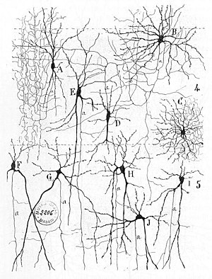
From "Texture of the Nervous System of Man and the Vertebrates" by Santiago Ramón y Cajal. The figure illustrates the diversity of neuronal morphologies in the auditory cortex.

Early treatments of neural networks can be found in Herbert Spencer's Principles of Psychology, 3rd edition (1872), Theodor Meynert's Psychiatry (1884), William James' Principles of Psychology (1890), and Sigmund Freud's Project for a Scientific Psychology (composed 1895). The first rule of neuronal learning was described by Hebb in 1949, in the Hebbian theory. Thus, Hebbian pairing of pre-synaptic and post-synaptic activity can substantially alter the dynamic characteristics of the synaptic connection and therefore either facilitate or inhibit signal transmission. In 1959, the neuroscientists, Warren Sturgis McCulloch and Walter Pitts published the first works on the processing of neural networks. They showed theoretically that networks of artificial neurons could implement logical, arithmetic, and symbolic functions. Simplified models of biological neurons were set up, now usually called perceptrons or artificial neurons. These simple models accounted for neural summation (i.e., potentials at the post-synaptic membrane will summate in the cell body). Later models also provided for excitatory and inhibitory synaptic transmission.

Connections between neurons

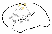
Proposed organization of motor-semantic neural circuits for action language comprehension. Gray dots represent areas of language comprehension, creating a network for comprehending all language. The semantic circuit of the motor system, particularly the motor representation of the legs (yellow dots), is incorporated when leg-related words are comprehended. Adapted from Shebani et al. (2013)

The connections between neurons in the brain are much more complex than those of the artificial neurons used in the connectionist neural computing models of artificial neural networks. The basic kinds of connections between neurons are synapses: both chemical and electrical synapses.

The establishment of synapses enables the connection of neurons into millions of overlapping, and interlinking neural circuits. Presynaptic proteins called neurexins are central to this process.

One principle by which neurons work is neural summationpotentials at the postsynaptic membrane will sum up in the cell body. If the depolarization of the neuron at the axon hillock goes above threshold an action potential will occur that travels down the axon to the terminal endings to transmit a signal to other neurons. Excitatory and inhibitory synaptic transmission is realized mostly by excitatory postsynaptic potentials (EPSPs), and inhibitory postsynaptic potentials (IPSPs).

On the electrophysiological level, there are various phenomena which alter the response characteristics of individual synapses (called synaptic plasticity) and individual neurons (intrinsic plasticity). These are often divided into short-term plasticity and long-term plasticity. Long-term synaptic plasticity is often contended to be the most likely memory substrate. Usually, the term "neuroplasticity" refers to changes in the brain that are caused by activity or experience.

Connections display temporal and spatial characteristics. Temporal characteristics refers to the continuously modified activity-dependent efficacy of synaptic transmission, called spike-timing-dependent plasticity. It has been observed in several studies that the synaptic efficacy of this transmission can undergo short-term increase (called facilitation) or decrease (depression) according to the activity of the presynaptic neuron. The induction of long-term changes in synaptic efficacy, by long-term potentiation (LTP) or depression (LTD), depends strongly on the relative timing of the onset of the excitatory postsynaptic potential and the postsynaptic action potential. LTP is induced by a series of action potentials which cause a variety of biochemical responses. Eventually, the reactions cause the expression of new receptors on the cellular membranes of the postsynaptic neurons or increase the efficacy of the existing receptors through phosphorylation.

Backpropagating action potentials cannot occur because after an action potential travels down a given segment of the axon, the m gates on voltage-gated sodium channels close, thus blocking any transient opening of the h gate from causing a change in the intracellular sodium ion (Na+) concentration, and preventing the generation of an action potential back towards the cell body. In some cells, however, neural backpropagation does occur through the dendritic branching and may have important effects on synaptic plasticity and computation.

A neuron in the brain requires a single signal to a neuromuscular junction to stimulate contraction of the postsynaptic muscle cell. In the spinal cord, however, at least 75 afferent neurons are required to produce firing. This picture is further complicated by variation in time constant between neurons, as some cells can experience their EPSPs over a wider period of time than others.

While in synapses in the developing brain synaptic depression has been particularly widely observed it has been speculated that it changes to facilitation in adult brains.

Circuitry

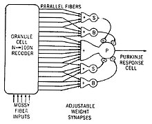
Model of a neural circuit in the cerebellum

An example of a neural circuit is the trisynaptic circuit in the hippocampus. Another is the Papez circuit linking the hypothalamus to the limbic lobe. There are several neural circuits in the cortico-basal ganglia-thalamo-cortical loop. These circuits carry information between the cortex, basal ganglia, thalamus, and back to the cortex. The largest structure within the basal ganglia, the striatum, is seen as having its own internal microcircuitry.

Neural circuits in the spinal cord called central pattern generators are responsible for controlling motor instructions involved in rhythmic behaviours. Rhythmic behaviours include walking, urination, and ejaculation. The central pattern generators are made up of different groups of spinal interneurons.

There are four principal types of neural circuits that are responsible for a broad scope of neural functions. These circuits are a diverging circuit, a converging circuit, a reverberating circuit, and a parallel after-discharge circuit.

In a diverging circuit, one neuron synapses with a number of postsynaptic cells. Each of these may synapse with many more making it possible for one neuron to stimulate up to thousands of cells. This is exemplified in the way that thousands of muscle fibers can be stimulated from the initial input from a single motor neuron.

In a converging circuit, inputs from many sources are converged into one output, affecting just one neuron or a neuron pool. This type of circuit is exemplified in the respiratory center of the brainstem, which responds to a number of inputs from different sources by giving out an appropriate breathing pattern.

A reverberating circuit produces a repetitive output. In a signalling procedure from one neuron to another in a linear sequence, one of the neurons may send a signal back to initiating neuron. Each time that the first neuron fires, the other neuron further down the sequence fires again sending it back to the source. This restimulates the first neuron and also allows the path of transmission to continue to its output. A resulting repetitive pattern is the outcome that only stops if one or more of the synapses fail, or if an inhibitory feed from another source causes it to stop. This type of reverberating circuit is found in the respiratory center that sends signals to the respiratory muscles, causing inhalation. When the circuit is interrupted by an inhibitory signal the muscles relax causing exhalation. This type of circuit may play a part in epileptic seizures.

In a parallel after-discharge circuit, a neuron inputs to several chains of neurons. Each chain is made up of a different number of neurons but their signals converge onto one output neuron. Each synapse in the circuit acts to delay the signal by about 0.5 msec so that the more synapses there are will produce a longer delay to the output neuron. After the input has stopped, the output will go on firing for some time. This type of circuit does not have a feedback loop as does the reverberating circuit. Continued firing after the stimulus has stopped is called after-discharge. This circuit type is found in the reflex arcs of certain reflexes.

Study methods

Different neuroimaging techniques have been developed to investigate the activity of neural circuits and networks. The use of "brain scanners" or functional neuroimaging to investigate the structure or function of the brain is common, either as simply a way of better assessing brain injury with high resolution pictures, or by examining the relative activations of different brain areas. Such technologies may include functional magnetic resonance imaging (fMRI), brain positron emission tomography (brain PET), and computed axial tomography (CAT) scans. Functional neuroimaging uses specific brain imaging technologies to take scans from the brain, usually when a person is doing a particular task, in an attempt to understand how the activation of particular brain areas is related to the task. In functional neuroimaging, especially fMRI, which measures hemodynamic activity (using BOLD-contrast imaging) which is closely linked to neural activity, PET, and electroencephalography (EEG) is used.

Connectionist models serve as a test platform for different hypotheses of representation, information processing, and signal transmission. Lesioning studies in such models, e.g. artificial neural networks, where parts of the nodes are deliberately destroyed to see how the network performs, can also yield important insights in the working of several cell assemblies. Similarly, simulations of dysfunctional neurotransmitters in neurological conditions (e.g., dopamine in the basal ganglia of Parkinson's patients) can yield insights into the underlying mechanisms for patterns of cognitive deficits observed in the particular patient group. Predictions from these models can be tested in patients or via pharmacological manipulations, and these studies can in turn be used to inform the models, making the process iterative.

The modern balance between the connectionist approach and the single-cell approach in neurobiology has been achieved through a lengthy discussion. In 1972, Barlow announced the single neuron revolution: "our perceptions are caused by the activity of a rather small number of neurons selected from a very large population of predominantly silent cells." This approach was stimulated by the idea of grandmother cell put forward two years earlier. Barlow formulated "five dogmas" of neuron doctrine. Recent studies of 'grandmother cell' and sparse coding phenomena develop and modify these ideas. The single cell experiments used intracranial electrodes in the medial temporal lobe (the hippocampus and surrounding cortex). Modern development of concentration of measure theory (stochastic separation theorems) with applications to artificial neural networks give mathematical background to unexpected effectiveness of small neural ensembles in high-dimensional brain.

Clinical significance

Sometimes neural circuitries can become pathological and cause problems such as in Parkinson's disease when the basal ganglia are involved. Problems in the Papez circuit can also give rise to a number of neurodegenerative disorders including Parkinson's.

Neurotechnology

From Wikipedia, the free encyclopedia

Neurotechnology is any technology that has a fundamental influence on how people understand the brain and various aspects of consciousness, thought, and higher order activities in the brain. It also includes technologies that are designed to improve and repair brain function and allow researchers and clinicians to visualize the brain.

Background

The field of neurotechnology has been around for nearly half a century but has only reached maturity in the last twenty years. The advent of brain imaging revolutionized the field, allowing researchers to directly monitor the brain's activities during experiments. Neurotechnology has made significant impact on society, though its presence is so commonplace that many do not realize its ubiquity. From pharmaceutical drugs to brain scanning, neurotechnology affects nearly all industrialized people either directly or indirectly, be it from drugs for depression, sleep, ADD, or anti-neurotics to cancer scanning, stroke rehabilitation, and much more.

As the field's depth increases it will potentially allow society to control and harness more of what the brain does and how it influences lifestyles and personalities. Commonplace technologies already attempt to do this; games like BrainAge, and programs like Fast ForWord that aim to improve brain function, are neurotechnologies.

Currently, modern science can image nearly all aspects of the brain as well as control a degree of the function of the brain. It can help control depression, over-activation, sleep deprivation, and many other conditions. Therapeutically it can help improve stroke victims' motor coordination, improve brain function, reduce epileptic episodes (see epilepsy), improve patients with degenerative motor diseases (Parkinson's disease, Huntington's disease, ALS), and can even help alleviate phantom pain perception. Advances in the field promise many new enhancements and rehabilitation methods for patients suffering from neurological problems. The neurotechnology revolution has given rise to the Decade of the Mind initiative, which was started in 2007. It also offers the possibility of revealing the mechanisms by which mind and consciousness emerge from the brain.

Current technologies

Live Imaging

Magnetoencephalography is a functional neuroimaging technique for mapping brain activity by recording magnetic fields produced by electrical currents occurring naturally in the brain, using very sensitive magnetometers. Arrays of SQUIDs (superconducting quantum interference devices) are the most common magnetometer. Applications of MEG include basic research into perceptual and cognitive brain processes, localizing regions affected by pathology before surgical removal, determining the function of various parts of the brain, and neurofeedback. This can be applied in a clinical setting to find locations of abnormalities as well as in an experimental setting to simply measure brain activity.

Magnetic resonance imaging (MRI) is used for scanning the brain for topological and landmark structure in the brain, but can also be used for imaging activation in the brain. While detail about how MRI works is reserved for the actual MRI article, the uses of MRI are far reaching in the study of neuroscience. It is a cornerstone technology in studying the mind, especially with the advent of functional MRI (fMRI). Functional MRI measures the oxygen levels in the brain upon activation (higher oxygen content = neural activation) and allows researchers to understand what loci are responsible for activation under a given stimulus. This technology is a large improvement to single cell or loci activation by means of exposing the brain and contact stimulation. Functional MRI allows researchers to draw associative relationships between different loci and regions of the brain and provides a large amount of knowledge in establishing new landmarks and loci in the brain.

Computed tomography (CT) is another technology used for scanning the brain. It has been used since the 1970s and is another tool used by neuroscientists to track brain structure and activation. While many of the functions of CT scans are now done using MRI, CT can still be used as the mode by which brain activation and brain injury are detected. Using an X-ray, researchers can detect radioactive markers in the brain that indicate brain activation as a tool to establish relationships in the brain as well as detect many injuries/diseases that can cause lasting damage to the brain such as aneurysms, degeneration, and cancer.

Positron emission tomography (PET) is another imaging technology that aids researchers. Instead of using magnetic resonance or X-rays, PET scans rely on positron emitting markers that are bound to a biologically relevant marker such as glucose. The more activation in the brain the more that region requires nutrients, so higher activation appears more brightly on an image of the brain. PET scans are becoming more frequently used by researchers because PET scans are activated due to metabolism whereas MRI is activated on a more physiological basis (sugar activation versus oxygen activation).

Transcranial magnetic stimulation

Transcranial magnetic stimulation (TMS) is essentially direct magnetic stimulation to the brain. Because electric currents and magnetic fields are intrinsically related, by stimulating the brain with magnetic pulses it is possible to interfere with specific loci in the brain to produce a predictable effect. This field of study is currently receiving a large amount of attention due to the potential benefits that could come out of better understanding this technology. Transcranial magnetic movement of particles in the brain shows promise for drug targeting and delivery as studies have demonstrated this to be noninvasive on brain physiology.

Transcranial direct current stimulation

Transcranial direct current stimulation (tDCS) is a form of neurostimulation which uses constant, low current delivered via electrodes placed on the scalp. The mechanisms underlying tDCS effects are still incompletely understood, but recent advances in neurotechnology allowing for in vivo assessment of brain electric activity during tDCS promise to advance understanding of these mechanisms. Research into using tDCS on healthy adults have demonstrated that tDCS can increase cognitive performance on a variety of tasks, depending on the area of the brain being stimulated. tDCS has been used to enhance language and mathematical ability (though one form of tDCS was also found to inhibit math learning), attention span, problem solving, memory, and coordination.

Cranial surface measurements

Electroencephalography (EEG) is a method of measuring brainwave activity non-invasively. A number of electrodes are placed around the head and scalp and electrical signals are measured. Typically EEGs are used when dealing with sleep, as there are characteristic wave patterns associated with different stages of sleep. Clinically EEGs are used to study epilepsy as well as stroke and tumor presence in the brain. EEGs are a different method to understand the electrical signaling in the brain during activation.

Magnetoencephalography (MEG) is another method of measuring activity in the brain by measuring the magnetic fields that arise from electrical currents in the brain. The benefit to using MEG instead of EEG is that these fields are highly localized and give rise to better understanding of how specific loci react to stimulation or if these regions over-activate (as in epileptic seizures).

Implant technologies

Neurodevices are any devices used to monitor or regulate brain activity. Currently there are a few available for clinical use as a treatment for Parkinson's disease. The most common neurodevices are deep brain stimulators (DBS) that are used to give electrical stimulation to areas stricken by inactivity. Parkinson's disease is known to be caused by an inactivation of the basal ganglia (nuclei) and recently DBS has become the more preferred form of treatment for Parkinson's disease, although current research questions the efficiency of DBS for movement disorders.

Neuromodulation is a relatively new field that combines the use of neurodevices and neurochemistry. The basis of this field is that the brain can be regulated using a number of different factors (metabolic, electrical stimulation, physiological) and that all these can be modulated by devices implanted in the neural network. While currently this field is still in the researcher phase, it represents a new type of technological integration in the field of neurotechnology. The brain is a very sensitive organ, so in addition to researching the amazing things that neuromodulation and implanted neural devices can produce, it is important to research ways to create devices that elicit as few negative responses from the body as possible. This can be done by modifying the material surface chemistry of neural implants.

Cell therapy

Researchers have begun looking at uses for stem cells in the brain, which recently have been found in a few loci. A large number of studies are being done to determine if this form of therapy could be used in a large scale. Experiments have successfully used stem cells in the brains of children who suffered from injuries in gestation and elderly people with degenerative diseases in order to induce the brain to produce new cells and to make more connections between neurons.

Pharmaceuticals

Pharmaceuticals play a vital role in maintaining stable brain chemistry, and are the most commonly used neurotechnology by the general public and medicine. Drugs like sertraline, methylphenidate, and zolpidem act as chemical modulators in the brain, and they allow for normal activity in many people whose brains cannot act normally under physiological conditions. While pharmaceuticals are usually not mentioned and have their own field, the role of pharmaceuticals is perhaps the most far-reaching and commonplace in modern society (the focus on this article will largely ignore neuropharmaceuticals, for more information, see neuropsychopharmacology). Movement of magnetic particles to targeted brain regions for drug delivery is an emerging field of study and causes no detectable circuit damage.

Low field magnetic stimulation

Stimulation with low-intensity magnetic fields is currently under study for depression at Harvard Medical School, and has previously been explored by Bell. It has FDA approval for treatment of depression. It is also being researched for other applications such as autism. One issue is that no two brains are alike and stimulation can cause either polarization or depolarization. (et al.),[21] Marino (et al.), and others.

How these help study the brain

Magnetic resonance imaging is a vital tool in neurological research in showing activation in the brain as well as providing a comprehensive image of the brain being studied. While MRIs are used clinically for showing brain size, it still has relevance in the study of brains because it can be used to determine extent of injuries or deformation. These can have a significant effect on personality, sense perception, memory, higher order thinking, movement, and spatial understanding. However, current research tends to focus more so on fMRI or real-time functional MRI (rtfMRI). These two methods allow the scientist or the participant, respectively, to view activation in the brain. This is incredibly vital in understanding how a person thinks and how their brain reacts to a person's environment, as well as understanding how the brain works under various stressors or dysfunctions. Real-time functional MRI is a revolutionary tool available to neurologists and neuroscientists because patients can see how their brain reacts to stressors and can perceive visual feedback. CT scans are very similar to MRI in their academic use because they can be used to image the brain upon injury, but they are more limited in perceptual feedback. CTs are generally used in clinical studies far more than in academic studies, and are found far more often in a hospital than a research facility. PET scans are also finding more relevance in academia because they can be used to observe metabolic uptake of neurons, giving researchers a wider perspective about neural activity in the brain for a given condition. Combinations of these methods can provide researchers with knowledge of both physiological and metabolic behaviors of loci in the brain and can be used to explain activation and deactivation of parts of the brain under specific conditions.

Transcranial magnetic stimulation is a relatively new method of studying how the brain functions and is used in many research labs focused on behavioral disorders and hallucinations. What makes TMS research so interesting in the neuroscience community is that it can target specific regions of the brain and shut them down or activate temporarily; thereby changing the way the brain behaves. Personality disorders can stem from a variety of external factors, but when the disorder stems from the circuitry of the brain TMS can be used to deactivate the circuitry. This can give rise to a number of responses, ranging from “normality” to something more unexpected, but current research is based on the theory that use of TMS could radically change treatment and perhaps act as a cure for personality disorders and hallucinations. Currently, repetitive transcranial magnetic stimulation (rTMS) is being researched to see if this deactivation effect can be made more permanent in patients suffering from these disorders. Some techniques combine TMS and another scanning method such as EEG to get additional information about brain activity such as cortical response.

Both EEG and MEG are currently being used to study the brain's activity under different conditions. Each uses similar principles but allows researchers to examine individual regions of the brain, allowing isolation and potentially specific classification of active regions. As mentioned above, EEG is very useful in analysis of immobile patients, typically during the sleep cycle. While there are other types of research that utilize EEG, EEG has been fundamental in understanding the resting brain during sleep. There are other potential uses for EEG and MEG such as charting rehabilitation and improvement after trauma as well as testing neural conductivity in specific regions of epileptics or patients with personality disorders.

Neuromodulation can involve numerous technologies combined or used independently to achieve a desired effect in the brain. Gene and cell therapy are becoming more prevalent in research and clinical trials and these technologies could help stunt or even reverse disease progression in the central nervous system. Deep brain stimulation is currently used in many patients with movement disorders and is used to improve the quality of life in patients. While deep brain stimulation is a method to study how the brain functions per se, it provides both surgeons and neurologists important information about how the brain works when certain small regions of the basal ganglia (nuclei) are stimulated by electrical currents.

Future technologies

The future of neurotechnologies lies in how they are fundamentally applied, and not so much on what new versions will be developed. Current technologies give a large amount of insight into the mind and how the brain functions, but basic research is still needed to demonstrate the more applied functions of these technologies. Currently, rtfMRI is being researched as a method for pain therapy have shown that there is a significant improvement in the way people perceive pain if they are made aware of how their brain is functioning while in pain. By providing direct and understandable feedback, researchers can help patients with chronic pain decrease their symptoms. This new type of bio/mechanical-feedback is a new development in pain therapy. Functional MRI is also being considered for a number of more applicable uses outside of the clinic. Research has been done on testing the efficiency of mapping the brain in the case when someone lies as a new way to detect lying. Along the same vein, EEG has been considered for use in lie detection as well. TMS is being used in a variety of potential therapies for patients with personality disorders, epilepsy, PTSD, migraine, and other brain-firing disorders, but has been found to have varying clinical success for each condition. The end result of such research would be to develop a method to alter the brain's perception and firing and train patients' brains to rewire permanently under inhibiting conditions (for more information see rTMS). In addition, PET scans have been found to be 93% accurate in detecting Alzheimer's disease nearly 3 years before conventional diagnosis, indicating that PET scanning is becoming more useful in both the laboratory and the clinic.

Stem cell technologies are always salient both in the minds of the general public and scientists because of their large potential. Recent advances in stem cell research have allowed researchers to ethically pursue studies in nearly every facet of the body, which includes the brain. Research has shown that while most of the brain does not regenerate and is typically a very difficult environment to foster regeneration, there are portions of the brain with regenerative capabilities (specifically the hippocampus and the olfactory bulbs). Much of the research in central nervous system regeneration is how to overcome this poor regenerative quality of the brain. It is important to note that there are therapies that improve cognition and increase the amount of neural pathways, but this does not mean that there is a proliferation of neural cells in the brain. Rather, it is called a plastic rewiring of the brain (plastic because it indicates malleability) and is considered a vital part of growth. Nevertheless, many problems in patients stem from death of neurons in the brain, and researchers in the field are striving to produce technologies that enable regeneration in patients with stroke, Parkinson's diseases, severe trauma, and Alzheimer's disease, as well as many others. While still in fledgling stages of development, researchers have recently begun making very interesting progress in attempting to treat these diseases. Researchers have recently successfully produced dopaminergic neurons for transplant in patients with Parkinson's diseases with the hopes that they will be able to move again with a more steady supply of dopamine. Many researchers are building scaffolds that could be transplanted into a patient with spinal cord trauma to present an environment that promotes growth of axons (portions of the cell attributed with transmission of electrical signals) so that patients unable to move or feel might be able to do so again. The potentials are wide-ranging, but it is important to note that many of these therapies are still in the laboratory phase and are slowly being adapted in the clinic. Some scientists remain skeptical with the development of the field, and warn that there is a much larger chance that electrical prosthesis will be developed to solve clinical problems such as hearing loss or paralysis before cell therapy is used in a clinic.

Novel drug delivery systems are being researched in order to improve the lives of those who struggle with brain disorders that might not be treated with stem cells, modulation, or rehabilitation. Pharmaceuticals play a very important role in society, and the brain has a very selective barrier that prevents some drugs from going from the blood to the brain. There are some diseases of the brain such as meningitis that require doctors to directly inject medicine into the spinal cord because the drug cannot cross the blood–brain barrier. Research is being conducted to investigate new methods of targeting the brain using the blood supply, as it is much easier to inject into the blood than the spine. New technologies such as nanotechnology are being researched for selective drug delivery, but these technologies have problems as with any other. One of the major setbacks is that when a particle is too large, the patient's liver will take up the particle and degrade it for excretion, but if the particle is too small there will not be enough drug in the particle to take effect. In addition, the size of the capillary pore is important because too large a particle might not fit or even plug up the hole, preventing adequate supply of the drug to the brain. Other research is involved in integrating a protein device between the layers to create a free-flowing gate that is unimpeded by the limitations of the body. Another direction is receptor-mediated transport, where receptors in the brain used to transport nutrients are manipulated to transport drugs across the blood–brain barrier. Some have even suggested that focused ultrasound opens the blood–brain barrier momentarily and allows free passage of chemicals into the brain. Ultimately the goal for drug delivery is to develop a method that maximizes the amount of drug in the loci with as little degraded in the blood stream as possible.

Neuromodulation is a technology currently used for patients with movement disorders, although research is currently being done to apply this technology to other disorders. Recently, a study was done on if DBS could improve depression with positive results, indicating that this technology might have potential as a therapy for multiple disorders in the brain. DBS is limited by its high cost however, and in developing countries the availability of DBS is very limited. A new version of DBS is under investigation and has developed into the novel field, optogenetics. Optogenetics is the combination of deep brain stimulation with fiber optics and gene therapy. Essentially, the fiber optic cables are designed to light up under electrical stimulation, and a protein would be added to a neuron via gene therapy to excite it under light stimuli. So by combining these three independent fields, a surgeon could excite a single and specific neuron in order to help treat a patient with some disorder. Neuromodulation offers a wide degree of therapy for many patients, but due to the nature of the disorders it is currently used to treat its effects are often temporary. Future goals in the field hope to alleviate that problem by increasing the years of effect until DBS can be used for the remainder of the patient's life. Another use for neuromodulation would be in building neuro-interface prosthetic devices that would allow quadriplegics the ability to maneuver a cursor on a screen with their thoughts, thereby increasing their ability to interact with others around them. By understanding the motor cortex and understanding how the brain signals motion, it is possible to emulate this response on a computer screen.

Ethics

Stem cells

The ethical debate about use of embryonic stem cells has stirred controversy both in the United States and abroad; although more recently these debates have lessened due to modern advances in creating induced pluripotent stem cells from adult cells. The greatest advantage for use of embryonic stem cells is the fact that they can differentiate (become) nearly any type of cell provided the right conditions and signals. However, recent advances by Shinya Yamanaka et al. have found ways to create pluripotent cells without the use of such controversial cell cultures. Using the patient's own cells and re-differentiating them into the desired cell type bypasses both possible patient rejection of the embryonic stem cells and any ethical concerns associated with using them, while also providing researchers a larger supply of available cells. However, induced pluripotent cells have the potential to form benign (though potentially malignant) tumors, and tend to have poor survivability in vivo (in the living body) on damaged tissue. Much of the ethics concerning use of stem cells has subsided from the embryonic/adult stem cell debate due to its rendered moot, but now societies find themselves debating whether or not this technology can be ethically used. Enhancements of traits, use of animals for tissue scaffolding, and even arguments for moral degeneration have been made with the fears that if this technology reaches its full potential a new paradigm shift will occur in human behavior.

Military application

New neurotechnologies have always garnered the appeal of governments, from lie detection technology and virtual reality to rehabilitation and understanding the psyche. Due to the Iraq War and War on Terror, American soldiers coming back from Iraq and Afghanistan are reported to have percentages up to 12% with PTSD. There are many researchers hoping to improve these peoples' conditions by implementing new strategies for recovery. By combining pharmaceuticals and neurotechnologies, some researchers have discovered ways of lowering the "fear" response and theorize that it may be applicable to PTSD. Virtual reality is another technology that has drawn much attention in the military. If improved, it could be possible to train soldiers how to deal with complex situations in times of peace, in order to better prepare and train a modern army.

Privacy

Finally, when these technologies are being developed society must understand that these neurotechnologies could reveal the one thing that people can always keep secret: what they are thinking. While there are large amounts of benefits associated with these technologies, it is necessary for scientists, citizens and policy makers alike to consider implications for privacy. This term is important in many ethical circles concerned with the state and goals of progress in the field of neurotechnology. Current improvements such as “brain fingerprinting” or lie detection using EEG or fMRI could give rise to a set fixture of loci/emotional relationships in the brain, although these technologies are still years away from full application. It is important to consider how all these neurotechnologies might affect the future of society, and it is suggested that political, scientific, and civil debates are heard about the implementation of these newer technologies that potentially offer a new wealth of once-private information. Some ethicists are also concerned with the use of TMS and fear that the technique could be used to alter patients in ways that are undesired by the patient.

Cognitive liberty

Cognitive liberty refers to a suggested right to self-determination of individuals to control their own mental processes, cognition, and consciousness including by the use of various neurotechnologies and psychoactive substances. This perceived right is relevant for reformation and development of associated laws.

Blood–brain barrier

From Wikipedia, the free encyclopedia
 
Blood-brain barrier
Protective barriers of the brain.jpg
Solute permeability at the BBB
vs. choroid plexus
Details
SystemNeuroimmune system
Identifiers
Acronym(s)BBB
MeSHD001812

The blood–brain barrier (BBB) is a highly selective semipermeable border of endothelial cells that prevents solutes in the circulating blood from non-selectively crossing into the extracellular fluid of the central nervous system where neurons reside. The blood-brain barrier is formed by endothelial cells of the capillary wall, astrocyte end-feet ensheathing the capillary, and pericytes embedded in the capillary basement membrane. This system allows the passage of some molecules by passive diffusion, as well as the selective and active transport of various nutrients, ions, organic anions, and macromolecules such as glucose, water and amino acids that are crucial to neural function.

The blood-brain barrier restricts the passage of pathogens, the diffusion of solutes in the blood, and large or hydrophilic molecules into the cerebrospinal fluid, while allowing the diffusion of hydrophobic molecules (O2, CO2, hormones) and small polar molecules. Cells of the barrier actively transport metabolic products such as glucose across the barrier using specific transport proteins. The barrier also restricts the passage of peripheral immune factors, like signaling molecules, antibodies, and immune cells, into the CNS, thus insulating the brain from damage due to peripheral immune events.

Specialized brain structures participating in sensory and secretory integration within brain neural circuits—the circumventricular organs and choroid plexus—have highly permeable capillaries.

Structure

Part of a network of capillaries supplying brain cells
 
The astrocytes type 1 surrounding capillaries in the brain
 
Sketch showing constitution of blood vessels inside the brain

The blood–brain barrier results from the selectivity of the tight junctions between the endothelial cells of brain capillaries, restricting the passage of solutes. At the interface between blood and the brain, endothelial cells are adjoined continuously by these tight junctions, which are composed of smaller subunits of transmembrane proteins, such as occludin, claudins, junctional adhesion molecule. Each of these transmembrane proteins is anchored into the endothelial cells by another protein complex that includes tight junction protein 1 and associated proteins.

The blood-brain barrier is composed of endothelial cells restricting passage of substances from the blood more selectively than endothelial cells of capillaries elsewhere in the body. Astrocyte cell projections called astrocytic feet (also known as "glia limitans") surround the endothelial cells of the BBB, providing biochemical support to those cells. The BBB is distinct from the quite similar blood-cerebrospinal fluid barrier, which is a function of the choroidal cells of the choroid plexus, and from the blood-retinal barrier, which can be considered a part of the whole realm of such barriers.

Several areas of the human brain are not on the brain side of the BBB. Some examples of this include the circumventricular organs, the roof of the third and fourth ventricles, capillaries in the pineal gland on the roof of the diencephalon and the pineal gland. The pineal gland secretes the hormone melatonin "directly into the systemic circulation", thus melatonin is not affected by the blood-brain barrier.

Development

The blood-brain barrier appears to be functional by the time of birth. P-glycoprotein, a transporter, exists already in the embryonal endothelium.

Measurement of brain uptake of various blood-borne solutes showed that newborn endothelial cells were functionally similar to those in adults, indicating that a selective BBB is operative at birth.

Function

The blood-brain barrier acts effectively to protect the brain from circulating pathogens. Accordingly, blood-borne infections of the brain are rare. Infections of the brain that do occur are often difficult to treat. Antibodies are too large to cross the blood-brain barrier, and only certain antibiotics are able to pass. In some cases, a drug has to be administered directly into the cerebrospinal fluid where it can enter the brain by crossing the blood-cerebrospinal fluid barrier.

The blood-brain barrier may become leaky in select neurological diseases, such as amyotrophic lateral sclerosis, epilepsy, brain trauma and edema, and in systemic diseases, such as liver failure. The blood-brain barrier becomes more permeable during inflammation, potentially allowing antibiotics and phagocytes to move across the BBB.

Circumventricular organs

Circumventricular organs (CVOs) are individual structures located adjacent to the fourth ventricle or third ventricle in the brain, and are characterized by dense capillary beds with permeable endothelial cells unlike those of the blood-brain barrier. Included among CVOs having highly permeable capillaries are the area postrema, subfornical organ, vascular organ of the lamina terminalis, median eminence, pineal gland, and three lobes of the pituitary gland.

Permeable capillaries of the sensory CVOs (area postrema, subfornical organ, vascular organ of the lamina terminalis) enable rapid detection of circulating signals in systemic blood, while those of the secretory CVOs (median eminence, pineal gland, pituitary lobes) facilitate transport of brain-derived signals into the circulating blood. Consequently, the CVO permeable capillaries are the point of bidirectional blood-brain communication for neuroendocrine function.

Specialized permeable zones

The border zones between brain tissue "behind" the blood-brain barrier and zones "open" to blood signals in certain CVOs contain specialized hybrid capillaries that are leakier than typical brain capillaries, but not as permeable as CVO capillaries. Such zones exist at the border of the area postrema—nucleus tractus solitarii (NTS), and median eminence—hypothalamic arcuate nucleus. These zones appear to function as rapid transit regions for brain structures involved in diverse neural circuits—like the NTS and arcuate nucleus—to receive blood signals which are then transmitted into neural output. The permeable capillary zone shared between the median eminence and hypothalamic arcuate nucleus is augmented by wide pericapillary spaces, facilitating bidirectional flow of solutes between the two structures, and indicating that the median eminence is not only a secretory organ, but may also be a sensory organ.

Therapeutic research

As a drug target

The blood-brain barrier is formed by the brain capillary endothelium and excludes from the brain 100% of large-molecule neurotherapeutics and more than 98% of all small-molecule drugs. Overcoming the difficulty of delivering therapeutic agents to specific regions of the brain presents a major challenge to treatment of most brain disorders. In its neuroprotective role, the blood-brain barrier functions to hinder the delivery of many potentially important diagnostic and therapeutic agents to the brain. Therapeutic molecules and antibodies that might otherwise be effective in diagnosis and therapy do not cross the BBB in adequate amounts to be clinically effective.

Mechanisms for drug targeting in the brain involve going either "through" or "behind" the BBB. Modalities for drug delivery to the brain in unit doses through the BBB entail its disruption by osmotic means, or biochemically by the use of vasoactive substances, such as bradykinin, or even by localized exposure to high-intensity focused ultrasound (HIFU).

Other methods used to get through the BBB may entail the use of endogenous transport systems, including carrier-mediated transporters, such as glucose and amino acid carriers, receptor-mediated transcytosis for insulin or transferrin, and the blocking of active efflux transporters such as p-glycoprotein. Some studies have shown that vectors targeting BBB transporters, such as the transferrin receptor, have been found to remain entrapped in brain endothelial cells of capillaries, instead of being ferried across the BBB into the targeted area.

Nanoparticles

Nanotechnology is under preliminary research for its potential to facilitate the transfer of drugs across the BBB. Capillary endothelial cells and associated pericytes may be abnormal in tumors and the blood-brain barrier may not always be intact in brain tumors. Other factors, such as astrocytes, may contribute to the resistance of brain tumors to therapy using nanoparticles. Fat soluble molecules less than 400 Daltons in weight can freely diffuse past the BBB through lipid mediated passive diffusion.

History

Paul Ehrlich was a bacteriologist studying staining, a procedure that is used in many microscopy studies to make fine biological structures visible using chemical dyes. As Ehrlich injected some of these dyes (notably the aniline dyes that were then widely used), the dye stained all of the organs of some kinds of animals except for their brains. At that time, Ehrlich attributed this lack of staining to the brain simply not picking up as much of the dye.

However, in a later experiment in 1913, Edwin Goldmann (one of Ehrlich's students) injected the dye directly into the cerebrospinal fluids of animal brains. He found then the brains did become dyed, but the rest of the body did not, demonstrating the existence of a compartmentalization between the two. At that time, it was thought that the blood vessels themselves were responsible for the barrier, since no obvious membrane could be found. The concept of the blood–brain barrier (then termed hematoencephalic barrier) was proposed by a Berlin physician, Lewandowsky, in 1900.


Archetype

From Wikipedia, the free encyclopedia https://en.wikipedia.org/wiki/Archetype The concept of an archetyp...