Search This Blog

Thursday, April 22, 2021

Placenta

From Wikipedia, the free encyclopedia
https://en.wikipedia.org/wiki/Placenta

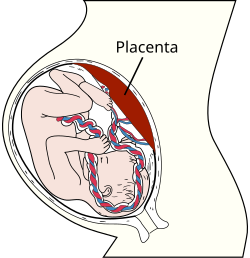
Placenta
Placenta.svg
Placenta
 
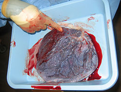
Human placenta baby side.jpg
Human placenta from just after birth with the umbilical cord in place
Details
Precursordecidua basalis, chorion frondosum
Identifiers
LatinPlacento
MeSHD010920
TEE5.11.3.1.1.0.5

The placenta is a temporary fetal organ that begins developing from the blastocyst shortly after implantation. It plays critical roles in facilitating nutrient, gas and waste exchange between the physically separate maternal and fetal circulations, and is an important endocrine organ producing hormones that regulate both maternal and fetal physiology during pregnancy. The placenta connects to the baby via the umbilical cord, and on the opposite aspect to the maternal uterus in a species dependent manner. In humans, a thin layer of maternal decidual (endometrial) tissue comes away with the placenta when it is expelled from the uterus following birth (sometimes incorrectly referred to as the 'maternal part' of the placenta). Placentas are a defining characteristic of placental mammals, but are also found in marsupials and some non-mammals with varying levels of development.

Mammal placentas probably first evolved about 150 million to 200 million years ago. The protein syncytin, found in the outer barrier of the placenta (the syncytiotrophoblast) between mother and baby, has a certain RNA signature in its genome that has led to the hypothesis that it originated from an ancient retrovirus: essentially a "good" virus that helped pave the transition from egg-laying to live-birth.

The word placenta comes from the Latin word for a type of cake, from Greek πλακόεντα/πλακοῦντα plakóenta/plakoúnta, accusative of πλακόεις/πλακούς plakóeis/plakoús, "flat, slab-like", in reference to its round, flat appearance in humans. The classical plural is placentae, but the form placentas is common in modern English and probably has the wider currency at present.

Phylogenetic diversity

Although all mammalian placentae have the same functions, there are important differences in structure and function in different groups of mammals. For example, human, bovine, equine and canine placentae are very different at the both gross and the microscopic levels. Placentae of these species also differ in their ability to provide maternal immunoglobulins to the fetus.

Structure

Placental mammals, such as humans, have a chorioallantoic placenta that forms from the chorion and allantois. In humans, the placenta averages 22 cm (9 inch) in length and 2–2.5 cm (0.8–1 inch) in thickness, with the center being the thickest, and the edges being the thinnest. It typically weighs approximately 500 grams (just over 1 lb). It has a dark reddish-blue or crimson color. It connects to the fetus by an umbilical cord of approximately 55–60 cm (22–24 inch) in length, which contains two umbilical arteries and one umbilical vein. The umbilical cord inserts into the chorionic plate (has an eccentric attachment). Vessels branch out over the surface of the placenta and further divide to form a network covered by a thin layer of cells. This results in the formation of villous tree structures. On the maternal side, these villous tree structures are grouped into lobules called cotyledons. In humans, the placenta usually has a disc shape, but size varies vastly between different mammalian species.

The placenta occasionally takes a form in which it comprises several distinct parts connected by blood vessels. The parts, called lobes, may number two, three, four, or more. Such placentas are described as bilobed/bilobular/bipartite, trilobed/trilobular/tripartite, and so on. If there is a clearly discernible main lobe and auxiliary lobe, the latter is called a succenturiate placenta. Sometimes the blood vessels connecting the lobes get in the way of fetal presentation during labor, which is called vasa previa.

Gene and protein expression

About 20,000 protein coding genes are expressed in human cells and 70% of these genes are expressed in the normal mature placenta. Some 350 of these genes are more specifically expressed in the placenta and fewer than 100 genes are highly placenta specific. The corresponding specific proteins are mainly expressed in trophoblasts and have functions related to female pregnancy. Examples of proteins with elevated expression in placenta compared to other organs and tissues are PEG10 and the cancer testis antigen PAGE4 and expressed in cytotrophoblasts, CSH1 and KISS1 expressed in syncytiotrophoblasts, and PAPPA2 and PRG2 expressed in extravillous trophoblasts.

Physiology

Development

Placenta
 
The initial stages of human embryogenesis.

The placenta begins to develop upon implantation of the blastocyst into the maternal endometrium. The outer layer of the blastocyst becomes the trophoblast, which forms the outer layer of the placenta. This outer layer is divided into two further layers: the underlying cytotrophoblast layer and the overlying syncytiotrophoblast layer. The syncytiotrophoblast is a multinucleated continuous cell layer that covers the surface of the placenta. It forms as a result of differentiation and fusion of the underlying cytotrophoblast cells, a process that continues throughout placental development. The syncytiotrophoblast (otherwise known as syncytium), thereby contributes to the barrier function of the placenta.

The placenta grows throughout pregnancy. Development of the maternal blood supply to the placenta is complete by the end of the first trimester of pregnancy week 14 (DM).

Placental circulation

Maternal blood fills the intervillous space, nutrients, water, and gases are actively and passively exchanged, then deoxygenated blood is displaced by the next maternal pulse.

Maternal placental circulation

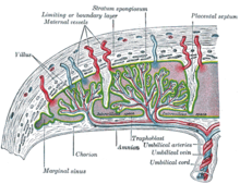
In preparation for implantation of the blastocyst, the endometrium undergoes decidualization. Spiral arteries in the decidua are remodeled so that they become less convoluted and their diameter is increased. The increased diameter and straighter flow path both act to increase maternal blood flow to the placenta. There is relatively high pressure as the maternal blood fills intervillous space through these spiral arteries which bathe the fetal villi in blood, allowing an exchange of gases to take place. In humans and other hemochorial placentals, the maternal blood comes into direct contact with the fetal chorion, though no fluid is exchanged. As the pressure decreases between pulses, the deoxygenated blood flows back through the endometrial veins.

Maternal blood flow is approximately 600–700 ml/min at term.

This begins at day 5 - day 12 

Fetoplacental circulation

Deoxygenated fetal blood passes through umbilical arteries to the placenta. At the junction of umbilical cord and placenta, the umbilical arteries branch radially to form chorionic arteries. Chorionic arteries, in turn, branch into cotyledon arteries. In the villi, these vessels eventually branch to form an extensive arterio-capillary-venous system, bringing the fetal blood extremely close to the maternal blood; but no intermingling of fetal and maternal blood occurs ("placental barrier").

Endothelin and prostanoids cause vasoconstriction in placental arteries, while nitric oxide causes vasodilation. On the other hand, there is no neural vascular regulation, and catecholamines have only little effect.

The fetoplacental circulation is vulnerable to persistent hypoxia or intermittent hypoxia and reoxygenation, which can lead to generation of excessive free radicals. This may contribute to pre-eclampsia and other pregnancy complications. It is proposed that melatonin plays a role as an antioxidant in the placenta.

This begins at day 17 - day 22 

Birth

Placental expulsion begins as a physiological separation from the wall of the uterus. The period from just after the child is born until just after the placenta is expelled is called the "third stage of labor". The placenta is usually expelled within 15–30 minutes of birth.

Placental expulsion can be managed actively, for example by giving oxytocin via intramuscular injection followed by cord traction to assist in delivering the placenta. Alternatively, it can be managed expectantly, allowing the placenta to be expelled without medical assistance. Blood loss and the risk of postpartum bleeding may be reduced in women offered active management of the third stage of labour, however there may be adverse effects and more research is necessary.

The habit is to cut the cord immediately after birth, but it is theorised that there is no medical reason to do this; on the contrary, it is theorised that not cutting the cord helps the baby in its adaptation to extrauterine life, especially in preterm infants.

Microbiome

The placenta is traditionally thought to be sterile, but recent research suggests that a resident, non-pathogenic, and diverse population of microorganisms may be present in healthy tissue. However, whether these microbes exist or are clinically important is highly controversial and is the subject of active research.

Functions

Nutrition and gas exchange

Maternal side of a placenta shortly after birth.

The placenta intermediates the transfer of nutrients between mother and fetus. The perfusion of the intervillous spaces of the placenta with maternal blood allows the transfer of nutrients and oxygen from the mother to the fetus and the transfer of waste products and carbon dioxide back from the fetus to the maternal blood. Nutrient transfer to the fetus can occur via both active and passive transport. Placental nutrient metabolism was found to play a key role in limiting the transfer of some nutrients. Adverse pregnancy situations, such as those involving maternal diabetes or obesity, can increase or decrease levels of nutrient transporters in the placenta potentially resulting in overgrowth or restricted growth of the fetus.

Animated schematic of the hearts and circulatory systems of a fetus and its mother – red and blue represent oxygenated and deoxygenated blood, respectively (animation)

Excretion

Waste products excreted from the fetus such as urea, uric acid, and creatinine are transferred to the maternal blood by diffusion across the placenta.

Immunity

IgG antibodies can pass through the human placenta, thereby providing protection to the fetus in utero. This transfer of antibodies begins as early as the 20th week of gestational age, and certainly by the 24th week. This passive immunity lingers for several months after birth, thus providing the newborn with a carbon copy of the mother's long-term humoral immunity to see the infant through the crucial first months of extrauterine life. IgM, however, cannot cross the placenta, which is why some infections acquired during pregnancy can be hazardous for the fetus.

Furthermore, the placenta functions as a selective maternal-fetal barrier against transmission of microbes. However, insufficiency in this function may still cause mother-to-child transmission of infectious diseases.

Endocrine function

  • The first hormone released by the placenta is called the human chorionic gonadotropin hormone. This is responsible for stopping the process at the end of menses when the Corpus luteum ceases activity and atrophies. If hCG did not interrupt this process, it would lead to spontaneous abortion of the fetus. The corpus luteum also produces and releases progesterone and estrogen, and hCG stimulates it to increase the amount that it releases. hCG is the indicator of pregnancy that pregnancy tests look for. These tests will work when menses has not occurred or after implantation has happened on days seven to ten. hCG may also have an anti-antibody effect, protecting it from being rejected by the mother's body. hCG also assists the male fetus by stimulating the testes to produce testosterone, which is the hormone needed to allow the sex organs of the male to grow.
  • Progesterone helps the embryo implant by assisting passage through the fallopian tubes. It also affects the fallopian tubes and the uterus by stimulating an increase in secretions necessary for fetal nutrition. Progesterone, like hCG, is necessary to prevent spontaneous abortion because it prevents contractions of the uterus and is necessary for implantation.
  • Estrogen is a crucial hormone in the process of proliferation. This involves the enlargement of the breasts and uterus, allowing for growth of the fetus and production of milk. Estrogen is also responsible for increased blood supply towards the end of pregnancy through vasodilation. The levels of estrogen during pregnancy can increase so that they are thirty times what a non-pregnant woman mid-cycles estrogen level would be.
  • Human placental lactogen is a hormone used in pregnancy to develop fetal metabolism and general growth and development. Human placental lactogen works with Growth hormone to stimulate Insulin-like growth factor production and regulating intermediary metabolism. In the fetus, hPL acts on lactogenic receptors to modulate embryonic development, metabolism and stimulate production of IGF, insulin, surfactant and adrenocortical hormones. hPL values increase with multiple pregnancies, intact molar pregnancy, diabetes and Rh incompatibility. They are decreased with toxemia, choriocarcinoma, and Placental insufficiency.

Immunological barrier

The placenta and fetus may be regarded as a foreign body inside the mother and must be protected from the normal immune response of the mother that would cause it to be rejected. The placenta and fetus are thus treated as sites of immune privilege, with immune tolerance.

For this purpose, the placenta uses several mechanisms:

However, the Placental barrier is not the sole means to evade the immune system, as foreign fetal cells also persist in the maternal circulation, on the other side of the placental barrier.

Other

The placenta also provides a reservoir of blood for the fetus, delivering blood to it in case of hypotension and vice versa, comparable to a capacitor.

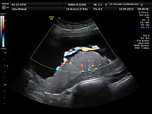
Ultrasound image of human placenta and umbilical cord (color Doppler rendering) with central cord insertion and three umbilical vessels, at 20 weeks of pregnancy

Clinical significance

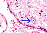
Micrograph of a cytomegalovirus (CMV) infection of the placenta (CMV placentitis). The characteristic large nucleus of a CMV-infected cell is seen off-centre at the bottom-right of the image. H&E stain.

Numerous pathologies can affect the placenta.

Society and culture

The placenta often plays an important role in various cultures, with many societies conducting rituals regarding its disposal. In the Western world, the placenta is most often incinerated.

Some cultures bury the placenta for various reasons. The Māori of New Zealand traditionally bury the placenta from a newborn child to emphasize the relationship between humans and the earth. Likewise, the Navajo bury the placenta and umbilical cord at a specially chosen site, particularly if the baby dies during birth. In Cambodia and Costa Rica, burial of the placenta is believed to protect and ensure the health of the baby and the mother. If a mother dies in childbirth, the Aymara of Bolivia bury the placenta in a secret place so that the mother's spirit will not return to claim her baby's life.

The placenta is believed by some communities to have power over the lives of the baby or its parents. The Kwakiutl of British Columbia bury girls' placentas to give the girl skill in digging clams, and expose boys' placentas to ravens to encourage future prophetic visions. In Turkey, the proper disposal of the placenta and umbilical cord is believed to promote devoutness in the child later in life. In Transylvania, and Japan, interaction with a disposed placenta is thought to influence the parents' future fertility.

Several cultures believe the placenta to be or have been alive, often a relative of the baby. Nepalese think of the placenta as a friend of the baby; Malaysian Orang Asli regard it as the baby's older sibling. Native Hawaiians believe that the placenta is a part of the baby, and traditionally plant it with a tree that can then grow alongside the child. Various cultures in Indonesia, such as Javanese, believe that the placenta has a spirit and needs to be buried outside the family house.

In some cultures, the placenta is eaten, a practice known as placentophagy. In some eastern cultures, such as China, the dried placenta (ziheche , literally "purple river car") is thought to be a healthful restorative and is sometimes used in preparations of traditional Chinese medicine and various health products. The practice of human placentophagy has become a more recent trend in western cultures and is not without controversy; its practice being considered cannibalism is debated.

Some cultures have alternative uses for placenta that include the manufacturing of cosmetics, pharmaceuticals and food.

Methylmercury

From Wikipedia, the free encyclopedia
 

Properties
CH3Hg
Molar mass 215.63 g/mol
Except where otherwise noted, data are given for materials in their standard state (at 25 °C [77 °F], 100 kPa).
☒ verify (what is check☒ ?)
Infobox references


Structures of two main types of complexes formed by "methylmercury". X = anion, L = neutral Lewis base.

Methylmercury (sometimes methyl mercury) is an extremely toxic organometallic cation with the formula [CH3Hg]+. Its derivatives are the major source of organic mercury for humans. It is a bioaccumulative environmental toxicant.

Structure and chemistry

"Methylmercury" is a shorthand for the hypothetical "methylmercury cation", sometimes written "methylmercury(1+) cation" or "methylmercury(II) cation". This functional group is composed of a methyl group bonded to a mercury. Its chemical formula is CH3Hg+ (sometimes written as MeHg+). Methylmercury exists as a substituent in many complexes of the type [MeHgL]+ (L = Lewis base) and MeHgX (X = anion).

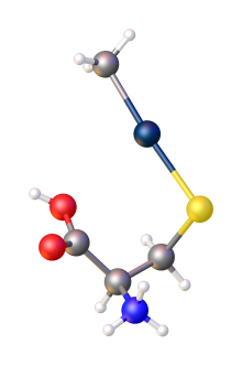
As a positively charged ion it readily combines with anions such as chloride (Cl), hydroxide (OH) and nitrate (NO3). It has particular affinity for sulfur-containing anions, particularly thiols (RS). Thiols are generated on the amino acid cysteine and the peptide glutathione form strong complexes with methylmercury:

[MeHg]+ + RSH → MeHg-SR + H+

Sources

Environmental sources

Structure of the complex of "methylmercury" and cysteine. Color code: dark blue = Hg, yellow = S.

Methylmercury is formed from inorganic mercury by the action of microbes that live in aquatic systems including lakes, rivers, wetlands, sediments, soils and the open ocean. This methylmercury production has been primarily attributed to anaerobic bacteria in the sediment. Significant concentrations of methylmercury in ocean water columns are strongly associated with nutrients and organic matter remineralization, which indicate that remineralization may contribute to methylmercury production. Direct measurements of methylmercury production using stable mercury isotopes have also been observed in toxic waters, but the microbes involved are still unknown. Flooding of soils associated with reservoir creation (e.g. for hydroelectric power generation) has been linked to increased methylmercury concentrations in reservoir water and fish.

There are various sources of inorganic mercury that may indirectly contribute to the production of methylmercury from microbes in the environment. Natural sources of mercury released to the atmosphere include volcanoes, forest fires, volatilization from the ocean and weathering of mercury-bearing rocks. Anthropogenic sources of mercury include the burning of wastes containing inorganic mercury and from the burning of fossil fuels, particularly coal. Although inorganic mercury is only a trace constituent of such fuels, their large scale combustion in utility and commercial/industrial boilers in the United States alone results in release of some 80.2 tons (73 tonnes) of elemental mercury to the atmosphere each year, out of total anthropogenic mercury emissions in the United States of 158 tons (144 tonnes)/year.

In the past, methylmercury was produced directly and indirectly as part of several industrial processes such as the manufacture of acetaldehyde. However, currently there are few direct anthropogenic sources of methylmercury pollution in the United States.

Whole-lake ecosystem experiments at IISD-ELA in Ontario, Canada showed that mercury falling directly on a lake had the fastest impacts on aquatic ecosystems as opposed to mercury falling on the surrounding land. This inorganic mercury is converted to methylmercury by bacteria. Different stable isotopes of mercury were added to lakes, wetlands, and uplands, simulating rain, and then mercury concentrations in fish were analyzed to find their source. The mercury applied to lakes was found in young-of-the-year yellow perch within two months, whereas the mercury applied to wetlands and uplands had a slower but longer influx.

Acute methylmercury poisoning can occur either directly from the release of methylmercury into the environment or indirectly from the release of inorganic mercury that is subsequently methylated in the environment. For example, methylmercury poisoning occurred at Grassy Narrows in Ontario, Canada (see Ontario Minamata disease) as a result of mercury released from the mercury-cell Chloralkali process, which uses liquid mercury as an electrode in a process that entails electrolytic decomposition of brine, followed by mercury methylation in the aquatic environment. An acute methylmercury poisoning tragedy occurred also in Minamata, Japan following release of methylmercury into Minamata Bay and its tributaries (see Minamata disease). In the Ontario case, inorganic mercury discharged into the environment was methylated in the environment; whereas in Minamata, Japan, there was direct industrial discharge of methylmercury.

Dietary sources

Because methylmercury is formed in aquatic systems and because it is not readily eliminated from organisms it is biomagnified in aquatic food chains from bacteria, to plankton, through macroinvertebrates, to herbivorous fish and to piscivorous (fish-eating) fish. At each step in the food chain, the concentration of methylmercury in the organism increases. The concentration of methylmercury in the top level aquatic predators can reach a level a million times higher than the level in the water. This is because methylmercury has a half-life of about 72 days in aquatic organisms resulting in its bioaccumulation within these food chains. Organisms, including humans, fish-eating birds, and fish-eating mammals such as otters and cetaceans (i.e. whales and dolphins) that consume fish from the top of the aquatic food chain receive the methylmercury that has accumulated through this process, plus the toxins in their habitat. Fish and other aquatic species are the main source of human methylmercury exposure.

The concentration of mercury in any given fish depends on the species of fish, the age and size of the fish and the type of water body in which it is found. In general, fish-eating fish such as shark, swordfish, marlin, larger species of tuna, walleye, largemouth bass, and northern pike, have higher levels of methylmercury than herbivorous fish or smaller fish such as tilapia and herring. Within a given species of fish, older and larger fish have higher levels of methylmercury than smaller fish. Fish that develop in water bodies that are more acidic also tend to have higher levels of methylmercury.

Biological impact

Human health effects

Ingested methylmercury is readily and completely absorbed by the gastrointestinal tract. It is mostly found complexed with free cysteine and with proteins and peptides containing that amino acid. The methylmercuric-cysteinyl complex is recognized as and/or by amino acids transporting proteins in the body as methionine, another essential amino acid. Because of this mimicry, it is transported freely throughout the body including across the blood–brain barrier and across the placenta, where it is absorbed by the developing fetus. Also for this reason as well as its strong binding to proteins, methylmercury is not readily eliminated. Methylmercury has a half-life in human blood of about 50 days.

Several studies indicate that methylmercury is linked to subtle developmental deficits in children exposed in utero such as loss of IQ points, and decreased performance in tests of language skills, memory function and attention deficits. Methylmercury exposure in adults has also been linked to increased risk of cardiovascular disease including heart attack. Some evidence also suggests that methylmercury can cause autoimmune effects in sensitive individuals. Despite some concerns about the relationship between methylmercury exposure and autism, there are few data that support such a link. Although there is no doubt that methylmercury is toxic in several respects, including through exposure of the developing fetus, there is still some controversy as to the levels of methylmercury in the diet that can result in adverse effects. Recent evidence suggests that the developmental and cardiovascular toxicity of methylmercury may be mitigated by co-exposures to omega-3 fatty acids and perhaps selenium, both found in fish and elsewhere.

There have been several episodes in which large numbers of people were severely poisoned by food contaminated with high levels of methylmercury, notably the dumping of industrial waste that resulted in the pollution and subsequent mass poisoning in Minamata and Niigata, Japan and the situation in Iraq in the 1960s and 1970s in which wheat treated with methylmercury as a preservative and intended as seed grain was fed to animals and directly consumed by people (see Basra poison grain disaster). These episodes resulted in neurological symptoms including paresthesias, loss of physical coordination, difficulty in speech, narrowing of the visual field, hearing impairment, blindness, and death. Children who had been exposed in-utero through their mothers' ingestion were also affected with a range of symptoms including motor difficulties, sensory problems and intellectual disability.

At present, exposures of this magnitude are rarely seen and are confined to isolated incidents. Accordingly, concern over methylmercury pollution is currently focused on more subtle effects that may be linked to levels of exposure presently seen in populations with high to moderate levels of dietary fish consumption. These effects are not necessarily identifiable on an individual level or may not be uniquely recognizable as due to methylmercury. However, such effects may be detected by comparing populations with different levels of exposure. There are isolated reports of various clinical health effects in individuals who consume large amounts of fish; however, the specific health effects and exposure patterns have not been verified with larger, controlled studies.

Many governmental agencies, the most notable ones being the United States Environmental Protection Agency (EPA), the United States Food and Drug Administration (FDA), Health Canada, and the European Union Health and Consumer Protection Directorate-General, as well as the World Health Organization (WHO) and the United Nations Food and Agriculture Organization (FAO), have issued guidance for fish consumers that is designed to limit methylmercury exposure from fish consumption. At present, most of this guidance is based on protection of the developing fetus; future guidance, however, may also address cardiovascular risk. In general, fish consumption advice attempts to convey the message that fish is a good source of nutrition and has significant health benefits, but that consumers, in particular pregnant women, women of child-bearing age, nursing mothers, and young children, should avoid fish with high levels of methylmercury, limit their intake of fish with moderate levels of methylmercury, and consume fish with low levels of methylmercury no more than twice a week.

Effects on fish and wildlife

Four vials of larvae of Jordanella after one month in normal water for the first batch, and in water containing 0.6PPB and 1.26PPB and 2.5PPB (parts per billion) of methylmercury for the three bottles at right.

In recent years, there has been increasing recognition that methylmercury affects fish and wildlife health, both in acutely polluted ecosystems and ecosystems with modest methylmercury levels. Two reviews document numerous studies of diminished reproductive success of fish, fish-eating birds, and mammals due to methylmercury contamination in aquatic ecosystems.

In public policy

Reported methylmercury levels in fish, along with fish consumption advisories, have the potential to disrupt people's eating habits, fishing traditions, and the livelihoods of the people involved in the capture, distribution, and preparation of fish as a foodstuff for humans. Furthermore, proposed limits on mercury emissions have the potential to add costly pollution controls on coal-fired utility boilers. Nevertheless, substantial benefits can be achieved globally by introducing mercury emission reduction measures because they reduce human and wildlife exposure to methyl mercury.

About 30% of the distributed mercury depositional input is from current anthropogenic sources, and 70% is from natural sources. The natural sources category includes re-emission of mercury previously deposited from anthropogenic sources. According to one study, based on modeled concentrations, pre-Anthropocene tissue-bound levels in fish may not have differed markedly from current levels. However, based on a comprehensive set of global measurements, the ocean contains about 60,000 to 80,000 tons of mercury from pollution, and mercury levels in the upper ocean have tripled since the beginning of the industrial revolution. Higher mercury levels in shallower ocean waters could increase the amount of the toxicant accumulating in food fish, exposing people to a greater risk of mercury poisoning.

Dimethylmercury

From Wikipedia, the free encyclopedia
 

Properties
HgC
2
H
6
Molar mass 230.66 g mol−1
Appearance Colorless liquid
Odor Sweet
Density 2.961 g mL−1
Melting point −43 °C (−45 °F; 230 K)
Boiling point 93 to 94 °C (199 to 201 °F; 366 to 367 K)
1.543
Thermochemistry
57.9–65.7 kJ mol−1
Hazards
GHS pictograms GHS06: ToxicGHS08: Health hazardGHS02: FlammableGHS09: Environmental hazard
GHS Signal word Danger
H300, H310, H330, H373, H410
P260, P264, P273, P280, P284, P301+310
NFPA 704 (fire diamond)
4
4
3
Flash point 5 °C (41 °F; 278 K)
Related compounds
Related compounds
Except where otherwise noted, data are given for materials in their standard state (at 25 °C [77 °F], 100 kPa).
☒ verify (what is check☒ ?)
Infobox references


Dimethylmercury ((CH3)2Hg) is an extremely toxic organomercury compound. A highly volatile, reactive, flammable, and colorless liquid, dimethylmercury is one of the strongest known neurotoxins, with a quantity of less than 0.1 mL capable of inducing severe mercury poisoning resulting in death, and is easily absorbed through the skin. Dimethylmercury is capable of permeating many materials, including plastic and rubber compounds. It has a slightly sweet odor, although inhaling enough of the chemical to notice this would be hazardous.

The acute toxicity of the compound was demonstrated by the 1997 death of heavy metal chemist Karen Wetterhahn, who died 10 months after a single exposure on August 14th, 1996 of only a few drops permeated through her disposable latex gloves which were at the time mistakenly believed to offer adequate protection.

Synthesis, structure, and reactions

The compound was one of the earliest organometallics reported, reflecting its considerable stability. The compound was first prepared by George Buckton in 1857 by a reaction of methylmercury iodide with potassium cyanide:

2 CH3HgI + 2 KCN → Hg(CH3)2 + 2 KI + (CN)2 + Hg

Later, Frankland discovered that it could be synthesized by treating sodium amalgam with methyl halides:

Hg + 2 Na + 2 CH3I → Hg(CH3)2 + 2 NaI

It can also be obtained by alkylation of mercuric chloride with methyllithium:

HgCl2 + 2 LiCH3 → Hg(CH3)2 + 2 LiCl

The molecule adopts a linear structure with Hg–C bond lengths of 2.083 Å.

Reactivity and physical properties

An unusual feature of this compound is its low reactivity towards proton sources, being stable to water and reacting with mineral acids at a significant rate only at elevated temperatures, whereas the corresponding organocadmium and organozinc compounds (and most metal alkyls in general) hydrolyze rapidly. The difference reflects the high electronegativity of Hg (Pauling EN = 2.00) low affinity of Hg(II) for oxygen ligands. The compound undergoes a redistribution reaction with mercuric chloride to give methylmercury chloride:

(CH3)2Hg + HgCl2 → 2 CH3HgCl

Whereas dimethylmercury is a volatile liquid, methylmercury chloride is a crystalline solid.

Use

Dimethylmercury currently has few applications because of the risks involved. As with many methyl-organometallics, it is a methylating agent that can donate its methyl groups to an organic molecule; however, the development of less acutely toxic nucleophiles such as dimethylzinc and trimethylaluminium, and the subsequent introduction of Grignard reagents (organometallic halides), has essentially rendered this compound obsolete in organic chemistry. It was formerly studied for reactions in which the methylmercury cation was bonded to the target molecule, forming potent bactericides; however, the bioaccumulation and ultimate toxicity of methylmercury has largely led it to be abandoned for this purpose in favor of the less toxic diethylmercury and ethylmercury compounds, which perform a similar function without the bioaccumulation hazard.

In toxicology, it was formerly used as a reference toxin. It has also been used to calibrate NMR instruments for detection of mercury (δ 0 ppm for 199Hg NMR), although diethylmercury and less toxic mercury salts are now preferred.

Safety

Dimethylmercury is extremely toxic and dangerous to handle. As early as 1865, two workers in the laboratory of Frankland died after exhibiting progressive neurological symptoms following accidental exposure to the compound. Absorption of doses as low as 0.1 mL can result in severe mercury poisoning. The risks are enhanced because of the high vapor pressure of the liquid.

Permeation tests showed that several types of disposable latex or polyvinyl chloride gloves (typically, about 0.1 mm thick), commonly used in most laboratories and clinical settings, had high and maximal rates of permeation by dimethylmercury within 15 seconds. The American Occupational Safety and Health Administration advises handling dimethylmercury with highly resistant laminated gloves with an additional pair of abrasion-resistant gloves worn over the laminate pair, and also recommends using a face shield and working in a fume hood.

Dimethylmercury is metabolized after several days to methylmercury. Methylmercury crosses the blood–brain barrier easily, probably owing to formation of a complex with cysteine. It is eliminated from the organism slowly, and therefore has a tendency to bioaccumulate. The symptoms of poisoning may be delayed by months, resulting in cases in which a diagnosis is ultimately discovered, but only at the point in which it is too late for an effective treatment regimen to be successful.

The toxicity of dimethylmercury was highlighted with the death of Karen Wetterhahn, a professor of chemistry at Dartmouth College, in 1997. Professor Wetterhahn specialized in heavy metal poisoning. After she spilled a few drops of this compound on her latex glove, the barrier was compromised, and within seconds it had permeated her gloves and was absorbed into her skin. It circulated through her body and accumulated in her brain, resulting in her death ten months later. This accident is a common toxicology case-study and directly resulted in improved safety procedures for chemical-protection clothing and fume hood use, which are now called for when any exposure to such severely toxic and/or highly penetrative substances is possible (e.g., in chemical munitions stockpiles and decontamination facilities).


RNA silencing suppressor p19

From Wikipedia, the free encyclopedia
 
RNA silencing suppressor p19
1R9F tombusvirus p19 dimer.png
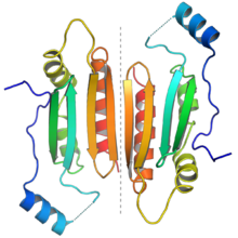
The protein dimer formed by two p19 molecules. Each monomer is colored from N-terminus (blue) to C-terminus (red) to illustrate the end-to-end orientation of the dimer. The dotted gray line in the center highlights the dimer interface. From PDB: 1R9F​.
 
Identifiers
SymbolTombus_p19
PfamPF03220
InterProIPR004905

RNA silencing suppressor p19 (also known as Tombusvirus P19 core protein and 19 kDa symptom severity modulator) is a protein expressed from the ORF4 gene in the genome of tombusviruses. These viruses are positive-sense single-stranded RNA viruses that infect plant cells, in which RNA silencing forms a widespread and robust antiviral defense system. The p19 protein serves as a counter-defense strategy, specifically binding the 19- to 21-nucleotide double-stranded RNAs that function as small interfering RNA (siRNA) in the RNA silencing system. By sequestering siRNA, p19 suppresses RNA silencing and promotes viral proliferation. The p19 protein is considered a significant virulence factor and a component of an evolutionary arms race between plants and their pathogens.

Structure

The structure of the tomato bushy stunt virus p19 protein bound to double-stranded RNA. The two p19 monomers are shown in blue and green; the RNA backbone is shown in orange. The alpha helices at the top and bottom interact with the ends of the RNA, ensuring that only RNA of the correct length is bound. This has been described as a "molecular caliper". From PDB: 1R9F​.

The p19 protein received its name from its size, being approximately 19 kilodaltons. It forms a functional homodimer. The crystal structures are available of p19 proteins from the tomato bushy stunt virus and Carnation Italian ringspot virus; the protein consists of a novel protein fold and exemplifies a previously unknown mechanism for binding RNA, using a binding surface formed by a beta sheet and flanked by alpha helices to interact with double-stranded RNAs of around 21 nucleotides in length in a non-sequence-specific manner.

Function

The p19 protein binds to double-stranded RNAs that function as short interfering RNA (siRNA) and is specialized for the 21-nucleotide product of the enzyme DCL4 (a member of a family of plant enzymes with homology to Dicer). By binding to siRNA, p19 sequesters these species and prevents them from interacting with the RNA-induced silencing complex (RISC), a protein complex that mediates the antiviral RNA silencing mechanism in the cell.

The p19 protein is also capable of binding to microRNA molecules that are endogenous to the host cell, as well as the siRNAs that are ultimately derived from the virus's own genome. Notably, an exception to this pattern is p19's inefficiency in interacting with the microRNA miR-168, a regulatory non-coding RNA that represses expression of argonaute-1 (AGO1). The AGO1 protein is required for RNA silencing, thus selectively sparing its repressor from p19's general sequestration of miRNA has the effect of reducing cellular AGO1 levels and is an additional mechanism by which p19 inhibits silencing. The two mechanisms are independent of one another and can be selectively abrogated by mutations.

Evolution

The gene encoding the p19 protein is an example of an overprinted gene, a genomic arrangement common in viruses in which multiple genes are encoded by the same portion of the genome read in alternate reading frames. The open reading frame ORF4, which encodes p19, is completely contained within the open reading frame of another gene, which is designated ORF3 and encodes the movement protein p22. Both genes, and their relative positions, are conserved within the tombusvirus family. P19 is thought to have originated de novo in this lineage.

Sequestration of dsRNA is a common viral counter-defense strategy against RNA silencing, evolved in a form of evolutionary arms race between virus and host. The p19 protein is not unique in this role; in an example of convergent evolution, this strategy appears to have evolved at least three times in distinct viral lineages using proteins with distinct structures and physical means of binding RNA.

History

The tomato bushy stunt virus, which is the type species of the tombusvirus family, is a long-standing model system for the study of plant viruses. The open reading frame encoding p19 was originally discovered in the late 1980s when the virus's genome was sequenced; it was subsequently demonstrated that the predicted protein was indeed expressed from the gene, although its role in promoting virulence and infectivity was initially underappreciated. Following the elucidation of its role as a suppressor of RNA silencing, p19 has also been used as a tool in molecular biology research on RNA silencing, RNA interference, and related processes.

Augustine of Hippo

From Wikipedia, the free encyclopedia https://en.wikipedia.org/wiki/Augustine_of_Hippo   Saint Augustine...