Search This Blog

Sunday, September 19, 2021

Arteriovenous malformation

From Wikipedia, the free encyclopedia
 
Arteriovenous malformation
Other namesAVM
Arteriovenous malformation - brain - low mag.jpg
Micrograph of an arteriovenous malformation in the brain. HPS stain.

Arteriovenous malformation is an abnormal connection between arteries and veins, bypassing the capillary system. This vascular anomaly is widely known because of its occurrence in the central nervous system (usually cerebral AVM), but can appear in any location. Although many AVMs are asymptomatic, they can cause intense pain or bleeding or lead to other serious medical problems.

AVMs are usually congenital and belong to the RASopathies. The genetic transmission patterns of AVMs are incomplete, but there are known genetic mutations (for instance in the epithelial line, tumor suppressor PTEN gene) which can lead to an increased occurrence throughout the body.

Signs and symptoms

Symptoms of AVM vary according to the location of the malformation. Roughly 88% of people with an AVM are asymptomatic; often the malformation is discovered as part of an autopsy or during treatment of an unrelated disorder (called in medicine an "incidental finding"); in rare cases, its expansion or a micro-bleed from an AVM in the brain can cause epilepsy, neurological deficit, or pain.

The most general symptoms of a cerebral AVM include headaches and epileptic seizures, with more specific symptoms occurring that normally depend on the location of the malformation and the individual. Such possible symptoms include:

Cerebral AVMs may present themselves in a number of different ways:

  • Bleeding (45% of cases)
  • Acute onset of severe headache. May be described as the worst headache of the patient's life. Depending on the location of bleeding, may be associated with new fixed neurologic deficit. In unruptured brain AVMs, the risk of spontaneous bleeding may be as low as 1% per year. After a first rupture, the annual bleeding risk may increase to more than 5%.
  • Seizure or brain seizure (46%) Depending on the place of the AVM, it can cause loss of vision in one place.
  • Headache (34%)
  • Progressive neurologic deficit (21%)
    • May be caused by mass effect or venous dilatations. Presence and nature of the deficit depend on location of lesion and the draining veins.
  • Pediatric patients

Pulmonary arteriovenous malformations

Pulmonary arteriovenous malformations are abnormal communications between the veins and arteries of the pulmonary circulation, leading to a right-to-left blood shunt. They have no symptoms in up to 29% of all cases, however they can give rise to serious complications including hemorrhage, and infection. They are most commonly associated with hereditary hemorrhagic telangiectasia.

Genetics

Can occur due to autosomal dominant diseases, such as hereditary hemorrhagic telangiectasia.

Pathophysiology

Arteries and veins are part of the vascular system. Arteries carry blood away from the heart to the lungs or the rest of the body, where the blood passes through capillaries, and veins return the blood to the heart. An AVM interferes with this process by forming a direct connection of the arteries and veins. AVMs can cause intense pain and lead to serious medical problems. Although AVMs are often associated with the brain and spinal cord, they can develop in any part of the body.

Normally, the arteries in the vascular system carry oxygen-rich blood, except in the case of the pulmonary artery. Structurally, arteries divide and sub-divide repeatedly, eventually forming a sponge-like capillary bed. Blood moves through the capillaries, giving up oxygen and taking up waste products, including CO
2
, from the surrounding cells. Capillaries in turn successively join together to form veins that carry blood away. The heart acts to pump blood through arteries and uptake the venous blood.

As an AVM lacks the dampening effect of capillaries on the blood flow, the AVM can get progressively larger over time as the amount of blood flowing through it increases, forcing the heart to work harder to keep up with the extra blood flow. It also causes the surrounding area to be deprived of the functions of the capillaries—removal of CO
2
and delivery of nutrients to the cells. The resulting tangle of blood vessels, often called a nidus (Latin for "nest"), has no capillaries. It can be extremely fragile and prone to bleeding because of the abnormally direct connections between high-pressure arteries and low-pressure veins. The resultant sign, audible via stethoscope, is a rhythmic, whooshing sound caused by excessively rapid blood flow through the arteries and veins. It has been given the term "bruit", French for noise. On some occasions, a patient with a brain AVM may become aware of the noise, which can compromise hearing and interfere with sleep in addition to causing psychological distress.

Diagnosis

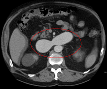
An arterial venous malformation of the left kidney and a simple cyst of the right kidney
 
An arterial venous malformation of the left kidney leading to aneurysmal dilatation of the left renal vein and inferior vena cava

AVMs are diagnosed primarily by the following imaging methods:

  • Computerized tomography (CT) scan is a noninvasive X-ray to view the anatomical structures within the brain to detect blood in or around the brain. A newer technology called CT angiography involves the injection of contrast into the blood stream to view the arteries of the brain. This type of test provides the best pictures of blood vessels through angiography and soft tissues through CT.
  • Magnetic resonance imaging (MRI) scan is a noninvasive test, which uses a magnetic field and radio-frequency waves to give a detailed view of the soft tissues of the brain.
  • Magnetic resonance angiography (MRA) – scans created using magnetic resonance imaging to specifically image the blood vessels and structures of the brain. A magnetic resonance angiogram can be an invasive procedure, involving the introduction of contrast dyes (e.g., gadolinium MR contrast agents) into the vasculature of a patient using a catheter inserted into an artery and passed through the blood vessels to the brain. Once the catheter is in place, the contrast dye is injected into the bloodstream and the MR images are taken. Additionally or alternatively, flow-dependent or other contrast-free magnetic resonance imaging techniques can be used to determine the location and other properties of the vasculature.

AVMs can occur in various parts of the body:

AVMs may occur in isolation or as a part of another disease (for example, Von Hippel-Lindau disease or hereditary hemorrhagic telangiectasia).

AVMs have been shown to be associated with aortic stenosis.

Bleeding from an AVM can be relatively mild or devastating. It can cause severe and less often fatal strokes. If a cerebral AVM is detected before a stroke occurs, usually the arteries feeding blood into the nidus can be closed off to avert the danger. However, interventional therapy may also be relatively risky.

Treatment

Treatment for brain AVMs can be symptomatic, and patients should be followed by a neurologist for any seizures, headaches, or focal neurologic deficits. AVM-specific treatment may also involve endovascular embolization, neurosurgery or radiosurgery. Embolization, that is, cutting off the blood supply to the AVM with coils, particles, acrylates, or polymers introduced by a radiographically guided catheter, may be used in addition to neurosurgery or radiosurgery, but is rarely successful in isolation except in smaller AVMs. Gamma knife may also be used.

Treatment of lung AVMs is typically performed with endovascular embolization alone, which is considered the standard of care. 

Epidemiology

The estimated detection rate of AVM in the US general population is 1.4/100,000 per year. This is approximately one fifth to one seventh the incidence of intracranial aneurysms. An estimated 300,000 Americans have AVMs, of whom 12% (approximately 36,000) will exhibit symptoms of greatly varying severity.

History

Luschka (1820–1875) and Virchow (1821–1902) first described arteriovenous malformations in the mid-1800s. Olivecrona (1891–1980) performed the first surgical excision of an intracranial AVM in 1932.

Society and culture

Notable cases

  • Indonesian actress Egidia Savitri [id] died from complications of AVM on November 29, 2013.
  • Phoenix Suns point guard AJ Price nearly died from AVM in 2004 while a student at the University of Connecticut.
  • On December 13, 2006, Senator Tim Johnson of South Dakota was diagnosed with AVM and treated at George Washington University Hospital.
  • On August 3, 2011, Mike Patterson of the Philadelphia Eagles collapsed on the field and suffered a seizure during a practice, leading to him being diagnosed with AVM.
  • Actor Ricardo Montalbán was born with spinal AVM. During the filming of the 1951 film Across the Wide Missouri, Montalbán was thrown from his horse, knocked unconscious, and trampled by another horse which aggravated his AVM and resulted in a painful back injury that never healed. The pain increased as he aged, and in 1993, Montalbán underwent 9½ hours of spinal surgery which left him paralyzed below the waist and using a wheelchair.
  • Actor/comedian T. J. Miller was diagnosed with AVM after filming Yogi Bear in New Zealand in 2010; Miller described his experience with the disease on the Pete Holmes podcast You Made It Weird on October 28, 2011, shedding his comedian side for a moment and becoming more philosophical, narrating his behaviors and inability to sleep during that time. He suffered a seizure upon return to Los Angeles and successfully underwent surgery that had a mortality rate of ten percent.
  • Jazz guitarist Pat Martino experienced an AVM and subsequently developed amnesia and manic depression. He eventually re-learned to play the guitar by listening to his own recordings from before the aneurysm. He still records and performs to this day.
  • YouTube vlogger Nikki Lilly (Nikki Christou), winner of the 2016 season of Junior Bake Off was born with AVM, which has resulted in some facial disfigurement.
  • Composer and lyricist William Finn was diagnosed with AVM and underwent Gamma Knife surgery in September 1992, soon after he won the 1992 Tony Award for best musical, awarded to "Falsettos". Finn wrote the 1998 Off-Broadway musical A New Brain about the experience.
  • Former Florida Gators and Oakland Raiders linebacker Neiron Ball was diagnosed with AVM in 2011 while playing for Florida, but recovered and was cleared to play. On September 16, 2018, Ball was placed in a medically induced coma due to complications of the disease, which lasted until his death on September 10, 2019.
  • Country music singer Drake White was diagnosed with AVM in January 2019, and is undergoing treatment.

Cultural Depictions

  • In the HBO series Six Feet Under (TV series), main character Nate Fisher discovers he has an AVM after being in a car accident and getting a precautionary cat scan at the hospital during Season 1. His AVM becomes a key focus during Season 2 and again in Season 5.

Research

Despite many years of research, the central question of whether to treat AVMs has not been answered. All treatments, whether involving surgery, radiation, or drugs, have risks and side-effects. Therefore, it might be better in some cases to avoid treatment altogether and simply accept a small risk of coming to harm from the AVM itself. This question is currently being addressed in clinical trials.

Frankenstein's monster

From Wikipedia, the free encyclopedia

Frankenstein's monster
Frankenstein, or the Modern Prometheus (Revised Edition, 1831) Creature.jpg
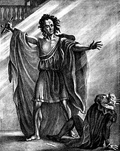
Steel engraving (993 × 78 mm), for the frontispiece of the 1831 revised edition of Mary Shelley's Frankenstein, published by Colburn and Bentley, London
First appearanceFrankenstein; or, The Modern Prometheus
Created byMary Shelley
Portrayed byBoris Karloff
Glenn Strange
Christopher Lee
Robert De Niro
Kevin James
Xavier Samuel
In-universe information
Nickname"Frankenstein's ", "The Monster", "The Creature", "The Wretch", "Adam Frankenstein" and others
SpeciesSimulacrum human
GenderMale
FamilyVictor Frankenstein (creator)
Bride of Frankenstein (companion/predecessor; in different adaptions)

Frankenstein's monster or Frankenstein's creature, often erroneously referred to as simply "Frankenstein", is a fictional character who first appeared in Mary Shelley's 1818 novel Frankenstein; or, The Modern Prometheus. Shelley's title thus compares the monster's creator, Victor Frankenstein, to the mythological character Prometheus, who fashioned humans out of clay and gave them fire.

In Shelley's Gothic story, Victor Frankenstein builds the creature in his laboratory through an ambiguous method based on a scientific principle he discovered. Shelley describes the monster as 8 feet (240 cm) tall and terribly hideous, but emotional. The monster attempts to fit into human society but is shunned, which leads him to seek revenge against Frankenstein. According to the scholar Joseph Carroll, the monster occupies "a border territory between the characteristics that typically define protagonists and antagonists".

Frankenstein's monster became iconic in popular culture, and has been featured in various forms of media, including films, television series, merchandise and video games. His most iconic version is his portrayal by Boris Karloff in the 1931 film Frankenstein, the 1935 sequel Bride of Frankenstein, and the 1939 sequel Son of Frankenstein.

Names

The actor T. P. Cooke as the monster in an 1823 stage production of Shelley's novel

Mary Shelley's original novel never gives the monster a name, although when speaking to his creator, Victor Frankenstein, the monster does say "I ought to be thy Adam" (in reference to the first man created in the Bible). Frankenstein refers to his creation as "creature", "fiend", "spectre", "the dæmon", "wretch", "devil", "thing", "being", and "ogre". Frankenstein's creation referred to himself as a "monster" at least once, as did the residents of a hamlet who saw the creature towards the end of the novel.

As in Shelley's story, the creature's namelessness became a central part of the stage adaptations in London and Paris during the decades after the novel's first appearance. In 1823, Shelley herself attended a performance of Richard Brinsley Peake's Presumption, the first successful stage adaptation of her novel. "The play bill amused me extremely, for in the list of dramatis personae came _________, by Mr T. Cooke," she wrote to her friend Leigh Hunt. "This nameless mode of naming the unnameable is rather good."

Within a decade of publication, the name of the creator—Frankenstein—was used to refer to the creature, but it did not become firmly established until much later. The story was adapted for the stage in 1927 by Peggy Webling, and Webling's Victor Frankenstein does give the creature his name. However, the creature has no name in the Universal film series starring Boris Karloff during the 1930s, which was largely based upon Webling's play. The 1931 Universal film treated the creature's identity in a similar way as Shelley's novel: in the opening credits, the character is referred to merely as "The Monster" (the actor's name is replaced by a question mark, but Karloff is listed in the closing credits). However, in the sequel Bride of Frankenstein (1935), the frame narration by a character representing Shelley's friend Lord Byron does refer to the monster as Frankenstein, although this scene takes place not quite in-universe. Nevertheless, the creature soon enough became best known in the popular imagination as "Frankenstein". This usage is sometimes considered erroneous, but some usage commentators regard the monster sense of "Frankenstein" as well-established and not an error.

Modern practice varies somewhat. For example, in Dean Koontz's Frankenstein, first published in 2004, the creature is named "Deucalion", after the character from Greek mythology, who is the son of the Titan Prometheus, a reference to the original novel's title. Another example is the second episode of Showtime's Penny Dreadful, which first aired in 2014; Victor Frankenstein briefly considers naming his creation "Adam", before deciding instead to let the monster "pick his own name". Thumbing through a book of the works of William Shakespeare, the monster chooses "Proteus" from The Two Gentlemen of Verona. It is later revealed that Proteus is actually the second monster Frankenstein has created, with the first, abandoned creation having been named "Caliban", from The Tempest, by the theatre actor who took him in and later, after leaving the theatre, named himself after the English poet John Clare. Another example is an attempt by Randall Munroe of webcomic xkcd to make "Frankenstein" the canonical name of the monster, by publishing a short derivative version which directly states that it is. In The Strange Case of the Alchemist's Daughter , the 2017 novel by Theodora Goss, the creature is named Adam.

Shelley's plot

Close-up of Charles Ogle as the monster in Thomas Edison's Frankenstein (1910)

Victor Frankenstein builds the creature in the attic of his boarding house in Ingolstadt after discovering a scientific principle which allows him to create life from non-living matter. Frankenstein is disgusted by his creation, however, and flees from it in horror. Frightened, and unaware of his own identity, the monster wanders through the wilderness.

He finds solace beside a remote cottage inhabited by an older, blind man and his two children. Eavesdropping, the creature familiarizes himself with their lives and learns to speak, whereby he becomes an eloquent, educated, and well-mannered individual. During this time, he also finds Frankenstein's journal in the pocket of the jacket he found in the laboratory and learns how he was created. The creature eventually introduces himself to the family's blind father, who treats him with kindness. When the rest of the family returns, however, they are frightened of him and drive him away. Enraged, the creature feels that humankind is his enemy and begins to hate his creator for abandoning him. However, although he despises Frankenstein, he sets out to find him, believing that he is the only person who will help him. On his journey, the creature rescues a young girl from a river but is shot in the shoulder by the child's father, believing the creature intended to harm his child. Enraged by this final act of cruelty, the creature swears revenge on humankind for the suffering they have caused him. He seeks revenge against his creator in particular for leaving him alone in a world where he is hated. Using the information in Frankenstein's notes, the creature resolves to find him.

The monster kills Victor's younger brother William upon learning of the boy's relation to his creator and makes it appear as if Justine Moritz, a young woman who lives with the Frankensteins, is responsible. When Frankenstein retreats to the Alps, the monster approaches him at the summit, recounts his experiences, and asks his creator to build him a female mate. He promises, in return, to disappear with his mate and never trouble humankind again, but threatens to destroy everything Frankenstein holds dear should he fail or refuse. Frankenstein agrees, and eventually constructs a female creature on a remote island in Orkney, but aghast at the possibility of creating a race of monsters, destroys the female creature before it is complete. Horrified and enraged, the creature immediately appears, and gives Frankenstein a final threat: "I will be with you on your wedding night."

After leaving his creator, the creature goes on to kill Victor's best friend, Henry Clerval, and later kills Frankenstein's bride, Elizabeth Lavenza, on their wedding night, whereupon Frankenstein's father dies of grief. With nothing left to live for but revenge, Frankenstein dedicates himself to destroying his creation, and the creature goads him into pursuing him north, through Scandinavia and into Russia, staying ahead of him the entire way.

As they reach the Arctic Circle and travel over the pack ice of the Arctic Ocean, Frankenstein, suffering from severe exhaustion and hypothermia, comes within a mile of the creature, but is separated from him when the ice he is traveling over splits. A ship exploring the region encounters the dying Frankenstein, who relates his story to the ship's captain, Robert Walton. Later, the monster boards the ship, but upon finding Frankenstein dead, is overcome by grief and pledges to incinerate himself at "the Northernmost extremity of the globe". He then departs, never to be seen again.

Appearance

Boris Karloff in Bride of Frankenstein (1935) in a variation of the classic 1931 film version with an assist from make-up artist Jack Pierce. Karloff had gained weight since the original iteration and much of the monster's hair has been burned off to indicate having been caught in a fire.
 
Frankenstein's monster in an editorial cartoon, 1896, an allegory on the Silverite movement displacing other progressive factions in late 19th century U.S.

Shelley described Frankenstein's monster as an 8-foot-tall (2.4 m) creature of hideous contrasts:

His limbs were in proportion, and I had selected his features as beautiful. Beautiful! Great God! His yellow skin scarcely covered the work of muscles and arteries beneath; his hair was of a lustrous black, and flowing; his teeth of a pearly whiteness; but these luxuriances only formed a more horrid contrast with his watery eyes, that seemed almost of the same colour as the dun-white sockets in which they were set, his shrivelled complexion and straight black lips.

A picture of the creature appeared in the 1831 edition. Early stage portrayals dressed him in a toga, shaded, along with the monster's skin, a pale blue. Throughout the 19th century, the monster's image remained variable according to the artist.

Portrayals in film

The best-known image of Frankenstein's monster in popular culture derives from Boris Karloff's portrayal in the 1931 movie Frankenstein, in which he wore makeup applied and designed by Jack P. Pierce. Universal Studios, which released the film, was quick to secure ownership of the copyright for the makeup format. Karloff played the monster in two more Universal films, Bride of Frankenstein and Son of Frankenstein; Lon Chaney Jr. took over the part from Karloff in The Ghost of Frankenstein; Bela Lugosi portrayed the role in Frankenstein Meets the Wolf Man; and Glenn Strange played the monster in the last three Universal Studios films to feature the character – House of Frankenstein, House of Dracula, and Abbott and Costello Meet Frankenstein. But their makeup replicated the iconic look first worn by Karloff. In modern times the image of Karloff's face is owned by his daughter's company, Karloff Enterprises, secured for her in a lawsuit for which she was represented by attorney Bela G. Lugosi (Bela Lugosi's son), after which Universal replaced Karloff's features with Glenn Strange's in most of their marketing. The New York Times mistakenly ran a photograph of Strange for Karloff's obituary.

Since Karloff's portrayal, the creature almost always appears as a towering, undead-like figure, often with a flat-topped angular head and bolts on his neck to serve as electrical connectors or grotesque electrodes. He wears a dark, usually tattered, suit having shortened coat sleeves and thick, heavy boots, causing him to walk with an awkward, stiff-legged gait (as opposed to the novel, in which he is described as much more flexible than a human). The tone of his skin varies (although shades of green or gray are common), and his body appears stitched together at certain parts (such as around the neck and joints). This image has influenced the creation of other fictional characters, such as the Hulk.

In the 1965 Toho film Frankenstein Conquers the World, the heart of Frankenstein's Monster was transported from Germany to Hiroshima as World War II neared its end, only to be irradiated during the atomic bombing of the city, granting it miraculous regenerative capabilities. Over the ensuing 20 years, it grows into a complete human child, who then rapidly matures into a giant, 20-metre-tall man. After escaping a laboratory in the city, he is blamed for the crimes of the burrowing Kaiju Baragon, and the two monsters face off in a showdown that ends with Frankenstein victorious, though he falls into the depths of the Earth after the ground collapses beneath his feet.

In the 1973 TV miniseries Frankenstein: The True Story, a different approach was taken in depicting the monster: Michael Sarrazin appears as a strikingly handsome man who later degenerates into a grotesque monster due to a flaw in the creation process.

In the 1994 film Mary Shelley's Frankenstein, the creature is played by Robert De Niro in a nearer approach to the original source, except this version gives the creature balding grey hair and a body covered in bloody stitches. He is, as in the novel, motivated by pain and loneliness. In this version, Frankenstein gives the monster the brain of his mentor, Doctor Waldman, while his body is made from a man who killed Waldman while resisting a vaccination. The monster retains Waldman's "trace memories" that apparently help him quickly learn to speak and read.

In the 2004 film Van Helsing, the monster is shown in a modernized version of the Karloff design. He is 8 to 9 feet (240–270 cm) tall, has a square bald head, gruesome scars, and pale green skin. The electricity is emphasized with one electrified dome in the back of his head and another over his heart. It also has hydraulic pistons in its legs, essentially rendering the design as a steam-punk cyborg. Although not as eloquent as in the novel, this version of the creature is intelligent and relatively nonviolent.

In 2004, a TV miniseries adaptation of Frankenstein was made by Hallmark. Luke Goss plays The Creature. This adaptation more closely resembles the monster as described in the novel: intelligent and articulate, with flowing, dark hair and watery eyes.

The 2005 film Frankenstein Reborn portrays the Creature as a paraplegic man who tries to regain the ability to walk by having a computer chip implanted. Instead, the surgeon kills him and resurrects his corpse as a reanimated zombie creature. he has stitches on his face where he was shot. with strains of brown hair black pants a dark hoodie and black jacket with brown fur collar.

The 2014 TV series Penny Dreadful also rejects the Karloff design in favour of Shelley's description. This version of the creature has the flowing dark hair described by Shelley, although he departs from her description by having pale grey skin and obvious scars along the right side of his face. Additionally, he is of average height, being even shorter than other characters in the series. In this series, the monster names himself "Caliban", after the character in William Shakespeare's The Tempest. In the series, Victor Frankenstein makes a second and third creature, each more indistinguishable from normal human beings.

Personality

Lon Chaney Jr. as the monster and Bela Lugosi as Ygor in The Ghost of Frankenstein (1942)
 
Glenn Strange as Frankenstein's monster with Boris Karloff, this time playing another character, in the 1944 film The House of Frankenstein
 
Christopher Lee as the creature in the Hammer's The Curse of Frankenstein (1957)

As depicted by Shelley, the monster is a sensitive, emotional creature whose only aim is to share his life with another sentient being like himself. The novel portrayed him as versed in Paradise Lost, Plutarch's Lives, and The Sorrows of Young Werther.

From the beginning, the monster is rejected by everyone he meets. He realizes from the moment of his "birth" that even his own creator cannot stand the sight of him; this is obvious when Frankenstein says "…one hand was stretched out, seemingly to detain me, but I escaped…". Upon seeing his own reflection, he realizes that he too is repulsed by his appearance. His greatest desire is to find love and acceptance; but when that desire is denied, he swears revenge on his creator.

The monster is a vegetarian. While speaking to Frankenstein, he tells him, "My food is not that of man; I do not destroy the lamb and the kid to glut my appetite; acorns and berries afford me sufficient nourishment...The picture I present to you is peaceful and human." At the time the novel was written, many writers, including Percy Shelley in A Vindication of Natural Diet, argued that practicing vegetarianism was the morally right thing to do.

Contrary to many film versions, the creature in the novel is very articulate and eloquent in his speech. Almost immediately after his creation, he dresses himself; and within 11 months, he can speak and read German and French. By the end of the novel, the creature is able to speak English fluently as well. The Van Helsing and Penny Dreadful interpretations of the character have similar personalities to the literary original, although the latter version is the only one to retain the character's violent reactions to rejection. In the 1931 film adaptation, the monster is depicted as mute and bestial; it is implied that this is because he is accidentally implanted with a criminal's "abnormal" brain. In the subsequent sequel, Bride of Frankenstein, the monster learns to speak, albeit in short, stunted sentences. In the second sequel, Son of Frankenstein, the creature is again rendered inarticulate. Following a brain transplant in the third sequel, The Ghost of Frankenstein, the monster speaks with the voice and personality of the brain donor. This was continued after a fashion in the scripting for the fourth sequel, Frankenstein Meets the Wolf Man, but the dialogue was excised before release. The monster was effectively mute in later sequels, although he refers to Count Dracula as his "master" in Abbott and Costello Meet Frankenstein. The monster is often portrayed as being afraid of fire, although he is not afraid of it in the novel.

The monster as a metaphor

Frankenstein's monster's bust, based on Boris Karloff, in the National Museum of Cinema of Turin, Italy

Scholars sometimes look for deeper meaning in Shelley's story, and have drawn an analogy between the monster and a motherless child; Shelley's own mother died while giving birth to her. The monster has also been analogized to an oppressed class; Shelley wrote that the monster recognized "the division of property, of immense wealth and squalid poverty". Others see in the monster the dangers of uncontrolled scientific progress, especially as at the time of publishing; Galvanism had convinced many scientists that raising the dead through use of electrical currents was a scientific possibility.

Another proposal is that the Frankenstein was based on a real scientist who had a similar name, and who had been called a modern Prometheus – Benjamin Franklin. Accordingly, the monster would represent the new nation that Franklin helped to create out of remnants left by England. Victor Frankenstein's father "made also a kite, with a wire and string, which drew down that fluid from the clouds," wrote Shelley, similar to Franklin's famous kite experiment.

Racial Interpretations

In discussing the physical description of the monster, there has been some speculation about the potential his design is rooted in common perceptions of race during the 18th century. Three scholars have noted that Shelley's description of the monster seems to be racially coded; one argues that, "Shelley's portrayal of her monster drew upon contemporary attitudes towards non-whites, in particular on fears and hopes of the abolition of slavery in the West Indies." Of course, there is no evidence to suggest that the Monster's depiction is meant to mimic any race, and such interpretations are based in personal conjectural interpretations of Shelley's text rather than remarks from herself or any known intentions of the author.

In her article "Frankenstein, Racial Science, and the Yellow Peril," Anne Mellor claims that the monster's features share a lot in common with the Mongoloid race. This term, now out of fashion and carrying some negative connotations, is used to describe the "yellow" races of Asia as distinct from the Caucasian or white races. To support her claim, Mellor points out that both Mary and Percy Shelley were friends with William Lawrence, an early proponent of racial science and someone who Mary "continued to consult on medical matters and [met with] socially until his death in 1830." While Mellor points out to allusions to Orientalism and the Yellow Peril, John Malchow in his article "Frankenstein's Monster and Images of Race in Nineteenth-Century Britain" explores the possibility of the monster either being intentionally or unintentionally coded as black. Malchow argues that the Monster's depiction is based in an 18th century understanding of "popular racial discourse [which] managed to conflate such descriptions of particular ethnic characteristics into a general image of the "Negro" body in which repulsive features, brute-like strength and size of limbs featured prominently." Malchow makes it clear that it is difficult to tell if this alleged racial allegory was intentional on Shelley's part or if it was inspired by the society she lived in (or if it exists in the text at all outside of his interpretation), and he states that "There is no clear proof that Mary Shelley consciously set out to create a monster which suggested, explicitly, the Jamaican escaped slave or maroon, or that she drew directly from any person knowledge of either planter or abolitionist propaganda." In addition to the previous interpretations, Karen Lynnea Piper argues in her article, "Inuit Diasporas: Frankenstein and the Inuit in England” that the symbolism surrounding Frankenstein’s monster could stem from the Inuit people of the arctic. Piper argues that the monster accounts for the “missing presence" of any indigenous people during Waldon's expedition, and that he represents the fear of the savage, lurking on the outskirts of civilization.

Xenotransplantation

From Wikipedia, the free encyclopedia

Xenotransplantation
File:Xenotransplantation-of-Human-Cardiomyocyte-Progenitor-Cells-Does-Not-Improve-Cardiac-Function-in-a-pone.0143953.s010.ogv
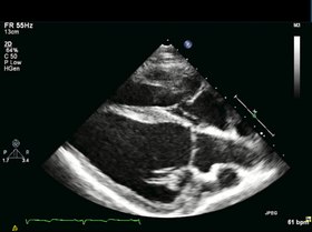
Long axis echocardiography. Representative long axis view echocardiography, 4 weeks after myocardial infarction (MI), right before CMPC/placebo infusion. Thinning and akinesia of the septal apical wall due to MI can be appreciated.
MeSHD014183

Xenotransplantation (xenos- from the Greek meaning "foreign" or strange), or heterologous transplant, is the transplantation of living cells, tissues or organs from one species to another. Such cells, tissues or organs are called xenografts or xenotransplants. It is contrasted with allotransplantation (from other individual of same species), syngeneic transplantation or isotransplantation (grafts transplanted between two genetically identical individuals of the same species) and autotransplantation (from one part of the body to another in the same person).

Xenotransplantation of human tumor cells into immunocompromised mice is a research technique frequently used in pre-clinical oncology research.

Human xenotransplantation offers a potential treatment for end-stage organ failure, a significant health problem in parts of the industrialized world. It also raises many novel medical, legal and ethical issues. A continuing concern is that many animals, such as pigs, have a shorter lifespan than humans, meaning that their tissues age at a quicker rate. Disease transmission (xenozoonosis) and permanent alteration to the genetic code of animals are also causes for concern. Similarly to objections to animal testing, animal rights activists have also objected to xenotransplantation on ethical grounds. A few temporarily successful cases of xenotransplantation are published.

It is common for patients and physicians to use the term "allograft" imprecisely to refer to either allograft (human-to-human) or xenograft (animal-to-human), but it is helpful scientifically (for those searching or reading the scientific literature) to maintain the more precise distinction in usage.

History

The first serious attempts at xenotransplantation (then called heterotransplantation) appeared in the scientific literature in 1905, when slices of rabbit kidney were transplanted into a child with chronic kidney disease. In the first two decades of the 20th century, several subsequent efforts to use organs from lambs, pigs, and primates were published.

Scientific interest in xenotransplantation declined when the immunological basis of the organ rejection process was described. The next waves of studies on the topic came with the discovery of immunosuppressive drugs. Even more studies followed Dr. Joseph Murray's first successful renal transplantation in 1954 and scientists, facing the ethical questions of organ donation for the first time, accelerated their effort in looking for alternatives to human organs.

In 1963, doctors at Tulane University attempted chimpanzee-to-human renal transplantations in six people who were near death; after this and several subsequent unsuccessful attempts to use primates as organ donors and the development of a working cadaver organ procuring program, interest in xenotransplantation for kidney failure dissipated. Out of 13 such transplants performed by Keith Reemtsma, one kidney recipient lived for 9 months, returning to work as a schoolteacher. At autopsy, the chimpanzee kidneys appeared normal and showed no signs of acute or chronic rejection.

An American infant girl known as "Baby Fae" with hypoplastic left heart syndrome was the first infant recipient of a xenotransplantation, when she received a baboon heart in 1984. The procedure was performed by Leonard Lee Bailey at Loma Linda University Medical Center in Loma Linda, California. Fae died 21 days later due to a humoral-based graft rejection thought to be caused mainly by an ABO blood type mismatch, considered unavoidable due to the rarity of type O baboons. The graft was meant to be temporary, but unfortunately a suitable allograft replacement could not be found in time. While the procedure itself did not advance the progress on xenotransplantation, it did shed a light on the insufficient amount of organs for infants. The story grew so big that it made such an impact that the crisis of infant organ shortage improved for that time.

Xenotransplantation of human tumor cells into immunocompromised mice is a research technique frequently used in oncology research. It is used to predict the sensitivity of the transplanted tumor to various cancer treatments; several companies offer this service, including the Jackson Laboratory.

Human organs have been transplanted into animals as a powerful research technique for studying human biology without harming human patients. This technique has also been proposed as an alternative source of human organs for future transplantation into human patients. For example, researchers from the Ganogen Research Institute transplanted human fetal kidneys into rats which demonstrated life supporting function and growth.

Potential uses

A worldwide shortage of organs for clinical implantation causes about 20–35% of patients who need replacement organs to die on the waiting list. Certain procedures, some of which are being investigated in early clinical trials, aim to use cells or tissues from other species to treat life-threatening and debilitating illnesses such as cancer, diabetes, liver failure and Parkinson's disease. If vitrification can be perfected, it could allow for long-term storage of xenogenic cells, tissues and organs so that they would be more readily available for transplant.

Xenotransplants could save thousands of patients waiting for donated organs. The animal organ, probably from a pig or baboon could be genetically altered with human genes to trick a patient's immune system into accepting it as a part of its own body. They have re-emerged because of the lack of organs available and the constant battle to keep immune systems from rejecting allotransplants. Xenotransplants are thus potentially a more effective alternative.

Xenotransplantation also is and has been a valuable tool used in research laboratories to study developmental biology.

Patient derived tumor xenografts in animals can be used to test treatments.

Potential animal organ donors

Since they are the closest relatives to humans, non-human primates were first considered as a potential organ source for xenotransplantation to humans. Chimpanzees were originally considered the best option since their organs are of similar size, and they have good blood type compatibility with humans, which makes them potential candidates for xenotransfusions. However, since chimpanzees are listed as an endangered species, other potential donors were sought. Baboons are more readily available, but impractical as potential donors. Problems include their smaller body size, the infrequency of blood group O (the universal donor), their long gestation period, and their typically small number of offspring. In addition, a major problem with the use of nonhuman primates is the increased risk of disease transmission, since they are so closely related to humans.

Pigs (Sus scrofa domesticus) are currently thought to be the best candidates for organ donation. The risk of cross-species disease transmission is decreased because of their increased phylogenetic distance from humans. Pigs have relatively short gestation periods, large litters, and are easy to breed making them readily available. They are inexpensive and easy to maintain in pathogen-free facilities, and current gene editing tools are adapted to pigs to combat rejection and potential zoonoses. Pig organs are anatomically comparable in size, and new infectious agents are less likely since they have been in close contact with humans through domestication for many generations. Treatments sourced from pigs have proven to be successful such as porcine-derived insulin for patients with diabetes mellitus. Increasingly, genetically engineered pigs are becoming the norm, which raises moral qualms, but also increases the success rate of the transplant. Current experiments in xenotransplantation most often use pigs as the donor, and baboons as human models.

In the field of regenerative medicine, pancreatogenesis- or nephrogenesis-disabled pig embryos, unable to form a specific organ, allow experimentation toward the in vivo generation of functional organs from xenogenic pluripotent stem cells in large animals via compensation for an empty developmental niche (blastocyst complementation). Such experiments provide the basis for potential future application of blastocyst complementation to generate transplantable human organs from the patient's own cells, using livestock animals, to increase quality of life for those with end-stage organ failure.

Barriers and issues

Immunologic barriers

To date, no xenotransplantation trials have been entirely successful due to the many obstacles arising from the response of the recipient's immune system. "Xenozoonoses" are one of the biggest threats to rejections, as they are xenogenetic infections. The introduction of these microorganisms are a big issue that lead to the fatal infections and then rejection of the organs. This response, which is generally more extreme than in allotransplantations, ultimately results in rejection of the xenograft, and can in some cases result in the immediate death of the recipient. There are several types of rejection organ xenografts are faced with, these include hyperacute rejection, acute vascular rejection, cellular rejection, and chronic rejection.

A rapid, violent, and hyperacute response comes as a result of antibodies present in the host organism. These antibodies are known as xenoreactive natural antibodies (XNAs).

Hyperacute rejection

This rapid and violent type of rejection occurs within minutes to hours from the time of the transplant. It is mediated by the binding of XNAs (xenoreactive natural antibodies) to the donor endothelium, causing activation of the human complement system, which results in endothelial damage, inflammation, thrombosis and necrosis of the transplant. XNAs are first produced and begin circulating in the blood in neonates, after colonization of the bowel by bacteria with galactose moieties on their cell walls. Most of these antibodies are the IgM class, but also include IgG, and IgA.

The epitope XNAs target is an α-linked galactose moiety, Gal-α-1,3Gal (also called the α-Gal epitope), produced by the enzyme α-galactosyl transferase. Most non-primates contain this enzyme thus, this epitope is present on the organ epithelium and is perceived as a foreign antigen by primates, which lack the galactosyl transferase enzyme. In pig to primate xenotransplantation, XNAs recognize porcine glycoproteins of the integrin family.

The binding of XNAs initiate complement activation through the classical complement pathway. Complement activation causes a cascade of events leading to: destruction of endothelial cells, platelet degranulation, inflammation, coagulation, fibrin deposition, and hemorrhage. The end result is thrombosis and necrosis of the xenograft.

Overcoming hyperacute rejection

Since hyperacute rejection presents such a barrier to the success of xenografts, several strategies to overcome it are under investigation:

Interruption of the complement cascade

  • The recipient's complement cascade can be inhibited through the use of cobra venom factor (which depletes C3), soluble complement receptor type 1, anti-C5 antibodies, or C1 inhibitor (C1-INH). Disadvantages of this approach include the toxicity of cobra venom factor, and most importantly these treatments would deprive the individual of a functional complement system.

Transgenic organs (Genetically engineered pigs)

  • 1,3 galactosyl transferase gene knockouts – These pigs don't contain the gene that codes for the enzyme responsible for expression of the immunogeneic gal-α-1,3Gal moiety (the α-Gal epitope).
  • Increased expression of H-transferase (α 1,2 fucosyltransferase), an enzyme that competes with galactosyl transferase. Experiments have shown this reduces α-Gal expression by 70%.
  • Expression of human complement regulators (CD55, CD46, and CD59) to inhibit the complement cascade.
  • Plasmaphoresis, on humans to remove 1,3 galactosyltransferase, reduces the risk of activation of effector cells such as CTL (CD8 T cells), complement pathway activation and delayed type hypersensitivity (DTH).

Acute vascular rejection

Also known as delayed xenoactive rejection, this type of rejection occurs in discordant xenografts within 2 to 3 days, if hyperacute rejection is prevented. The process is much more complex than hyperacute rejection and is currently not completely understood. Acute vascular rejection requires de novo protein synthesis and is driven by interactions between the graft endothelial cells and host antibodies, macrophages, and platelets. The response is characterized by an inflammatory infiltrate of mostly macrophages and natural killer cells (with small numbers of T cells), intravascular thrombosis, and fibrinoid necrosis of vessel walls.

Binding of the previously mentioned XNAs to the donor endothelium leads to the activation of host macrophages as well as the endothelium itself. The endothelium activation is considered type II since gene induction and protein synthesis are involved. The binding of XNAs ultimately leads to the development of a procoagulant state, the secretion of inflammatory cytokines and chemokines, as well as expression of leukocyte adhesion molecules such as E-selectin, intercellular adhesion molecule-1 (ICAM-1), and vascular cell adhesion molecule-1 (VCAM-1).

This response is further perpetuated as normally binding between regulatory proteins and their ligands aid in the control of coagulation and inflammatory responses. However, due to molecular incompatibilities between the molecules of the donor species and recipient (such as porcine major histocompatibility complex molecules and human natural killer cells), this may not occur.

Overcoming acute vascular rejection

Due to its complexity, the use of immunosuppressive drugs along with a wide array of approaches are necessary to prevent acute vascular rejection, and include administering a synthetic thrombin inhibitor to modulate thrombogenesis, depletion of anti-galactose antibodies (XNAs) by techniques such as immunoadsorption, to prevent endothelial cell activation, and inhibiting activation of macrophages (stimulated by CD4+ T cells) and NK cells (stimulated by the release of Il-2). Thus, the role of MHC molecules and T cell responses in activation would have to be reassessed for each species combo.

Accommodation

If hyperacute and acute vascular rejection are avoided accommodation is possible, which is the survival of the xenograft despite the presence of circulating XNAs. The graft is given a break from humoral rejection when the complement cascade is interrupted, circulating antibodies are removed, or their function is changed, or there is a change in the expression of surface antigens on the graft. This allows the xenograft to up-regulate and express protective genes, which aid in resistance to injury, such as heme oxygenase-1 (an enzyme that catalyzes the degradation of heme).

Cellular rejection

Rejection of the xenograft in hyperacute and acute vascular rejection is due to the response of the humoral immune system, since the response is elicited by the XNAs. Cellular rejection is based on cellular immunity, and is mediated by natural killer cells that accumulate in and damage the xenograft and T-lymphocytes which are activated by MHC molecules through both direct and indirect xenorecognition.

In direct xenorecognition, antigen presenting cells from the xenograft present peptides to recipient CD4+ T cells via xenogeneic MHC class II molecules, resulting in the production of interleukin 2 (IL-2). Indirect xenorecognition involves the presentation of antigens from the xenograft by recipient antigen presenting cells to CD4+ T cells. Antigens of phagocytosed graft cells can also be presented by the host's class I MHC molecules to CD8+ T cells.

The strength of cellular rejection in xenografts remains uncertain, however, it is expected to be stronger than in allografts due to differences in peptides among different animals. This leads to more antigens potentially recognized as foreign, thus eliciting a greater indirect xenogenic response.

Overcoming cellular rejection

A proposed strategy to avoid cellular rejection is to induce donor non-responsiveness using hematopoietic chimerism. Donor stem cells are introduced into the bone marrow of the recipient, where they coexist with the recipient's stem cells. The bone marrow stem cells give rise to cells of all hematopoietic lineages, through the process of hematopoiesis. Lymphoid progenitor cells are created by this process and move to the thymus where negative selection eliminates T cells found to be reactive to self. The existence of donor stem cells in the recipient's bone marrow causes donor reactive T cells to be considered self and undergo apoptosis.

Chronic rejection

Chronic rejection is slow and progressive, and usually occurs in transplants that survive the initial rejection phases. Scientists are still unclear how chronic rejection exactly works, research in this area is difficult since xenografts rarely survive past the initial acute rejection phases. Nonetheless, it is known that XNAs and the complement system are not primarily involved. Fibrosis in the xenograft occurs as a result of immune reactions, cytokines (which stimulate fibroblasts), or healing (following cellular necrosis in acute rejection). Perhaps the major cause of chronic rejection is arteriosclerosis.  Lymphocytes, which were previously activated by antigens in the vessel wall of the graft, activate macrophages to secrete smooth muscle growth factors. This results in a build up of smooth muscle cells on the vessel walls, causing the hardening and narrowing of vessels within the graft. Chronic rejection leads to pathologic changes of the organ, and is why transplants must be replaced after so many years. It is also anticipated that chronic rejection will be more aggressive in xenotransplants as opposed to allotransplants.

Dysregulated coagulation

Successful efforts have been made to create knockout mice without α1,3GT; the resulting reduction in the highly immunogenic αGal epitope has resulted in the reduction of the occurrence of hyperacute rejection, but has not eliminated other barriers to xenotransplantation such as dysregulated coagulation, also known as coagulopathy.

Different organ xenotransplants result in different responses in clotting. For example, kidney transplants result in a higher degree of coagulopathy, or impaired clotting, than cardiac transplants, whereas liver xenografts result in severe thrombocytopenia, causing recipient death within a few days due to bleeding. An alternate clotting disorder, thrombosis, may be initiated by preexisting antibodies that affect the protein C anticoagulant system. Due to this effect, porcine donors must be extensively screened before transplantation. Studies have also shown that some porcine transplant cells are able to induce human tissue factor expression, thus stimulating platelet and monocyte aggregation around the xenotransplanted organ, causing severe clotting. Additionally, spontaneous platelet accumulation may be caused by contact with pig von Willebrand factor.

Just as the α1,3G epitope is a major problem in xenotransplantation, so too is dysregulated coagulation a cause of concern. Transgenic pigs that can control for variable coagulant activity based on the specific organ transplanted would make xenotransplantation a more readily available solution for the 70,000 patients per year who do not receive a human donation of the organ or tissue they need.

Physiology

Extensive research is required to determine whether animal organs can replace the physiological functions of human organs. Many issues include size – differences in organ size limit the range of potential recipients of xenotransplants; longevity – The lifespan of most pigs is roughly 15 years, currently it is unknown whether or not a xenograft may be able to last longer than that; hormone and protein differences – some proteins will be molecularly incompatible, which could cause malfunction of important regulatory processes. These differences also make the prospect of hepatic xenotransplantation less promising, since the liver plays an important role in the production of so many proteins; environment – for example, pig hearts work in a different anatomical site and under different hydrostatic pressure than in humans; temperature – the body temperature of pigs is 39 °C (2 °C above the average human body temperature). Implications of this difference, if any, on the activity of important enzymes are currently unknown.

Xenozoonosis

Xenozoonosis, also known as zoonosis or xenosis, is the transmission of infectious agents between species via xenograft. Animal to human infection is normally rare, but has occurred in the past. An example of such is the avian influenza, when an influenza A virus was passed from birds to humans. Xenotransplantation may increase the chance of disease transmission for 3 reasons: (1) implantation breaches the physical barrier that normally helps to prevent disease transmission, (2) the recipient of the transplant will be severely immunosuppressed, and (3) human complement regulators (CD46, CD55, and CD59) expressed in transgenic pigs have been shown to serve as virus receptors, and may also help to protect viruses from attack by the complement system.

Examples of viruses carried by pigs include porcine herpesvirus, rotavirus, parvovirus, and circovirus. Porcine herpesviruses and rotaviruses can be eliminated from the donor pool by screening, however others (such as parvovirus and circovirus) may contaminate food and footwear then re-infect the herd. Thus, pigs to be used as organ donors must be housed under strict regulations and screened regularly for microbes and pathogens. Unknown viruses, as well as those not harmful in the animal, may also pose risks. Of particular concern are PERVS (porcine endogenous retroviruses), vertically transmitted microbes that embed in swine genomes. The risks with xenosis are twofold, as not only could the individual become infected, but a novel infection could initiate an epidemic in the human population. Because of this risk, the FDA has suggested any recipients of xenotransplants shall be closely monitored for the remainder of their life, and quarantined if they show signs of xenosis.

Baboons and pigs carry myriad transmittable agents that are harmless in their natural host, but extremely toxic and deadly in humans. HIV is an example of a disease believed to have jumped from monkeys to humans. Researchers also do not know if an outbreak of infectious diseases could occur and if they could contain the outbreak even though they have measures for control. Another obstacle facing xenotransplants is that of the body's rejection of foreign objects by its immune system. These antigens (foreign objects) are often treated with powerful immunosuppressive drugs that could, in turn, make the patient vulnerable to other infections and actually aid the disease. This is the reason the organs would have to be altered to fit the patients' DNA (histocompatibility).

In 2005, the Australian National Health and Medical Research Council (NHMRC) declared an eighteen-year moratorium on all animal-to-human transplantation, concluding that the risks of transmission of animal viruses to patients and the wider community had not been resolved. This was repealed in 2009 after an NHMRC review stated "... the risks, if appropriately regulated, are minimal and acceptable given the potential benefits.", citing international developments on the management and regulation of xenotransplantation by the World Health Organisation and the European Medicines Agency.

Porcine endogenous retroviruses

Endogenous retroviruses are remnants of ancient viral infections, found in the genomes of most, if not all, mammalian species. Integrated into the chromosomal DNA, they are vertically transferred through inheritance. Due to the many deletions and mutations they accumulate over time, they usually are not infectious in the host species, however the virus may become infectious in another species. PERVS were originally discovered as retrovirus particles released from cultured porcine kidney cells. Most breeds of swine harbor approximately 50 PERV genomes in their DNA. Although it is likely that most of these are defective, some may be able to produce infectious viruses so every proviral genome must be sequenced to identify which ones pose a threat. In addition, through complementation and genetic recombination, two defective PERV genomes could give rise to an infectious virus. There are three subgroups of infectious PERVs (PERV-A, PERV-B, and PERV-C). Experiments have shown that PERV-A and PERV-B can infect human cells in culture. To date no experimental xenotransplantations have demonstrated PERV transmission, yet this does not mean PERV infections in humans are impossible. Pig cells have been engineered to inactivate all 62 PERVs in the genome using CRISPR Cas9 genome editing technology, and eliminated infection from the pig to human cells in culture.

Ethics

Xenografts have been a controversial procedure since they were first attempted. Many, including animal rights groups, strongly oppose killing animals to harvest their organs for human use. In the 1960s, many organs came from the chimpanzees, and were transferred into people that were deathly ill, and in turn, did not live much longer afterwards. Modern scientific supporters of xenotransplantation argue that the potential benefits to society outweigh the risks, making pursuing xenotransplantation the moral choice. None of the major religions object to the use of genetically modified pig organs for life-saving transplantation. Religions such as Buddhism and Jainism, however, have long espoused non-violence towards all living creatures. In general, the use of pig and cow tissue in humans has been met with little resistance, save some religious beliefs and a few philosophical objections. Experimentation without consent doctrines are now followed, which was not the case in the past, which may lead to new religious guidelines to further medical research on pronounced ecumenical guidelines. The "Common Rule" is the United States bio-ethics mandate as of 2011.

History of Xenotransplantation in Ethics

At the beginning of the 20th century when studies in Xenotransplantation were just beginning, few questioned the morality of it, turning to animals as a "natural" alternative to allografts. While satirical plays mocked Xenografters such as Serge Voronoff, and some images showing emotionally distraught primates appeared - who Voronoff had deprived of their testicles - no serious attempts were yet made to question the science based on animal rights concerns. Xenotransplantation was not taken seriously, at least in France, during the first half of the 20th century.

With the Baby Fae incident of 1984 as the impetus, animal rights activists began to protest, gathering media attention and proving that some people felt that it was unethical and a violation of the animal's own rights to use its organs to preserve a sick human's life. Treating animals as mere tools for the slaughter on demand by human will would lead to a world they would not prefer. Supporters of the transplant pushed back, claiming that saving a human life justifies the sacrifice of an animal one. Most animal rights activists found the use of primate organs more reprehensible than those of, for example, pigs. As Peter Singer et al. have expressed, many primates exhibit greater social structure, communication skills, and affection than mentally deficient humans and human infants. Despite this, it is considerably unlikely that animal suffering will provide sufficient impetus for regulators to prevent xenotransplantation.

Informed consent of patient

Autonomy and informed consent are important when considering the future uses of xenotransplantation. A patient undergoing xenotransplantation should be fully aware of the procedure and should have no outside force influencing their choice. The patient should understand the risks and benefits of such a transplantation. However, it has been suggested that friends and family members should also give consent, because the repercussions of transplantation are high, with the potential of diseases and viruses crossing over to humans from the transplantation. Close contacts are at risk for such infections. Monitoring of close relations may also be required to ensure that xenozoonosis is not occurring. The question then becomes: does the autonomy of the patient become limited based on the willingness or unwillingness of friends and family to give consent, and are the principles of confidentiality broken?

The safety of public health is a factor to be considered. If there is any risk to the public at all for an outbreak from transplantation there must be procedures in place to protect the public. Not only does the recipient of the transplantation have to understand the risks and benefits, but society must also understand and consent to such an agreement.

The Ethics Committee of the International Xenotransplantation Association points out one major ethical issue is the societal response to such a procedure. The assumption is that the recipient of the transplantation will be asked to undergo lifelong monitoring, which would deny the recipient the ability to terminate the monitoring at any time, which is in direct opposition of the Declaration of Helsinki and the US Code of Federal Regulations. In 2007, xenotransplantation was banned under ethical grounds in all countries but Argentina, Russia and New Zealand. Since then, the practice has only been carried out to treatment for diabetes type 1 to serve as a substitute for insulin injections.

The application of the four bioethics principal is found to be everywhere because it is now standardized in the moral conducts of a laboratory. The four principles emphasize on the informed consent, the Hippocratic Oath to do no harm, apply one's skill to help others, and protecting the rights of others to quality care.

Problems with xenotransplantation is that even though it has future medical benefits, it also has the serious risk of introducing and spreading the infectious diseases, into the human population. There have been guidelines that have been drafted by the government that have the purpose of forming the foundation of infectious disease surveillance. In the United Kingdom, the guideline that were introduced state that first, "the periodic provision of bodily samples that would then be archived for epidemiological purposes;" second, "post-mortem analysis in case of death, the storage of samples post-mortem, and the disclosure of this agreement to their family;" third, "refrain from donating blood, tissue or organs;" fourth, "the use of barrier contraception when engaging in sexual intercourse;" fifth, keep both name and current address on register and to notify the relevant health authorities when moving abroad;" and lastly "divulge confidential information, including one's status as a xenotransplantation recipient to researchers, all health care professionals from whom one seeks professional services, and close contacts such as current and future sexual partners." With these guidelines in place the patient has to abide to these rules until either their lifetime or until the government determines that there is no need for public health safe guards.

Xenotransplantation guidelines in the United States

The Food and Drug Administration (FDA) has also stated that if a transplantation takes place the recipient must undergo monitoring for the rest of that recipient's lifetime and waive their right to withdraw. The reason for requiring lifelong monitoring is due to the risk of acute infections that may occur. The FDA suggests that a passive screening program should be implemented and should extend for the life of the recipient.

Operator (computer programming)

From Wikipedia, the free encyclopedia https://en.wikipedia.org/wiki/Operator_(computer_programmin...