Search This Blog

Thursday, June 20, 2019

Pre-eclampsia

From Wikipedia, the free encyclopedia

Pre-eclampsia
Other namesPre-eclampsia toxaemia (PET), pre-eclampsia
Hypertrophic decidual vasculopathy high mag.jpg
A micrograph showing hypertrophic decidual vasculopathy, a finding seen in gestational hypertension and pre-eclampsia. H&E stain.
SpecialtyObstetrics
SymptomsHigh blood pressure, protein in the urine
ComplicationsRed blood cell breakdown, low blood platelet count, impaired liver function, kidney problems, swelling, shortness of breath due to fluid in the lungs, eclampsia
Usual onsetAfter 20 weeks of pregnancy
Risk factorsObesity, prior hypertension, older age, diabetes mellitus
Diagnostic methodBP > 140 mmHg systolic or 90 mmHg diastolic at two separate times
PreventionAspirin, calcium supplementation, treatment of prior hypertension
TreatmentDelivery, medications
MedicationLabetalol, methyldopa, magnesium sulfate
Frequency2–8% of pregnancies
Deaths46,900 hypertensive disorders in pregnancy (2015)

Pre-eclampsia (PE) is a disorder of pregnancy characterized by the onset of high blood pressure and often a significant amount of protein in the urine. When it arises, the condition begins after 20 weeks of pregnancy. In severe disease there may be red blood cell breakdown, a low blood platelet count, impaired liver function, kidney dysfunction, swelling, shortness of breath due to fluid in the lungs, or visual disturbances. Pre-eclampsia increases the risk of poor outcomes for both the mother and the baby. If left untreated, it may result in seizures at which point it is known as eclampsia.

Risk factors for pre-eclampsia include obesity, prior hypertension, older age, and diabetes mellitus. It is also more frequent in a woman's first pregnancy and if she is carrying twins. The underlying mechanism involves abnormal formation of blood vessels in the placenta amongst other factors. Most cases are diagnosed before delivery. Rarely, pre-eclampsia may begin in the period after delivery. While historically both high blood pressure and protein in the urine were required to make the diagnosis, some definitions also include those with hypertension and any associated organ dysfunction. Blood pressure is defined as high when it is greater than 140 mmHg systolic or 90 mmHg diastolic at two separate times, more than four hours apart in a woman after twenty weeks of pregnancy. Pre-eclampsia is routinely screened for during prenatal care.

Recommendations for prevention include: aspirin in those at high risk, calcium supplementation in areas with low intake, and treatment of prior hypertension with medications. In those with pre-eclampsia delivery of the baby and placenta is an effective treatment. When delivery becomes recommended depends on how severe the pre-eclampsia and how far along in pregnancy a woman is. Blood pressure medication, such as labetalol and methyldopa, may be used to improve the mother's condition before delivery. Magnesium sulfate may be used to prevent eclampsia in those with severe disease. Bedrest and salt intake have not been found to be useful for either treatment or prevention.

Pre-eclampsia affects 2–8% of pregnancies worldwide. Hypertensive disorders of pregnancy (which include pre-eclampsia) are one of the most common causes of death due to pregnancy. They resulted in 46,900 deaths in 2015. Pre-eclampsia usually occurs after 32 weeks; however, if it occurs earlier it is associated with worse outcomes. Women who have had pre-eclampsia are at increased risk of heart disease and stroke later in life. The word "eclampsia" is from the Greek term for lightning. The first known description of the condition was by Hippocrates in the 5th century BC.

Signs and symptoms

Swelling (especially in the hands and face) was originally considered an important sign for a diagnosis of pre-eclampsia. However, because swelling is a common occurrence in pregnancy, its utility as a distinguishing factor in pre-eclampsia is not high. Pitting edema (unusual swelling, particularly of the hands, feet, or face, notable by leaving an indentation when pressed on) can be significant, and should be reported to a health care provider. 

In general, none of the signs of pre-eclampsia are specific, and even convulsions in pregnancy are more likely to have causes other than eclampsia in modern practice. Further, a symptom such as epigastric pain may be misinterpreted as heartburn. Diagnosis, therefore, depends on finding a coincidence of several pre-eclamptic features, the final proof being their regression after delivery.

Causes

There is no definitive known cause of pre-eclampsia, though it is likely related to a number of factors. Some of these factors include:
  • Abnormal placentation (formation and development of the placenta)
  • Immunologic factors
  • Prior or existing maternal pathology—pre-eclampsia is seen more at a higher incidence in individuals with pre-existing hypertension, obesity, antiphospholipid antibody syndrome, and those with history of pre-eclampsia
  • Dietary factors, e.g. calcium supplementation in areas where dietary calcium intake is low has been shown to reduce the risk of pre-eclampsia
  • Environmental factors, e.g. air pollution
Those with long term high blood pressure have a risk 7 to 8 times higher than those without.

Physiologically, research has linked pre-eclampsia to the following physiologic changes: alterations in the interaction between the maternal immune response and the placenta, placental injury, endothelial cell injury, altered vascular reactivity, oxidative stress, imbalance among vasoactive substances, decreased intravascular volume, and disseminated intravascular coagulation.

While the exact cause of pre-eclampsia remains unclear, there is strong evidence that a major cause predisposing a susceptible woman to pre-eclampsia is an abnormally implanted placenta. This abnormally implanted placenta may result in poor uterine and placental perfusion, yielding a state of hypoxia and increased oxidative stress and the release of anti-angiogenic proteins along with inflammatory mediators into the maternal plasma. A major consequence of this sequence of events is generalized endothelial dysfunction. The abnormal implantation may stem from the maternal immune system's response to the placenta, specifically a lack of established immunological tolerance in pregnancy. Endothelial dysfunction results in hypertension and many of the other symptoms and complications associated with pre-eclampsia. Those with pre-eclampsia may have a lower risk of breast cancer.

Abnormal chromosome 19 microRNA cluster (C19MC) impairs extravillus trophoblast cell invasion to the spiral arteries, causing high resistance, low blood flow, and low nutrient supply to the fetus.

Risk factors

Known risk factors for pre-eclampsia include:

Pathogenesis

Although much research into mechanism of pre-eclampsia has taken place, its exact pathogenesis remains uncertain. Pre-eclampsia is thought to result from an abnormal placenta, the removal of which ends the disease in most cases. During normal pregnancy, the placenta vascularizes to allow for the exchange of water, gases, and solutes, including nutrients and wastes, between maternal and fetal circulations. Abnormal development of the placenta leads to poor placental perfusion. The placenta of women with pre-eclampsia is abnormal and characterized by poor trophoblastic invasion. It is thought that this results in oxidative stress, hypoxia, and the release of factors that promote endothelial dysfunction, inflammation, and other possible reactions.

The clinical manifestations of pre-eclampsia are associated with general endothelial dysfunction, including vasoconstriction and end-organ ischemia. Implicit in this generalized endothelial dysfunction may be an imbalance of angiogenic and anti-angiogenic factors. Both circulating and placental levels of soluble fms-like tyrosine kinase-1 (sFlt-1) are higher in women with pre-eclampsia than in women with normal pregnancy. sFlt-1 is an anti-angiogenic protein that antagonizes vascular endothelial growth factor (VEGF) and placental growth factor (PIGF), both of which are proangiogenic factors. Soluble endoglin (sEng) has also been shown to be elevated in women with pre-eclampsia and has anti-angiogenic properties, much like sFlt-1 does.

Both sFlt-1 and sEng are upregulated in all pregnant women to some extent, supporting the idea that hypertensive disease in pregnancy is a normal pregnancy adaptation gone awry. As natural killer cells are intimately involved in placentation and placentation involves a degree of maternal immune tolerance for a foreign placenta, it is not surprising that the maternal immune system might respond more negatively to the arrival of some placentae under certain circumstances, such as a placenta which is more invasive than normal. Initial maternal rejection of the placental cytotrophoblasts may be the cause of the inadequately remodeled spiral arteries in those cases of pre-eclampsia associated with shallow implantation, leading to downstream hypoxia and the appearance of maternal symptoms in response to upregulated sFlt-1 and sEng. 

Oxidative stress may also play an important part in the pathogenesis of pre-eclampsia. The main source of reactive oxygen species (ROS) is the enzyme xanthine oxidase (XO) and this enzyme mainly occurs in the liver. One hypothesis is that the increased purine catabolism from placental hypoxia results in increased ROS production in the maternal liver and release into the maternal circulation that causes endothelial cell damage.

Abnormalities in the maternal immune system and insufficiency of gestational immune tolerance seem to play major roles in pre-eclampsia. One of the main differences found in pre-eclampsia is a shift toward Th1 responses and the production of IFN-γ. The origin of IFN-γ is not clearly identified and could be the natural killer cells of the uterus, the placental dendritic cells modulating responses of T helper cells, alterations in synthesis of or response to regulatory molecules, or changes in the function of regulatory T cells in pregnancy. Aberrant immune responses promoting pre-eclampsia may also be due to an altered fetal allorecognition or to inflammatory triggers. It has been documented that fetal cells such as fetal erythroblasts as well as cell-free fetal DNA are increased in the maternal circulation in women who develop pre-eclampsia. These findings have given rise to the hypothesis that pre-eclampsia is a disease process by which a placental lesion such as hypoxia allows increased fetal material into the maternal circulation, that in turn leads to an immune response and endothelial damage, and that ultimately results in pre-eclampsia and eclampsia.

One hypothesis for vulnerability to pre-eclampsia is the maternal-fetal conflict between the maternal organism and fetus. After the first trimester trophoblasts enter the spiral arteries of the mother to alter the spiral arteries and thereby gain more access to maternal nutrients. Occasionally there is impaired trophoblast invasion that results in inadequate alterations to the uterine spiral arteries. It is hypothesized that the developing embryo releases biochemical signals that result in the woman developing hypertension and pre-eclampsia so that the fetus can benefit from a greater amount of maternal circulation of nutrients due to increased blood flow to the impaired placenta. This results in a conflict between maternal and fetal fitness and survival because the fetus is invested in only its survival and fitness while the mother is invested in this and subsequent pregnancies.

Another evolutionary hypothesis for vulnerability to pre-eclampsia is the idea of ensuring pair-bonding between the mother and father and paternal investment in the fetus. Researchers posit that pre-eclampsia is an adaptation for the mother to terminate investment in a fetus that might have an unavailable paternal donor, as determined by repeated semen exposure of the paternal donor to the mother. Various studies have shown that women who frequently had exposure to partners' semen before conception had a reduced risk of pre-eclampsia. Also, subsequent pregnancies by the same paternal donor had a reduced risk of pre-eclampsia while subsequent pregnancies by a different paternal donor had a higher risk of developing pre-eclampsia.

In normal early embryonic development, the outer epithelial layer contains cytotrophoblast cells, a stem cell type found in the trophoblast that later differentiates into the fetal placenta. These cells differentiate into many placental cells types, including extravillous trophoblast cells. Extravillous trophoblast cells are an invasive cell type which remodel the maternal spiral arteries by replacing the maternal epithelium and smooth muscle lining the spiral arteries causing artery dilation. This prevents maternal vasoconstriction in the spiral arteries and allows for continued blood and nutrient supply to the growing fetus with low resistance and high blood flow.

In pre-eclampsia, abnormal expression of chromosome 19 microRNA cluster (C19MC) in placental cell lines reduces extravillus trophoblast migration. Specific microRNAs in this cluster which might cause abnormal spiral artery invasion include miR-520h, miR-520b, and 520c-3p. This impairs extravillus trophoblast cells invasion to the maternal spiral arteries, causing high resistance and low blood flow and low nutrient supply to the fetus. There is tentative evidence that vitamin supplementation can decrease the risk.

Immune factors may also play a role.

Diagnosis

Pre-eclampsia laboratory values
Medical diagnostics
LDH/Uric Acid/AST/ALT/Plt/Cr
Shorthand for laboratory values commonly used in pre-eclampsia. LDH=Lactate dehydrogenase, Uric acid=Uric acid, AST=Aspartate aminotransferase, ALT=Alanine aminotransferase, Plt=Platelets, Cr=Creatinine.
Reference rangeLDH: 105–333 IU/L
Uric Acid: 2.4–6.0 mg/dL
AST: 5–40 U/L
ALT: 7–56 U/L
Plt: 140–450 x 109/L
Cr: 0.6–1.2 mg/dL
MeSHD007770
LOINCCodes for pre-eclampsia

Testing for pre-eclampsia is recommended throughout pregnancy via measuring a woman's blood pressure.

Diagnostic criteria

Pre-eclampsia is diagnosed when a pregnant woman develops:
  • Blood pressure ≥140 mmHg systolic or ≥90 mmHg diastolic on two separate readings taken at least four to six hours apart after 20 weeks' gestation in an individual with previously normal blood pressure.
  • In a woman with essential hypertension beginning before 20 weeks' gestational age, the diagnostic criteria are: an increase in systolic blood pressure (SBP) of ≥30 mmHg or an increase in diastolic blood pressure (DBP) of ≥15 mmHg.
  • Proteinuria ≥ 0.3 grams (300 mg) or more of protein in a 24-hour urine sample or a SPOT urinary protein to creatinine ratio ≥0.3 or a urine dipstick reading of 1+ or greater (dipstick reading should only be used if other quantitative methods are not available).
Suspicion for pre-eclampsia should be maintained in any pregnancy complicated by elevated blood pressure, even in the absence of proteinuria. Ten percent of individuals with other signs and symptoms of pre-eclampsia and 20% of individuals diagnosed with eclampsia show no evidence of proteinuria. In the absence of proteinuria, the presence of new-onset hypertension (elevated blood pressure) and the new onset of one or more of the following is suggestive of the diagnosis of pre-eclampsia:
Pre-eclampsia is a progressive disorder and these signs of organ dysfunction are indicative of severe pre-eclampsia. A systolic blood pressure ≥160 or diastolic blood pressure ≥110 and/or proteinuria more than 5g in a 24-hour period is also indicative of severe pre-eclampsia. Clinically, individuals with severe pre-eclampsia may also present epigastric/right upper quadrant abdominal pain, headaches, and vomiting. Severe pre-eclampsia is a significant risk factor for intrauterine fetal death. 

A rise in baseline blood pressure (BP) of 30 mmHg systolic or 15 mmHg diastolic, while not meeting the absolute criteria of 140/90, is important to note but is not considered diagnostic.

Predictive tests

There have been many assessments of tests aimed at predicting pre-eclampsia, though no single biomarker is likely to be sufficiently predictive of the disorder. Predictive tests that have been assessed include those related to placental perfusion, vascular resistance, kidney dysfunction, endothelial dysfunction, and oxidative stress. Examples of notable tests include:
  • Doppler ultrasonography of the uterine arteries to investigate for signs of inadequate placental perfusion. This test has a high negative predictive value among those individuals with a history of prior pre-eclampsia.
  • Elevations in serum uric acid (hyperuricemia) is used by some to "define" pre-eclampsia, though it has been found to be a poor predictor of the disorder. Elevated levels in the blood (hyperuricemia) are likely due to reduced uric acid clearance secondary to impaired kidney function.
  • Angiogenic proteins such as vascular endothelial growth factor (VEGF) and placental growth factor (PIGF) and anti-angiogenic proteins such as soluble fms-like tyrosine kinase-1 (sFlt-1) have shown promise for potential clinical use in diagnosing pre-eclampsia, though evidence is sufficient to recommend a clinical use for these markers.
  • Recent studies have shown that looking for podocytes (specialized cells of the kidney) in the urine has the potential to aid in the prediction of pre-eclampsia. Studies have demonstrated that finding podocytes in the urine may serve as an early marker of and diagnostic test for pre-eclampsia.

Differential diagnosis

Pre-eclampsia can mimic and be confused with many other diseases, including chronic hypertension, chronic renal disease, primary seizure disorders, gallbladder and pancreatic disease, immune or thrombotic thrombocytopenic purpura, antiphospholipid syndrome and hemolytic-uremic syndrome. It must be considered a possibility in any pregnant woman beyond 20 weeks of gestation. It is particularly difficult to diagnose when pre-existing conditions such as hypertension are present. Women with acute fatty liver of pregnancy may also present with elevated blood pressure and protein in the urine, but differ by the extent of liver damage. Other disorders that can cause high blood pressure include thyrotoxicosis, pheochromocytoma, and drug misuse.

Prevention

Preventive measures against pre-eclampsia have been heavily studied. Because the pathogenesis of pre-eclampsia is not completely understood, prevention remains a complex issue. Below are some of the currently accepted recommendations.

Diet

Supplementation with a balanced protein and energy diet does not appear to reduce the risk of pre-eclampsia. Further, there is no evidence that changing salt intake has an effect.

Supplementation with antioxidants such as vitamin C, D and E has no effect on pre-eclampsia incidence; therefore, supplementation with vitamins C, E, and D is not recommended for reducing the risk of pre-eclampsia.

Calcium supplementation of at least 1 gram per day is recommended during pregnancy as it prevents pre-eclampsia where dietary calcium intake is low, especially for those at high risk. Low selenium status is associated with higher incidence of pre-eclampsia.

Aspirin

Taking aspirin is associated with a 1 to 5% reduction in pre-eclampsia and a 1 to 5% reduction in premature births in women at high risk. The World Health Organization recommends low-dose aspirin for the prevention of pre-eclampsia in women at high risk and recommends it be started before 20 weeks of pregnancy. The United States Preventive Services Task Force recommends a low-dose regimen for women at high risk beginning in the 12th week. Benefits are less if started after 16 weeks.

Physical activity

There is insufficient evidence to recommend either exercise or strict bedrest as preventive measures of pre-eclampsia.

Smoking cessation

In low-risk pregnancies, the association between cigarette smoking and a reduced risk of pre-eclampsia has been consistent and reproducible across epidemiologic studies. High-risk pregnancies (those with pregestational diabetes, chronic hypertension, history of pre-eclampsia in a previous pregnancy, or multifetal gestation) showed no significant protective effect. The reason for this discrepancy is not definitively known; research supports speculation that the underlying pathology increases the risk of pre-eclampsia to such a degree that any measurable reduction of risk due to smoking is masked. However, the damaging effects of smoking on overall health and pregnancy outcomes outweighs the benefits in decreasing the incidence of pre-eclampsia. It is recommended that smoking be stopped prior to, during and after pregnancy.

Treatment

The definitive treatment for pre-eclampsia is the delivery of the baby and placenta. The timing of delivery should balance the desire for optimal outcomes for the baby while reducing risks for the mother. The severity of disease and the maturity of the baby are primary considerations. These considerations are situation-specific and management will vary with situation, location, and institution. Treatment can range from expectant management to expedited delivery by induction of labor or Caesarean section, in addition to medications. Important in management is the assessment of the mothers organ systems, management of severe hypertension, and prevention and treatment of eclamptic seizures. Separate interventions directed at the baby may also be necessary. Bed rest has not been found to be useful and is thus not routinely recommended.

Blood pressure

The World Health Organization recommends that women with severe hypertension during pregnancy should receive treatment with anti-hypertensive agents. Severe hypertension is generally considered systolic BP of at least 160 or diastolic BP of at least 110. Evidence does not support the use of one anti-hypertensive over another. The choice of which agent to use should be based on the prescribing clinician's experience with a particular agent, its cost, and its availability. Diuretics are not recommended for prevention of pre-eclampsia and its complications. Labetalol, Hydralazine and Nifedipine are commonly used antihypertensive agents for hypertension in pregnancy. ACE inhibitors and angiotensin receptor blockers are contraindicated as they affect fetal development.

The goal of treatment of severe hypertension in pregnancy is to prevent cardiovascular, kidney, and cerebrovascular complications. The target blood pressure has been proposed to be 140–160 mmHg systolic and 90–105 mmHg diastolic, although values are variable.

Prevention of eclampsia

The intrapartum and postpartum administration of magnesium sulfate is recommended in severe pre-eclampsia for the prevention of eclampsia. Further, magnesium sulfate is recommended for the treatment of eclampsia over other anticonvulsants. Magnesium sulfate acts by interacting with NMDA receptors.

Epidemiology

Pre-eclampsia affects approximately 2–8% of all pregnancies worldwide, The incidence of pre-eclampsia has risen in the U.S. since the 1990s, possibly as a result of increased prevalence of predisposing disorders, such as chronic hypertension, diabetes, and obesity.

Pre-eclampsia is one of the leading causes of maternal and perinatal morbidity and mortality worldwide. Nearly one-tenth of all maternal deaths in Africa and Asia and one-quarter in Latin America are associated with hypertensive diseases in pregnancy, a category that encompasses pre-eclampsia.

Pre-eclampsia is much more common in women who are pregnant for the first time. Women who have previously been diagnosed with pre-eclampsia are also more likely to experience pre-eclampsia in subsequent pregnancies. Pre-eclampsia is also more common in women who have pre-existing hypertension, obesity, diabetes, autoimmune diseases such as lupus, various inherited thrombophilias such as Factor V Leiden, renal disease, multiple gestation (twins or multiple birth), and advanced maternal age. Women who live at high altitude are also more likely to experience pre-eclampsia. Pre-eclampsia is also more common in some ethnic groups (e.g. African-Americans, Sub-Saharan Africans, Latin Americans, African Caribbeans, and Filipinos). Change of paternity in a subsequent pregnancy has been implicated as affecting risk, except in those with a family history of hypertensive pregnancy.

Eclampsia is a major complication of pre-eclampsia. Eclampsia affects 0.56 per 1,000 pregnant women in developed countries and almost 10 to 30 times as many women in low-income countries as in developed countries.

Complications

Complications of pre-eclampsia can affect both the mother and the fetus. Acutely, pre-eclampsia can be complicated by eclampsia, the development of HELLP syndrome, hemorrhagic or ischemic stroke, liver damage and dysfunction, acute kidney injury, and acute respiratory distress syndrome (ARDS).

Pre-eclampsia is also associated with increased frequency of Caesarean section, preterm delivery, and placental abruption. Furthermore, an elevation in blood pressure can occur in some individuals in the first week postpartum attributable to volume expansion and fluid mobilization. Fetal complications include fetal growth restriction and potential fetal or perinatal death.

Long-term, an individual with pre-eclampsia is at increased risk for recurrence of pre-eclampsia in subsequent pregnancies.

Eclampsia

Eclampsia is the development of new convulsions in a pre-eclamptic patient that may not be attributed to other cause. It is a sign that the underlying pre-eclamptic condition is severe and is associated with high rates of perinatal and maternal morbidity and mortality. Warning symptoms for eclampsia in an individual with current pre-eclampsia may include headaches, visual disturbances, and right upper quadrant or epigastric abdominal pain, with a headache being the most consistent symptom. Magnesium sulfate is used to prevent convulsions in cases of severe pre-eclampsia.

HELLP Syndrome

HELLP syndrome is defined as hemolysis (microangiopathic), elevated liver enzymes (liver dysfunction), and low platelets (thrombocytopenia). This condition may occur in 10–20% of patients with severe pre-eclampsia and eclampsia and is associated with increased maternal and fetal morbidity and mortality. In 50% of instances, HELLP syndrome develops preterm, while 20% of cases develop in late gestation and 30% during the post-partum period.

Long term

There is also an increased risk for cardiovascular complications, including hypertension and ischemic heart disease, and kidney disease. Other risks include stroke and venous thromboembolism. It seems pre-eclampsia does not increase the risk of cancer.

Lowered blood supply to the fetus in pre-eclampsia causes lowered nutrient supply, which could result in intrauterine growth restriction (IUGR) and low birth weight. The fetal origins hypothesis states that fetal undernutrition is linked with coronary heart disease later in adult life due to disproportionate growth.

Because pre-eclampsia leads to a mismatch between the maternal energy supply and fetal energy demands, pre-eclampsia can lead to IUGR in the developing fetus. Infants suffering from IUGR are prone to suffer from poor neuronal development and in increased risk for adult disease according to the Barker hypothesis. Associated adult diseases of the fetus due to IUGR include, but are not limited to, coronary artery disease (CAD), type 2 diabetes mellitus (T2DM), cancer, osteoporosis, and various psychiatric illnesses.

The risk of pre-eclampsia and development of placental dysfunction has also been shown to be recurrent cross-generationally on the maternal side and most likely on the paternal side. Fetuses born to mothers that were born small for gestational age (SGA) were 50% more likely to develop pre-eclampsia while fetuses born to both SGA parents were three-fold more likely to develop pre-eclampsia in future pregnancies.

History

The word "eclampsia" is from the Greek term for lightning. The first known description of the condition was by Hippocrates in the 5th century BC.

An outdated medical term for pre-eclampsia is toxemia of pregnancy, a term that originated in the mistaken belief that the condition was caused by toxins.

Research

Some studies have suggested the importance of a woman's immunological tolerance to her baby's father, as the baby and father share genetics. There is tentative evidence that ongoing exposure either by vaginal or oral sex to the same semen that resulted in the pregnancy decreases the risk of pre-eclampsia. As one early study described, "although pre-eclampsia is a disease of first pregnancies, the protective effect of multiparity is lost with change of partner". The study also concluded that although women with changing partners are strongly advised to use condoms to prevent sexually transmitted diseases, "a certain period of sperm exposure within a stable relation, when pregnancy is aimed for, is associated with protection against pre-eclampsia".

Several other studies have since investigated the decreased incidence of pre-eclampsia in women who had received blood transfusions from their partner, those with long preceding histories of sex without barrier contraceptives, and in women who had been regularly performing oral sex.

Having already noted the importance of a woman's immunological tolerance to her baby's paternal genes, several Dutch reproductive biologists decided to take their research a step further. Consistent with the fact that human immune systems tolerate things better when they enter the body via the mouth, the Dutch researchers conducted a series of studies that confirmed a surprisingly strong correlation between a diminished incidence of pre-eclampsia and a woman's practice of oral sex, and noted that the protective effects were strongest if she swallowed her partner's semen. A team from the University of Adelaide has also investigated to see if men who have fathered pregnancies which have ended in miscarriage or pre-eclampsia had low seminal levels of critical immune modulating factors such as TGF-beta. The team has found that certain men, dubbed "dangerous males", are several times more likely to father pregnancies that would end in either pre-eclampsia or miscarriage. Among other things, most of the "dangerous males" seemed to lack sufficient levels of the seminal immune factors necessary to induce immunological tolerance in their partners.

As the theory of immune intolerance as a cause of pre-eclampsia has become accepted, women who with repeated pre-eclampsia, miscarriages, or in vitro fertilization failures could potentially be administered key immune factors such as TGF-beta along with the father's foreign proteins, possibly either orally, as a sublingual spray, or as a vaginal gel to be applied onto the vaginal wall before intercourse.

A Congo Red Dot Paper Test is being studied for the rapid identification of preeclampsia.

Preterm birth

From Wikipedia, the free encyclopedia

Preterm birth
Other namesPremature birth, preemies, premmies
Premature infant with ventilator.jpg
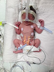
Intubated preterm baby in an incubator
SpecialtyObstetrics, pediatrics
SymptomsBirth of a baby at younger than 37 weeks' gestational age
ComplicationsCerebral palsy, delays in development, hearing problems, sight problems
CausesOften unknown
Risk factorsDiabetes, high blood pressure, being pregnant with more than one baby, obesity or underweight, a number of vaginal infections, celiac disease, tobacco smoking, psychological stress
PreventionProgesterone
TreatmentCorticosteroids, keeping the baby warm through skin to skin contact, supporting breastfeeding, treating infections, supporting breathing
Frequency~15 million a year (12% of deliveries)
Deaths805,800

Preterm birth, also known as premature birth, is the birth of a baby at fewer than 37 weeks' gestational age. These babies are known as preemies or premies. Symptoms of preterm labor include uterine contractions which occur more often than every ten minutes or the leaking of fluid from the vagina. Premature infants are at greater risk for cerebral palsy, delays in development, hearing problems and sight problems. These risks are greater the earlier a baby is born.

The cause of preterm birth is often not known. Risk factors include diabetes, high blood pressure, being pregnant with more than one baby, being either obese or underweight, a number of vaginal infections, tobacco smoking and psychological stress, among others. It is recommended that labor not be medically induced before 39 weeks unless required for other medical reasons. The same recommendation applies to cesarean section. Medical reasons for early delivery include preeclampsia.

In those at risk, the hormone progesterone, if taken during pregnancy, may prevent preterm birth. Evidence does not support the usefulness of bed rest. It is estimated that at least 75% of preterm infants would survive with appropriate treatment, and the survival rate is highest among the infants born the latest. In women who might deliver between 24 and 37 weeks, corticosteroids improve outcomes. A number of medications, including nifedipine, may delay delivery so that a mother can be moved to where more medical care is available and the corticosteroids have a greater chance to work. Once the baby is born, care includes keeping the baby warm through skin to skin contact, supporting breastfeeding, treating infections and supporting breathing.

Preterm birth is the most common cause of death among infants worldwide. About 15 million babies are preterm each year (5% to 18% of all deliveries). Approximately 0.5% of births are extremely early periviable births, and these account for most of the deaths. In many countries, rates of premature births have increased between the 1990s and 2010s. Complications from preterm births resulted in 0.81 million deaths in 2015 down from 1.57 million in 1990. The chance of survival at 22 weeks is about 6%, while at 23 weeks it is 26%, 24 weeks 55% and 25 weeks about 72%. The chances of survival without any long-term difficulties are lower.

Signs and symptoms

A new mother holds her premature baby at Kapiolani Medical Center NICU in Honolulu, Hawaii
 
Preterm birth causes a range of problems.

The main categories of causes of preterm birth are preterm labor induction and spontaneous preterm labor. Signs and symptoms of preterm labor include four or more uterine contractions in one hour. In contrast to false labour, true labor is accompanied by cervical dilatation and effacement. Also, vaginal bleeding in the third trimester, heavy pressure in the pelvis, or abdominal or back pain could be indicators that a preterm birth is about to occur. A watery discharge from the vagina may indicate premature rupture of the membranes that surround the baby. While the rupture of the membranes may not be followed by labor, usually delivery is indicated as infection (chorioamnionitis) is a serious threat to both fetus and mother. In some cases, the cervix dilates prematurely without pain or perceived contractions, so that the mother may not have warning signs until very late in the birthing process. 

A review into using uterine monitoring at home to detect contractions and possible preterm births in women at higher risk of having a preterm baby found that it did not reduce the number of preterm births. The research included in the review was poor quality but it showed that home monitoring may increase the number of unplanned antenatal visits and may reduce the number of babies admitted to special care when compared with women receiving normal antenatal care.

Complications

Mortality and morbidity

In the U.S. where many neonatal infections and other causes of neonatal death have been markedly reduced, prematurity is the leading cause of neonatal mortality at 25%. Prematurely born infants are also at greater risk for having subsequent serious chronic health problems as discussed below. 

The earliest gestational age at which the infant has at least a 50% chance of survival is referred to as the limit of viability. As NICU care has improved over the last 40 years, the limit of viability has reduced to approximately 24 weeks. Most newborns who die, and 40% of older infants who die, were born between 20 and 25.9 weeks (gestational age), during the second trimester.

As risk of brain damage and developmental delay is significant at that threshold even if the infant survives, there are ethical controversies over the aggressiveness of the care rendered to such infants. The limit of viability has also become a factor in the abortion debate.

Specific risks for the preterm neonate

Preterm infants usually show physical signs of prematurity in reverse proportion to the gestational age. As a result, they are at risk for numerous medical problems affecting different organ systems.
A study of 241 children born between 22 and 25 weeks who were currently of school age found that 46 percent had severe or moderate disabilities such as cerebral palsy, vision or hearing loss and learning problems. Thirty-four percent were mildly disabled and 20 percent had no disabilities, while 12 percent had disabling cerebral palsy.

Risk factors

The exact cause of preterm birth is difficult to determine and it may be multi-factorial. Labor is a complex process involving many factors. Four different pathways have been identified that can result in preterm birth and have considerable evidence: precocious fetal endocrine activation, uterine overdistension (placental abruption), decidual bleeding, and intrauterine inflammation/infection.

Identifying women at high risk of giving birth early would enable the health services to provide specialized care for these women to delay the birth or make sure they are in the best place to give birth (for example a hospital with a special care baby unit). Risk scoring systems have been suggested as a possible way of identifying these women. However, there is no research in this area so it is unclear whether the risk scoring systems would prolong pregnancy and reduce the numbers of preterm births or not.

Maternal factors

Risk factor Relative risk or odds ratio 95% confidence
interval
Black ethnicity/race 2.0 1.8–2.2
Filipino ancestry 1.7 1.5–2.1
High or low BMI 0.96 0.66–1.4
Large or small pregnancy weight gain 1.8 1.5–2.3
Short maternal height 1.8 1.3–2.5
History of spontaneous preterm birth 3.6 3.2–4.0
Being single/unmarried 1.2 1.03–1.28
Bacterial vaginosis 2.2 1.5–3.1
Asymptomatic bacteriuria 1.1 0.8–1.5
Periodontitis 1.6 1.1–2.3
Low socio-economic status 1.9 1.7–2.2
Short cervical length 2.9 2.1–3.9
Fetal fibronectin 4.0 2.9–5.5
Chlamydia 2.2 1.0–4.8
Celiac disease 1.4 1.2–1.6
Percentage premature births in England and Wales 2011, by age of mother and whether single or multiple birth.
 
A number of factors have been identified that are linked to a higher risk of a preterm birth such as being less than 18 years of age. Maternal height and weight can play a role.

Further, in the U.S. and the UK, black women have preterm birth rates of 15–18%, more than double than that of the white population. Filipinos are also at high risk of premature birth, and it is believed that nearly 11–15% of Filipinos born in the U.S. (compared to other Asians at 7.6% and whites at 7.8%) are premature. Filipinos being a big risk factor is evidenced with the Philippines being the 8th highest ranking in the world for preterm births, the only non-African country in the top 10. This discrepancy is not seen in comparison to other Asian groups or Hispanic immigrants and remains unexplained.

Pregnancy interval makes a difference as women with a six-month span or less between pregnancies have a two-fold increase in preterm birth. Studies on type of work and physical activity have given conflicting results, but it is opined that stressful conditions, hard labor, and long hours are probably linked to preterm birth.

A history of spontaneous (i.e., miscarriage) or surgical abortion has been associated with a small increase in the risk of preterm birth, with an increased risk with increased number of abortions, although it is unclear whether the increase is caused by the abortion or by confounding risk factors (e.g., socioeconomic status). Increased risk has not been shown in women who terminated their pregnancies medically. Pregnancies that are unwanted or unintended are also a risk factor for preterm birth.

Adequate maternal nutrition is important. Women with a low BMI are at increased risk for preterm birth. Further, women with poor nutrition status may also be deficient in vitamins and minerals. Adequate nutrition is critical for fetal development and a diet low in saturated fat and cholesterol may help reduce the risk of a preterm delivery. Obesity does not directly lead to preterm birth; however, it is associated with diabetes and hypertension which are risk factors by themselves. To some degree those individuals may have underlying conditions (i.e., uterine malformation, hypertension, diabetes) that persist. 

Women with celiac disease have an increased risk of the development of preterm birth. The risk of preterm birth is more elevated when celiac disease remains undiagnosed and untreated.

Marital status is associated with risk for preterm birth. A study of 25,373 pregnancies in Finland revealed that unmarried mothers had more preterm deliveries than married mothers (P=0.001). Pregnancy outside of marriage was associated overall with a 20% increase in total adverse outcomes, even at a time when Finland provided free maternity care. A study in Quebec of 720,586 births from 1990 to 1997 revealed less risk of preterm birth for infants with legally married mothers compared with those with common-law wed or unwed parents.

Genetic make-up is a factor in the causality of preterm birth. Genetics has been a big factor into why Filipinos have a high risk of premature birth as the Filipinos have a large prevalence of mutations that help them be predisposed to premature births. An intra- and transgenerational increase in the risk of preterm delivery has been demonstrated. No single gene has been identified. 

Subfertility is associated with preterm birth. Couples who have tried more than 1 year versus those who have tried less than 1 year before achieving a spontaneous conception have an adjusted odds ratio of 1.35 (95% confidence interval 1.22-1.50) of preterm birth. Pregnancies after IVF confers a greater risk of preterm birth than spontaneous conceptions after more than 1 year of trying, with an adjusted odds ratio of 1.55 (95% CI 1.30-1.85).

Factors during pregnancy

The use of fertility medication that stimulates the ovary to release multiple eggs and of IVF with embryo transfer of multiple embryos has been implicated as an important factor in preterm birth. Maternal medical conditions increase the risk of preterm birth. Often labor has to be induced for medical reasons; such conditions include high blood pressure, pre-eclampsia, maternal diabetes, asthma, thyroid disease, and heart disease.

In a number of women anatomical issues prevent the baby from being carried to term. Some women have a weak or short cervix (the strongest predictor of premature birth). Women with vaginal bleeding during pregnancy are at higher risk for preterm birth. While bleeding in the third trimester may be a sign of placenta previa or placental abruption – conditions that occur frequently preterm – even earlier bleeding that is not caused by these conditions is linked to a higher preterm birth rate. Women with abnormal amounts of amniotic fluid, whether too much (polyhydramnios) or too little (oligohydramnios), are also at risk. The mental status of the women is of significance. Anxiety and depression have been linked to preterm birth.

Finally, the use of tobacco, cocaine, and excessive alcohol during pregnancy increases the chance of preterm delivery. Tobacco is the most commonly abused drug during pregnancy and contributes significantly to low birth weight delivery. Babies with birth defects are at higher risk of being born preterm.

Passive smoking and/or smoking before the pregnancy influences the probability of a preterm birth. The World Health Organization published an international study in March 2014.

Presence of anti-thyroid antibodies is associated with an increased risk preterm birth with an odds ratio of 1.9 and 95% confidence interval of 1.1–3.5.

A 2004 systematic review of 30 studies on the association between intimate partner violence and birth outcomes concluded that preterm birth and other adverse outcomes, including death, are higher among abused pregnant women than among non-abused women.

The Nigerian cultural method of abdominal massage has been shown to result in 19% preterm birth among women in Nigeria, plus many other adverse outcomes for the mother and baby. This ought not be confused with massage conducted by a fully trained and licensed massage therapist or by significant others trained to provide massage during pregnancy, which has been shown to have numerous positive results during pregnancy, including the reduction of preterm birth, less depression, lower cortisol, and reduced anxiety.

Infection

The frequency of infection in preterm birth is inversely related to the gestational age. Mycoplasma genitalium infection is associated with increased risk of preterm birth, and spontaneous abortion.

Infectious microorganisms can be ascending, hematogeneous, iatrogenic by a procedure, or retrograde through the Fallopian tubes. From the deciduas they may reach the space between the amnion and chorion, the amniotic fluid, and the fetus. A chorioamnionitis also may lead to sepsis of the mother. Fetal infection is linked to preterm birth and to significant long-term handicap including cerebral palsy.

It has been reported that asymptomatic colonization of the decidua occurs in up to 70% of women at term using a DNA probe suggesting that the presence of micro-organism alone may be insufficient to initiate the infectious response. 

As the condition is more prevalent in black women in the US and the UK, it has been suggested to be an explanation for the higher rate of preterm birth in these populations. It is opined that bacterial vaginosis before or during pregnancy may affect the decidual inflammatory response that leads to preterm birth. The condition known as aerobic vaginitis can be a serious risk factor for preterm labor; several previous studies failed to acknowledge the difference between aerobic vaginitis and bacterial vaginosis, which may explain some of the contradiction in the results.

Untreated yeast infections are associated with preterm birth.

A review into prophylactic antibiotics (given to prevent infection) in the second and third trimester of pregnancy (13–42 weeks of pregnancy) found a reduction in the number of preterm births in women with bacterial vaginosis. These antibiotics also reduced the number of waters breaking before labor in full-term pregnancies, reduced the risk of infection of the lining of the womb after delivery (endometritis), and rates of gonococcal infection. However, the women without bacterial vaginosis did not have any reduction in preterm births or pre-labor preterm waters breaking. Much of the research included in this review lost participants during follow-up so did not report the long-term effects of the antibiotics on mothers or babies. More research in this area is needed to find the full effects of giving antibiotics throughout the second and third trimesters of pregnancy.

A number of maternal bacterial infections are associated with preterm birth including pyelonephritis, asymptomatic bacteriuria, pneumonia, and appendicitis. A review into giving antibiotics in pregnancy for asymptomatic bacteriuria (urine infection with no symptoms) found the research was of very low quality but that it did suggest that taking antibiotics reduced the numbers of preterm births and babies with low birth weight. Another review found that one dose of antibiotics did not seem as effective as a course of antibiotics but fewer women reported side effects from one dose. This review recommended that more research is needed to discover the best way of treating asymptomatic bacteriuria.

A different review found that preterm births happened less for pregnant women who had routine testing for low genital tract infections than for women who only had testing when they showed symptoms of low genital tract infections. The women being routinely tested also gave birth to fewer babies with a low birth weight. Even though these results look promising, the review was only based on one study so more research is needed into routine screening for low genital tract infections.

Also periodontal disease has been shown repeatedly to be linked to preterm birth. In contrast, viral infections, unless accompanied by a significant febrile response, are considered not to be a major factor in relation to preterm birth.

Genetics

There is believed to be a maternal genetic component in preterm birth. Estimated heritability of timing-of-birth in women was 34%. However, the occurrence of preterm birth in families does not follow a clear inheritance pattern, thus supporting the idea that preterm birth is a non-Mendelian trait with a polygenic nature.

Diagnosis

Placental alpha microglobulin-1

Placental alpha microglobulin-1 (PAMG-1) has been the subject of several investigations evaluating its ability to predict imminent spontaneous preterm birth in women with signs, symptoms, or complaints suggestive of preterm labor. In one investigation comparing this test to fetal fibronectin testing and cervical length measurement via transvaginal ultrasound, the test for PAMG-1 (commercially known as the PartoSure test) has been reported to be the single best predictor of imminent spontaneous delivery within 7 days of a patient presenting with signs, symptoms, or complaints of preterm labor. Specifically, the PPV, or positive predictive value, of the tests were 76%, 29%, and 30% for PAMG-1, fFN and CL, respectively (P less than 0.01).

Fetal fibronectin

Fetal fibronectin (fFN) has become an important biomarker—the presence of this glycoprotein in the cervical or vaginal secretions indicates that the border between the chorion and deciduas has been disrupted. A positive test indicates an increased risk of preterm birth, and a negative test has a high predictive value. It has been shown that only 1% of women in questionable cases of preterm labor delivered within the next week when the test was negative.

Ultrasound

Obstetric ultrasound has become useful in the assessment of the cervix in women at risk for premature delivery. A short cervix preterm is undesirable: A cervical length of less than 25 mm at or before 24 weeks of gestational age is the most common definition of cervical incompetence.

Classification

Stages in prenatal development, with weeks and months numbered from last menstruation.

In humans, the usual definition of preterm birth is birth before a gestational age of 37 complete weeks. In the normal human fetus, several organ systems mature between 34 and 37 weeks, and the fetus reaches adequate maturity by the end of this period. One of the main organs greatly affected by premature birth is the lungs. The lungs are one of the last organs to mature in the womb; because of this, many premature babies spend the first days and weeks of their lives on ventilators. Therefore, a significant overlap exists between preterm birth and prematurity. Generally, preterm babies are premature and term babies are mature. Preterm babies born near 37 weeks often have no problems relating to prematurity if their lungs have developed adequate surfactant, which allows the lungs to remain expanded between breaths. Sequelae of prematurity can be reduced to a small extent by using drugs to accelerate maturation of the fetus, and to a greater extent by preventing preterm birth.

Prevention

Historically efforts have been primarily aimed to improve survival and health of preterm infants (tertiary intervention). Such efforts, however, have not reduced the incidence of preterm birth. Increasingly primary interventions that are directed at all women, and secondary intervention that reduce existing risks are looked upon as measures that need to be developed and implemented to prevent the health problems of premature infants and children. Smoking bans are effective in decreasing preterm births.

Before pregnancy

Adoption of specific professional policies can immediately reduce risk of preterm birth as the experience in assisted reproduction has shown when the number of embryos during embryo transfer was limited. Many countries have established specific programs to protect pregnant women from hazardous or night-shift work and to provide them with time for prenatal visits and paid pregnancy-leave. The EUROPOP study showed that preterm birth is not related to type of employment, but to prolonged work (over 42 hours per week) or prolonged standing (over 6 hours per day). Also, night work has been linked to preterm birth. Health policies that take these findings into account can be expected to reduce the rate of preterm birth. Preconceptional intake of folic acid is recommended to reduce birth defects. There is significant evidence that long-term (more than one year) use of folic acid supplement preconceptionally may reduce premature birth. Reducing smoking is expected to benefit pregnant women and their offspring.

During pregnancy

Healthy eating can be instituted at any stage of the pregnancy including nutritional adjustments, use of vitamin supplements, and smoking cessation. Calcium supplementation in women who have low dietary calcium may reduce the number of negative outcomes including preterm birth, pre-eclampsia, and maternal death. The World Health Organization (WHO) suggests 1.5–2 g of calcium supplements daily, for pregnant women who have low levels calcium in their diet. Supplemental intake of C and E vitamins have not been found to reduce preterm birth rates. Different strategies are used in the administration of prenatal care, and future studies need to determine if the focus can be on screening for high-risk women, or widened support for low-risk women, or to what degree these approaches can be merged. While periodontal infection has been linked with preterm birth, randomized trials have not shown that periodontal care during pregnancy reduces preterm birth rates.

Screening of low risk women

Screening for asymptomatic bacteriuria followed by appropriate treatment reduces pyelonephritis and reduces the risk of preterm birth. Extensive studies have been carried out to determine if other forms of screening in low-risk women followed by appropriate intervention are beneficial, including: Screening for and treatment of Ureaplasma urealyticum, group B streptococcus, Trichomonas vaginalis, and bacterial vaginosis did not reduce the rate of preterm birth. Routine ultrasound examination of the length of the cervix identifies patients at risk, but cerclage is not proven useful, and the application of a progestogen is under study. Screening for the presence of fibronectin in vaginal secretions is not recommended at this time in women at low risk.

Self-care

Self-care methods to reduce the risk of preterm birth include proper nutrition, avoiding stress, seeking appropriate medical care, avoiding infections, and the control of preterm birth risk factors (e.g. working long hours while standing on feet, carbon monoxide exposure, domestic abuse, and other factors). Self-monitoring vaginal pH followed by yogurt treatment or clindamycin treatment if the pH was too high all seem to be effective at reducing the risk of preterm birth. Additional support during pregnancy does not appear to prevent low birthweight or preterm birth.

Reducing existing risks

Women are identified to be at increased risk for preterm birth on the basis of their past obstetrical history or the presence of known risk factors. Preconception intervention can be helpful in selected patients in a number of ways. Patients with certain uterine anomalies may have a surgical correction (i.e. removal of a uterine septum), and those with certain medical problems can be helped by optimizing medical therapies prior to conception, be it for asthma, diabetes, hypertension and others.

Multiple pregnancies

In multiple pregnancies, which often result from use of assisted reproductive technology, there is a high risk of preterm birth. Selective reduction is used to reduce the number of fetuses to two or three.

Reducing indicated preterm birth

A number of agents have been studied for the secondary prevention of indicated preterm birth. Trials using low-dose aspirin, fish oil, vitamin C and E, and calcium to reduce preeclampsia demonstrated some reduction in preterm birth only when low-dose aspirin was used. Even if agents such as calcium or antioxidants were able to reduce preeclampsia, a resulting decrease in preterm birth was not observed.

Reducing spontaneous preterm birth

Reduction in activity by the mother—pelvic rest, limited work, bed rest—may be recommended although there is no evidence it is useful with some concerns it is harmful. Increasing medical care by more frequent visits and more education has not been shown to reduce preterm birth rates. Use of nutritional supplements such as omega-3 polyunsaturated fatty acids is based on the observation that populations who have a high intake of such agents are at low risk for preterm birth, presumably as these agents inhibit production of proinflammatory cytokines. A randomized trial showed a significant decline in preterm birth rates, and further studies are in the making.
Antibiotics
While antibiotics can get rid of bacterial vaginosis in pregnancy, this does not appear to change the risk of preterm birth. It has been suggested that chronic chorioamnionitis is not sufficiently treated by antibiotics alone (and therefore they cannot ameliorate the need for preterm delivery in this condition).
Progestogens
Progestogens, often given in the form of progesterone or hydroxyprogesterone caproate, relaxes the uterine musculature, maintains cervical length, and has anti-inflammatory properties, and thus exerts activities expected to be beneficial in reducing preterm birth. Two meta-analyses demonstrated a reduction in the risk of preterm birth in women with recurrent preterm birth by 40–55%.

Progestogen supplementation also reduces the frequency of preterm birth in pregnancies where there is a short cervix. However, progestogens are not effective in all populations, as a study involving twin gestations failed to see any benefit.
Cervical cerclage
In preparation for childbirth, the woman's cervix shortens. Preterm cervical shortening is linked to preterm birth and can be detected by ultrasonography. Cervical cerclage is a surgical intervention that places a suture around the cervix to prevent its shortening and widening. Numerous studies have been performed to assess the value of cervical cerclage and the procedure appears helpful primarily for women with a short cervix and a history of preterm birth. Instead of a prophylactic cerclage, women at risk can be monitored during pregnancy by sonography, and when shortening of the cervix is observed, the cerclage can be performed.

Management

Preterm birth at 32 weeks and 4 days with a weight of 2,000 g attached to medical equipment
 
About 75% of nearly a million deaths due to preterm deliver would survive if provided warmth, breastfeeding, treatments for infection, and breathing support. If a baby has cardiac arrest at birth and is before 23 weeks or less than 400 g attempts at resuscitation are not indicated.

Tertiary interventions are aimed at women who are about to go into preterm labor, or rupture the membranes or bleed preterm. The use of the fibronectin test and ultrasonography improves the diagnostic accuracy and reduces false-positive diagnosis. While treatments to arrest early labor where there is progressive cervical dilatation and effacement will not be effective to gain sufficient time to allow the fetus to grow and mature further, it may defer delivery sufficiently to allow the mother to be brought to a specialized center that is equipped and staffed to handle preterm deliveries. In a hospital setting women are hydrated via intravenous infusion (as dehydration can lead to premature uterine contractions).

Steroids

Severely premature infants may have underdeveloped lungs because they are not yet producing their own surfactant. This can lead directly to respiratory distress syndrome, also called hyaline membrane disease, in the neonate. To try to reduce the risk of this outcome, pregnant mothers with threatened premature delivery prior to 34 weeks are often administered at least one course of glucocorticoids, a steroid that crosses the placental barrier and stimulates the production of surfactant in the lungs of the baby. Steroid use up to 37 weeks is also recommended by the American Congress of Obstetricians and Gynecologists. Typical glucocorticoids that would be administered in this context are betamethasone or dexamethasone, often when the pregnancy has reached viability at 23 weeks. 

In cases where premature birth is imminent, a second "rescue" course of steroids may be administered 12 to 24 hours before the anticipated birth. There are still some concerns about the efficacy and side effects of a second course of steroids, but the consequences of RDS are so severe that a second course is often viewed as worth the risk. A 2015 Cochrane review supports the use of repeat dose(s) of prenatal corticosteroids for women still at risk of preterm birth seven days or more after an initial course.

Beside reducing respiratory distress, other neonatal complications are reduced by the use of glucocorticosteroids, namely intraventricular bleeding, necrotising enterocolitis, and patent ductus arteriosus. A single course of antenatal corticosteroids could be considered routine for preterm delivery, but there are some concerns about applicability of this recommendation to low-resource settings with high rates of infections. It remains unclear whether one corticosteroid (or one particular regimen) has advantages over another.

Concerns about adverse effects of prenatal corticosteroids include increased risk for maternal infection, difficulty with diabetic control, and possible long-term effects on neurodevelopmental outcomes for the infants. There is ongoing discussion about when steroids should be given (i.e. only antenatally or postnatally too) and for how long (i.e. single course or repeated administration). Despite these unknowns, there is a consensus that the benefits of a single course of prenatal glucocorticosteroids vastly outweigh the potential risks.

Antibiotics

The routine administration of antibiotics to all women with threatened preterm labor reduces the risk of the baby to get infected with group B streptococcus and has been shown to reduce related mortality rates.

When membranes rupture prematurely, obstetrical management looks for development of labor and signs of infection. Prophylactic antibiotic administration has been shown to prolong pregnancy and reduced neonatal morbidity with rupture of membranes at less than 34 weeks. Because of concern about necrotizing enterocolitis, amoxicillin or erythromycin has been recommended, but not amoxicillin + clavulanic acid (co-amoxiclav).

Tocolysis

A number of medications may be useful to delay delivery including: nonsteroidal anti-inflammatory drugs, calcium channel blockers, beta mimetics, and atosiban. Tocolysis rarely delays delivery beyond 24–48 hours. This delay, however, may be sufficient to allow the pregnant woman to be transferred to a center specialized for management of preterm deliveries and give administered corticosteroids to reduce neonatal organ immaturity. Meta-analyses indicate that calcium-channel blockers and an oxytocin antagonist can delay delivery by 2–7 days, and β2-agonist drugs delay by 48 hours but carry more side effects. Magnesium sulfate does not appear to be useful to prevent preterm birth. Its use before delivery, however, does appear to decrease the risk of cerebral palsy.

Mode of delivery

The routine use of caesarean section for early delivery of infants expected to have very low birth weight is controversial, and a decision concerning the route and time of delivery probably needs to be made on a case by case basis.

Neonatal care

Incubator for preterm baby
 
After delivery, plastic wraps or warm mattresses are useful to keep the infant warm on their way to the neonatal intensive care unit (NICU). In developed countries premature infants are usually cared for in an NICU. The physicians who specialize in the care of very sick or premature babies are known as neonatologists. In the NICU, premature babies are kept under radiant warmers or in incubators (also called isolettes), which are bassinets enclosed in plastic with climate control equipment designed to keep them warm and limit their exposure to germs. Modern neonatal intensive care involves sophisticated measurement of temperature, respiration, cardiac function, oxygenation, and brain activity. Treatments may include fluids and nutrition through intravenous catheters, oxygen supplementation, mechanical ventilation support, and medications. In developing countries where advanced equipment and even electricity may not be available or reliable, simple measures such as kangaroo care (skin to skin warming), encouraging breastfeeding, and basic infection control measures can significantly reduce preterm morbidity and mortality. Bili lights may also be used to treat newborn jaundice (hyperbilirubinemia). 

Water can be carefully provided to prevent dehydration but no so much to increase risks of side effects.

In a 2012 policy statement, the American Academy of Pediatrics recommended feeding preterm infants human milk, finding "significant short- and long-term beneficial effects," including lower rates of necrotizing enterocolitis (NEC). It is unclear if fortification of breast milk improves outcomes in preterm babies, though it may speed growth. There is limited evidence to support prescribing a preterm formula for the preterm babies after hospital discharge.

Prognosis

Preterm infants survival rates
 
The chance of survival at 22 weeks is about 6%, while at 23 weeks it is 26%, 24 weeks 55% and 25 weeks about 72%. The chances of survival without long-term difficulties is less. In the developed world overall survival is about 90% while in low-income countries survival rates are about 10%.

Some children will adjust well during childhood and adolescence, although disability is more likely nearer the limits of viability. A large study followed children born between 22 and 25 weeks until the age of 6 years old. Of these children, 46 percent had moderate to severe disabilities such as cerebral palsy, vision or hearing loss and learning disabilities, 34 percent had mild disabilities, and 20 percent had no disabilities. Twelve percent had disabling cerebral palsy.

As survival has improved, the focus of interventions directed at the newborn has shifted to reduce long-term disabilities, particularly those related to brain injury. Some of the complications related to prematurity may not be apparent until years after the birth. A long-term study demonstrated that the risks of medical and social disabilities extend into adulthood and are higher with decreasing gestational age at birth and include cerebral palsy, intellectual disability, disorders of psychological development, behavior, and emotion, disabilities of vision and hearing, and epilepsy. Standard intelligence tests showed that 41 percent of children born between 22 and 25 weeks had moderate or severe learning disabilities when compared to the test scores of a group of similar classmates who were born at full-term. It is also shown that higher levels of education were less likely to be obtained with decreasing gestational age at birth. People born prematurely may be more susceptible to developing depression as teenagers. Some of these problems can be described as being within the executive domain and have been speculated to arise due to decreased myelinization of the frontal lobes. Studies of people born premature and investigated later with MRI brain imaging, demonstrate qualitative anomalies of brain structure and grey matter deficits within temporal lobe structures and the cerebellum that persist into adolescence. Throughout life they are more likely to require services provided by physical therapists, occupational therapists, or speech therapists.

Despite the neurosensory, mental and educational problems studied in school age and adolescent children born extremely preterm, the majority of preterm survivors born during the early years of neonatal intensive care are found to do well and to live fairly normal lives in young adulthood. Young adults born preterm seem to acknowledge that they have more health problems than their peers, yet feel the same degree of satisfaction with their quality of life.

Beyond the neurodevelopmental consequences of prematurity, infants born preterm have a greater risk for many other health problems. For instance, children born prematurely have an increased risk for developing chronic kidney disease.

Epidemiology

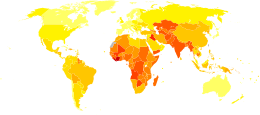
Disability-adjusted life year for prematurity and low birth weight per 100,000 inhabitants in 2004.
 
  no data
  less than 120
  120-240
  240-360
  360-480
  480-600
  600-720
  720-840
  840-960
  960-1080
  1080-1200
  1200-1500
  more than 1500

Preterm birth complicates the births of infants worldwide affecting 5% to 18% of births. In Europe and many developed countries the preterm birth rate is generally 5–9%, and in the USA it has even risen to 12–13% in the last decades.

As weight is easier to determine than gestational age, the World Health Organization tracks rates of low birth weight (less than 2,500 grams), which occurred in 16.5 percent of births in less developed regions in 2000. It is estimated that one third of these low birth weight deliveries are due to preterm delivery. Weight generally correlates to gestational age, however, infants may be underweight for other reasons than a preterm delivery. Neonates of low birth weight (LBW) have a birth weight of less than 2500 g (5 lb 8 oz) and are mostly but not exclusively preterm babies as they also include small for gestational age (SGA) babies. Weight-based classification further recognizes Very Low Birth Weight (VLBW) which is less than 1,500 g, and Extremely Low Birth Weight (ELBW) which is less than 1,000 g. Almost all neonates in these latter two groups are born preterm. 

Complications from preterm births resulted in 740,000 deaths in 2013, down from 1.57 million in 1990.

Society and culture

Economics

Preterm birth is a significant cost factor in healthcare, not even considering the expenses of long-term care for individuals with disabilities due to preterm birth. A 2003 study in the US determined neonatal costs to be $224,400 for a newborn at 500–700 g versus $1,000 at over 3,000 g. The costs increase exponentially with decreasing gestational age and weight. The 2007 Institute of Medicine report Preterm Birth found that the 550,000 premature babies born each year in the U.S. run up about $26 billion in annual costs, mostly related to care in neonatal intensive care units, but the real tab may top $50 billion.

Notable cases

James Elgin Gill (born on 20 May 1987 in Ottawa, Ontario, Canada) was the earliest premature baby in the world, until that record was broken in 2014. He was 128 days premature (21 weeks and 5 days' gestation) and weighed 1 pound 6 ounces (624 g). He survived.

In 2014, Lyla Stensrud, born in San Antonio, Texas, U.S. became the youngest premature baby in the world. She was born at 21 weeks 4 days and weighed 410 grams (less than a pound). Kaashif Ahmad resuscitated the baby after she was born. As of November 2018, Lyla was attending preschool. She had a slight delay in speech, but no other known medical issues or disabilities.

Amillia Taylor is also often cited as the most premature baby. She was born on 24 October 2006 in Miami, Florida, U.S. at 21 weeks and 6 days' gestation. This report has created some confusion as her gestation was measured from the date of conception (through in vitro fertilization) rather than the date of her mother's last menstrual period, making her appear 2 weeks younger than if gestation was calculated by the more common method. At birth, she was 9 inches (22.9 cm) long and weighed 10 ounces (280 g). She suffered digestive and respiratory problems, together with a brain hemorrhage. She was discharged from the Baptist Children's Hospital on 20 February 2007.

The record for the smallest premature baby to survive was held for a considerable amount of time by Madeline Mann, who was born in 1989 at 26 weeks, weighing 9.9 ounces (280 g) and measuring 9.5 inches (241.3 mm) long. This record was broken in September 2004 by Rumaisa Rahman, who was born in the same hospital, Loyola University Medical Center in Maywood, Illinois. at 25 weeks' gestation. At birth, she was 8 inches (200 mm) long and weighed 261 grams (9.2 oz). Her twin sister was also a small baby, weighing 563 grams (1 lb 3.9 oz) at birth. During pregnancy their mother had pre-eclampsia, requiring birth by caesarean section. The larger twin left the hospital at the end of December, while the smaller remained there until 10 February 2005 by which time her weight had increased to 1.18 kg (2.6 lb). Generally healthy, the twins had to undergo laser eye surgery to correct vision problems, a common occurrence among premature babies. 

In May 2019, Sharp Mary Birch Hospital for Women & Newborns in San Diego announced that a baby nicknamed "Saybie" had been discharged almost five months after being born at 23 weeks gestation and weighing 244 grams (8.6 oz). Saybie was confirmed by Dr. Edward Bell of the University of Iowa to be the new smallest surviving premature baby.

The world's smallest premature boy to survive was born in February 2009 at Children's Hospitals and Clinics of Minnesota in Minneapolis, Minnesota, U.S.. Jonathon Whitehill was born at 25 weeks' gestation with a weight of 310 grams (11 oz). He was hospitalized in a neonatal intensive care unit for five months, and then discharged.

Historical figures who were born prematurely include Johannes Kepler (born in 1571 at seven months' gestation), Isaac Newton (born in 1642, small enough to fit into a quart mug, according to his mother), Winston Churchill (born in 1874 at seven months' gestation), and Anna Pavlova (born in 1885 at seven months' gestation),

Israel and apartheid

From Wikipedia, the free encyclopedia https://en.wikipedia.org/wiki/Israel_and_apartheid A Palestinian c...