From Wikipedia, the free encyclopedia
| Parkinson's disease |
|---|
| Other names | Parkinson disease, idiopathic or primary parkinsonism, hypokinetic rigid syndrome, paralysis agitans, shaking palsy |
|---|
 |
| Illustration of Parkinson's disease by William Richard Gowers, first published in A Manual of Diseases of the Nervous System (1886) |
| Specialty | Neurology |
|---|
| Symptoms | Shaking, rigidity, slowness of movement, difficulty walking |
|---|
| Complications | Dementia, depression, anxiety |
|---|
| Usual onset | Age over 60 |
|---|
| Causes | Unknown |
|---|
| Risk factors | Pesticide exposure, head injuries |
|---|
| Diagnostic method | Based on symptoms |
|---|
| Differential diagnosis | Dementia with Lewy bodies, progressive supranuclear palsy, essential tremor, antipsychotic use |
|---|
| Treatment | Medications, surgery |
|---|
| Medication | L-DOPA, dopamine agonists |
|---|
| Prognosis | Life expectancy ~ 15 years |
|---|
| Frequency | 6.2 million (2015) |
|---|
| Deaths | 117,400 (2015) |
|---|
Parkinson's disease (
PD) is a long-term
degenerative disorder of the
central nervous system that mainly affects the
motor system. As the disease worsens, non-motor symptoms become more common. The symptoms usually emerge slowly. Early in the disease, the most obvious symptoms are
shaking,
rigidity,
slowness of movement, and
difficulty with walking.
Thinking and behavioral problems may also occur.
Dementia becomes common in the advanced stages of the disease.
Depression and anxiety are also common, occurring in more than a third of people with PD. Other symptoms include sensory,
sleep, and
emotional problems. The main motor symptoms are collectively called "
parkinsonism", or a "parkinsonian syndrome".
The cause of Parkinson's disease is
unknown, but is believed to involve both
genetic and environmental factors. Those with a family member affected are more likely to get the disease themselves. There is also an increased risk in people exposed to certain
pesticides and among those who have had prior
head injuries, while there is a reduced risk in
tobacco smokers and those who drink
coffee or
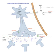
tea. The motor symptoms of the disease result from the
death of cells in the
substantia nigra, a region of the
midbrain. This results in not enough
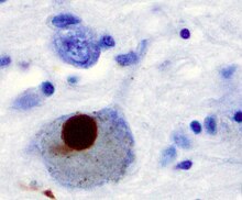
dopamine in this region of the brain. The cause of this cell death is poorly understood, but it involves the build-up of
proteins into
Lewy bodies in the
neurons. Diagnosis of typical cases is mainly based on symptoms, with tests such as
neuroimaging used to rule out other diseases.
There is no
cure for Parkinson's disease. Treatment aims to improve the symptoms. Initial treatment is typically with the
antiparkinson medication levodopa (
L-DOPA), followed by
dopamine agonists when levodopa becomes less effective.
As the disease progresses and neurons continue to be lost, these
medications become less effective while at the same time they produce a
complication marked by
involuntary writhing movements. Diet and some forms of rehabilitation have shown some effectiveness at improving symptoms.
Surgery to place
microelectrodes for
deep brain stimulation has been used to reduce motor symptoms in severe cases where drugs are ineffective.
Evidence for treatments for the non-movement-related symptoms of PD,
such as sleep disturbances and emotional problems, is less strong.
In 2015, PD affected 6.2 million people and resulted in about 117,400 deaths globally. Parkinson's disease typically occurs in people over the age of 60, of whom about one percent are affected. Males are more often affected than females at a ratio of around 3:2. When it is seen in people before the age of 50, it is called early-onset PD. The average
life expectancy following diagnosis is between 7 and 15 years. The disease is named after the English doctor
James Parkinson, who published the first detailed description in
An Essay on the Shaking Palsy, in 1817. Public awareness campaigns include World Parkinson's Day (on the birthday of James Parkinson, 11 April) and the use of a red
tulip as the symbol of the disease. People with Parkinson's who have increased the public's awareness of the condition include actor
Michael J. Fox, Olympic cyclist
Davis Phinney, professional boxer
Muhammad Ali, and actor
Alan Alda.
Classification
The movement difficulties found in PD are called
parkinsonism, which is defined as
bradykinesia
(slowness in initiating voluntary movements, with progressive reduction
in speed and range of repetitive actions such as voluntary
finger-tapping) in combination with one of three other physical signs: muscular (
lead-pipe or
cogwheel) rigidity,
tremor at rest, and
postural instability. A number of different disorders can have parkinsonism type movement issues.
Parkinson's disease is the most common form of parkinsonism and
is sometimes called "idiopathic parkinsonism", meaning parkinsonism with
no identifiable cause.
Identifiable causes of parkinsonism include toxins, infections, side
effects of drugs, metabolic derangement, and brain lesions such as
strokes. Several neurodegenerative disorders also may present with
parkinsonism and are sometimes referred to as "atypical parkinsonism" or
"Parkinson plus" syndromes (illnesses with parkinsonism plus some other features distinguishing them from PD). They include
multiple system atrophy,
progressive supranuclear palsy,
corticobasal degeneration, and
dementia with Lewy bodies (DLB).
Scientists sometimes refer to Parkinson’s disease as a
synucleinopathy (due to an abnormal accumulation of
alpha-synuclein protein in the brain) to distinguish it from other neurodegenerative diseases, such as
Alzheimer's disease where the brain accumulates
tau protein. Considerable clinical and pathological overlap exists between
tauopathies
and synucleinopathies. In contrast to Parkinson's disease, Alzheimer's
disease presents most commonly with memory loss, and the cardinal signs
of Parkinson's disease (slowness, tremor, stiffness, and postural
instability) are not normal features of Alzheimer's.
Dementia with Lewy bodies is another synucleinopathy and it has
close pathological similarities with PD, especially with the subset of
PD cases with
dementia known as
Parkinson's disease dementia. The relationship between PD and DLB is complex and incompletely understood.
They may represent parts of a continuum with variable distinguishing
clinical and pathological features or they may prove to be separate
diseases.
Signs and symptoms
A man with Parkinson's disease displaying a flexed walking posture pictured in 1892
Handwriting of a person affected by PD
The most recognizable symptoms in Parkinson's disease are movement ("motor") related. Non-motor symptoms, which include
autonomic dysfunction,
neuropsychiatric
problems (mood, cognition, behavior or thought alterations), and
sensory (especially altered sense of smell) and sleep difficulties, are
also common. Some of these non-motor symptoms may be present at the time
of diagnosis.
Motor
Four motor symptoms are considered
cardinal in PD: tremor, slowness of movement (bradykinesia), rigidity, and postural instability.
The most common presenting sign is a coarse slow
tremor of the hand at rest which disappears during voluntary movement of the affected arm and in the deeper stages of sleep. It typically appears in only one hand, eventually affecting both hands as the disease progresses. Frequency of PD tremor is between 4 and 6
hertz (cycles per second). A feature of tremor is
pill-rolling, the tendency of the index finger and thumb to touch and perform together a circular movement.
The term derives from the similarity between the movement of people
with PD and the early pharmaceutical technique of manually making pills.
Bradykinesia (slowness of movement) is found in every case of PD, and is due to disturbances in
motor planning
of movement initiation, and associated with difficulties along the
whole course of the movement process, from planning to initiation to
execution of a movement. Performance of sequential and simultaneous
movement is impaired. Bradykinesia is the most handicapping symptom of
Parkinson’s disease leading to difficulties with everyday tasks such as
dressing, feeding, and bathing. It leads to particular difficulty in
carrying out two independent motor activities at the same time and can
be made worse by emotional stress or concurrent illnesses. Paradoxically
patients with Parkinson's disease can often ride a bicycle or climb
stairs more easily than walk on a level. While most physicians may
readily notice bradykinesia, formal assessment requires a patient to do
repetitive movements with their fingers and feet.
Rigidity is stiffness and resistance to limb movement caused by increased
muscle tone, an excessive and continuous contraction of muscles. In parkinsonism the rigidity can be uniform ("
lead-pipe rigidity") or ratchety ("
cogwheel rigidity"). The combination of tremor and increased tone is considered to be at the origin of cogwheel rigidity. Rigidity may be associated with joint pain; such pain being a frequent initial manifestation of the disease.
In early stages of Parkinson's disease, rigidity is often asymmetrical
and it tends to affect the neck and shoulder muscles prior to the
muscles of the face and extremities. With the progression of the disease, rigidity typically affects the whole body and reduces the ability to move.
Postural instability is typical in the later stages of the disease, leading to impaired balance and frequent falls, and secondarily to
bone fractures, loss of confidence, and reduced mobility.
Instability is often absent in the initial stages, especially in
younger people, especially prior to the development of bilateral
symptoms.
Up to 40% of people diagnosed with PD may experience falls and around
10% may have falls weekly, with the number of falls being related to the
severity of PD.
Other recognized motor signs and symptoms include gait and posture disturbances such as
festination (rapid shuffling steps and a
forward-flexed posture
when walking with no flexed arm swing). Freezing of gait (brief arrests
when the feet seem to get stuck to the floor, especially on turning or
changing direction), a slurred monotonous quiet voice, mask-like facial
expression, and handwriting that gets smaller and smaller are other
common signs.
Neuropsychiatric
Parkinson's disease can cause
neuropsychiatric disturbances, which can range from mild to severe. This includes disorders of cognition, mood, behavior, and thought.
Cognitive disturbances can occur in the early stages of the
disease and sometimes prior to diagnosis, and increase in prevalence
with duration of the disease. The most common cognitive deficit in PD is
executive dysfunction, which can include problems with planning,
cognitive flexibility, abstract thinking, rule acquisition,
inhibiting inappropriate actions, initiating appropriate actions,
working memory, and
control of attention. Other cognitive difficulties include
slowed cognitive processing speed, impaired
recall and impaired perception and estimation of time. Nevertheless, improvement appears when recall is aided by cues.
Visuospatial difficulties are also part of the disease, seen for
example when the individual is asked to perform tests of facial
recognition and perception of the orientation of drawn lines.
A person with PD has two to six times the risk of dementia compared to the general population. Up to 78% of people with PD have Parkinson's disease dementia. The prevalence of dementia increases with age and, to a lesser degree, duration of the disease. Dementia is associated with a reduced
quality of life in people with PD and their
caregivers, increased mortality, and a higher probability of needing
nursing home care.
Impulse control disorders including pathological gambling,
compulsive sexual behavior, binge eating, compulsive shopping and
reckless generosity can be caused by medication, particularly orally
active dopamine agonists. The
dopamine dysregulation syndrome – with wanting of medication leading to overusage – is a rare complication of levodopa use.
Behavior and mood alterations are more common in PD without
cognitive impairment than in the general population, and are usually
present in PD with dementia. The most frequent mood difficulties are
depression,
apathy, and
anxiety.
Establishing the diagnosis of depression is complicated by the fact
that the body language of depression may masquerade as PD including a
sad expressionless anxious face, a hang dog appearance, slow movement,
and monotonous speech. Up to 30% of people with PD may experience
symptoms of anxiety, ranging from a
generalized anxiety disorder to
social phobia,
panic disorders and
obsessive compulsive disorders.
They contribute to impaired quality of life and increased severity of
motor symptoms such as on/off fluctuations or freezing episodes.
Punding in which complicated repetitive aimless
stereotyped behaviors occur for many hours is another disturbance caused by anti-Parkinson medication.
Hallucinations or
delusions
occur in approximately 50% of people with PD over the course of the
illness, and may herald the emergence of dementia. These range from
minor hallucinations – "sense of passage" (something quickly passing
beside the person) or "sense of presence" (the perception of
something/someone standing just to the side or behind the person) – to
full blown vivid,
formed visual hallucinations and
paranoid
ideation. Auditory hallucinations are uncommon in PD, and are rarely
described as voices. It is now believed that psychosis is an integral
part of the disease. A psychosis with delusions and associated
delirium
is a recognized complication of anti-Parkinson drug treatment and may
also be caused by urinary tract infections (as frequently occurs in the
fragile elderly), but drugs and infection are not the only factors, and
underlying brain pathology or changes in neurotransmitters or their
receptors (e.g., acetylcholine, serotonin) are also thought to play a
role in psychosis in PD.
Other
In addition to neuropsychiatric and motor symptoms, PD can impair other functions.
Sleep disorders are a feature of the disease and can be worsened by medications. Symptoms can manifest as daytime
drowsiness (including sudden sleep attacks resembling
narcolepsy), disturbances in
REM sleep, or
insomnia.
REM behavior disorder
(RBD), in which patients act out dreams, sometimes injuring themselves
or their bed partner, may begin many years before the development of
motor or cognitive features of PD or
DLB.
Causes
Many risk factors have been proposed, sometimes in relation to
theories concerning possible mechanisms of the disease; however, none
have been conclusively proven.
The most frequently replicated relationships are an increased risk in
those exposed to pesticides, and a reduced risk in smokers. There is a possible link between PD and
H. pylori infection that can prevent the absorption of some drugs including levodopa.
Environmental factors
Exposure to
pesticides
and a history of head injury have each been linked with Parkinson
disease (PD), but the risks are modest. Never having smoked cigarettes,
and never drinking caffeinated beverages, are also associated with small
increases in risk of developing PD.
Low concentrations of
urate in the blood
serum is associated with an increased risk of PD.
Genetics
Research indicates that PD is the product of a complex interaction of genetic and
environmental factors. Around 15% of individuals with PD have a
first-degree relative who has the disease, and 5–10% of people with PD are known to have forms of the disease that occur because of a
mutation in one of several specific genes.
Harboring one of these gene mutations may not lead to the disease;
susceptibility factors put the individual at an increased risk, often in
combination with other risk factors, which also affect age of onset,
severity and progression. At least 17
autosomal dominant and
autosomal recessive gene mutations have been implicated in the development of PD, including
SNCA,
LRRK2/PARK8,
GBA,
PRKN,
PINK1,
DJ1/PARK7,
VPS35,
EIF4G1,
DNAJC13,
CHCHD2 and
UCHL1.
About 5% of people with PD have mutations in the
GBA1 gene.
These mutations are present in less than 1% of the unaffected
population. The risk of developing PD is increased 20-30 fold if these
mutations are present. PD associated with these mutations has the same
clinical features, but an earlier age of onset and a more rapid
cognitive and motor decline.
Mutations in some genes, including SNCA, LRRK2 and GBA, have been found to be risk factors for "sporadic" (non-familial) PD.
Mutations in the gene LRRK2 are the most common known cause of familial
and sporadic PD, accounting for approximately 5% of individuals with a
family history of the disease and 3% of sporadic cases. A mutation in GBA presents the greatest genetic risk of developing Parkinsons disease.
Several Parkinson-related genes are involved in the function of
lysosomes, organelles that digest cellular waste products. It has been suggested that some cases of PD may be caused by
lysosomal disorders that reduce the ability of cells to break down
alpha-synuclein.
Pathophysiology
- Schematic
initial progression of Lewy body deposits in the first stages of
Parkinson's disease, as proposed by Braak and colleagues
- Localization of the area of significant brain volume reduction in initial PD compared with a group of participants without the disease in a neuroimaging study, which concluded that brainstem damage may be the first identifiable stage of PD neuropathology
There are five major pathways in the brain connecting other brain areas with the basal ganglia. These are known as the
motor,
oculo-motor,
associative,
limbic and
orbitofrontal circuits, with names indicating the main projection area of each circuit.
All of them are affected in PD, and their disruption explains many of
the symptoms of the disease, since these circuits are involved in a wide
variety of functions, including movement, attention and learning. Scientifically, the motor circuit has been examined the most intensively.
A particular conceptual model of the motor circuit and its
alteration with PD has been of great influence since 1980, although some
limitations have been pointed out which have led to modifications.
In this model, the basal ganglia normally exert a constant inhibitory
influence on a wide range of motor systems, preventing them from
becoming active at inappropriate times. When a decision is made to
perform a particular action,
inhibition
is reduced for the required motor system, thereby releasing it for
activation. Dopamine acts to facilitate this release of inhibition, so
high levels of dopamine function tend to promote motor activity, while
low levels of dopamine function, such as occur in PD, demand greater
exertions of effort for any given movement. Thus, the net effect of
dopamine depletion is to produce
hypokinesia, an overall reduction in motor output.
Drugs that are used to treat PD, conversely, may produce excessive
dopamine activity, allowing motor systems to be activated at
inappropriate times and thereby producing
dyskinesias.
Brain cell death
There is speculation of several mechanisms by which the brain cells could be lost. One mechanism consists of an abnormal accumulation of the protein
alpha-synuclein bound to
ubiquitin in the damaged cells. This insoluble
protein accumulates inside neurones forming inclusions called Lewy bodies. According to the
Braak staging, a classification of the disease based on pathological findings proposed by
Heiko Braak, Lewy bodies first appear in the
olfactory bulb,
medulla oblongata and
pontine tegmentum;
individuals at this stage may be asymptomatic or may have early
non-motor symptoms (such as loss of sense of smell, or some sleep or
automatic dysfunction). As the disease progresses, Lewy bodies develop
in the substantia nigra, areas of the
midbrain and basal
forebrain and, finally, the
neocortex.
These brain sites are the main places of neuronal degeneration in PD;
however, Lewy bodies may not cause cell death and they may be protective
(with the abnormal protein sequestered or walled off). Other forms of
alpha-synuclein (e.g.,
oligomers) that are not aggregated in Lewy bodies and
Lewy neurites may actually be the toxic forms of the protein. In people with dementia, a generalized presence of Lewy bodies is common in cortical areas.
Neurofibrillary tangles and
senile plaques, characteristic of
Alzheimer's disease, are not common unless the person is demented.
Other cell-death mechanisms include
proteasomal and
lysosomal system dysfunction and reduced
mitochondrial activity.
Iron accumulation in the substantia nigra is typically observed in
conjunction with the protein inclusions. It may be related to
oxidative stress,
protein aggregation and neuronal death, but the mechanisms are not fully understood.
Diagnosis
A physician will initially assess for Parkinson's disease with a careful
medical history and
neurological examination.
People may be given levodopa, with any resulting improvement in motor
impairment helping to confirm the PD diagnosis. The finding of Lewy
bodies in the midbrain on
autopsy
is usually considered final proof that the person had PD. The clinical
course of the illness over time may reveal it is not Parkinson's
disease, requiring that the clinical presentation be periodically
reviewed to confirm accuracy of the diagnosis.
Other causes that can secondarily produce parkinsonism are
stroke and drugs.
Parkinson plus syndromes such as
progressive supranuclear palsy and
multiple system atrophy must be
ruled out. Anti-Parkinson's medications are typically less effective at controlling symptoms in Parkinson plus syndromes.
Faster progression rates, early cognitive dysfunction or postural
instability, minimal tremor or symmetry at onset may indicate a
Parkinson plus disease rather than PD itself. Genetic forms with an autosomal
dominant or
recessive pattern of inheritance are sometimes referred to as familial Parkinson's disease or familial parkinsonism.
Medical organizations have created
diagnostic criteria
to ease and standardize the diagnostic process, especially in the early
stages of the disease. The most widely known criteria come from the UK
Queen Square Brain Bank for Neurological Disorders and the U.S.
National Institute of Neurological Disorders and Stroke.
The Queen Square Brain Bank criteria require slowness of movement
(bradykinesia) plus either rigidity, resting tremor, or postural
instability. Other possible causes of these symptoms need to be ruled
out. Finally, three or more of the following supportive features are
required during onset or evolution: unilateral onset, tremor at rest,
progression in time, asymmetry of motor symptoms, response to levodopa
for at least five years, clinical course of at least ten years and
appearance of
dyskinesias induced by the intake of excessive levodopa.
When PD diagnoses are checked by autopsy, movement disorders
experts are found on average to be 79.6% accurate at initial assessment
and 83.9% accurate after they have refined their diagnosis at a
follow-up examination. When clinical diagnoses performed mainly by
nonexperts are checked by autopsy, average accuracy is 73.8%. Overall,
80.6% of PD diagnoses are accurate, and 82.7% of diagnoses using the
Brain Bank criteria are accurate.
Imaging
Computed tomography (CT) scans of people with PD usually appear normal.
MRI has become more accurate in diagnosis of the disease over time, specifically through iron-sensitive
T2* and
SWI
sequences at a magnetic field strength of at least 3T, both of which
can demonstrate absence of the characteristic 'swallow tail' imaging
pattern in the
dorsolateral substantia nigra. In a meta-analysis, absence of this pattern was highly
sensitive and
specific for the disease.
Diffusion MRI
has shown potential in distinguishing between PD and Parkinson plus
syndromes, though its diagnostic value is still under investigation. CT and MRI are also used to rule out other diseases that can be secondary causes of parkinsonism, most commonly
encephalitis and
chronic ischemic insults, as well as less frequent entities such as basal ganglia
tumors and
hydrocephalus.
The
metabolic activity of
dopamine transporters in the basal ganglia can be directly measured with
PET and
SPECT scans, with the
DaTSCAN being a common proprietary version of this study. It has shown high agreement with clinical diagnoses of Parkinson's.
Reduced dopamine-related activity in the basal ganglia can help exclude
drug-induced Parkinsonism. This finding is not entirely specific,
however, and can be seen with both PD and Parkinson-plus disorders. In the United States, DaTSCANs are only
FDA approved to distinguish Parkinson’s disease or Parkinsonian syndromes from
essential tremor.
Differential diagnosis
Other conditions that can have similar presentations to PD include:
Prevention
Exercise in middle age may reduce the risk of Parkinson's disease later in life.
Caffeine also appears protective with a greater decrease in risk occurring with a larger intake of caffeinated beverages such as coffee. People who smoke cigarettes or use
smokeless tobacco
are less likely than non-smokers to develop PD, and the more they have
used tobacco, the less likely they are to develop PD. It is not known
what underlies this effect. Tobacco use may actually protect against PD,
or it may be that an unknown factor both increases the risk of PD and
causes an aversion to tobacco or makes it easier to quit using tobacco.
Antioxidants, such as
vitamins C and
E,
have been proposed to protect against the disease, but results of
studies have been contradictory and no positive effect has been proven. The results regarding fat and
fatty acids have been contradictory, with various studies reporting protective effects, risk-increasing effects or no effects. There have been preliminary indications that the use of
anti-inflammatory drugs and
calcium channel blockers may be protective. A 2010
meta-analysis found that
nonsteroidal anti-inflammatory drugs (apart from
aspirin),
have been associated with at least a 15 percent (higher in long-term
and regular users) reduction of incidence of the development of
Parkinson's disease.
Management
Pharmacological treatment of Parkinson's disease
Braak staging
of Parkinson's disease gives six stages, that can be used to identify
early stages, later stages, and late stages. The initial stage in which
some disability has already developed and requires pharmacological
treatment is followed by later stages associated with the development of
complications related to levodopa usage, and a third stage when
symptoms unrelated to dopamine deficiency or levodopa treatment may
predominate.
Treatment in the first stage aims for an optimal
trade-off
between symptom control and treatment side-effects. The start of
levodopa treatment may be postponed by initially using other medications
such as MAO-B inhibitors and dopamine agonists instead, in the hope of
delaying the onset of complications due to levodopa use.
However, levodopa is still the most effective treatment for the motor
symptoms of PD and should not be delayed in patients when their quality
of life is impaired. Levodopa-related dyskinesias correlate more
strongly with duration and severity of the disease than duration of
levodopa treatment, so delaying this therapy may not provide much longer
dyskinesia-free time than early use.
In later stages the aim is to reduce PD symptoms while
controlling fluctuations in the effect of the medication. Sudden
withdrawals from medication or its overuse have to be managed. When oral medications are not enough to control symptoms, surgery,
deep brain stimulation, subcutaneous waking day
apomorphine infusion and
enteral dopa pumps can be of use. Late stage PD presents many challenges requiring a variety of treatments including those for psychiatric symptoms particularly
depression,
orthostatic hypotension, bladder dysfunction and
erectile dysfunction. In the final stages of the disease,
palliative care is provided to improve quality of life.
Medications
Levodopa
The motor symptoms of PD are the result of reduced dopamine
production in the brain's basal ganglia. Dopamine does not cross the
blood-brain barrier, so it cannot be taken as a medicine to boost the brain's depleted levels of dopamine. However a
precursor
of dopamine, levodopa, can pass through to the brain where it is
readily converted to dopamine, and administration of levodopa
temporarily diminishes the motor symptoms of PD. Levodopa has been the
most widely used PD treatment for over 40 years.
Only 5–10% of levodopa crosses the blood–brain barrier. Much of
the remainder is metabolized to dopamine elsewhere in the body, causing a
variety of side effects including
nausea, vomiting and
orthostatic hypotension.
Carbidopa and
benserazide are
dopa decarboxylase inhibitors
which do not cross the blood-brain barrier and inhibit the conversion
of levodopa to dopamine outside the brain, reducing side effects and
improving the availability of levodopa for passage into the brain. One
of these drugs is usually taken along with levodopa, often combined with
levodopa in the same pill.
Levodopa-use leads in the long term to the development of complications: involuntary movements called
dyskinesias, and fluctuations in the effectiveness of the medication.
When fluctuations occur, a person can cycle through phases with good
response to medication and reduced PD symptoms ("on" state), and phases
with poor response to medication and significant PD symptoms ("off"
state). Using lower doses of levodopa may reduce the risk and severity of these levodopa-induced complications.
A former strategy to reduce levodopa-related dyskinesia and
fluctuations was to withdraw levodopa medication for some time. This is
now discouraged since it can bring on dangerous side effects such as
neuroleptic malignant syndrome. Most people with PD will eventually need levodopa and will later develop levodopa-induced fluctuations and dyskinesias.
There are
controlled-release versions of levodopa. Older controlled-release levodopa preparations have poor and unreliable
absorption and
bioavailability
and have not demonstrated improved control of PD motor symptoms or a
reduction in levodopa-related complications when compared to immediate
release preparations. A newer extended-release levodopa preparation does
seem to be more effective in reducing fluctuations but in many patients
problems persist. Intestinal infusions of levodopa (Duodopa) can result
in striking improvements in fluctuations compared to oral levodopa when
the fluctuations are due to insufficient uptake caused by
gastroparesis. Other oral, longer acting formulations are under study and other modes of delivery (inhaled, transdermal) are being developed.
COMT inhibitors
Tolcapone inhibits the activity
COMT, an enzyme which degrades dopamine. It has been used to complement levodopa; however, its usefulness is limited by possible complications such as liver damage. A similarly effective drug,
entacapone, has not been shown to cause significant alterations of liver function. Licensed preparations of entacapone contain entacapone alone or in combination with carbidopa and levodopa.
Dopamine agonists
Several
dopamine agonists that bind to dopamine receptors in the brain have similar effects to levodopa.
These were initially used as a complementary therapy to levodopa for
individuals experiencing levodopa complications (on-off fluctuations and
dyskinesias); they are now mainly used on their own as first therapy
for the motor symptoms of PD with the aim of delaying the initiation of
levodopa therapy and so delaying the onset of levodopa's complications. Dopamine agonists include
bromocriptine,
pergolide,
pramipexole,
ropinirole,
piribedil,
cabergoline,
apomorphine and
lisuride.
Though dopamine agonists are less effective than levodopa at
controlling PD motor symptoms, they are usually effective enough to
manage these symptoms in the first years of treatment.
Dyskinesias due to dopamine agonists are rare in younger people who
have PD but, along with other complications, become more common with
older age at onset. Thus dopamine agonists are the preferred initial treatment for younger onset PD, and levodopa is preferred for older onset PD.
Dopamine agonists produce significant, although usually mild, side effects including
drowsiness, hallucinations, insomnia, nausea, and constipation.
Sometimes side effects appear even at a minimal clinically effective
dose, leading the physician to search for a different drug.
Agonists have been related to impulse control disorders (such as
compulsive sexual activity, eating, gambling and shopping) even more
strongly than levodopa. They tend to be more expensive than levodopa.
Apomorphine, a non-orally administered dopamine agonist, may be used to reduce off periods and dyskinesia in late PD. It is administered by intermittent injections or continuous
subcutaneous infusions.
Since secondary effects such as confusion and hallucinations are
common, individuals receiving apomorphine treatment should be closely
monitored. Two dopamine agonists that are administered through skin patches (
lisuride and
rotigotine) and are useful for people in the initial stages and possibly to control off states in those in the advanced state.
MAO-B inhibitors
MAO-B inhibitors (
safinamide,
selegiline and
rasagiline) increase the amount of dopamine in the basal ganglia by inhibiting the activity of
monoamine oxidase B (MAO-B), an enzyme which breaks down dopamine.
Like dopamine agonists, their use may delay the commencement of
levodopa therapy in early disease, but MAO-B inhibitors produce more
adverse effects and are less effective than levodopa at controlling PD
motor symptoms. There are few studies of their effectiveness in the
advanced stage, although results suggest that they are useful to reduce
fluctuations between on and off periods.
An initial study indicated that selegiline in combination with levodopa
increased the risk of death, but this was later disproven.
Other drugs
Other drugs such as
amantadine and
anticholinergics
may be useful as treatment of motor symptoms. However, the evidence
supporting them lacks quality, so they are not first choice treatments.
In addition to motor symptoms, PD is accompanied by a diverse range of
symptoms. A number of drugs have been used to treat some of these
problems. Examples are the use of
quetiapine for psychosis,
cholinesterase inhibitors for dementia, and
modafinil for
daytime sleepiness. In 2016
pimavanserin was approved for the management of Parkinson's disease psychosis.
Doxepin and rasagline may reduce physical fatigue in PD.
Surgery
Placement of an electrode into the brain. The head is stabilised in a frame for stereotactic surgery.
Treating motor symptoms with surgery was once a common practice, but
since the discovery of levodopa, the number of operations has declined.
Studies in the past few decades have led to great improvements in
surgical techniques, so that surgery is again being used in people with
advanced PD for whom drug therapy is no longer sufficient. Surgery for PD can be divided in two main groups: lesional and
deep brain stimulation (DBS). Target areas for DBS or lesions include the
thalamus, the
globus pallidus or the
subthalamic nucleus. Deep brain stimulation is the most commonly used surgical treatment, developed in the 1980s by
Alim Louis Benabid and others. It involves the implantation of a medical device called a
neurostimulator,
which sends electrical impulses to specific parts of the brain. DBS is
recommended for people who have PD with motor fluctuations and tremor
inadequately controlled by medication, or to those who are intolerant to
medication, as long as they do not have severe
neuropsychiatric problems. Other, less common, surgical therapies involve intentional formation of lesions to suppress overactivity of specific
subcortical areas. For example,
pallidotomy involves surgical destruction of the globus pallidus to control dyskinesia.
Four areas of the brain have been treated with neural stimulators in PD. These are the
globus pallidus interna,
thalamus,
subthalamic nucleus and the
pedunculopontine nucleus.
DBS of the globus pallidus interna improves motor function while DBS of
the thalamic DBS improves tremor but has little effect on bradykinesia
or rigidity. DBS of the subthalamic nucleus is usually avoided if a
history of depression or neurocognitive impairment is present. DBS of
the subthalamic nucleus is associated with reduction in medication.
Pedunculopontine nucleus DBS remains experimental at present. Generally
DBS is associated with 30–60% improvement in motor score evaluations.
Rehabilitation
Exercise programs are recommended in people with Parkinson's disease.
There is some evidence that speech or mobility problems can improve
with rehabilitation, although studies are scarce and of low quality. Regular
physical exercise with or without
physical therapy can be beneficial to maintain and improve mobility, flexibility, strength, gait speed, and quality of life.
When an exercise program is performed under the supervision of a
physiotherapist, there are more improvements in motor symptoms, mental
and emotional functions, daily living activities, and quality of life
compared to a self-supervised exercise program at home. In terms of improving flexibility and range of motion for people experiencing
rigidity,
generalized relaxation techniques such as gentle rocking have been
found to decrease excessive muscle tension. Other effective techniques
to promote relaxation include slow rotational movements of the
extremities and trunk, rhythmic initiation,
diaphragmatic breathing, and
meditation techniques. As for gait and addressing the challenges associated with the disease such as
hypokinesia
(slowness of movement), shuffling and decreased arm swing;
physiotherapists have a variety of strategies to improve functional
mobility and safety. Areas of interest with respect to gait during
rehabilitation programs focus on, but are not limited to improving gait
speed, the base of support, stride length, trunk and arm swing movement.
Strategies include utilizing assistive equipment (pole walking and
treadmill walking), verbal cueing (manual, visual and auditory),
exercises (marching and PNF patterns) and altering environments
(surfaces, inputs, open vs. closed).
Strengthening exercises have shown improvements in strength and motor
function for people with primary muscular weakness and weakness related
to inactivity with mild to moderate Parkinson's disease. However,
reports show a significant interaction between strength and the time the
medications was taken. Therefore, it is recommended that people with PD
should perform exercises 45 minutes to one hour after medications when
they are at their best.
Also, due to the forward flexed posture, and respiratory dysfunctions
in advanced Parkinson's disease, deep diaphragmatic breathing exercises
are beneficial in improving chest wall mobility and vital capacity. Exercise may improve constipation. It is unclear if exercise reduces physical fatigue in PD.
One of the most widely practiced
treatments for speech disorders associated with Parkinson's disease is the
Lee Silverman voice treatment (LSVT). Speech therapy and specifically LSVT may improve speech.
Occupational therapy (OT) aims to promote health and quality of life by helping people with the disease to participate in as many of their
daily living activities as possible.
There have been few studies on the effectiveness of OT and their
quality is poor, although there is some indication that it may improve
motor skills and quality of life for the duration of the therapy.
Palliative care
Palliative care
is specialized medical care for people with serious illnesses,
including Parkinson's. The goal of this speciality is to improve quality
of life for both the person with Parkinson's and the family by
providing relief from the symptoms, pain, and stress of illnesses.
As Parkinson's is not a curable disease, all treatments are focused on
slowing decline and improving quality of life, and are therefore
palliative in nature.
Palliative care should be involved earlier, rather than later in the disease course.
Palliative care specialists can help with physical symptoms, emotional
factors such as loss of function and jobs, depression, fear, and
existential concerns.
Along with offering emotional support to both the patient and
family, palliative care serves an important role in addressing goals of
care. People with Parkinson's may have many difficult decisions to make
as the disease progresses such as wishes for
feeding tube,
non-invasive ventilator, and
tracheostomy; wishes for or against
cardiopulmonary resuscitation; and when to use
hospice care.
Palliative care team members can help answer questions and guide people
with Parkinson's on these complex and emotional topics to help them
make the best decision based on their own values.
Muscles and nerves that control the digestive process may be affected by PD, resulting in
constipation and
gastroparesis (food remaining in the stomach for a longer period than normal).
A balanced diet, based on periodical nutritional assessments, is
recommended and should be designed to avoid weight loss or gain and
minimize consequences of gastrointestinal dysfunction. As the disease advances, swallowing difficulties (
dysphagia) may appear. In such cases it may be helpful to use
thickening agents for liquid intake and an upright posture when eating, both measures reducing the risk of choking.
Gastrostomy to deliver food directly into the stomach is possible in severe cases.
Levodopa and
proteins use the same transportation system in the intestine and the blood–brain barrier, thereby competing for access. When they are taken together, this results in a reduced effectiveness of the drug. Therefore, when levodopa is introduced, excessive
protein consumption is discouraged and well balanced
Mediterranean diet
is recommended. In advanced stages, additional intake of low-protein
products such as bread or pasta is recommended for similar reasons. To minimize interaction with proteins, levodopa should be taken 30 minutes before meals. At the same time, regimens for PD restrict proteins during breakfast and lunch, allowing protein intake in the evening.
Prognosis
Global burden of Parkinson's disease, measured in disability-adjusted life years per 100,000 inhabitants in 2004
|
no data
< 5
5–12.5
12.5–20
20–27.5
27.5–35
35–42.5
|
42.5–50
50–57.5
57.5–65
65–72.5
72.5–80
> 80
|
PD invariably progresses with time. A severity rating method known as the
Unified Parkinson's disease rating scale
(UPDRS) is the most commonly used metric for clinical study. A modified
version known as the MDS-UPDRS is also sometimes used. An older scaling
method known as the
Hoehn and Yahr scale
(originally published in 1967), and a similar scale known as the
Modified Hoehn and Yahr scale, have also been commonly used. The Hoehn
and Yahr scale defines five basic stages of progression.
Motor symptoms, if not treated, advance aggressively in the early
stages of the disease and more slowly later. Untreated, individuals are
expected to lose independent
ambulation after an average of eight years and be bedridden after ten years.
However, it is uncommon to find untreated people nowadays. Medication
has improved the prognosis of motor symptoms, while at the same time it
is a new source of disability, because of the undesired effects of
levodopa after years of use. In people taking levodopa, the progression time of symptoms to a stage of high dependency from caregivers may be over 15 years. However, it is hard to predict what course the disease will take for a given individual. Age is the best predictor of disease progression.
The rate of motor decline is greater in those with less impairment at
the time of diagnosis, while cognitive impairment is more frequent in
those who are over 70 years of age at symptom onset.
Since current therapies improve motor symptoms, disability at present is mainly related to non-motor features of the disease.
Nevertheless, the relationship between disease progression and
disability is not linear. Disability is initially related to motor
symptoms.
As the disease advances, disability is more related to motor symptoms
that do not respond adequately to medication, such as swallowing/speech
difficulties, and gait/balance problems; and also to levodopa-induced
complications, which appear in up to 50% of individuals after 5 years of
levodopa usage.
Finally, after ten years most people with the disease have autonomic
disturbances, sleep problems, mood alterations and cognitive decline. All of these symptoms, especially cognitive decline, greatly increase disability.
The
life expectancy of people with PD is reduced.
Mortality ratios are around twice those of unaffected people.
Cognitive decline and dementia, old age at onset, a more advanced
disease state and presence of swallowing problems are all mortality
risk factors. On the other hand, a disease pattern mainly characterized by tremor as opposed to rigidity predicts an improved survival. Death from
aspiration pneumonia is twice as common in individuals with PD as in the healthy population.
In 2013 PD resulted in about 103,000 deaths globally, up from 44,000 deaths in 1990. The death rate increased from an average of 1.5 to 1.8 per 100,000 during that time.
Epidemiology
Deaths from Parkinson disease per million persons in 2012
0–1
2–4
5–6
7–8
9–10
11–12
13–17
18–36
37–62
63–109
PD is the second most common
neurodegenerative disorder after
Alzheimer's disease and affects approximately seven million people globally and one million people in the United States. The
proportion in a population at a given time
is about 0.3% in industrialized countries. PD is more common in the
elderly and rates rise from 1% in those over 60 years of age to 4% of
the population over 80.
The mean age of onset is around 60 years, although 5–10% of cases,
classified as young onset PD, begin between the ages of 20 and 50. Males are more often affected than females at a ratio of around 3:2. PD may be less prevalent in those of African and Asian ancestry, although this finding is disputed.
Some studies have proposed that it is more common in men than women,
but others failed to detect any differences between the two sexes. The
number of new cases per year of PD is between 8 and 18 per 100,000 person–years. The age adjusted rate of Parkinson's disease in Estonia is 28.0/100,000 person years. The Estonian rate has been stable between 2000 and 2019.
History
Several early sources, including an
Egyptian papyrus, an
Ayurvedic medical treatise, the
Bible, and
Galen's writings, describe symptoms resembling those of PD. After Galen there are no references unambiguously related to PD until the 17th century. In the 17th and 18th centuries, several authors wrote about elements of the disease, including
Sylvius,
Gaubius,
Hunter and
Chomel.
In 1817 an English doctor,
James Parkinson, published his essay reporting six cases of paralysis agitans.
An Essay on the Shaking Palsy
described the characteristic resting tremor, abnormal posture and gait,
paralysis and diminished muscle strength, and the way that the disease
progresses over time. Early neurologists who made further additions to the knowledge of the disease include
Trousseau,
Gowers,
Kinnier Wilson and
Erb, and most notably
Jean-Martin Charcot, whose studies between 1868 and 1881 were a landmark in the understanding of the disease. Among other advances, he made the distinction between rigidity, weakness and bradykinesia. He also championed the renaming of the disease in honor of James Parkinson.
Anticholinergics and surgery (lesioning of the
corticospinal pathway
or some of the basal ganglia structures) were the only treatments until
the arrival of levodopa, which reduced their use dramatically.
Levodopa was first synthesized in 1911 by
Casimir Funk, but it received little attention until the mid 20th century. It entered clinical practice in 1967 and brought about a revolution in the management of PD. By the late 1980s
deep brain stimulation introduced by
Alim Louis Benabid and colleagues at
Grenoble, France, emerged as a possible treatment.
Society and culture
Cost
"Parkinson's awareness" logo with red tulip symbol
The costs of PD to society are high, but precise calculations are
difficult due to methodological issues in research and differences
between countries.
The annual cost in the UK is estimated to be between £49 million and
£3.3 billion, while the cost per patient per year in the U.S. is
probably around $10,000 and the total burden around $23 billion. The largest share of direct cost comes from
inpatient care and nursing homes, while the share coming from medication is substantially lower. Indirect costs are high, due to reduced productivity and the burden on caregivers. In addition to economic costs, PD reduces
quality of life of those with the disease and their caregivers.
Advocacy
11 April, the birthday of James Parkinson, has been designated as World Parkinson's Day.
A red tulip was chosen by international organizations as the symbol of
the disease in 2005: it represents the James Parkinson Tulip
cultivar, registered in 1981 by a Dutch horticulturalist. Advocacy organizations include the
National Parkinson Foundation, which has provided more than $180 million in care, research and support services since 1982,
Parkinson's Disease Foundation,
which has distributed more than $115 million for research and nearly
$50 million for education and advocacy programs since its founding in
1957 by William Black; the
American Parkinson Disease Association, founded in 1961; and the
European Parkinson's Disease Association, founded in 1992.
Notable cases
Actor
Michael J. Fox has PD and has greatly increased the public awareness of the disease.
After diagnosis, Fox embraced his Parkinson's in television roles,
sometimes acting without medication, in order to further illustrate the
effects of the condition. He has written two autobiographies in which
his fight against the disease plays a major role, and appeared before the
United States Congress without medication to illustrate the effects of the disease.
The Michael J. Fox Foundation aims to develop a cure for Parkinson's disease. Fox received an
honorary doctorate in medicine from
Karolinska Institutet for his contributions to research in Parkinson's disease.
Professional cyclist and Olympic medalist
Davis Phinney, who was diagnosed with young onset Parkinson's at age 40, started the
Davis Phinney Foundation in 2004 to support Parkinson's research, focusing on quality of life for people with the disease.
Boxer
Muhammad Ali
showed signs of Parkinson's when he was 38, but was not diagnosed until
he was 42, and has been called the "world's most famous Parkinson's
patient". Whether he had PD or
parkinsonism related to boxing is unresolved.
Research
There is little prospect of significant new PD treatments in the near future. Currently active research directions include the search for new
animal models of the disease and studies of the potential usefulness of
gene therapy,
stem cell transplants and
neuroprotective agents.
Animal models
PD is not known to occur naturally in any species other than humans,
although animal models which show some features of the disease are used
in research. The appearance of parkinsonism in a group of drug addicts
in the early 1980s who consumed a contaminated batch of the synthetic
opiate MPPP led to the discovery of the chemical
MPTP as an agent that causes parkinsonism in non-human primates as well as in humans. Other predominant toxin-based models employ the insecticide
rotenone, the herbicide paraquat and the fungicide
maneb. Models based on toxins are most commonly used in
primates.
Transgenic rodent models that replicate various aspects of PD have been developed. The use of
neurotoxin 6-hydroxydopamine,
creates a model of Parkinson's disease in rats by targeting and
destroying dopaminergic neurons in the nigrostriatal pathway when
injected into the substantia nigra.
Gene therapy
Gene therapy typically involves the use of a non-infectious
virus (i.e., a
viral vector such as the
adeno-associated virus) to shuttle genetic material into a part of the brain. The gene used leads to the production of an
enzyme that helps to manage PD symptoms or protects the brain from further damage. In 2010 there were four clinical trials using gene therapy in PD.
There have not been important adverse effects in these trials although
the clinical usefulness of gene therapy is still unknown. One of these reported positive results in 2011, but the company filed for bankruptcy in March 2012.
Neuroprotective treatments
Several chemical compounds, such as GDNF (chemical structure pictured) have been proposed as neuroprotectors in PD, but their effectiveness has not been proven.
Investigations on
neuroprotection are at the forefront of PD research. Several molecules have been proposed as potential treatments. However, none of them have been conclusively demonstrated to reduce degeneration. Agents currently under investigation include,
antiglutamatergics,
monoamine oxidase inhibitors (
selegiline,
rasagiline),
promitochondrials (
coenzyme Q10,
creatine),
calcium channel blockers (
isradipine) and
growth factors (
GDNF). Preclinical research also targets
alpha-synuclein. A
vaccine that primes the human immune system to destroy
alpha-synuclein, PD01A (developed by Austrian company, Affiris), has entered clinical trials in humans. In 2018 another vaccine, PRX002/RG7935, has passed stage I trials and has been supported for stage II trials.
Cell-based therapies
Since early in the 1980s,
fetal,
porcine,
carotid or
retinal tissues have been used in
cell transplants,
in which dissociated cells are injected into the substantia nigra in
the hope that they will incorporate themselves into the brain in a way
that replaces the dopamine-producing cells that have been lost. Although there was initial evidence of
mesencephalic dopamine-producing cell transplants being beneficial,
double-blind trials to date indicate that cell transplants produce no long-term benefit. An additional significant problem was the excess release of dopamine by the transplanted tissue, leading to
dystonias.
Stem cell
transplants are a recent research target, because stem cells are easy
to manipulate and stem cells transplanted into the brains of rodents and
monkeys have been found to survive and reduce behavioral abnormalities. Nevertheless, use of fetal stem cells is
controversial. It has been proposed that effective treatments may be developed in a less controversial way by use of
induced pluripotent stem cells taken from adults.
Other
The role of the
gut–brain axis and the
gut flora in Parkinsons became a topic of study in the 2010s, starting with work in germ-free transgenic mice, in which
fecal transplants from people with PD had worse outcomes. Some studies in humans have shown a correlation between patterns of
dysbiosis
in the gut flora in the people with PD, and these patterns, along with a
measure of severity of constipation, could diagnose PD with a 90%
specificity but only a 67% sensitivity. As of 2017 some scientists
hypothesized that changes in the gut flora might be an early site of PD
pathology, or might be part of the pathology.


