Search This Blog

Saturday, May 9, 2020

Intersex in history

From Wikipedia, the free encyclopedia
 
Intersex, in humans and other animals, describes variations in sex characteristics including chromosomes, gonads, sex hormones, or genitals that, according to the UN Office of the High Commissioner for Human Rights, "do not fit typical binary notions of male or female bodies". Intersex people were historically termed hermaphrodites, "congenital eunuchs", or even congenitally "frigid". Such terms have fallen out of favor, now considered to be misleading and stigmatizing.

Intersex people have been treated in different ways by different cultures. Whether or not they were socially tolerated or accepted by any particular culture, the existence of intersex people was known to many ancient and pre-modern cultures and legal systems, and numerous historical accounts exist.

Ancient history

Sumer

A Sumerian creation myth from more than 4,000 years ago has Ninmah, a mother goddess, fashioning humanity out of clay. She boasts that she will determine the fate – good or bad – for all she fashions:
Enki answered Ninmah: "I will counterbalance whatever fate – good or bad – you happen to decide.
Ninmah took clay from the top of the abzu [ab: water; zu: far] in her hand and she fashioned from it first a man who could not bend his outstretched weak hands. Enki looked at the man who cannot bend his outstretched weak hands, and decreed his fate: he appointed him as a servant of the king. (Three men and one woman with atypical biology are formed and Enki gives each of them various forms of status to ensure respect for their uniqueness)
...Sixth, she fashioned one with neither penis nor vagina on its body. Enki looked at the one with neither penis nor vagina on its body and gave it the name Nibru (eunuch(?)), and decreed as its fate to stand before the king.

Ancient Judaism

In traditional Jewish culture, intersex individuals were either androgynos or tumtum and took on different gender roles, sometimes conforming to men's, sometimes to women's.

Ancient Islam

By the eighth century CE, records of Islamic legal rulings discuss individuals known in Arabic as khuntha. This term, which has been translated as "hermaphrodite," was used to apply to individuals with a range of intersex conditions, including mixed gonadal disgenesis, male hypospadias, partial androgen insensitivity syndrome, 5-alpha reductase deficiency, gonadal aplasia, and congenital adrenal hyperplasia.

In Islamic law, inheritance was determined based on sex, so it was sometimes necessary to attempt to determine the biological sex of sexually ambiguous heirs. The first recorded case of this sort has been attributed to the seventh-century Rashidun caliph named 'Ali, who attempted to settle an inheritance case between five brothers in which one brother had both a male and female urinary opening. 'Ali advised the brothers that sex could be determined by site of urination in a practice called hukm al-mabal; if urine exited the male opening, the individual was male, and if it exited the female opening, the individual was female. If it exited both openings simultaneously, as it did in this case, the heir would be given half of a male inheritance and half of a female inheritance. Later, in the thirteenth century CE, Shafi'i law expert Abu Zakariya al-Nawawi ruled that an individual whose sex could not be determined by hukm al-mabal, such as those with urination from both openings or those with no identifiable sex organs, was assigned the intermediary sex category khuntha mushkil.

Both Hanafi and Hanbali lawmakers also recognized that puberty could clarify a new dominant sex in intersex individuals who were labeled khuntha, male, or female in childhood. If a khuntha or male developed female secondary sex characteristics, performed vaginal sex, lactacted, menstruated, or conceived, this person's legal sex could change to female. Conversely, if a khuntha or female developed male secondary sex characteristics, performed penetrative sex with a woman, or had an erection, their legal sex could change to male. This understanding of the effect of puberty on intersex conditions appears in Islamic law as early as the eleventh century CE, notably by Ibn Qudama.

In the sixteenth century CE, Ibrahim al-Halabi, a member of the Hanafi school of jurisprudence in Islam, directed slave owners to use special gender-neutral language when freeing intersex slaves. He recognized that language manumitting "males" or "females" would not directly apply to them.

Ancient South Asia

The Tirumantiram Tirumular recorded the relationship between intersex people and Shiva.

Ardhanarishvara, an androgynous composite form of male deity Shiva and female deity Parvati, originated in Kushan culture as far back as the first century CE. A statue depicting Ardhanarishvara is included in India's Meenkashi Temple; this statue clearly shows both male and female bodily elements.

Due to the presence of intersex traits, Ardhanarishvara is associated with the hijra, a third sex category that has been accepted in South Asia for centuries. After interviewing and studying the hijra for many years, Serena Nanda writes in her book Neither Man Nor Woman: The Hijras of India as follows: "There is a widespread belief in India that hijras are born hermaphrodites [intersex] and are taken away by the hijra community at birth or in childhood, but I found no evidence to support this belief among the hijras I met, all of whom joined the community voluntarily, often in their teens."

According to Gilbert Herdt, the hijra differentiate between "born" and "made" individuals, or those who have physical intersex traits by birth and those who become hijra through penectomy, respectively. According to Indian tradition, the hijra perform a traditional song and dance as part of a family's celebration of the birth of a male child; during the performance, they also inspect the newborn's genitals to verify its sex. Herdt states that it is widely accepted that if the child is intersex, the hijra have a right to claim it as part of their community. However, Warne and Raza argue that an association between intersex and hijra people is mostly unfounded but provokes parental fear. The hijra are mentioned in some versions of the Ramayana, a Hindu epic poem from around 300 BCE, in a myth about the hero Rama instructing his devotees to return to the city Ayodhya rather than follow him across the city's adjacent river into banishment. Since he gives this instruction specifically to "all you men and women," his hijra followers, being neither, remain on the banks of the river for fourteen years until Rama returns from exile.

In the Tantric sect of Hinduism, there is a belief that all individuals possess both male and female components. This belief can be seen explicitly in the Tantric concept of a Supreme Being with both male and female sex organs, which constitutes "one complete sex" and the ideal physical form.

Ancient Greece

According to Leah DeVun, a "traditional Hippocratic/Galenic model of sexual difference – popularized by the late antique physician Galen and the ascendant theory for much of the Middle Ages – viewed sex as a spectrum that encompassed masculine men, feminine women, and many shades in between, including hermaphrodites, a perfect balance of male and female". DeVun contrasts this with an Artistotelian view of intersex, which argued that "hermaphrodites were not an intermediate sex but a case of doubled or superfluous genitals", and this later influenced Aquinas.

In the mythological tradition, Hermaphroditus was a beautiful youth who was the son of Hermes (Roman Mercury) and Aphrodite (Venus). Ovid wrote the most influential narrative of how Hermaphroditus became androgynous, emphasizing that although the handsome youth was on the cusp of sexual adulthood, he rejected love as Narcissus had, and likewise at the site of a reflective pool. There the water nymph Salmacis saw and desired him. He spurned her, and she pretended to withdraw until, thinking himself alone, he undressed to bathe in her waters. She then flung herself upon him, and prayed that they might never be parted. The gods granted this request, and thereafter the body of Hermaphroditus contained both male and female. As a result, men who drank from the waters of the spring Salmacis supposedly "grew soft with the vice of impudicitia". The myth of Hylas, the young companion of Hercules who was abducted by water nymphs, shares with Hermaphroditus and Narcissus the theme of the dangers that face the beautiful adolescent male as he transitions to adult masculinity, with varying outcomes for each.

Ancient Rome

Hermaphroditus in a wall painting from Herculaneum (first half of the 1st century AD)
 
Pliny notes that "there are even those who are born of both sexes, whom we call hermaphrodites, at one time androgyni" (andr-, "man," and gyn-, "woman", from the Greek). However, the era also saw a historical account of a congenital eunuch.

The Sicilian historian Diodorus (latter 1st-century BC) wrote of "hermaphroditus" in the first century BCE:
Hermaphroditus, as he has been called, who was born of Hermes and Aphrodite and received a name which is a combination of those of both his parents. Some say that this Hermaphroditus is a god and appears at certain times among men, and that he is born with a physical body which is a combination of that of a man and that of a woman, in that he has a body which is beautiful and delicate like that of a woman, but has the masculine quality and vigour of man. But there are some who declare that such creatures of two sexes are monstrosities, and coming rarely into the world as they do they have the quality of presaging the future, sometimes for evil and sometimes for good.
Isidore of Seville (c. 560–636) described a hermaphrodite fancifully as those who "have the right breast of a man and the left of a woman, and after coitus in turn can both sire and bear children." Under Roman law, as many others, a hermaphrodite had to be classed as either male or female. Will Roscoe writes that the hermaphrodite represented a "violation of social boundaries, especially those as fundamental to daily life as male and female."

In traditional Roman religion, a hermaphroditic birth was a kind of prodigium, an occurrence that signalled a disturbance of the pax deorum, Rome's treaty with the gods. But Pliny observed that while hermaphrodites were once considered portents, in his day they had become objects of delight (deliciae) who were trafficked in an exclusive slave market.[31] According to historian John Clarke, depictions of Hermaphroditus were very popular among the Romans:
Artistic representations of Hermaphroditus bring to the fore the ambiguities in sexual differences between women and men as well as the ambiguities in all sexual acts. ... (A)rtists always treat Hermaphroditus in terms of the viewer finding out his/her actual sexual identity. ... Hermaphroditus is a highly sophisticated representation, invading the boundaries between the sexes that seem so clear in classical thought and representation.[32]
In c.400, Augustine wrote in The Literal Meaning of Genesis that humans were created in two sexes, despite "as happens in some births, in the case of what we call androgynes".

Historical accounts of intersex people include the sophist and philosopher Favorinus, described as a eunuch (εὐνοῦχος) by birth. Mason and others thus describe Favorinus as having an intersex trait.

A broad sense of the term "eunuch" is reflected in the compendium of ancient Roman laws collected by Justinian I in the 6th century known as the Digest or Pandects. Those texts distinguish between the general category of eunuchs (spadones, denoting "one who has no generative power, an impotent person, whether by nature or by castration", D 50.16.128) and the more specific subset of castrati (castrated males, physically incapable of procreation). Eunuchs (spadones) sold in the slave markets were deemed by the jurist Ulpian to be "not defective or diseased, but healthy", because they were anatomically able to procreate just like monorchids (D 21.1.6.2). On the other hand, as Julius Paulus pointed out, "if someone is a eunuch in such a way that he is missing a necessary part of his body" (D 21.1.7), then he would be deemed diseased. In these Roman legal texts, spadones (eunuchs) are eligible to marry women (D 23.3.39.1), institute posthumous heirs (D 28.2.6), and adopt children (Institutions of Justinian 1.11.9), unless they are castrati.

Middle Ages

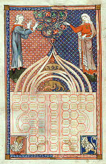
An illustration from a 13th-century manuscript of the Decretum Gratiani

In Abnormal (Les anormaux), Michel Foucault suggested it is likely that, "from the Middle Ages to the sixteenth century ... hermaphrodites were considered to be monsters and were executed, burnt at the stake and their ashes thrown to the winds."

However, Christof Rolker disputes this, arguing that "Contrary to what has been claimed, there is no evidence for hermaphrodites being persecuted in the Middle Ages, and the learned laws did certainly not provide any basis for such persecution". Canon Law sources provide evidence of alternative perspectives, based upon prevailing visual indications and the performance of gendered roles. The 12th-century Decretum Gratiani states that "Whether an hermaphrodite may witness a testament, depends on which sex prevails" ("Hermafroditus an ad testamentum adhiberi possit, qualitas sexus incalescentis ostendit.").

In the late twelfth century, the canon lawyer Huguccio stated that, "If someone has a beard, and always wishes to act like a man (excercere virilia) and not like a female, and always wishes to keep company with men and not with women, it is a sign that the male sex prevails in him and then he is able to be a witness, where a woman is not allowed". Concerning the ordination of 'hermaphrodites', Huguccio concluded: "If therefore the person is drawn to the feminine more than the male, the person does not receive the order. If the reverse, the person is able to receive but ought not to be ordained on account of deformity and monstrosity."

Henry de Bracton's De Legibus et Consuetudinibus Angliae ("On the Laws and Customs of England"), c. 1235, classifies mankind as "male, female, or hermaphrodite", and "A hermaphrodite is classed with male or female according to the predominance of the sexual organs."

The thirteenth-century canon lawyer Henry of Segusio argued that a "perfect hermaphrodite" where no sex prevailed should choose their legal gender under oath.

Early modern period

The 17th-century English jurist and judge Edward Coke (Lord Coke), wrote in his Institutes of the Lawes of England on laws of succession stating, "Every heire is either a male, a female, or an hermaphrodite, that is both male and female. And an hermaphrodite (which is also called Androgynus) shall be heire, either as male or female, according to that kind of sexe which doth prevaile." The Institutes are widely held to be a foundation of common law

A few historical accounts of intersex people exist due primarily to the discovery of relevant legal records, including those of Thomas(ine) Hall (17th-century United States), Eleno de Céspedes, a 16th-century intersex person in Spain (in Spanish), and Fernanda Fernández (18th-century Spain). 

In 2019 the Smithsonian Channel aired a documentary "American's Hidden Stories: The General was Female?" with evidence that Casimir Pulaski, the important American Revolutionary War hero, may have been intersex. 

In a court case, heard at the Castellania in 1774 during the Order of St. John in Malta, 17-year-old Rosa Mifsud from Luqa, later described in the British Medical Journal as a pseudo-hermaphrodite, petitioned for a change in sex classification from female. Two clinicians were appointed by the court to perform an examination. They found that "the male sex is the dominant one". The examiners were the Physician-in-Chief and a senior surgeon, both working at the Sacra Infermeria. The Grandmaster himself who took the final decision for Mifsud to wear only men clothes from then on.

Maria Dorothea Derrier/Karl Dürrge was a German intersex person who made their living for 30 years as a human research subject. Born in Potsdam in 1780, and designated as female at birth, they assumed a male identity around 1807. Traveling intersex persons, like Derrier and Katharina/Karl Hohmann, who allowed themselves to be examined by physicians were instrumental in the development of codified standards for sexing.

Mid modern period

A golden-coloured statue of a man in a gown on a seat with a sword on his knees. In front there is a polished wooden table with goldleaf and a blue and white porcelain vase with yellow flowers. Behind him is a wooden altar with lights and incense holders. The altar has the same design as the table. The wall is cream-coloured.
Bronze statue of Lê Văn Duyệt in his tomb
 
During the Victorian era, medical authors introduced the terms "true hermaphrodite" for an individual who has both ovarian and testicular tissue, verified under a microscope, "male pseudo-hermaphrodite" for a person with testicular tissue, but either female or ambiguous sexual anatomy, and "female pseudo-hermaphrodite" for a person with ovarian tissue, but either male or ambiguous sexual anatomy.


Historical accounts including those of Vietnamese general Lê Văn Duyệt (18th/19th-century) who helped to unify Vietnam; Gottlieb Göttlich, a 19th-century German travelling medical case; and Levi Suydam, an intersex person in 19th-century USA whose capacity to vote in male-only elections was questioned. 


The memoirs of 19th-century intersex Frenchwoman Herculine Barbin were published by Michel Foucault in 1980. Her birthday is marked in Intersex Day of Remembrance on 8 November.

Contemporary period

The Phall-O-Meter satirizes clinical assessments of appropriate clitoris and penis length at birth.
 
The term intersexuality was coined by Richard Goldschmidt in the 1917 paper Intersexuality and the endocrine aspect of sex. The first suggestion to replace the term 'hermaphrodite' with 'intersex' came from British specialist Cawadias in the 1940s. This suggestion was taken up by specialists in the UK during the 1960s. Historical accounts from the early twentieth century include that of Australian Florrie Cox, whose marriage was annulled due to "malformation frigidity".

Since the rise of modern medical science in Western societies, some intersex people with ambiguous external genitalia have had their genitalia surgically modified to resemble either female or male genitals. Surgeons pinpointed intersex babies as a "social emergency" once they were born. The parents of the intersex babies were not content about the situation. Psychologists, sexologists, and researchers frequently still believe that it is better for a baby's genitalia to be changed when they were younger than when they were a mature adult. These scientists believe that early intervention helped avoid gender identity confusion. This was called the 'Optimal Gender Policy', and it was initially developed in the 1950s by John Money. Money and others controversially believed that children were more likely to develop a gender identity that matched sex of rearing than might be determined by chromosomes, gonads, or hormones. The primary goal of assignment was to choose the sex that would lead to the least inconsistency between external anatomy and assigned psyche (gender identity).

Since advances in surgery have made it possible for intersex conditions to be concealed, many people are not aware of how frequently intersex conditions arise in human beings or that they occur at all. Dialog between what were once antagonistic groups of activists and clinicians has led to only slight changes in medical policies and how intersex patients and their families are treated in some locations. Numerous civil society organizations and human rights institutions now call for an end to unnecessary "normalizing" interventions.

The first public demonstration by intersex people took place in Boston on October 26, 1996, outside the venue in Boston where the American Academy of Pediatrics was holding its annual conference. The group demonstrated against "normalizing" treatments, and carried a sign saying "Hermaphrodites With Attitude". The event is now commemorated by Intersex Awareness Day.

In 2011, Christiane Völling became the first intersex person known to have successfully sued for damages in a case brought for non-consensual surgical intervention. In April 2015, Malta became the first country to outlaw non-consensual medical interventions to modify sex anatomy, including that of intersex people.

True hermaphroditism

From Wikipedia, the free encyclopedia
 
True hermaphroditism
Other namesDiseasesDB = 29664
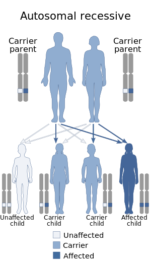
Autosomal recessive - en.svg
This condition is inherited in an autosomal recessive manner
SpecialtyObstetrics and gynaecology, endocrinology 

True hermaphroditism, clinically known as ovotesticular disorder of sex development, is a medical term for an intersex condition in which an individual is born with ovarian and testicular tissue. More commonly one or both gonads is an ovotestis containing both types of tissue.

Although similar in some ways to mixed gonadal dysgenesis, the conditions can be distinguished histologically.

Presentation

External genitalia are often ambiguous, the degree depending mainly on the amount of testosterone produced by the testicular tissue between 8 and 16 weeks of gestation.

Causes

There are several ways in which this may occur.
  • It can be caused by the division of one ovum, followed by fertilization of each haploid ovum and fusion of the two zygotes early in development.
  • Alternately, an ovum can be fertilized by two sperm followed by trisomic rescue in one or more daughter cells.
  • Two ova fertilized by two sperm cells will occasionally fuse to form a tetragametic chimera. If one male zygote and one female zygote fuse, a hermaphroditic individual may result.
  • It can be associated with a mutation in the SRY gene.

Karyotypes

Encountered karyotypes include 46XX/46XY, or 46XX/47XXY or XX & XY with SRY mutations, mixed chromosomal anomalies or hormone deficiency/excess disorders, 47XXY, and various degrees of mosaicism of these and a variety of others. The 3 primary karyotypes for true hermaphroditism are XX with genetic anomalies (55-70% of cases), XX/XY (20-30% of cases) & XY (5-15% of cases) with the remainder being a variety of other chromosomal anomalies and mosaicisms.

Prevalence

Only 500 cases have been reported in medical literature.

Fertility

There are extremely rare cases of fertility in "truly hermaphroditic" humans. These individuals typically have functional ovarian tissue, but underdeveloped testes that are unable to engage in spermatogenesis. As a result, these individuals are fertile, but not auto-fertile.

There is evidence that 50% of individuals with ovotestes can go through ovulation but spermatogenesis is rare.

There is a hypothetical scenario, though, in which it could be possible for a human to self fertilize. If a human chimera is formed from a male and female zygote fusing into a single embryo, giving an individual functional gonadal tissue of both types, such a self-fertilization is feasible. Indeed, it is known to occur in non-human species where hermaphroditic animals are common, including some mammals. However, no such case of functional self-fertilization has ever been documented in humans.

As of 2010, there have been at least 11 reported cases of fertility in true hermaphrodite humans in the scientific literature, with one case of a person with XY-predominant (96%) mosaic giving birth. Estimated frequency of ovotestes is one in 83,000 births (0.0012%).

Etymology

The term derives from the Latin: hermaphroditus, from Ancient Greek: ἑρμαφρόδιτος, romanizedhermaphroditos, which derives from Hermaphroditos ( Ἑρμαϕρόδιτος), the son of Hermes and Aphrodite in Greek mythology. According to Ovid, he fused with the nymph Salmacis resulting in one individual possessing physical traits of both sexes; according to the earlier Diodorus Siculus, he was born with a physical body combining both sexes. The word hermaphrodite entered the English lexicon in the late fourteenth century.

Society and culture

Having ovotesticular disorder of sex development can make one inadmissible for service in the United States Armed Forces.

M.C. v. Aaronson

The U.S. legal case of M.C. v. Aaronson, advanced by intersex civil society organization interACT with the Southern Poverty Law Center was brought before the courts in 2013. The child in the case was born in December 2004 with ovotestes, initially determined as male, but subsequently assigned female and placed in the care of South Carolina Department of Social Services in February 2005. Physicians responsible for M.C. initially concluded that surgery was not urgent or necessary and M.C. had potential to identify as male or female, but, in April 2006, M.C. was subjected to feminizing medical interventions. He was adopted in December 2006. Aged 8 at the time the case was taken, he now identifies as male. The Southern Poverty Law Center state: "In M.C.’s condition, there is no way to tell whether the child will ultimately identify as a boy or a girl. Instead, the doctors decided to assign M.C. female and change his body to fit their stereotype of how a girl should look." The defendant in the case, Dr Ian Aaronson, had written in 2001 that "feminizing genitoplasty on an infant who might eventually identify herself as a boy would be catastrophic".

The defendants sought to dismiss the case and seek a defense of qualified immunity, but these were denied by the District Court for the District of South Carolina. In January 2015, the Court of Appeals for the Fourth Circuit reversed this decision and dismissed the complaint, stating that, "it did not “mean to diminish the severe harm that M.C. claims to have suffered” but that a reasonable official in 2006 did not have fair warning from then-existing precedent that performing sex assignment surgery on sixteen-month-old M.C. violated a clearly established constitutional right." The Court did not rule on whether or not the surgery violated M.C.'s constitutional rights.

State suits were subsequently filed. In July 2017, it was reported that the case had been settled out of court by the Medical University of South Carolina for $440,000. The University denied negligence, but agreed to a "compromise" settlement to avoid "costs of litigation."

Klinefelter syndrome

From Wikipedia, the free encyclopedia

Klinefelter syndrome
Other namesXXY syndrome, Klinefelter's syndrome, Klinefelter-Reifenstein-Albright syndrome
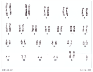
Human chromosomesXXY01.png
47,XXY karyotype
Pronunciation
SpecialtyMedical genetics
SymptomsOften few
Usual onsetAt fertilisation
DurationLong term
CausesTwo or more X chromosomes in males
Risk factorsOlder mother
Diagnostic methodGenetic testing (karyotype)
TreatmentPhysical therapy, speech and language therapy, counseling
PrognosisNearly normal life expectancy
Frequency1:500 to 1:1,000 males

Klinefelter syndrome (KS), also known as 47, XXY is the set of symptoms that result from two or more X chromosomes in males. The primary features are infertility and small poorly functioning testicles. Often, symptoms may be subtle and many people do not realize they are affected. Sometimes, symptoms are more prominent and may include weaker muscles, greater height, poor coordination, less body hair, breast growth, and less interest in sex. Often it is only at puberty that these symptoms are noticed. Intelligence is usually normal; however, reading difficulties and problems with speech are more common. Symptoms are typically more severe if three or more X chromosomes are present (48,XXXY syndrome or 49,XXXXY syndrome).

Klinefelter syndrome occurs randomly. The extra X chromosome comes from the father and mother nearly equally. An older mother may have a slightly increased risk of a child with KS. The condition is not typically inherited from one's parents. The underlying mechanisms involves at least one extra X chromosome in addition to a Y chromosome such that the total chromosome number is 47 or more rather than the usual 46. KS is diagnosed by the genetic test known as a karyotype.

While no cure is known, a number of treatments may help. Physical therapy, speech and language therapy, counselling, and adjustments of teaching methods may be useful. Testosterone replacement may be used in those who have significantly lower levels. Enlarged breasts may be removed by surgery. About half of affected males have a chance of fathering children with the help of assisted reproductive technology, but this is expensive and not risk free. XXY males appear to have a higher risk of breast cancer than typical, but still lower than that of females. People with the condition have a nearly normal life expectancy.

Klinefelter syndrome is one of the most common chromosomal disorders, occurring in one to two per 1,000 live male births. It is named after American endocrinologist Harry Klinefelter, who identified the condition in the 1940s. In 1956, identification of the extra X chromosome was first noticed. Mice can also have the XXY syndrome, making them a useful research model.

Signs and symptoms

A person with typical untreated (surgery/hormones) Klinefelter 46,XY/47,XXY mosaic, diagnosed at age 19 - the scar from biopsy may be visible on left nipple.
 
The primary features are infertility and small poorly functioning testicles. Often, symptoms may be subtle and many people do not realize they are affected. Sometimes, symptoms are more prominent and may include weaker muscles, greater height, poor coordination, less body hair, breast growth, and less interest in sex. Often it is only at puberty that these symptoms are noticed.

Physical

As babies and children, XXY males may have weaker muscles and reduced strength. As they grow older, they tend to become taller than average. They may have less muscle control and coordination than other boys of their age.

During puberty, the physical traits of the syndrome become more evident; because these boys do not produce as much testosterone as other boys, they have a less muscular body, less facial and body hair, and broader hips. As teens, XXY males may develop breast tissue and also have weaker bones, and a lower energy level than other males.

By adulthood, XXY males look similar to males without the condition, although they are often taller. In adults, possible characteristics vary widely and include little to no sign of affectedness, a lanky, youthful build and facial appearance, or a rounded body type with some degree of gynecomastia (increased breast tissue). Gynecomastia is present to some extent in about a third of affected individuals, a slightly higher percentage than in the XY population. About 10% of XXY males have gynecomastia noticeable enough that they may choose to have cosmetic surgery.

Affected males are often infertile, or may have reduced fertility. Advanced reproductive assistance is sometimes possible.

The term hypogonadism in XXY symptoms is often misinterpreted to mean "small testicles" when it means decreased testicular hormone/endocrine function. Because of this (primary) hypogonadism, individuals often have a low serum testosterone level, but high serum follicle-stimulating hormone and luteinizing hormone levels. Despite this misunderstanding of the term, however, XXY men may also have microorchidism (i.e., small testicles).

The testicle size of affected males are usually less than 2 cm in length (and always shorter than 3.5 cm), 1 cm in width, and 4 ml in volume.

XXY males are also more likely than other men to have certain health problems, such as autoimmune disorders, breast cancer, venous thromboembolic disease, and osteoporosis. In contrast to these potentially increased risks, rare X-linked recessive conditions are thought to occur less frequently in XXY males than in normal XY males, since these conditions are transmitted by genes on the X chromosome, and people with two X chromosomes are typically only carriers rather than affected by these X-linked recessive conditions.

Cognitive and developmental

Some degree of language learning or reading impairment may be present, and neuropsychological testing often reveals deficits in executive functions, although these deficits can often be overcome through early intervention. Also, delays in motor development may occur, which can be addressed through occupational and physical therapies. XXY males may sit up, crawl, and walk later than other infants; they may also struggle in school, both academically and with sports.

Endocrine

Men with Klinefelter's syndrome have testosterone levels that are significantly lower (−260 ng/dL difference), estradiol levels that are slightly higher (+6.8 pg/mL) though not significantly different when restricted to high-accuracy assays (+5.5 pg/mL difference), and the ratio of estradiol to testosterone is significantly higher (+0.06).

Cause

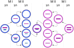
Birth of a cell with karyotype XXY due to a nondisjunction event of one X chromosome from a Y chromosome during meiosis I in the male
 
Birth of a cell with karyotype XXY due to a nondisjunction event of one X chromosome during meiosis II in the female
 
Maternal age is the only known risk factor. Women at 40 years have a four times higher risk for a child with Klinefelter syndrome than women aged 24 years.

The extra chromosome is retained because of a nondisjunction event during paternal meiosis I, maternal meiosis I, or maternal meiosis II (gametogenesis). The relevant nondisjunction in meiosis I occurs when homologous chromosomes, in this case the X and Y or two X sex chromosomes, fail to separate, producing a sperm with an X and a Y chromosome or an egg with two X chromosomes. Fertilizing a normal (X) egg with this sperm produces an XXY offspring (Klinefelter). Fertilizing a double X egg with a normal sperm also produces an XXY offspring (Klinefelter).

Another mechanism for retaining the extra chromosome is through a nondisjunction event during meiosis II in the egg. Nondisjunction occurs when sister chromatids on the sex chromosome, in this case an X and an X, fail to separate. An XX egg is produced, which when fertilized with a Y sperm, yields an XXY offspring. This XXY chromosome arrangement is one of the most common genetic variations from the XY karyotype, occurring in about one in 500 live male births.

In mammals with more than one X chromosome, the genes on all but one X chromosome are not expressed; this is known as X inactivation. This happens in XXY males, as well as normal XX females. However, in XXY males, a few genes located in the pseudoautosomal regions of their X chromosomes have corresponding genes on their Y chromosome and are capable of being expressed.

Variations

48,XXYY or 48,XXXY occurs in one in 18,000–50,000 male births. The incidence of 49,XXXXY is one in 85,000 to 100,000 male births. These variations are extremely rare. Additional chromosomal material can contribute to cardiac, neurological, orthopedic, and other anomalies.

Males with KS may have a mosaic 47,XXY/46,XY constitutional karyotype and varying degrees of spermatogenic failure. Mosaicism 47,XXY/46,XX with clinical features suggestive of KS is very rare. Thus far, only about 10 cases have been described in literature.

Analogous XXY syndromes are known to occur in cats—specifically, the presence of calico or tortoiseshell markings in male cats is an indicator of the relevant abnormal karyotype. As such, male cats with calico or tortoiseshell markings are a model organism for KS, because a color gene involved in cat tabby coloration is on the X chromosome.

Diagnosis

The standard diagnostic method is the analysis of the chromosomes' karyotype on lymphocytes. A small blood sample is sufficient as test material. In the past, the observation of the Barr body was common practice, as well. To investigate the presence of a possible mosaicism, analysis of the karyotype using cells from the oral mucosa is performed. Physical characteristics of a Klinefelter syndrome can be tall stature, low body hair and occasionally an enlargement of the breast. There is usually a small testicle volume of 1–5 ml per testicle (standard values: 12–30 ml). During puberty and adulthood, low testosterone levels with increased levels of the pituitary hormones FSH and LH in the blood can indicate the presence of Klinefelter syndrome. A spermiogram can also be part of the further investigation. Often there is an azoospermia present, rarely an oligospermia. Furthermore, Klinefelter syndrome can be diagnosed as a coincidental prenatal finding in the context of invasive prenatal diagnosis (amniocentesis, chorionic villus sampling). About 10% of KS cases are found by prenatal diagnosis.

The symptoms of KS are often variable; therefore, a karyotype analysis should be ordered when small testes, infertility, gynecomastia, long arms/legs, developmental delay, speech/language deficits, learning disabilities/academic issues, and/or behavioral issues are present in an individual.

Treatment

The genetic variation is irreversible, thus there is no causal therapy. From the onset of puberty, the existing testosterone deficiency can be compensated by appropriate hormone replacement therapy. Testosterone preparations are available in the form of syringes, patches or gel. If gynecomastia is present, the surgical removal of the breast may be considered for both the psychological reasons and to reduce the risk of breast cancer.

The use of behavioral therapy can mitigate any language disorders, difficulties at school, and socialization. An approach by occupational therapy is useful in children, especially those who have dyspraxia.

Infertility treatment

Methods of reproductive medicine, such as intracytoplasmic sperm injection (ICSI) with previously conducted testicular sperm extraction (TESE), have led to men with Klinefelter's syndrome to produce biological offspring. By 2010, over 100 successful pregnancies have been reported using IVF technology with surgically removed sperm material from males with KS.

Prognosis

The lifespan of individuals with Klinefelter syndrome appears to be reduced by approximately 2.1 years compared to the general male population. These results are still questioned data, are not absolute, and need further testing.

Epidemiology

This syndrome, evenly distributed in all ethnic groups, has a prevalence of one to two subjects per every 1000 males in the general population. However, it is estimated that only 25% of the individuals with Klinefelter syndrome are diagnosed throughout their lives. 3.1% of infertile males have Klinefelter syndrome. The syndrome is also the main cause of male hypogonadism.

History

The syndrome was named after American endocrinologist Harry Klinefelter, who in 1942 worked with Fuller Albright and E. C. Reifenstein at Massachusetts General Hospital in Boston, Massachusetts, and first described it in the same year. The account given by Klinefelter came to be known as Klinefelter syndrome as his name appeared first on the published paper, and seminiferous tubule dysgenesis was no longer used. Considering the names of all three researchers, it is sometimes also called Klinefelter–Reifenstein–Albright syndrome. In 1956 it was discovered that Klinefelter syndrome resulted from an extra chromosome. Plunkett and Barr found the sex chromatin body in cell nuclei of the body. This was further clarified as XXY in 1959 by Patricia Jacobs and John Anderson Strong. The first published report of a man with a 47,XXY karyotype was by Patricia Jacobs and John Strong at Western General Hospital in Edinburgh, Scotland, in 1959. This karyotype was found in a 24-year-old man who had signs of KS. Jacobs described her discovery of this first reported human or mammalian chromosome aneuploidy in her 1981 William Allan Memorial Award address.

Other animals

Klinefelter syndrome can also occur in other animals. In cats it can result in a male tortoiseshell and calico cat (patches of different colored fur), a pattern that is usually only seen in female cats. Not all cats with Klinefelter syndrome have tortoiseshell or calico patterns.

Turner syndrome

From Wikipedia, the free encyclopedia
 
Turner syndrome
Other namesUllrich–Turner syndrome; Bonnevie–Ullrich–Turner syndrome; gonadal dysgenesis; 45X; 45X0
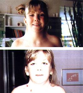
Neck of girl with Turner Syndrome (before and after).jpg
Girl with Turner syndrome before and after an operation for neck-webbing
SpecialtyPediatrics, medical genetics
SymptomsWebbed neck, short stature, swollen hands and feet
ComplicationsHeart defects, diabetes, low thyroid hormone
Usual onsetAt birth
DurationLong term
CausesMissing X chromosome
Diagnostic methodPhysical signs, genetic testing
MedicationHuman growth hormone, estrogen replacement therapy
PrognosisShorter life expectancy
Frequency1 in 2,000 to 5,000

Turner syndrome (TS), also known 45,X, or 45,X0, is a genetic condition in which a female is partly or completely missing an X chromosome. Signs and symptoms vary among those affected. Often, a short and webbed neck, low-set ears, low hairline at the back of the neck, short stature, and swollen hands and feet are seen at birth. Typically, they develop menstrual periods and breasts only with hormone treatment, and are unable to have children without reproductive technology. Heart defects, diabetes, and low thyroid hormone occur more frequently. Most people with TS have normal intelligence. Many have troubles with spatial visualization that may be needed for mathematics. Vision and hearing problems occur more often.

Turner syndrome is not usually inherited; rather, it occurs as a result of a genetic defect arising during formation of the reproductive cells in a parent or in early cell division during development. No environmental risks are known, and the mother's age does not play a role. Turner syndrome is due to a chromosomal abnormality in which all or part of one of the X chromosomes is missing or altered. While most people have 46 chromosomes, people with TS usually have 45. The chromosomal abnormality may be present in just some cells in which case it is known as TS with mosaicism. In these cases, the symptoms are usually fewer and possibly none occur at all. Diagnosis is based on physical signs and genetic testing.

No cure for Turner syndrome is known. Treatment may help with symptoms. Human growth hormone injections during childhood may increase adult height. Estrogen replacement therapy can promote development of the breasts and hips. Medical care is often required to manage other health problems with which TS is associated.

Turner syndrome occurs in between one in 2,000 and one in 5,000 females at birth. All regions of the world and cultures are affected about equally. Generally people with TS have a shorter life expectancy, mostly due to heart problems and diabetes. Henry Turner first described the condition in 1938. In 1964, it was determined to be due to a chromosomal abnormality.

Signs and symptoms

Lymphedema, puffy legs of a newborn with Turner syndrome
 
Of the following common symptoms of Turner syndrome, an individual may have any combination of symptoms and is unlikely to have all symptoms.
Other features may include a small lower jaw (micrognathia), cubitus valgus, soft upturned nails, palmar crease, and drooping eyelids. Less common are pigmented moles, hearing loss, and a high-arch palate (narrow maxilla). Turner syndrome manifests itself differently in each female affected by the condition; therefore, no two individuals share the same features.

While most of the physical findings are harmless, significant medical problems can be associated with the syndrome. Most of these significant conditions are treatable with surgery and medication.

Prenatal

Despite the excellent postnatal prognosis, 99% of Turner syndrome conceptions are thought to end in miscarriage or stillbirth, and as many as 15% of all spontaneous abortions have the 45,X karyotype. Among cases that are detected by routine amniocentesis or chorionic villus sampling, one study found that the prevalence of Turner syndrome among tested pregnancies was 5.58 and 13.3 times higher, respectively, than among live neonates in a similar population.

Cardiovascular

The rate of cardiovascular malformations among patients with Turner syndrome ranges from 17% to 45%. The variations found in the different studies are mainly attributable to variations in noninvasive methods used for screening and the types of lesions that they can characterize. However, it could be simply attributable to the small number of subjects in most studies. 

Different karyotypes may have differing rates of cardiovascular malformations. Two studies found a rate of cardiovascular malformations of 30% and 38% in a group of pure 45,X monosomy. Considering other karyotype groups, though, they reported a prevalence of 24.3% and 11% in people with mosaic X monosomy, and a rate of 11% in people with X chromosomal structural abnormalities.

The higher rate in the group of pure 45,X monosomy is primarily due to a difference in the rate of aortic valve abnormalities and coarctation of the aorta, the two most common cardiovascular malformations.

Congenital heart disease

The most commonly observed are congenital obstructive lesions of the left side of the heart, leading to reduced flow on this side of the heart. This includes bicuspid aortic valve and coarctation (narrowing) of the aorta. More than 50% of the cardiovascular malformations of individuals with Turner syndrome in one study were bicuspid aortic valves or coarctation of the aorta (usually preductal), alone or in combination.

Other congenital cardiovascular malformations, such as partial anomalous venous drainage and aortic valve stenosis or aortic regurgitation, are also more common in Turner syndrome than in the general population. Hypoplastic left heart syndrome represents the most severe reduction in left-sided structures.
Bicuspid aortic valve
Up to 15% of adults with Turner syndrome have bicuspid aortic valves, meaning only two, instead of three, parts to the valves in the main blood vessel leading from the heart are present. Since bicuspid valves are capable of regulating blood flow properly, this condition may go undetected without regular screening. However, bicuspid valves are more likely to deteriorate and later fail. Calcification also occurs in the valves, which may lead to a progressive valvular dysfunction as evidenced by aortic stenosis or regurgitation.

With a rate from 12.5% to 17.5% (Dawson-Falk et al., 1992), bicuspid aortic valve is the most common congenital malformation affecting the heart in this syndrome. It is usually isolated, but it may be seen in combination with other anomalies, particularly coarctation of the aorta.
Coarctation of the aorta
Between 5% and 10% of those born with Turner syndrome have coarctation of the aorta, a congenital narrowing of the descending aorta, usually just distal to the origin of the left subclavian artery (the artery that branches off the arch of the aorta to the left arm) and opposite to the ductus arteriosus (termed "juxtaductal"). Estimates of the prevalence of this malformation in patients with Turner syndrome range from 6.9 to 12.5%. A coarctation of the aorta in a female is suggestive of Turner syndrome and suggests the need for further tests, such as a karyotype.
Partial anomalous venous drainage
This abnormality is a relatively rare congenital heart disease in the general population. The prevalence of this abnormality also is low (around 2.9%) in Turner syndrome. However, its relative risk is 320 in comparison with the general population. Strangely, Turner syndrome seems to be associated with unusual forms of partial anomalous venous drainage.

In a patient with Turner syndrome, these left-sided cardiovascular malformations can result in an increased susceptibility to bacterial endocarditis. Therefore, prophylactic antibiotics should be considered when procedures with a high risk of endocarditis are performed, such as dental cleaning.

Turner syndrome is often associated with persistent hypertension, sometimes in childhood. In the majority of Turner syndrome patients with hypertension, no specific cause is known. In the remainder, it is usually associated with cardiovascular or kidney abnormalities, including coarctation of the aorta.

Aortic dilation, dissection, and rupture

Two studies have suggested aortic dilatation in Turner syndrome, typically involving the root of the ascending aorta and occasionally extending through the aortic arch to the descending aorta, or at the site of previous coarctation of the aorta repair.
  • A study that evaluated 28 girls with Turner syndrome found a greater mean aortic root diameter in people with Turner syndrome than in the control group (matched for body surface area). Nonetheless, the aortic root diameters found in Turner syndrome patients were still well within the limits.
  • This has been confirmed by a study that evaluated 40 patients with Turner syndrome. The study presented basically the same findings: a greater mean aortic root diameter, which nevertheless remains within the normal range for body surface area.
Whether aortic root diameters that are relatively large for body surface area but still well within normal limits imply a risk for progressive dilatation remains unproven.
Rate of aortic abnormalities
The prevalence of aortic root dilatation ranges from 8.8 to 42% in patients with Turner syndrome. Even if not every aortic root dilatation necessarily goes on to an aortic dissection (circumferential or transverse tear of the intima), complications such as dissection, aortic rupture resulting in death may occur. The natural history of aortic root dilatation is still unknown, but it is linked to aortic dissection and rupture, which has a high mortality rate.

Aortic dissection affects 1 to 2% of patients with Turner syndrome. As a result, any aortic root dilatation should be seriously taken into account, as it could become a fatal aortic dissection. Routine surveillance is highly recommended.
Risk factors for aortic rupture
Cardiovascular malformations (typically bicuspid aortic valve, coarctation of the aorta, and some other left-sided cardiac malformations) and hypertension predispose to aortic dilatation and dissection in the general population. Indeed, these same risk factors are found in more than 90% of patients with Turner syndrome who develop aortic dilatation. Only a small number of patients (around 10%) have no apparent predisposing risk factors. The risk of hypertension is increased three-fold in patients with Turner syndrome. Because of its relation to aortic dissection, blood pressure must be regularly monitored and hypertension should be treated aggressively with an aim to keep blood pressure below 140/80 mmHg. As with the other cardiovascular malformations, complications of aortic dilatation is commonly associated with 45,X karyotype.
Pathogenesis of aortic dissection and rupture
The exact role that these risk factors play in the process leading to rupture is unclear. Aortic root dilatation is thought to be due to a mesenchymal defect as pathological evidence of cystic medial necrosis has been found by several studies. The association between a similar defect and aortic dilatation is well established in such conditions such as Marfan syndrome. Also, abnormalities in other mesenchymal tissues (bone matrix and lymphatic vessels) suggests a similar primary mesenchymal defect in patients with Turner syndrome. However, no evidence suggests that patients with Turner syndrome have a significantly higher risk of aortic dilatation and dissection in absence of predisposing factors. So, the risk of aortic dissection in Turner syndrome appears to be a consequence of structural cardiovascular malformations and hemodynamic risk factors rather than a reflection of an inherent abnormality in connective tissue. The natural history of aortic root dilatation is unknown, but because of its lethal potential, this aortic abnormality needs to be carefully followed.

Skeletal

Normal skeletal development is inhibited due to a large variety of factors, mostly hormonal. The average height of a woman with Turner syndrome, in the absence of growth hormone treatment, is 4 ft 7 in (140 cm). Patients with Turner's mosaicism can reach normal average height. 

The fourth metacarpal bone (fourth toe and ring finger) may be unusually short, as may the fifth.

Due to inadequate production of estrogen, many of those with Turner syndrome develop osteoporosis. This can decrease height further, as well as exacerbate the curvature of the spine, possibly leading to scoliosis. It is also associated with an increased risk of bone fractures.

Kidney

About one-third of all women with Turner syndrome have one of three kidney abnormalities:
  1. A single, horseshoe-shaped kidney on one side of the body
  2. An abnormal urine-collecting system
  3. Poor blood flow to the kidneys
Some of these conditions can be corrected surgically. Even with these abnormalities, the kidneys of most women with Turner syndrome function normally. However, as noted above, kidney problems may be associated with hypertension.

Thyroid

Approximately one-third of all women with Turner syndrome have a thyroid disorder. Usually it is hypothyroidism, specifically Hashimoto's thyroiditis. If detected, it can be easily treated with thyroid hormone supplements.

Diabetes

Women with Turner syndrome are at a moderately increased risk of developing type 1 diabetes in childhood and a substantially increased risk of developing type 2 diabetes by adult years. The risk of developing type 2 diabetes can be substantially reduced by maintaining a healthy weight.

Cognitive

People with Turner syndrome have normal intelligence, and demonstrate relative strengths in verbal skills, but may exhibit weaker nonverbal skills – particularly in arithmetic, select visuospatial skills, and processing speed. Turner syndrome does not typically cause intellectual disability or impair cognition. However, learning difficulties are common among women with Turner syndrome, particularly a specific difficulty in perceiving spatial relationships, such as nonverbal learning disorder. This may also manifest itself as a difficulty with motor control or with mathematics. While it is not correctable, in most cases it does not cause difficulty in daily living. Most Turner syndrome patients are employed as adults and lead productive lives. 

Also, a rare variety of Turner syndrome, known as "Ring-X Turner syndrome", has about a 60% association with intellectual disability. This variety accounts for around 2–4% of all Turner syndrome cases.

Psychological

Social difficulties appear to be an area of vulnerability for young women. Counseling affected individuals and their families about the need to carefully develop social skills and relationships may prove useful in advancing social adaptation. Women with Turner syndrome may experience adverse psychosocial outcomes which can be improved through early intervention and the provision of appropriate psychological and psychiatric care. Genetic, hormonal, and medical problems associated with TS are likely to affect psychosexual development of female adolescent patients, and thus influence their psychological functioning, behavior patterns, social interactions, and learning ability. Although TS constitutes a chronic medical condition, with possible physical, social, and psychological complications in a woman's life, hormonal and estrogen replacement therapy, and assisted reproduction, are treatments that can be helpful for TS patients and improve their quality of life. Research shows a possible association between age at diagnosis and increased substance use and depressive symptoms.

Reproductive

Women with Turner syndrome are almost universally infertile. While some women with Turner syndrome have successfully become pregnant and carried their pregnancies to term, this is very rare and is generally limited to those women whose karyotypes are not 45,X. Even when such pregnancies do occur, there is a higher than average risk of miscarriage or birth defects, including Turner syndrome or Down syndrome. Some women with Turner syndrome who are unable to conceive without medical intervention may be able to use IVF or other fertility treatments.

Usually, estrogen replacement therapy is used to spur the growth of secondary sexual characteristics at the time when puberty should onset. While very few women with Turner syndrome menstruate spontaneously, estrogen therapy requires a regular shedding of the uterine lining ("withdrawal bleeding") to prevent its overgrowth. Withdrawal bleeding can be induced monthly, like menstruation, or less often, usually every three months, if the patient desires. Estrogen therapy does not make a woman with nonfunctional ovaries fertile, but it plays an important role in assisted reproduction; the health of the uterus must be maintained with estrogen if an eligible woman with Turner Syndrome wishes to use IVF (using donated oocytes).

Especially in mosaic cases of Turner syndrome that contains Y-chromosome (e.g. 45,X/46,XY) due to the risk of development of ovarian malignancy (most common is gonadoblastoma) gonadectomy is recommended. Turner syndrome is characterized by primary amenorrhoea, premature ovarian failure (hypergonadotropic hypogonadism), streak gonads and infertility (however, technology (especially oocyte donation) provides the opportunity of pregnancy in these patients). Failure to develop secondary sex characteristics (sexual infantilism) is typical.

As more women with Turner syndrome complete pregnancy thanks to modern techniques to treat infertility, it has to be noted that pregnancy may be a risk of cardiovascular complications for the mother. Indeed, several studies had suggested an increased risk for aortic dissection in pregnancy. The influence of estrogen has been examined but remains unclear. It seems that the high risk of aortic dissection during pregnancy in women with Turner syndrome may be due to the increased hemodynamic load rather than the high estrogen rate. Of course, these findings are important and need to be remembered while following a pregnant patient with Turner syndrome.

Hearing

Recurrent acute otitis media (AOM) and otitis media with effusion (OME) commonly occur in children with Turner syndrome during the preschool age, which can persist or develop later in childhood. The recurring AOM can also be a predisposition to cholesteatomas. People with the monosomy 45, X karyotype have an increased rate of hearing loss over other TS karyotype variants. Conductive hearing losses are more commonly seen with children than adults and becomes more of a sensorineural pattern once in the adolescence age. There seems to be an apparent linear relation between hearing loss and age in TS. About 75% of people with Turner syndrome have some hearing loss, with the most common presenting as a high frequency sensorineural hearing loss (HFSNHL) across all ages. People with TS tend to have more of a progressive hearing loss with a higher decline rate than those in their corresponding age groups with hearing loss. The increased decline tends to occur in the higher frequency range, with a rate of around 0.8-2.2 dB a year.

Cause

Turner syndrome is caused by the absence of one complete or partial copy of the X chromosome in some or all the cells. The abnormal cells may have only one X (monosomy) (45,X) or they may be affected by one of several types of partial monosomy like a deletion of the short p arm of one X chromosome (46,X,del(Xp)) or the presence of an isochromosome with two q arms (46,X,i(Xq)). Turner syndrome has distinct features due to the lack of pseudoautosomal regions, which are typically spared from X-inactivation. In mosaic individuals, cells with X monosomy (45,X) may occur along with cells that are normal (46,XX), cells that have partial monosomies, or cells that have a Y chromosome (46,XY). The presence of mosaicism is estimated to be relatively common in affected individuals (67–90%).

Inheritance

In the majority of cases where monosomy occurs, the X chromosome comes from the mother. This may be due to a nondisjunction in the father. Meiotic errors that lead to the production of X with p arm deletions or abnormal Y chromosomes are also mostly found in the father. Isochromosome X or ring chromosome X on the other hand are formed equally often by both parents. Overall, the functional X chromosome usually comes from the mother. 

In most cases, Turner syndrome is a sporadic event, and for the parents of an individual with Turner syndrome the risk of recurrence is not increased for subsequent pregnancies. Rare exceptions may include the presence of a balanced translocation of the X chromosome in a parent, or where the mother has 45,X mosaicism restricted to her germ cells.

Diagnosis

Prenatal

45,X karyotype, showing an unpaired X at the lower right

Turner syndrome may be diagnosed by amniocentesis or chorionic villus sampling during pregnancy.
Usually, fetuses with Turner syndrome can be identified by abnormal ultrasound findings (i.e., heart defect, kidney abnormality, cystic hygroma, ascites). In a study of 19 European registries, 67.2% of prenatally diagnosed cases of Turner syndrome were detected by abnormalities on ultrasound. 69.1% of cases had one anomaly present, and 30.9% had two or more anomalies.

An increased risk of Turner syndrome may also be indicated by abnormal triple or quadruple maternal serum screen. The fetuses diagnosed through positive maternal serum screening are more often found to have a mosaic karyotype than those diagnosed based on ultrasonographic abnormalities, and conversely, those with mosaic karyotypes are less likely to have associated ultrasound abnormalities.

Postnatal

Turner syndrome can be diagnosed postnatally at any age. Often, it is diagnosed at birth due to heart problems, an unusually wide neck or swelling of the hands and feet. However, it is also common for it to go undiagnosed for several years, often until the girl reaches the age of puberty and fails to develop typically (the changes associated with puberty do not occur). In childhood, a short stature can be indicative of Turner syndrome.

A test called a karyotype, also known as a chromosome analysis, analyzes the chromosomal composition of the individual. This is the test of choice to diagnose Turner syndrome.

Treatment

As a chromosomal condition, there is no cure for Turner syndrome. However, much can be done to minimize the symptoms. For example:
  • Growth hormone, either alone or with a low dose of androgen, will increase growth and probably final adult height. Growth hormone is approved by the U.S. Food and Drug Administration for treatment of Turner syndrome and is covered by many insurance plans. There is evidence that this is effective, even in toddlers.
  • Estrogen replacement therapy such as the birth control pill, has been used since the condition was described in 1938 to promote development of secondary sexual characteristics. Estrogens are crucial for maintaining good bone integrity, cardiovascular health and tissue health. Women with Turner syndrome who do not have spontaneous puberty and who are not treated with estrogen are at high risk for osteoporosis and heart conditions.
  • Modern reproductive technologies have also been used to help women with Turner syndrome become pregnant if they desire. For example, a donor egg can be used to create an embryo, which is carried by the Turner syndrome woman.
  • Uterine maturity is positively associated with years of estrogen use, history of spontaneous menarche, and negatively associated with the lack of current hormone replacement therapy.

Epidemiology

Turner syndrome occurs in between one in 2000 and one in 5000 females at birth.

Approximately 99 percent of fetuses with Turner syndrome spontaneously terminate during the first trimester. Turner syndrome accounts for about 10 percent of the total number of spontaneous abortions in the United States.

History

The syndrome is named after Henry Turner, an endocrinologist from Illinois, who described it in 1938. In Europe, it is often called Ullrich–Turner syndrome or even Bonnevie–Ullrich–Turner syndrome to acknowledge that earlier cases had also been described by European doctors. In Russian and USSR literature it is called Shereshevsky–Turner syndrome to acknowledge that the condition was first described as hereditary in 1925 by the Soviet endocrinologist Nikolai Shereshevsky [ru], who believed that it was due to the underdevelopment of the gonads and the anterior pituitary gland and was combined with congenital malformations of internal development.

The first published report of a female with a 45,X karyotype was in 1959 by Dr. Charles Ford and colleagues in Harwell near Oxford, and Guy's Hospital in London. It was found in a 14-year-old girl with signs of Turner syndrome.

Inequality (mathematics)

From Wikipedia, the free encyclopedia https://en.wikipedia.org/wiki/Inequality...