Search This Blog

Sunday, July 7, 2019

Andreas Vesalius

From Wikipedia, the free encyclopedia

Andreas Vesalius
Vesalius Portrait pg xii - c.png
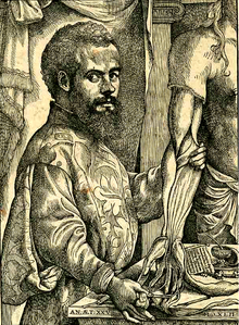
A portrait of Vesalius from De humani corporis fabrica
Born31 December 1514
Died15 October 1564 (aged 49)
Alma materUniversity of Pavia
University of Padua
Known forDe humani corporis fabrica (On the Fabric of the Human Body)
Scientific career
FieldsAnatomy
ThesisParaphrasis in nonum librum Rhazae medici Arabis clarissimi ad regem Almansorem, de affectuum singularum corporis partium curatione (1537)
Doctoral advisorJohannes Winter von Andernach Gemma Frisius
Doctoral studentsMatteo Realdo Colombo
InfluencesGalen Jacques Dubois Jean Fernel
InfluencedGabriele Falloppio

Andreas Vesalius was a 16th-century Flemish anatomist, physician, and author of one of the most influential books on human anatomy, De humani corporis fabrica (On the Fabric of the Human Body). Vesalius is often referred to as the founder of modern human anatomy. He was born in Brussels, which was then part of the Habsburg Netherlands. He was professor at the University of Padua and later became Imperial physician at the court of Emperor Charles V.

Andreas Vesalius is the Latinized form of the Dutch Andries van Wesel. It was a common practice among European scholars in his time to Latinize their names. His name is also given as Andrea Vesalius, André Vésale, Andrea Vesalio, Andreas Vesal, André Vesalio and Andre Vesale.

Early life and education

Vesalius was born as Andries van Wesel to his father Andries van Wesel and mother Isabel Crabbe on 31 December 1514 in Brussels, which was then part of the Habsburg Netherlands. His great grandfather, Jan van Wesel, probably born in Wesel, received a medical degree from the University of Pavia and taught medicine in 1528 at the University of Leuven. His grandfather, Everard van Wesel, was the Royal Physician of Emperor Maximilian, while his father, Anders van Wesel, served as apothecary to Maximilian, and later valet de chambre to his successor Charles V. Anders encouraged his son to continue in the family tradition and enrolled him in the Brethren of the Common Life in Brussels to learn Greek and Latin prior to learning medicine, according to standards of the era.

In 1528 Vesalius entered the University of Leuven (Pedagogium Castrense) taking arts, but when his father was appointed as the Valet de Chambre in 1532, he decided instead to pursue a career in the military at the University of Paris, where he relocated in 1533. There he studied the theories of Galen under the auspices of Jacques Dubois (Jacobus Sylvius) and Jean Fernel. It was during this time that he developed an interest in anatomy, and he was often found examining excavated bones in the charnel houses at the Cemetery of the Innocents

Vesalius was forced to leave Paris in 1536 owing to the opening of hostilities between the Holy Roman Empire and France and returned to Leuven. He completed his studies there under Johann Winter von Andernach and graduated the following year. His thesis, Paraphrasis in nonum librum Rhazae medici arabis clariss. ad regem Almansorum de affectuum singularum corporis partium curatione, was a commentary on the ninth book of Rhazes. He remained at Leuven for a while, before leaving after a dispute with his professor. After settling briefly in Venice in 1536, he moved to the University of Padua (Universitas artistarum) to study for his medical doctorate, which he received in 1537.

Medical career and accomplishments

On the day of his graduation he was immediately offered the chair of surgery and anatomy (explicator chirurgiae) at Padua. He also guest-lectured at the Bologna and the Pisa. Prior to taking up his position in Padua, Vesalius traveled through Italy, and assisted the future Pope Paul IV and Ignatius of Loyola to heal those afflicted by Hansen’s disease (leprosy). In Venice, he met the illustrator Johan van Calcar, a student of Titian. It was with van Calcar that Vesalius published his first anatomical text, Tabulae Anatomicae Sex, in 1538. Previously these topics had been taught primarily from reading classical texts, mainly Galen, followed by an animal dissection by a barber–surgeon whose work was directed by the lecturer. No attempt was made to confirm Galen's claims, which were considered unassailable. Vesalius, in contrast, performed dissection as the primary teaching tool, handling the actual work himself and urging students to perform dissection themselves. He considered hands-on direct observation to be the only reliable resource. 

Vesalius created detailed illustrations of anatomy for students in the form of six large woodcut posters. When he found that some of them were being widely copied, he published them all in 1538 under the title Tabulae anatomicae sex. He followed this in 1539 with an updated version of Guinter's anatomical handbook, Institutiones anatomicae.
 
In 1539 he also published his Venesection letter on bloodletting. This was a popular treatment for almost any illness, but there was some debate about where to take the blood from. The classical Greek procedure, advocated by Galen, was to collect blood from a site near the location of the illness. However, the Muslim and medieval practice was to draw a smaller amount of blood from a distant location. Vesalius' pamphlet generally supported Galen's view, but with qualifications that rejected the infiltration of Galen. 

In 1541 while in Bologna, Vesalius discovered that all of Galen's research had to be restricted to animals; since dissection had been banned in ancient Rome. Galen had dissected Barbary macaques instead, which he considered structurally closest to man. Even though Galen produced many errors due to the anatomical material available to him, he was a qualified examiner, but his research was weakened by stating his findings philosophically, so his findings were based on religious precepts rather than science. Vesalius contributed to the new Giunta edition of Galen's collected works and began to write his own anatomical text based on his own research. Until Vesalius pointed out Galen's substitution of animal for human anatomy, it had gone unnoticed and had long been the basis of studying human anatomy. However, some people still chose to follow Galen and resented Vesalius for calling attention to the difference.

Galen had assumed that arteries carried the purest blood to higher organs such as the brain and lungs from the left ventricle of the heart, while veins carried blood to the lesser organs such as the stomach from the right ventricle. In order for this theory to be correct, some kind of opening was needed to interconnect the ventricles, and Galen claimed to have found them. So paramount was Galen's authority that for 1400 years a succession of anatomists had claimed to find these holes, until Vesalius admitted he could not find them. Nonetheless, he did not venture to dispute Galen on the distribution of blood, being unable to offer any other solution, and so supposed that it diffused through the unbroken partition between the ventricles.

Other famous examples of Vesalius disproving Galen's assertions were his discoveries that the lower jaw (mandible) was composed of only one bone, not two (which Galen had assumed based on animal dissection) and that humans lack the rete mirabile, a network of blood vessels at the base of the brain that is found in sheep and other ungulates

In 1543, Vesalius conducted a public dissection of the body of Jakob Karrer von Gebweiler, a notorious felon from the city of Basel, Switzerland. He assembled and articulated the bones, finally donating the skeleton to the University of Basel. This preparation ("The Basel Skeleton") is Vesalius' only well-preserved skeletal preparation, and also the world's oldest surviving anatomical preparation. It is still displayed at the Anatomical Museum of the University of Basel.

In the same year Vesalius took residence in Basel to help Johannes Oporinus publish the seven-volume De humani corporis fabrica (On the fabric of the human body), a groundbreaking work of human anatomy that he dedicated to Charles V. Many believe it was illustrated by Titian's pupil Jan Stephen van Calcar, but evidence is lacking, and it is unlikely that a single artist created all 273 illustrations in a period of time so short. At about the same time he published an abridged edition for students, Andrea Vesalii suorum de humani corporis fabrica librorum epitome, and dedicated it to Philip II of Spain, the son of the Emperor. That work, now collectively referred to as the Fabrica of Vesalius, was groundbreaking in the history of medical publishing and is considered to be a major step in the development of scientific medicine. Because of this, it marks the establishment of anatomy as a modern descriptive science.

Though Vesalius' work was not the first such work based on actual dissection, nor even the first work of this era, the production quality, highly detailed and intricate plates, and the likelihood that the artists who produced it were clearly present in person at the dissections made it an instant classic. Pirated editions were available almost immediately, an event Vesalius acknowledged in a printer's note would happen. Vesalius was 28 years old when the first edition of Fabrica was published.

Imperial physician and death

The Holy Roman Emperor, Charles V, who was an important patron of Vesalius
 
Soon after publication, Vesalius was invited to become imperial physician to the court of Emperor Charles V. He informed the Venetian Senate that he would leave his post in Padua, which prompted Duke Cosimo I de' Medici to invite him to move to the expanding university in Pisa, which he declined. Vesalius took up the offered position in the imperial court, where he had to deal with other physicians who mocked him for being a mere barber surgeon instead of an academic working on the respected basis of theory.

In the 1540s, shortly after entering in service of the emperor, Vesalius married Anne van Hamme, from Vilvorde, Belgium. They had one daughter, named Anne, who died in 1588.

Over the next eleven years Vesalius traveled with the court, treating injuries caused in battle or tournaments, performing postmortems, administering medication, and writing private letters addressing specific medical questions. During these years he also wrote the Epistle on the China root, a short text on the properties of a medical plant whose efficacy he doubted, as well as a defense of his anatomical findings. This elicited a new round of attacks on his work that called for him to be punished by the emperor. In 1551, Charles V commissioned an inquiry in Salamanca to investigate the religious implications of his methods. Although Vesalius' work was cleared by the board, the attacks continued. Four years later one of his main detractors and one-time professors, Jacobus Sylvius, published an article that claimed that the human body itself had changed since Galen had studied it. 

After the abdication of Emperor Charles V, Vesalius continued at court in great favor with his son Philip II, who rewarded him with a pension for life by making him a count palatine. In 1555 he published a revised edition of De humani corporis fabrica.

In 1564 Vesalius went on a pilgrimage to the Holy Land, some said, in penance after being accused of dissecting a living body. He sailed with the Venetian fleet under James Malatesta via Cyprus. When he reached Jerusalem he received a message from the Venetian senate requesting him again to accept the Paduan professorship, which had become vacant on the death of his friend and pupil Fallopius

After struggling for many days with adverse winds in the Ionian Sea, he was shipwrecked on the island of Zakynthos. Here he soon died, in such debt that a benefactor kindly paid for his funeral. At the time of his death he was 49 years of age. He was buried somewhere on the island of Zakynthos (Zante).

For some time, it was assumed that Vesalius's pilgrimage was due to the pressures imposed on him by the Inquisition. Today, this assumption is generally considered to be without foundation and is dismissed by modern biographers. It appears the story was spread by Hubert Languet, a diplomat under Emperor Charles V and then under the Prince of Orange, who claimed in 1565 that Vesalius had performed an autopsy on an aristocrat in Spain while the heart was still beating, leading to the Inquisition's condemning him to death. The story went on to claim that Philip II had the sentence commuted to a pilgrimage. That story re-surfaced several times, until it was more recently revised.

Publications

De Humani Corporis Fabrica

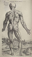
Vesalius's Fabrica contained many intricately detailed drawings of human dissections, often in allegorical poses.
 
In 1543, Vesalius asked Johannes Oporinus to publish the book De humani corporis fabrica (On the fabric of the human body), a groundbreaking work of human anatomy he dedicated to Charles V and which many believe was illustrated by Titian's pupil Jan Stephen van Calcar

About the same time he published another version of his great work, entitled De humani corporis fabrica librorum epitome (Abridgement of the Structure of the Human Body) more commonly known as the Epitome, with a stronger focus on illustrations than on text, so as to help readers, including medical students, to easily understand his findings. The actual text of the Epitome was an abridged form of his work in the Fabrica, and the organization of the two books was quite varied. He dedicated it to Philip II of Spain, son of the Emperor.

The Fabrica emphasized the priority of dissection and what has come to be called the "anatomical" view of the body, seeing human internal functioning as a result of an essentially corporeal structure filled with organs arranged in three-dimensional space. His book contains drawings of several organs on two leaves. This allows for the creation of three-dimensional diagrams by cutting out the organs and pasting them on flayed figures. This was in stark contrast to many of the anatomical models used previously, which had strong Galenic/Aristotelean elements, as well as elements of astrology. Although modern anatomical texts had been published by Mondino and Berenger, much of their work was clouded by reverence for Galen and Arabian doctrines. 

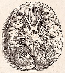
Besides the first good description of the sphenoid bone, he showed that the sternum consists of three portions and the sacrum of five or six, and described accurately the vestibule in the interior of the temporal bone. He not only verified Estienne's observations on the valves of the hepatic veins, but also described the vena azygos, and discovered the canal which passes in the fetus between the umbilical vein and the vena cava, since named the ductus venosus. He described the omentum and its connections with the stomach, the spleen and the colon; gave the first correct views of the structure of the pylorus; observed the small size of the caecal appendix in man; gave the first good account of the mediastinum and pleura and the fullest description of the anatomy of the brain up to that time. He did not understand the inferior recesses, and his account of the nerves is confused by regarding the optic as the first pair, the third as the fifth, and the fifth as the seventh. 

In this work, Vesalius also becomes the first person to describe mechanical ventilation. It is largely this achievement that has resulted in Vesalius being incorporated into the Australian and New Zealand College of Anaesthetists college arms and crest.

Excerpts

When I undertake the dissection of a human pelvis I pass a stout rope tied like a noose beneath the lower jaw and through the zygomas up to the top of the head... The lower end of the noose I run through a pulley fixed to a beam in the room so that I may raise or lower the cadaver as it hangs there or turn around in any direction to suit my purpose; ... You must take care not to put the noose around the neck, unless some of the muscles connected to the occipital bone have already been cut away.

Other publications

In 1538, Vesalius wrote Epistola, docens venam axillarem dextri cubiti in dolore laterali secandam (A letter, teaching that in cases of pain in the side, the axillary vein of the right elbow be cut), commonly known as the Venesection Letter, which demonstrated a revived venesection, a classical procedure in which blood was drawn near the site of the ailment. He sought to locate the precise site for venesection in pleurisy within the framework of the classical method. The real significance of the book is his attempt to support his arguments by the location and continuity of the venous system from his observations rather than appeal to earlier published works. With this novel approach to the problem of venesection, Vesalius posed the then striking hypothesis that anatomical dissection might be used to test speculation. 

In 1546, three years after the Fabrica, he wrote his Epistola rationem modumque propinandi radicis Chynae decocti, commonly known as the Epistle on the China Root. Ostensibly an appraisal of a popular but ineffective treatment for gout, syphilis, and stone, this work is especially important as a continued polemic against Galenism and a reply to critics in the camp of his former professor Jacobus Sylvius, now an obsessive detractor. 

In February 1561, Vesalius was given a copy of Gabriele Fallopio's Observationes anatomicae, friendly additions and corrections to the Fabrica. Before the end of the year Vesalius composed a cordial reply, Anatomicarum Gabrielis Fallopii observationum examen, generally referred to as the Examen. In this work he recognizes in Fallopio a true equal in the science of dissection he had done so much to create. Vesalius' reply to Fallopio was published in May 1564, a month after Vesalius' death on the Greek island of Zante (now called Zakynthos).

Scientific findings

Skeletal system

  • Vesalius believed the skeletal system to be the framework of the human body. It was in this opening chapter or book of De fabrica that Vesalius made several of his strongest claims against Galen's theories and writings which he had put in his anatomy books. In his extensive study of the skull, Vesalius claimed that the mandible consisted of one bone, whereas Galen had thought it to be two separate bones. He accurately described the vestibule in the interior of the temporal bone of the skull.
  • In Galen's observation of the ape, he had discovered that their sternum consisted of seven parts which he assumed also held true for humans. Vesalius discovered that the human sternum consisted of only three parts.
  • He also disproved the common belief that men had one rib fewer than women and noted that the fibula and tibia bones of the leg were indeed larger than the humerus bone of the arm, unlike Galen's original findings.

Muscular system

  • Vesalius' most impressive contribution to the study of the muscular system may be the illustrations that accompany the text in De fabrica, which would become known as the "muscle men". He describes the source and position of each muscle of the body and provides information on their respective operation.

Vascular and circulatory systems

  • Vesalius' work on the vascular and circulatory systems was his greatest contribution to modern medicine. In his dissections of the heart, Vesalius became convinced that Galen's claims of a porous Interventricular septum were false. This fact was previously described by Michael Servetus, a fellow of Vesalius, but never reached the public, for it was written down in the "Manuscript of Paris",[15] in 1546, and published later in his Christianismi Restitutio (1553), a book regarded as heretical by the Inquisition. Only three copies survived, but these remained hidden for decades, the rest having been burned shortly after publication. In the second edition Vesalius published that the septum was indeed waterproof, discovering (and naming), the mitral valve to explain the blood flow.
  • Vesalius believed that cardiac systole is synchronous with the arterial pulse.
  • He not only verified Estienne's findings on the valves of the hepatic veins, but also described the azygos vein, and discovered the canal which passes into the fetus between the umbilical vein and vena cava.

Nervous system

  • Vesalius defined a nerve as the mode of transmitting sensation and motion and thus refuted his contemporaries' claims that ligaments, tendons and aponeuroses were three types of nerve units.
  • He believed that the brain and the nervous system are the center of the mind and emotion in contrast to the common Aristotelian belief that the heart was the center of the body. He correspondingly believed that nerves themselves do not originate from the heart, but from the brain.
  • Upon studying the optic nerve, Vesalius came to the conclusion that nerves were not hollow.

Abdominal organs

  • In De fabrica, he corrected an earlier claim he made in Tabulae about the right kidney being set higher than the left. Vesalius claimed that the kidneys were not a filter device for urine to pass through, but rather that the kidneys serve to filter blood as well, and that excretions from the kidneys travelled through the ureters to the bladder.
  • He described the omentum, and its connections with the stomach, the spleen and the colon gave the first correct views of the structure of the pylorus.
  • He also observed the small size of the caecal appendix in man and gave the first good account of the mediastinum and pleura.
  • Vesalius admitted that due to a lack of pregnant cadavers he was unable to come to a significant understanding of the reproductive organs. However, he did find that the uterus had been falsely identified as having two distinct sections.

Heart

  • Through his work with muscles, Vesalius believed that a criterion for muscles was their voluntary motion. On this claim, he deduced that the heart was not a true muscle due to the obvious involuntary nature of its motion.
  • He identified two chambers and two atria. The right atrium was considered a continuation of the inferior and superior venae cavae, and the left atrium was considered a continuation of the pulmonary vein.
  • He also addressed the controversial issue of the heart being the centre of the soul. He wished to avoid drawing any conclusions due to possible conflict with contemporary religious beliefs.
  • Other achievements

    • Vesalius disproved Galen's assertion that men have more teeth than women.
    • Vesalius introduced the notion of induction of the extraction of empyema through surgical means.
    • Due to his impressive study of the human skull and the variations in its features he is said to have been responsible for the launch of the study of physical anthropology.
    • Vesalius always encouraged his students to check their findings, and even his own findings, so that they could better understand the structure of the human body.
    • In addition to his continual efforts to study anatomy he also worked on medicinal remedies and came to such conclusions as treating syphilis with chinaroot.
    • Vesalius claimed that medicine had three aspects: drugs, diet, and 'the use of hands'—mainly suggesting surgery and the knowledge of anatomy and physiology gained through dissection.
    • Vesalius was a supporter of 'parallel dissections' in which an animal cadaver and a human cadaver are dissected simultaneously in order to demonstrate the anatomical differences and thus correct Galenic errors.

    Scientific and historical impact

    The influence of Vesalius' plates representing the partial dissections of the human figure posing in a landscape setting is apparent in the anatomical plates prepared by the Baroque painter Pietro da Cortona (1596–1669), who executed anatomical plates with figures in dramatic poses, most of them with architectural or landscape backdrops.
    During the 20th century, the American artist, Jacob Lawrence created his Vesalius Suite based on the anatomical drawings of Andreas Vesalius.

    "De Fabrica received a mixed reception when it first appeared; strict Galenists deplored its attacks on their master, while other anatomists, particularly in Italy, praised it as an important contribution—the reaction that was ultimately to carry the day," concludes Katharine Park her Commentary in the 1998 De Fabrica.

    Vesalius was going up against the towering authority of a tradition stretching back to the ancients—here specifically the work of Galen — with only his experience on his side. He knew what his eyes saw and his hands felt, and concluded that traditional belief was wrong. In his publications we see Vesalius doing everything he can think of to bolster his authoritative image: publishing a huge monument to himself, but presenting the work using Galen’s own flowchart; presenting himself as personal physician to the emperor and having himself depicted in a commanding position on the title page of the book; augmenting his words with illustration after illustration and recommending the experiential road to all his students far and wide. His guarantee: if you doubt what I say and show here, do your own anatomy, see for yourself. Simultaneously, Vesalius’s work was part of one of the earliest known public health programs. The Council of Doges in Venice responded to the Bubonic Plague in the mid-14th century by directing the University of Padua Medical School to devote itself to discovering the causes of plague, how it spreads, how it develops in the individual, and if possible how victims might be cured. It ultimately took three centuries to find the solution, but with Venice leading the way plague was eventually eliminated as a major killer across Europe.

    Cognitive neuroscience

    From Wikipedia, the free encyclopedia

    Cognitive neuroscience is the scientific field that is concerned with the study of the biological processes and aspects that underlie cognition, with a specific focus on the neural connections in the brain which are involved in mental processes. It addresses the questions of how cognitive activities are affected or controlled by neural circuits in the brain. Cognitive neuroscience is a branch of both neuroscience and psychology, overlapping with disciplines such as behavioral neuroscience, cognitive psychology, physiological psychology and affective neuroscience. Cognitive neuroscience relies upon theories in cognitive science coupled with evidence from neurobiology, and computational modeling.

    Parts of the brain play an important role in this field. Neurons play the most vital role, since the main point is to establish an understanding of cognition from a neural perspective, along with the different lobes of the cerebral cortex.

    Methods employed in cognitive neuroscience include experimental procedures from psychophysics and cognitive psychology, functional neuroimaging, electrophysiology, cognitive genomics, and behavioral genetics.

    Studies of patients with cognitive deficits due to brain lesions constitute an important aspect of cognitive neuroscience. The damages in lesioned brains provide a comparable basis with regards to healthy and fully functioning brains. These damages change the neural circuits in the brain and cause it to malfunction during basic cognitive processes, such as memory or learning. With the damage, we can compare how the healthy neural circuits are functioning, and possibly draw conclusions about the basis of the affected cognitive processes.

    Also, cognitive abilities based on brain development are studied and examined under the subfield of developmental cognitive neuroscience. This shows brain development over time, analyzing differences and concocting possible reasons for those differences.

    Theoretical approaches include computational neuroscience and cognitive psychology.

    Historical origins

    Timeline of development of field of cognitive neuroscience
    Timeline showing major developments in science that led to the emergence of the field cognitive neuroscience.
     
    Cognitive neuroscience is an interdisciplinary area of study that has emerged from neuroscience and psychology. There were several stages in these disciplines that changed the way researchers approached their investigations and that led to the field becoming fully established. 

    Although the task of cognitive neuroscience is to describe how the brain creates the mind, historically it has progressed by investigating how a certain area of the brain supports a given mental faculty. However, early efforts to subdivide the brain proved to be problematic. The phrenologist movement failed to supply a scientific basis for its theories and has since been rejected. The aggregate field view, meaning that all areas of the brain participated in all behavior, was also rejected as a result of brain mapping, which began with Hitzig and Fritsch’s experiments and eventually developed through methods such as positron emission tomography (PET) and functional magnetic resonance imaging (fMRI). Gestalt theory, neuropsychology, and the cognitive revolution were major turning points in the creation of cognitive neuroscience as a field, bringing together ideas and techniques that enabled researchers to make more links between behavior and its neural substrates.

    Origins in philosophy

    Philosophers have always been interested in the mind: "the idea that explaining a phenomenon involves understanding the mechanism responsible for it has deep roots in the History of Philosophy from atomic theories in 5th century B.C. to its rebirth in the 17th and 18th century in the works of Galileo, Descartes, and Boyle. Among others, it’s Descartes’ idea that machines humans build could work as models of scientific explanation." For example, Aristotle thought the brain was the body’s cooling system and the capacity for intelligence was located in the heart. It has been suggested that the first person to believe otherwise was the Roman physician Galen in the second century AD, who declared that the brain was the source of mental activity, although this has also been accredited to Alcmaeon. However, Galen believed that personality and emotion were not generated by the brain, but rather by other organs. Andreas Vesalius, an anatomist and physician, was the first to believe that the brain and the nervous system are the center of the mind and emotion. Psychology, a major contributing field to cognitive neuroscience, emerged from philosophical reasoning about the mind.

    19th century

    Phrenology

    A page from the American Phrenological Journal

    One of the predecessors to cognitive neuroscience was phrenology, a pseudoscientific approach that claimed that behavior could be determined by the shape of the scalp. In the early 19th century, Franz Joseph Gall and J. G. Spurzheim believed that the human brain was localized into approximately 35 different sections. In his book, The Anatomy and Physiology of the Nervous System in General, and of the Brain in Particular, Gall claimed that a larger bump in one of these areas meant that that area of the brain was used more frequently by that person. This theory gained significant public attention, leading to the publication of phrenology journals and the creation of phrenometers, which measured the bumps on a human subject's head. While phrenology remained a fixture at fairs and carnivals, it did not enjoy wide acceptance within the scientific community. The major criticism of phrenology is that researchers were not able to test theories empirically.

    Localizationist view

    The localizationist view was concerned with mental abilities being localized to specific areas of the brain rather than on what the characteristics of the abilities were and how to measure them. Studies performed in Europe, such as those of John Hughlings Jackson, supported this view. Jackson studied patients with brain damage, particularly those with epilepsy. He discovered that the epileptic patients often made the same clonic and tonic movements of muscle during their seizures, leading Jackson to believe that they must be occurring in the same place every time. Jackson proposed that specific functions were localized to specific areas of the brain, which was critical to future understanding of the brain lobes.

    Aggregate field view

    According to the aggregate field view, all areas of the brain participate in every mental function.

    Pierre Flourens, a French experimental psychologist, challenged the localizationist view by using animal experiments. He discovered that removing the cerebellum in rabbits and pigeons affected their sense of muscular coordination, and that all cognitive functions were disrupted in pigeons when the cerebral hemispheres were removed. From this he concluded that the cerebral cortex, cerebellum, and brainstem functioned together as a whole. His approach has been criticised on the basis that the tests were not sensitive enough to notice selective deficits had they been present.

    Emergence of neuropsychology

    Perhaps the first serious attempts to localize mental functions to specific locations in the brain was by Broca and Wernicke. This was mostly achieved by studying the effects of injuries to different parts of the brain on psychological functions. In 1861, French neurologist Paul Broca came across a man who was able to understand language but unable to speak. The man could only produce the sound "tan". It was later discovered that the man had damage to an area of his left frontal lobe now known as Broca's area. Carl Wernicke, a German neurologist, found a patient who could speak fluently but non-sensibly. The patient had been the victim of a stroke, and could not understand spoken or written language. This patient had a lesion in the area where the left parietal and temporal lobes meet, now known as Wernicke's area. These cases, which suggested that lesions caused specific behavioral changes, strongly supported the localizationist view.

    Mapping the brain

    In 1870, German physicians Eduard Hitzig and Gustav Fritsch published their findings about the behavior of animals. Hitzig and Fritsch ran an electric current through the cerebral cortex of a dog, causing different muscles to contract depending on which areas of the brain were electrically stimulated. This led to the proposition that individual functions are localized to specific areas of the brain rather than the cerebrum as a whole, as the aggregate field view suggests. Brodmann was also an important figure in brain mapping; his experiments based on Franz Nissl’s tissue staining techniques divided the brain into fifty-two areas.

    20th century

    Cognitive revolution

    At the start of the 20th century, attitudes in America were characterised by pragmatism, which led to a preference for behaviorism as the primary approach in psychology. J.B. Watson was a key figure with his stimulus-response approach. By conducting experiments on animals he was aiming to be able to predict and control behaviour. Behaviourism eventually failed because it could not provide realistic psychology of human action and thought – it focused primarily on stimulus-response associations at the expense of explaining phenomena like thought and imagination. This led to what is often termed as the "cognitive revolution".

    Neuron doctrine

    In the early 20th century, Santiago Ramón y Cajal and Camillo Golgi began working on the structure of the neuron. Golgi developed a silver staining method that could entirely stain several cells in a particular area, leading him to believe that neurons were directly connected with each other in one cytoplasm. Cajal challenged this view after staining areas of the brain that had less myelin and discovering that neurons were discrete cells. Cajal also discovered that cells transmit electrical signals down the neuron in one direction only. Both Golgi and Cajal were awarded a Nobel Prize in Physiology or Medicine in 1906 for this work on the neuron doctrine.

    Mid-late 20th century

    Several findings in the 20th century continued to advance the field, such as the discovery of ocular dominance columns, recording of single nerve cells in animals, and coordination of eye and head movements. Experimental psychology was also significant in the foundation of cognitive neuroscience. Some particularly important results were the demonstration that some tasks are accomplished via discrete processing stages, the study of attention, and the notion that behavioural data do not provide enough information by themselves to explain mental processes. As a result, some experimental psychologists began to investigate neural bases of behaviour. Wilder Penfield created maps of primary sensory and motor areas of the brain by stimulating cortices of patients during surgery. The work of Sperry and Michael Gazzaniga on split brain patients in the 1950s was also instrumental in the progress of the field. The term cognitive neuroscience itself was coined by Gazzaniga and cognitive psychologist George Armitage Miller while sharing a taxi in 1976.

    Brain mapping

    New brain mapping technology, particularly fMRI and PET, allowed researchers to investigate experimental strategies of cognitive psychology by observing brain function. Although this is often thought of as a new method (most of the technology is relatively recent), the underlying principle goes back as far as 1878 when blood flow was first associated with brain function. Angelo Mosso, an Italian psychologist of the 19th century, had monitored the pulsations of the adult brain through neurosurgically created bony defects in the skulls of patients. He noted that when the subjects engaged in tasks such as mathematical calculations the pulsations of the brain increased locally. Such observations led Mosso to conclude that blood flow of the brain followed function.

    Emergence of a new discipline

    Birth of cognitive science

    On September 11, 1956, a large-scale meeting of cognitivists took place at the Massachusetts Institute of Technology. George A. Miller presented his "The Magical Number Seven, Plus or Minus Two" paper while Noam Chomsky and Newell & Simon presented their findings on computer science. Ulric Neisser commented on many of the findings at this meeting in his 1967 book Cognitive Psychology. The term "psychology" had been waning in the 1950s and 1960s, causing the field to be referred to as "cognitive science". Behaviorists such as Miller began to focus on the representation of language rather than general behavior. David Marr concluded that one should understand any cognitive process at three levels of analysis. These levels include computational, algorithmic/representational, and physical levels of analysis.

    Combining neuroscience and cognitive science

    Before the 1980s, interaction between neuroscience and cognitive science was scarce. Cognitive neuroscience began to integrate the newly laid theoretical ground in cognitive science, that emerged between the 1950s and 1960s, with approaches in experimental psychology, neuropsychology and neuroscience. (Neuroscience was not established as a unified discipline until 1971). In the very late 20th century new technologies evolved that are now the mainstay of the methodology of cognitive neuroscience, including TMS (1985) and fMRI (1991). Earlier methods used in cognitive neuroscience include EEG (human EEG 1920) and MEG (1968). Occasionally cognitive neuroscientists utilize other brain imaging methods such as PET and SPECT. An upcoming technique in neuroscience is NIRS which uses light absorption to calculate changes in oxy- and deoxyhemoglobin in cortical areas. In some animals Single-unit recording can be used. Other methods include microneurography, facial EMG, and eye tracking. Integrative neuroscience attempts to consolidate data in databases, and form unified descriptive models from various fields and scales: biology, psychology, anatomy, and clinical practice. In 2014, Stanislas Dehaene, Giacomo Rizzolatti and Trevor Robbins, were awarded the Brain Prize "for their pioneering research on higher brain mechanisms underpinning such complex human functions as literacy, numeracy, motivated behaviour and social cognition, and for their efforts to understand cognitive and behavioural disorders". Brenda Milner, Marcus Raichle and John O'Keefe received the Kavli Prize in Neuroscience “for the discovery of specialized brain networks for memory and cognition" and O'Keefe shared the Nobel Prize in Physiology or Medicine in the same year with May-Britt Moser and Edvard Moser "for their discoveries of cells that constitute a positioning system in the brain". In 2017, Wolfram Schultz, Peter Dayan and Ray Dolan were awarded the Brain Prize "for their multidisciplinary analysis of brain mechanisms that link learning to reward, which has far-reaching implications for the understanding of human behaviour, including disorders of decision-making in conditions such as gambling, drug addiction, compulsive behaviour and schizophrenia".

    Major contributors to the field

    Hubel and Wiesel – 1960s

    David H. Hubel and Torsten Wiesel, both neurophysiologists, studied the visual system in cats to better understand sensory processing. They performed experiments which demonstrated the specificity of the responding of neurons. Their experiments showed that neurons fired rapidly at some angles, and not so much at others. A difference was also found in light and dark settings. Their studies gave rise to the idea of complex visual representations being formed from relatively simple stimuli. 

    They also discovered the simple cell and complex cell. These exist in the primary visual cortex and respond differentially to differently oriented presentations of light.

    Recent trends

    Recently the foci of research have expanded from the localization of brain area(s) for specific functions in the adult brain using a single technology, studies have been diverging in several different directions: exploring the interactions between different brain areas, using multiple technologies and approaches to understand brain functions, and using computational approaches. Advances in non-invasive functional neuroimaging and associated data analysis methods have also made it possible to use highly naturalistic stimuli and tasks such as feature films depicting social interactions in cognitive neuroscience studies.

    Atmospheric escape

    From Wikipedia, the free encyclopedia
     
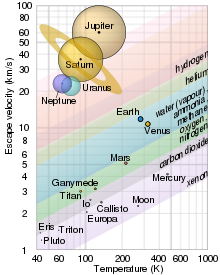
    Graphs of escape velocity against surface temperature of some Solar System objects showing which gases are retained. The objects are drawn to scale, and their data points are at the black dots in the middle.
     
    Atmospheric escape is the loss of planetary atmospheric gases to outer space. A number of different mechanisms can be responsible for atmospheric escape; these processes can be divided into thermal escape, non-thermal (or suprathermal) escape, and impact erosion. The relative importance of each loss process depends on the planet's escape velocity, its atmosphere composition, and its distance from its sun. Escape occurs when molecular kinetic energy overcomes gravitational energy; in other words, a molecule can escape when it is moving faster than the escape velocity of its planet. Categorizing the rate of atmospheric escape in exoplanets is important to determining whether an atmosphere persists, and so the exoplanet's habitability and likelihood of life.

    Thermal escape mechanisms

    Thermal escape occurs if the molecular velocity due to thermal energy is sufficiently high. Thermal escape happens at all scales, from the molecular level (Jeans escape) to bulk atmospheric outflow (hydrodynamic escape). 

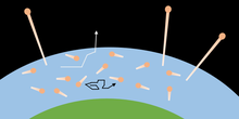
    A visualization of Jeans escape. Temperature defines a range of molecular energy. Above the exobase, molecules with enough energy escape, while in the lower atmosphere, molecules are trapped by collisions with other molecules.

    Jeans Escape

    One classical thermal escape mechanism is Jeans escape, named after British astronomer Sir James Jeans, who first described this process of atmospheric loss. In a quantity of gas, the average velocity of any one molecule is measured by the gas's temperature, but the velocities of individual molecules change as they collide with one another, gaining and losing kinetic energy. The variation in kinetic energy among the molecules is described by the Maxwell distribution. The kinetic energy (), mass (), and velocity () of a molecule are related by . Individual molecules in the high tail of the distribution (where a few particles have much higher speeds than the average) may reach escape velocity and leave the atmosphere, provided they can escape before undergoing another collision; this happens predominantly in the exosphere, where the mean free path is comparable in length to the pressure scale height. The number of particles able to escape depends on the molecular concentration at the exobase, which is limited by diffusion through the thermosphere

    Three factors strongly contribute to the relative importance of Jeans escape: mass of the molecule, escape velocity of the planet, and heating of the upper atmosphere by radiation from the parent star. Heavier molecules are less likely to escape because they move slower than lighter molecules at the same temperature. This is why hydrogen escapes from an atmosphere more easily than carbon dioxide. Second, a planet with a larger mass has more gravity, so the escape velocity is greater, and fewer particles will gain the energy required to escape. This is why the gas giant planets still retain significant amounts of hydrogen, which escape more readily from Earth's atmosphere. Finally, the distance a planet orbits from a star also plays a part; a close planet has a hotter atmosphere, with higher velocities and hence, a greater likelihood of escape. A distant body has a cooler atmosphere, with lower velocities, and less chance of escape. 

    A visualization of hydrodynamic escape. At some level in the atmosphere, the bulk gas will be heated and begin to expand. As the gas expands, it accelerates and escapes the atmosphere. In this process, lighter, faster molecules drag heavier, slower molecules out of the atmosphere.

    Hydrodynamic escape

    An atmosphere with high pressure and temperature can also undergo hydrodynamic escape. In this case, a large amount of thermal energy, usually through extreme ultraviolet radiation, is absorbed by the atmosphere. As molecules are heated, they expand upwards and are further accelerated until they reach escape velocity. In this process, lighter molecules can drag heavier molecules with them through collisions as a larger quantity of gas escapes. Hydrodynamic escape has been observed for exoplanets close to their host star, including the hot Jupiter HD 209458b.

    Non-thermal (suprathermal) escape

    Escape can also occur due to non-thermal interactions. Most of these processes occur due to photochemistry or charged particle (ion) interactions.

    Photochemical escape

    In the upper atmosphere, high energy ultraviolet photons can react more readily with molecules. Photodissociation can break a molecule into smaller components and provide enough energy for those components to escape. Photoionization produces ions, which can get trapped in the planet's magnetosphere or undergo dissociative recombination. In the first case, these ions may undergo escape mechanisms described below. In the second case, the ion recombines with an electron, releases energy, and can escape.

    Sputtering escape

    Excess kinetic energy from the solar wind can impart sufficient energy to eject atmospheric particles, similar to sputtering from a solid surface. This type of interaction is more pronounced in the absence of a planetary magnetosphere, as the electrically charged solar wind is deflected by magnetic fields, which mitigates the loss of atmosphere.

    The fast ion captures an electron from a slow neutral in a charge exchange collision. The new, fast neutral can escape the atmosphere, and the new, slow ion is trapped on magnetic field lines.

    Charge exchange escape

    Ions in the solar wind or magnetosphere can charge exchange with molecules in the upper atmosphere. A fast-moving ion can capture the electron from a slow atmospheric neutral, creating a fast neutral and a slow ion. The slow ion is trapped on the magnetic field lines, but the fast neutral can escape.

    Polar wind escape

    Atmospheric molecules can also escape from the polar regions on a planet with a magnetosphere, due to the polar wind. Near the poles of a magnetosphere, the magnetic field lines are open, allowing a pathway for ions in the atmosphere to exhaust into space.

    Atmospheric escape from impact erosion is concentrated in a cone (red dash-dotted line) centered at the impact site. The angle of this cone increases with impact energy to eject a maximum of all the atmosphere above a tangent plane (orange dotted line).

    Impact erosion

    The impact of a large meteoroid can lead to the loss of atmosphere. If a collision is sufficiently energetic, it is possible for ejecta, including atmospheric molecules, to reach escape velocity. 

    In order to have a significant effect on atmospheric escape, the radius of the impacting body must be larger than the scale height. The projectile can impart momentum, and thereby facilitate escape of the atmosphere, in three main ways: (a) the meteroid heats and accelerates the gas it encounters as it travels through the atmosphere, (b) solid ejecta from the impact crater heat atmospheric particles through drag as they are ejected, and (c) the impact creates vapor which expands away from the surface. In the first case, the heated gas can escape in a manner similar to hydrodynamic escape, albeit on a more localized scale. Most of the escape from impact erosion occurs due to the third case. The maximum atmosphere that can be ejected is above a plane tangent to the impact site.

    Dominant atmospheric escape and loss processes in the Solar System

    Earth

    Atmospheric escape of hydrogen on Earth is due to Jeans escape (~10 - 40%), charge exchange escape (~ 60 - 90%), and polar wind escape (~ 10 - 15%), currently losing about 3 kg/s of hydrogen. The Earth additionally loses approximately 50 g/s of helium primarily through polar wind escape. Escape of other atmospheric constituents is much smaller. A Japanese research team in 2017 found evidence of a small number of oxygen ions on the moon that came from the Earth.

    In 1 billion years, the Sun will be 10% brighter than it is now, making it hot enough for Earth to lose enough hydrogen to space to cause it to lose all of its water.

    Venus

    Recent models indicate that hydrogen escape on Venus is almost entirely due to suprathermal mechanisms, primarily photochemical reactions and charge exchange with the solar wind. Oxygen escape is dominated by charge exchange and sputtering escape. Venus Express measured the effect of coronal mass ejections on the rate of atmospheric escape of Venus, and researchers found a factor of 1.9 increase in escape rate during periods of increased coronal mass ejections compared with calmer space weather.

    Mars

    Primordial Mars also suffered from the cumulative effects of multiple small impact erosion events, and recent observations with MAVEN suggest that 66% of the 36Ar in the Martian atmosphere has been lost over the last 4 billion years due to suprathermal escape, and the amount of CO2 lost over the same time period is around 0.5 bar or more.

    The MAVEN mission has also explored the current rate of atmospheric escape of Mars. Jeans escape plays an important role in the continued escape of hydrogen on Mars, contributing to a loss rate that varies between 160 - 1800 g/s. Oxygen loss is dominated by suprathermal methods: photochemical (~ 1300 g/s), charge exchange (~ 130 g/s), and sputtering (~ 80 g/s) escape combine for a total loss rate of ~ 1500 g/s. Other heavy atoms, such as carbon and nitrogen, are primarily lost due to photochemical reactions and interactions with the solar wind.

    Titan and Io

    Saturn's moon Titan and Jupiter's moon Io have atmospheres and are subject to atmospheric loss processes. They have no magnetic fields of their own, but orbit planets with powerful magnetic fields, which protects these moons from the solar wind when its orbit is within the bow shock. However Titan spends roughly half of its transit time outside of the bow-shock, subjected to unimpeded solar winds. The kinetic energy gained from pick-up and sputtering associated with the solar winds increases thermal escape throughout the transit of Titan, causing neutral hydrogen to escape. The escaped hydrogen maintains an orbit following in the wake of Titan, creating a neutral hydrogen torus around Saturn. Io, in its transit around Jupiter, encounters a plasma cloud. Interaction with the plasma cloud induces sputtering, kicking off sodium particles. The interaction produces a stationary banana-shaped charged sodium cloud along a part of the orbit of Io.

    Observations of exoplanet atmospheric escape

    Studies of exoplanets have measured atmospheric escape as a means of determining atmospheric composition and habitability. The most common method is Lyman-alpha line absorption. Much as exoplanets are discovered using the dimming of a distant star's brightness (transit), looking specifically at wavelengths corresponding to hydrogen absorption describes the amount of hydrogen present in a sphere around the exoplanet. This method indicates that the hot Jupiters HD209458b and HD189733b and Hot Neptune GJ436b are experiencing significant atmospheric escape.

    Other atmospheric loss mechanisms

    Sequestration is not a form of escape from the planet, but a loss of molecules from the atmosphere and into the planet. It occurs on Earth when water vapor condenses to form rain or glacial ice, when carbon dioxide is sequestered in sediments or cycled through the oceans, or when rocks are oxidized (for example, by increasing the oxidation states of ferric rocks from Fe2+ to Fe3+). Gases can also be sequestered by adsorption, where fine particles in the regolith capture gas which adheres to the surface particles.

    Operator (computer programming)

    From Wikipedia, the free encyclopedia https://en.wikipedia.org/wiki/Operator_(computer_programmin...