Given
that many fundamental questions in neuroscience are still open, it
seems pertinent to explore whether the brain might use other physical
modalities than the ones that have been discovered so far. In particular
it is well established that neurons can emit photons, which prompts the
question whether these biophotons could serve as signals between
neurons, in addition to the well-known electro-chemical signals. For
such communication to be targeted, the photons would need to travel in
waveguides. Here we show, based on detailed theoretical modeling, that
myelinated axons could serve as photonic waveguides, taking into account
realistic optical imperfections. We propose experiments, both in vivo and in vitro,
to test our hypothesis. We discuss the implications of our results,
including the question whether photons could mediate long-range quantum
entanglement in the brain.
Introduction
The
human brain is a dynamic physical system of unparalleled complexity.
While neuroscience has made great strides, many fundamental questions
are still unanswered, including the processes underlying memory formation, the working principle of anesthesia, and–most fundamentally–the generation of conscious experience.
It therefore seems pertinent to explore whether the brain might
generate, transmit and store information using other physical modalities
than the ones that have been discovered so far.
In the present work we focus on the question whether biophotons could serve as a supplementary information
carrier in the brain in addition to the well established
electro-chemical signals. Biophotons are the quanta of light spanning
the near-UV to near-IR frequency range. They are produced mostly by
electronically excited molecular species in a variety of oxidative
metabolic processes in cells. They may play a role in cell to cell communication, and have been observed in many organisms, including humans, and in different parts of the body, including the brain.
Photons in the brain could serve as ideal candidates for information
transfer. They travel tens of millions of times faster than a typical
electrical neural signal and are not prone to thermal noise at body
temperature owing to their relatively high energies. It is conceivable
that evolution might have found a way to utilize these precious
high-energy resources for information transfer, even if they were just
the by–products of metabolism to begin with. Most of the required
molecular machinery seems to exist in living cells such as neurons. Mitochondrial respiration or lipid oxidation could serve as sources, and centrosomes or chromophores in the mitochondria could serve as detectors.
However,
one crucial element for optical communication is not well established,
namely the existence of physical links to connect all of these spatially
separated agents in a selective way. The only viable way to achieve
targeted optical communication in the dense and (seemingly) disordered
brain environment is for the photons to travel in waveguides.
Mitochondria and microtubules in neurons have been hypothesized to serve
as waveguides. However, these structures are too small and inhomogeneous to guide light efficiently over significant distances.
Here
we propose myelinated axons as potential biophoton waveguides in the
brain, and we support this hypothesis with detailed theoretical
modeling. These axons are tightly wrapped by a lamellar structure called
the myelin sheath, which has a higher refractive index than both the inside of the axon and the interstitial fluid outside (see Fig. 1a).
This compact sheath could therefore also serve as a waveguide, in
addition to increasing the propagation speed of an action potential (via
saltatory conduction) based on its insulating property. There is some indirect experimental evidence for light conduction by axons,
including the observation of increased transmission along the axes of
the white matter tracts, which consist of myelinated axons.
Myelin is formed in the central nervous system (CNS) by a kind of glia
cell called oligodendrocyte. Interestingly, certain glia cells, known as
Müller cells, have been shown to guide light in mammalian eyes.
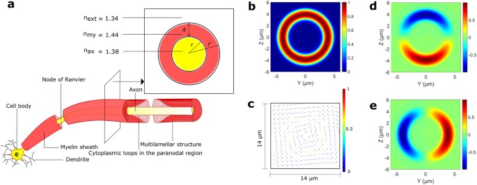
Figure 1
3-D schematic representation of a segment of a neuron, and an eigenmode of a cylindrical myelinated axon.
(a)
Different parts of a segment of a neuron whose myelinated axon is
sliced longitudinally near the end of the segment. The inset depicts the
cross section in the transverse plane. Here r and r′ are the inner and outer radii of the myelin sheath, d is the thickness of the myelin sheath, and nmy, nax, and next
are the refractive indices of the myelin sheath, the inside of the
axon, and the interstitial fluid outside respectively. The compact
myelin (shown in red) terminates in the paranodal region near the Node
of Ranvier, with each closely apposed layer of myelin ending in a
cytoplasm filled loop (shown in light red). (b) Magnitude of the electric field of a cylindrically symmetric eigenmode (λ = 0.612 μm) of a (cylindrical) myelinated axon, with r = 3 μm, and r′ = 5 μm. (c)
A vector plot of the electric field showing the azimuthal polarization
of the input mode. For clarity in the depiction of the direction of the
field at different points, the arrow length is renormalised to the same
value everywhere. The adjacent color bar depicts the actual field
magnitude. (d,e) Electric field components along the Y (Ey), and Z axes (Ez) respectively.
An
interesting feature of photonic communication channels is that they can
transmit quantum information as well. The potential role of quantum
effects in biological systems is currently being investigated in several
areas, including olfaction, avian magnetoreception, and photosynthesis.
There is also growing speculation about the role of fundamental quantum
features such as superposition and entanglement in certain higher level
brain functions.
Of particular relevance is the “binding problem” of consciousness,
which questions how a single integrated experience arises from the
activities of individual molecules in billions of neurons. The answer to
this question might be provided by quantum entanglement, where the whole is more than the sum of its parts in a well-defined physical and mathematical sense.
The
main challenge in envisioning a “quantum brain” is environmental
decoherence, which destroys quantum effects very rapidly at room
temperature for most physical degrees of freedom. However, nuclear spins can have coherence times of tens of milliseconds in the brain, and much longer times are imaginable. Long-lived nuclear spin entanglement has also been demonstrated in other condensed-matter systems at room temperature. A recent proposal on “quantum cognition”
is based on nuclear spins, but relies on the physical transport of
molecules to carry quantum information, which is very slow. In contrast,
photons are well suited for transmitting quantum information over long
distances, which is why currently envisioned man-made quantum networks
rely on optical communication channels (typically optical fibers)
between spins.
Efficient
light guidance therefore seems necessary for both classical and quantum
optical networks in the brain. Is this possible in myelinated axons
with all their “imperfections” from a waveguide perspective? In an
attempt to answer this question, we have developed a detailed
theoretical model of light guidance in axons. We show in the next
section that the answer seems to be in the affirmative.