Search This Blog

Thursday, April 16, 2015

Raman spectroscopy


From Wikipedia, the free encyclopedia


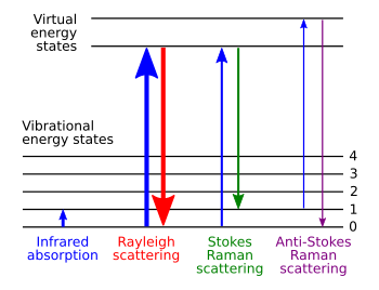
Energy-level diagram showing the states involved in Raman signal. The line thickness is roughly proportional to the signal strength from the different transitions.

Raman spectroscopy (/ˈrɑːmən/; named after Sir C. V. Raman) is a spectroscopic technique used to observe vibrational, rotational, and other low-frequency modes in a system.[1] Raman spectroscopy is commonly used in chemistry to provide a fingerprint by which molecules can be identified.

It relies on inelastic scattering, or Raman scattering, of monochromatic light, usually from a laser in the visible, near infrared, or near ultraviolet range. The laser light interacts with molecular vibrations, phonons or other excitations in the system, resulting in the energy of the laser photons being shifted up or down. The shift in energy gives information about the vibrational modes in the system. Infrared spectroscopy yields similar, but complementary, information.

Typically, a sample is illuminated with a laser beam. Electromagnetic radiation from the illuminated spot is collected with a lens and sent through a monochromator. Elastic scattered radiation at the wavelength corresponding to the laser line (Rayleigh scattering) is filtered out, while the rest of the collected light is dispersed onto a detector by either a notch filter or a band pass filter.

Spontaneous Raman scattering is typically very weak, and as a result the main difficulty of Raman spectroscopy is separating the weak inelastically scattered light from the intense Rayleigh scattered laser light. Historically, Raman spectrometers used holographic gratings and multiple dispersion stages to achieve a high degree of laser rejection. In the past, photomultipliers were the detectors of choice for dispersive Raman setups, which resulted in long acquisition times. However, modern instrumentation almost universally employs notch or edge filters for laser rejection and spectrographs either axial transmissive (AT), Czerny–Turner (CT) monochromator, or FT (Fourier transform spectroscopy based), and CCD detectors.

There are a number of advanced types of Raman spectroscopy, including surface-enhanced Raman, resonance Raman, tip-enhanced Raman, polarised Raman, stimulated Raman (analogous to stimulated emission), transmission Raman, spatially offset Raman, and hyper Raman.

Theoretical basis

The Raman effect occurs when electromagnetic radiation impinges on a molecule and interacts with the polarizable electron density and the bonds of the molecule in the phase (solid, liquid or gaseous) and environment in which the molecule finds itself. For the spontaneous Raman effect, which is a form of inelastic light scattering, a photon (electromagnetic radiation of a specific wavelength) excites (interacts with) the molecule in either the ground rovibronic state (lowest rotational and vibrational energy level of the ground electronic state) or an excited rovibronic state. This results in the molecule being in a so-called virtual energy state for a short period of time before an inelastically scattered photon results. The resulting inelastically scattered photon which is "emitted"/"scattered" can be of either lower (Stokes) or higher (anti-Stokes) energy than the incoming photon. In Raman scattering the resulting rovibronic state of the molecule is a different rotational or vibrational state than the one in which the molecule was originally, before interacting with the incoming photon (electromagnetic radiation). The difference in energy between the original rovibronic state and this resulting rovibronic state leads to a shift in the emitted photon's frequency away from the excitation wavelength, the so-called Rayleigh line. The Raman effect is due to inelastic scattering and should not be confused with emission (fluorescence or phosphorescence) where a molecule in an excited electronic state emits a photon of energy and returns to the ground electronic state, in many cases to a vibrationally excited state on the ground electronic state potential energy surface.

If the final vibrational state of the molecule is more energetic than the initial state, the inelastically scattered photon will be shifted to a lower frequency for the total energy of the system to remain balanced. This shift in frequency is designated as a Stokes shift. If the final vibrational state is less energetic than the initial state, then the inelastically scattered photon will be shifted to a higher frequency, and this is designated as an anti-Stokes shift. Raman scattering is an example of inelastic scattering because of the energy and momentum transfer between the photons and the molecules during the interaction. Rayleigh scattering is an example of elastic scattering, the energy of the scattered Rayleigh scattering is of the same frequency (wavelength) as the incoming electromagnetic radiation.

A change in the molecular electric dipole-electric polarizability with respect to the vibrational coordinate corresponding to the rovibronic state is required for a molecule to exhibit a Raman effect. The intensity of the Raman scattering is proportional to the electric dipole-electric dipole polarizability change. The Raman spectra (Raman scattering intensity as a function of the Stokes and anti-Stokes frequency shifts) is dependent on the rovibronic (rotational and vibrational energy levels of the ground electronic state) states of the sample. This dependence on the electric dipole-electric dipole polarizability derivative differs from infrared spectroscopy where the interaction between the molecule and light is determined by the electric dipole moment derivative, the so-called atomic polar tensor (APT); this contrasting feature allows one to analyze transitions that might not be IR active via Raman spectroscopy, as exemplified by the rule of mutual exclusion in centrosymmetric molecules. Bands which have large Raman intensities in many cases have weak infrared intensities and vice versa. For very symmetric molecules, certain vibrations may be both infrared and Raman inactive (within the harmonic approximation). In those instances, one can use a technique called inelastic incoherent neutron scattering to determine the vibrational frequencies. The selection rules for inelastic incoherent neutron scattering (IINS) are different from those of both infrared and Raman scattering. Hence the three types of vibrational spectroscopy are complementary, all giving in theory the same frequency for a given vibrational transion, but the relative intensities giving different information due to the types of interaction between the molecule and the electromagnetic radiation for infrared and Raman spectroscopy and with the neutron beam for IINS.

History

Although the inelastic scattering of light was predicted by Adolf Smekal in 1923,[2] it was not until 1928 that it was observed in practice. The Raman effect was named after one of its discoverers, the Indian scientist Sir C. V. Raman who observed the effect by means of sunlight (1928, together with K. S. Krishnan and independently by Grigory Landsberg and Leonid Mandelstam).[1] Raman won the Nobel Prize in Physics in 1930 for this discovery accomplished using sunlight, a narrow band photographic filter to create monochromatic light, and a "crossed filter" to block this monochromatic light. He found that a small amount of light had changed frequency and passed through the "crossed" filter.

Systematic pioneering theory of the Raman effect was developed by Czechoslovak physicist George Placzek between 1930 and 1934.[3] The mercury arc became the principal light source, first with photographic detection and then with spectrophotometric detection.

In the years following its discovery, Raman spectroscopy was used to provide the first catalog of molecular vibrational frequencies. Originally, heroic measures were required to obtain Raman spectra due to the low sensitivity of the technique. Typically, the sample was held in a long tube and illuminated along its length with a beam of filtered monochromatic light generated by a gas discharge lamp. The photons that were scattered by the sample were collected through an optical flat at the end of the tube. To maximize the sensitivity, the sample was highly concentrated (1 M or more) and relatively large volumes (5 mL or more) were used. Consequently, the use of Raman spectroscopy dwindled when commercial IR spectrophotometers became available in the 1940s. However, the advent of the laser in the 1960s resulted in simplified Raman spectroscopy instruments and also boosted the sensitivity of the technique. This has revived the use of Raman spectroscopy as a common analytical technique.

Raman shift

Raman shifts are typically reported in wavenumbers, which have units of inverse length, as this value is directly related to energy. In order to convert between spectral wavelength and wavenumbers of shift in the Raman spectrum, the following formula can be used:
\Delta w = \left( \frac{1}{\lambda_0} - \frac{1}{\lambda_1} \right) \ ,
where \Delta w is the Raman shift expressed in wavenumber, λ0 is the excitation wavelength, and λ1 is the Raman spectrum wavelength. Most commonly, the unit chosen for expressing wavenumber in Raman spectra is inverse centimeters (cm−1). Since wavelength is often expressed in units of nanometers (nm), the formula above can scale for this unit conversion explicitly, giving
\Delta w (\text{cm}^{-1}) = \left( \frac{1}{\lambda_0 (\text{nm})} - \frac{1}{\lambda_1 (\text{nm})} \right) \times \frac{(10^{7}\text{nm})}{(\text{cm})} .

Applications

Raman spectroscopy is commonly used in chemistry, since vibrational information is specific to the chemical bonds and symmetry of molecules. Therefore, it provides a fingerprint by which the molecule can be identified. For instance, the vibrational frequencies of SiO, Si2O2, and Si3O3 were identified and assigned on the basis of normal coordinate analyses using infrared and Raman spectra.[4] The fingerprint region of organic molecules is in the (wavenumber) range 500–2000 cm−1. Another way that the technique is used is to study changes in chemical bonding, as when a substrate is added to an enzyme.

Raman gas analyzers have many practical applications. For instance, they are used in medicine for real-time monitoring of anesthetic and respiratory gas mixtures during surgery.

In solid state chemistry and the bio-pharmaceutical industry, Raman spectroscopy can be used to not only identify (ID) active pharmaceutical ingredients (APIs), but in the case of multiple polymorphic forms, it can also be used to identify the polymorphic form of the API. For example there are 4 different polymorphic forms of the API (aztreonam) in Cayston, a drug marketed by Gilead Sciences for cystic fibrosis[citation needed]. Both infrared and Raman spectroscopy can be used to identify and characterize the API which is used in the formulation of Cayston.
In bio-pharmaceutical formulations, one must use not only the correct molecule, but the correct polymorphic form, as different polymorphic forms have different physical properties, for example, solubility, melting point, and Raman/infrared spectra.

In solid-state physics, spontaneous Raman spectroscopy is used to, among other things, characterize materials, measure temperature, and find the crystallographic orientation of a sample. As with single molecules, a given solid material has characteristic phonon modes that can help an experimenter identify it. In addition, Raman spectroscopy can be used to observe other low frequency excitations of the solid, such as plasmons, magnons, and superconducting gap excitations. The spontaneous Raman signal gives information on the population of a given phonon mode in the ratio between the Stokes (downshifted) intensity and anti-Stokes (upshifted) intensity.
Raman scattering by an anisotropic crystal gives information on the crystal orientation. The polarization of the Raman scattered light with respect to the crystal and the polarization of the laser light can be used to find the orientation of the crystal, if the crystal structure (to be specific, its point group) is known.

Raman spectroscopy is the basis for distributed temperature sensing (DTS) along optical fibers, which uses the Raman-shifted backscatter from laser pulses to determine the temperature along optical fibers.

Raman active fibers, such as aramid and carbon, have vibrational modes that show a shift in Raman frequency with applied stress. Polypropylene fibers also exhibit similar shifts. The radial breathing mode is a commonly used technique to evaluate the diameter of carbon nanotubes. In nanotechnology, a Raman microscope can be used to analyze nanowires to better understand the composition of the structures.

Spatially offset Raman spectroscopy (SORS), which is less sensitive to surface layers than conventional Raman, can be used to discover counterfeit drugs without opening their packaging, and for non-invasive monitoring of biological tissue.[5] Raman spectroscopy can be used to investigate the chemical composition of historical documents such as the Book of Kells and contribute to knowledge of the social and economic conditions at the time the documents were produced.[6] This is especially helpful because Raman spectroscopy offers a non-invasive way to determine the best course of preservation or conservation treatment for such materials.

Several research projects demonstrated usage of Raman spectroscopy as a means to detect explosives using laser beams from safe distance (Portendo, 2008,[7] TU Vienna, 2012[8]).[9]

Raman spectroscopy has also been used to confirm the prediction of existence of low-frequency phonons [10] in proteins and DNA (see, e.g., [11] [12] [13] [14]) greatly stimulating the studies of low-frequency collective motion in proteins and DNA and their biological functions.[15][16]

Raman reporter molecules with olefin or alkyne moieties are being developed to allow for tissue imaging with SERS-labeled antibodies.[17] Raman spectroscopy has also been used as a noninvasive technique for real-time, in situ biochemical characterization of healing wounds and multivariate analysis of Raman spectra has enabled a quantitative measure of wound healing progress.[18] Raman spectroscopy has a wide usage in studies of biominerals.[19]

Microspectroscopy

Raman spectroscopy offers several advantages for microscopic analysis. Since it is a scattering technique, specimens do not need to be fixed or sectioned. Raman spectra can be collected from a very small volume (< 1 µm in diameter); these spectra allow the identification of species present in that volume. Water does not generally interfere with Raman spectral analysis. Thus, Raman spectroscopy is suitable for the microscopic examination of minerals, materials such as polymers and ceramics, cells, proteins and forensic trace evidence. A Raman microscope begins with a standard optical microscope, and adds an excitation laser, a monochromator, and a sensitive detector (such as a charge-coupled device (CCD), or photomultiplier tube (PMT)). FT-Raman has also been used with microscopes. Ultraviolet microscopes and UV enhanced optics must be used when a UV laser source is used for Raman microspectroscopy.

In direct imaging, the whole field of view is examined for scattering over a small range of wavenumbers (Raman shifts). For instance, a wavenumber characteristic for cholesterol could be used to record the distribution of cholesterol within a cell culture.

The other approach is hyperspectral imaging or chemical imaging, in which thousands of Raman spectra are acquired from all over the field of view. The data can then be used to generate images showing the location and amount of different components. Taking the cell culture example, a hyperspectral image could show the distribution of cholesterol, as well as proteins, nucleic acids, and fatty acids. Sophisticated signal- and image-processing techniques can be used to ignore the presence of water, culture media, buffers, and other interference.

Raman microscopy, and in particular confocal microscopy, has very high spatial resolution. For example, the lateral and depth resolutions were 250 nm and 1.7 µm, respectively, using a confocal Raman microspectrometer with the 632.8 nm line from a helium–neon laser with a pinhole of 100 µm diameter. Since the objective lenses of microscopes focus the laser beam to several micrometres in diameter, the resulting photon flux is much higher than achieved in conventional Raman setups. This has the added benefit of enhanced fluorescence quenching. However, the high photon flux can also cause sample degradation, and for this reason some setups require a thermally conducting substrate (which acts as a heat sink) in order to mitigate this process.

Another approach called global Raman imaging[20] uses complete monochromatic images instead of reconstruction of images from acquired spectra. This technique is being used for the characterization of large scale devices, mapping of different compounds and dynamics study. It has already been use for the characterization of graphene layers,[21] J-aggregated dyes inside carbon nanotubes[22] and multiple other 2D materials such as MoS2 and WSe2. Since the excitation beam is dispersed over the whole field of view, those measurements can be done without damaging the sample.

By using Raman microspectroscopy, in vivo time- and space-resolved Raman spectra of microscopic regions of samples can be measured. As a result, the fluorescence of water, media, and buffers can be removed. Consequently in vivo time- and space-resolved Raman spectroscopy is suitable to examine proteins, cells and organs.

Raman microscopy for biological and medical specimens generally uses near-infrared (NIR) lasers (785 nm diodes and 1064 nm Nd:YAG are especially common). This reduces the risk of damaging the specimen by applying higher energy wavelengths. However, the intensity of NIR Raman is low (owing to the ω4 dependence of Raman scattering intensity), and most detectors require very long collection times. Recently, more sensitive detectors have become available, making the technique better suited to general use. Raman microscopy of inorganic specimens, such as rocks and ceramics and polymers, can use a broader range of excitation wavelengths.[23]

Polarized analysis

The polarization of the Raman scattered light also contains useful information. This property can be measured using (plane) polarized laser excitation and a polarization analyzer. Spectra acquired with the analyzer set at both perpendicular and parallel to the excitation plane can be used to calculate the depolarization ratio. Study of the technique is useful in teaching the connections between group theory, symmetry, Raman activity, and peaks in the corresponding Raman spectra.[24] Polarized light only gives access to some of the Raman active modes. By rotating the polarization you can gain access to the other modes. Each mode is separated according to its symmetry.[25]

The spectral information arising from this analysis gives insight into molecular orientation and vibrational symmetry. In essence, it allows the user to obtain valuable information relating to the molecular shape, for example in synthetic chemistry or polymorph analysis. It is often used to understand macromolecular orientation in crystal lattices, liquid crystals or polymer samples.[26]

It is convenient in polarised Raman spectroscopy to describe the propagation and polarisation directions using Porto's notation,[27] described by and named after Brazilian physicist Sergio Pereira da Silva Porto.

Variations

Several variations of Raman spectroscopy have been developed. The usual purpose is to enhance the sensitivity (e.g., surface-enhanced Raman), to improve the spatial resolution (Raman microscopy), or to acquire very specific information (resonance Raman).
  • Surface-enhanced Raman spectroscopy (SERS) – Normally done in a silver or gold colloid or a substrate containing silver or gold. Surface plasmons of silver and gold are excited by the laser, resulting in an increase in the electric fields surrounding the metal. Given that Raman intensities are proportional to the electric field, there is large increase in the measured signal (by up to 1011). This effect was originally observed by Martin Fleischmann but the prevailing explanation was proposed by Van Duyne in 1977.[28] A comprehensive theory of the effect was given by Lombardi and Birke.[29]
  • Resonance Raman spectroscopy – The excitation wavelength is matched to an electronic transition of the molecule or crystal, so that vibrational modes associated with the excited electronic state are greatly enhanced. This is useful for studying large molecules such as polypeptides, which might show hundreds of bands in "conventional" Raman spectra. It is also useful for associating normal modes with their observed frequency shifts.[30]
  • Surface-enhanced resonance Raman spectroscopy (SERRS) – A combination of SERS and resonance Raman spectroscopy that uses proximity to a surface to increase Raman intensity, and excitation wavelength matched to the maximum absorbance of the molecule being analysed.
  • Angle-resolved Raman spectroscopy – Not only are standard Raman results recorded but also the angle with respect to the incident laser. If the orientation of the sample is known then detailed information about the phonon dispersion relation can also be gleaned from a single test.[31]
  • Hyper Raman – A non-linear effect in which the vibrational modes interact with the second harmonic of the excitation beam. This requires very high power, but allows the observation of vibrational modes that are normally "silent". It frequently relies on SERS-type enhancement to boost the sensitivity.[32]
  • Spontaneous Raman spectroscopy (SRS) – Used to study the temperature dependence of the Raman spectra of molecules.
  • Optical tweezers Raman spectroscopy (OTRS) – Used to study individual particles, and even biochemical processes in single cells trapped by optical tweezers.
  • Stimulated Raman spectroscopy – A spatially coincident, two color pulse (with polarization either parallel or perpendicular) transfers the population from ground to a rovibrationally excited state, if the difference in energy corresponds to an allowed Raman transition, and if neither frequency corresponds to an electronic resonance. Two photon UV ionization, applied after the population transfer but before relaxation, allows the intra-molecular or inter-molecular Raman spectrum of a gas or molecular cluster (indeed, a given conformation of molecular cluster) to be collected. This is a useful molecular dynamics technique.
  • Spatially offset Raman spectroscopy (SORS) – The Raman scattering beneath an obscuring surface is retrieved from a scaled subtraction of two spectra taken at two spatially offset points
  • Coherent anti-Stokes Raman spectroscopy (CARS) – Two laser beams are used to generate a coherent anti-Stokes frequency beam, which can be enhanced by resonance.
  • Raman optical activity (ROA) – Measures vibrational optical activity by means of a small difference in the intensity of Raman scattering from chiral molecules in right- and left-circularly polarized incident light or, equivalently, a small circularly polarized component in the scattered light.[33]
  • Transmission Raman – Allows probing of a significant bulk of a turbid material, such as powders, capsules, living tissue, etc. It was largely ignored following investigations in the late 1960s (Schrader and Bergmann, 1967)[34] but was rediscovered in 2006 as a means of rapid assay of pharmaceutical dosage forms.[35] There are medical diagnostic applications particularly in the detection of cancer.[9][36][37]
  • Inverse Raman spectroscopy.
  • Tip-enhanced Raman spectroscopy (TERS) – Uses a metallic (usually silver-/gold-coated AFM or STM) tip to enhance the Raman signals of molecules situated in its vicinity. The spatial resolution is approximately the size of the tip apex (20–30 nm). TERS has been shown to have sensitivity down to the single molecule level and holds some promise for bioanalysis applications.[38]
  • Surface plasmon polariton enhanced Raman scattering (SPPERS) – This approach exploits apertureless metallic conical tips for near field excitation of molecules. This technique differs from the TERS approach due to its inherent capability of suppressing the background field. In fact, when an appropriate laser source impinges on the base of the cone, a TM0 mode [39] (polaritonic mode) can be locally created, namely far away from the excitation spot (apex of the tip). The mode can propagate along the tip without producing any radiation field up to the tip apex where it interacts with the molecule. In this way, the focal plane is separated from the excitation plane by a distance given by the tip length, and no background plays any role in the Raman excitation of the molecule.[40][41][42][43]
  • Micro-Cavity Substrates – A method that improves the detection limit of conventional Raman spectra using micro-Raman in a micro-cavity coated with reflective Au or Ag. The micro-cavity has a radius of several micrometers and enhances the entire Raman signal by providing multiple excitations of the sample and couples the forward-scattered Raman photons toward the collection optics in the back-scattered Raman geometry.[44]
  • Stand-off Remote Raman – Standoff Raman detection offers a fast-Raman mode of analyzing large areas such as a football field in minutes. A pulsed laser source and gated detector allow Raman spectra measurements in the daylight and reduces the long-lived fluorescent background generated by transition ions and rare earth ions. Another way to avoid fluorescence, first demonstrated by Sandy Asher in 1984, is to use a UV laser probe beam. At wavelengths of 260 nm, there is effectively no fluorescence interference and the UV signal is inherently strong.[9][45][46] A 10X beam expander mounted in front of the laser allows focusing of the beam and a telescope is directly coupled through the camera lens for signal collection. With the system's time-gating capability it is possible to measure remote Raman of your distant target and the atmosphere between the laser and target.[9]
50 years of DNA research turned upside down as scientists discover second programming language within genetic code


Scientists have discovered a second code hiding within DNA. This second code contains information that changes how scientists read the instructions contained in DNA and interpret mutations to make sense of health and disease.

A research team led by Dr. John Stamatoyannopoulos, University of Washington associate professor of genome sciences and of medicine, made the discovery. The findings are reported in the Dec. 13 issue of Science.

Read the research paper. Also see commentary in Science, "The Hidden Codes that Shape Protein Evolution."

The work is part of the Encyclopedia of DNA Elements Project, also known as ENCODE. The National Human Genome Research Institute funded the multi-year, international effort. ENCODE aims to discover where and how the directions for biological functions are stored in the human genome.

Since the genetic code was deciphered in the 1960s, scientists have assumed that it was used exclusively to write information about proteins. UW scientists were stunned to discover that genomes use the genetic code to write two separate languages. One describes how proteins are made, and the other instructs the cell on how genes are controlled. One language is written on top of the other, which is why the second language remained hidden for so long.

"For over 40 years we have assumed that DNA changes affecting the genetic code solely impact how proteins are made," said Stamatoyannopoulos. "Now we know that this basic assumption about reading the human genome missed half of the picture. These new findings highlight that DNA is an incredibly powerful information storage device, which nature has fully exploited in unexpected ways."

The genetic code uses a 64-letter alphabet called codons. The UW team discovered that some codons, which they called duons, can have two meanings, one related to protein sequence, and one related to gene control. These two meanings seem to have evolved in concert with each other. The gene control instructions appear to help stabilize certain beneficial features of proteins and how they are made.

The discovery of duons has major implications for how scientists and physicians interpret a patient's genome and will open new doors to the diagnosis and treatment of disease.

"The fact that the genetic code can simultaneously write two kinds of information means that many DNA changes that appear to alter protein sequences may actually cause disease by disrupting gene control programs or even both mechanisms simultaneously," said Stamatoyannopoulos.

Grants from the National Institutes of Health U54HG004592, U54HG007010, and UO1E51156 and National Institute of Diabetes and Digestive and Kidney Diseases FDK095678A funded the research.

In addition to Stamatoyannopoulos, the research team included Andrew B. Stergachis, Eric Haugen, Anthony Shafer, Wenqing Fu, Benjamin Vernot, Alex Reynolds, and Joshua M. Akey, all from the UW Department of Genome Sciences, Anthony Raubitschek of the UW Department of Immunology and Benaroya Research Institute, Steven Ziegler of Benaroya Research Institute, and Emily M. LeProust, formerly of Agilent Technologists and now with Twist Bioscience.
About the author

Stephanie H. Seiler heads the communications agency Gemini BioProjects LLC.

Wednesday, April 15, 2015

Irradiating formamide with meteorite dust can lead to synthesis of prebiotic compounds

Apr 14, 2015 by Bob Yirka report

Original link:  http://phys.org/news/2015-04-irradiating-formamide-meteorite-synthesis-prebiotic.html?hootPostID=d95ad0fc125ca2d23616dc56a18eca5d


Irradiating formamide with meteorite dust can lead to synthesis of prebiotic compounds 
Neat FA mixed with meteorite powder was irradiated at 243 K with
170-MeV protons for 3 min. The uniform proton field was bounded
10 × 10 cm2 by the collimator system. The averaged linear energy
transfer (LET) was 0.57 keV/μm, and the calculated absorbed dose
was 6 Gy. Eleven meteorites classified into the iron (Canyon Diablo
and Campo del Cielo), stony-iron (NWA 4482), chondrite (NWA 2828,
Gold Basin, Dhofar 959, Orgueil, NWA 1465, and Chelyabinsk), and
achondrite (NWA 5357 and Al Haggounia 001) families were used in
the FA irradiations. The products were analyzed by gas
chromatography–mass spectrometry (GC-MS) after formation of the
corresponding trimethylsilyl ethers (TMS).
Credit: (c) PNAS, doi: 10.1073/pnas.1422225112

(Phys.org)—A combined team of researchers from Italy and Russia has shown that prebiotic compounds can be synthesized by irradiating liquid formamide (aka methanamide) mixed with meteorite dust. In their paper published in Proceedings of the National Academy of Sciences, the team members describe their experiments, the results they found and what their findings suggest about the origins of life on Earth.

Formamide, an amide derived from , is a compound abundant in (here on Earth it is used to make a variety of products) and because of that scientists are eager to find out if it might have played a role in the development of life on our planet. Prior research has shown that if it is heated it will break down into ammonia and and eventually into and .

In this new effort, the combined team treated it differently—they took a logical approach to finding the answer to whether it might have served as a precursor to the development of compounds necessary for life to have formed, by attempting to duplicate the conditions that might have existed on a meteorite during Earth's early history. They ground up samples from the four major classes of meteorites and added them to liquid formamide and kept the mixtures at the extremely low temperatures of space. Next, they shot a proton laser beam at the different mixes as a means of simulating the . Close examination of the various materials afterwards revealed the presence of nucleobases, carboxylic acids, sugars, amino acids and four nucleosides.

The researchers suggest their findings indicate that prebiotic compounds could have come about on planet Earth courtesy of meteorites carrying formamide that had been exposed to the solar wind. When mixed with phosphates on the planet's surface, the result could very well have been the building blocks of life. Their findings also suggest that because the process of producing the prebiotic compounds was so simple, and done with materials abundant in space, the chances of life evolving on other planets would seem to be high, at least for one residing in a habitable zone.


More information: Meteorite-catalyzed syntheses of nucleosides and of other prebiotic compounds from formamide under proton irradiation, Raffaele Saladino, PNAS, DOI: 10.1073/pnas.1422225112

Abstract
Liquid formamide has been irradiated by high-energy proton beams in the presence of powdered meteorites, and the products of the catalyzed resulting syntheses were analyzed by mass spectrometry. Relative to the controls (no radiation, or no formamide, or no catalyst), an extremely rich, variegate, and prebiotically relevant panel of compounds was observed. The meteorites tested were representative of the four major classes: iron, stony iron, chondrites, and achondrites. The products obtained were amino acids, carboxylic acids, nucleobases, sugars, and, most notably, four nucleosides: cytidine, uridine, adenosine, and thymidine. In accordance with theoretical studies, the detection of HCN oligomers suggests the occurrence of mechanisms based on the generation of radical cyanide species (CN·) for the synthesis of nucleobases. Given that many of the compounds obtained are key components of extant organisms, these observations contribute to outline plausible exogenous high-energy–based prebiotic scenarios and their possible boundary conditions, as discussed.

Tuesday, April 14, 2015

Anti-GMO groups obsess about superweeds, the non-existent glyphosate-created pest

& | April 14, 2015 |

http://www.geneticliteracyproject.org/2015/04/anti-gmo-groups-obsess-about-superweeds-the-non-existent-glyphosate-created-pest/
Screen Shot 2015-04-14 at 1.41.08 AM
Read the latest “analysis” on GMOs from Consumer Reports and you’ll “learn” that glyphosate, the chemical developed by Monsanto (it’s trademark is now expired), as Roundup–often but not exclusively paired with herbicide tolerant GM seeds—has led to an “explosion” in what are popularly known as “superweeds.”
The use of genetically modified seeds has … led to about a 10-fold increase in farmers’ use of glyphosate. But that in turn has created a new problem for farmers to battle: a rising number of “superweeds” that have now become immune to glyphosate. “This defeats one of the major reasons why GMOs were introduced in the first place,” says [Michael] Hansen, Ph.D., senior scientist at Consumers Union.
Doug Gurian-Sherman, a activist scientist at the Center for Food Safety, made identical claims when he was a lead scientist at the Union of Concerned Scientists before he was eased out of UCS almost a year ago. Superweeds are a “plague”, he has contended:
It sounds like a bad sci-fi movie or something out of The Twilight Zone. But ‘superweeds’ are real and they’re infesting America’s croplands, Overuse of Monsanto’s ‘Roundup Ready’ seeds and herbicides in our industrial farming system is largely to blame. And if we’re not careful, the industry’s proposed ‘solutions’ could make this epidemic much worse.
Well, Gurian-Sherman and Consumer Reports are over the top wrong.

Let’s start with the use of the term “superweed.” You now see it in stories all the time, even in the mainstream media and in usually reliable sources. But for the most part it’s a meaningless term. Andrew Kniss, associate professor of weed biology & ecology at the University of Wyoming responding to a story in the journal Nature:
I was a little disappointed to see the term “superweeds” in any type of scientific publication. I have repeatedly expressed my displeasure with this term, and my graduate students know better than to ever use the word around me. To see it in a publication as reputable as Nature is exceptionally frustrating.
Many activists use the word to describe a weed that can out compete with other plants, particularly food crops, in ways never seen before on farms. They conjured images out of the Little Shop of Horrors: monstrous, hideous creatures leaving carnage in their wake, as Missouri Farm Bureau head, Blake Hurst once wrote. But that’s not the case. In fact, attempts by farmers to stave off bugs, fungus and weeds reach back thousands of years. Hardy weeds that have developed resistance to herbicides, including organic herbicides, have always “plagued” modern farming.
Weeds, just like humans and all living things, have a fierce survival instinct. When a chemical–natural or synthetic–is applied to fields to kill them, random “protective” mutations allow a handful to survive. Over the years, that hardy handful of weeds propagate, and eventually the farmer is faced with a so-called “superweed”. It’s how evolution works.

So let’s dump the scare jargon: superweeds are not ‘super’ in any real sense of the word; they are just weeds that have evolved to evade a particular weed management strategy. If you have ever seen a dandelion so short that it has almost no stem, you’ve seen a superweed. That dandelion’s super power is crouching down, so that lawnmowers can’t get it before it goes to seed. But really, we are talking about weeds that have evolved to withstand applications of our most commonly used herbicides, including glyphosate.

Periodic resistance to pesticides has been growing for about 40 years–in parallel with the rise of large scale farming, both conventional and organic. Contradicting the dire picture painted by Hansen and Gurian-Sherman, there’s been no sudden increase in resistance since genetically modified crops were introduced. In fact, as we’ve reported at Genetic Literacy Project, the level has fallen somewhat since glyphosate resistant crops were introduced. Glyphosate has actually improved the situation with herbicide resistant weeds by decreasing the use of atrazine which was the most popular herbicide before RR crops came along.

Some weeds have even managed to evolve ways to resist many different herbicides. In an herbicide-free environment, certain weeds will grow faster and bigger, physically crowding out other plants (including desirable crops). They’ll take water and soil nutrients as well as physical space. It doesn’t matter whether the crop is conventional, organic or genetically modified; weeds will act exactly the same way. Resistance comes from using an herbicide that has one mechanism of action—a small number of weeds will be able to survive the herbicide, and return to plague the crops that received the herbicide.

In other words, there is no crisis; weed management is complex; glyphosate is not the devil in plant form. All of which makes the hyperbolic comments spewed by Consumer Reports, once a reliable independent consumer-focused magazine, all the more disappointing. CR is now claiming dozens of weeds now resist glyphosate, which means, it concludes, that the herbicide is “losing its effectiveness.”

While the International Survey of Herbicide Resistant Weeds does count 32 weeds that resist the main action of glyphosate and similarly acting herbicides, the same survey shows there are 150 weeds overall that resist some kind of herbicide. In other words, disease resistance is not a problem unique to glyphosate. It’s relationship to genetic engineering is minimal. And here’s something you won’t read in Consumer Reports: Independent international agencies tracking weed resistance show that it has subsided somewhat since genetically modified crops were introduced. The down trend is modest, so it’s not a reason to stand up and cheer. But the facts belie the hysteria generated by campaigning scientists—let’s call it, in Al Gore’s words, an “inconvenient truth” to hard-edged ideologues.

Instead of promoting fear and perpetuating the myth of Godzilla-like monster plants wrecking havoc oh humanity,,the Weed Science Society of America recommends a number of practices that any farm (organic, genetically modified, or conventional non-GMO) can take:
• Apply integrated weed management practices, including multiple herbicide modes-of-action with overlapping weed spectrums in rotation, sequences, or mixtures.
• Use the full recommended herbicide rate and proper application timing for the hardest to control weed species.
• Scout fields after herbicide application to ensure control has been achieved. Avoid allowing weeds to reproduce by seed or to proliferate vegetatively.
• Monitor site and clean equipment between sites.
Many farmers are using these practices. But they will never entirely eliminate the damage wrought by weeds, fungi, insects and other pests and the collateral problems that flow from agents used to combat these pests. Let’s not lose sight of the fact that pesticide control is a central component of modern agriculture, whether you run a conventional or organic farm. Crop losses before harvesting average 35 percent worldwide currently. What would those losses be without pesticides? More like 70 percent, say agricultural experts.

Andrew Porterfield is a writer, editor and communications consultant for academic institutions, companies and non-profits in the life sciences. He is based in Camarillo, California. Follow @AMPorterfield on Twitter.

Jon Entine, executive director of the Genetic Literacy Project, is a senior fellow at the World Food Center Institute for Food and Agricultural Literacy, University of California-Davis. Follow @JonEntine on Twitter

Sunday, April 12, 2015

Clean Energy Revolution Is Ahead of Schedule


competitive


The most important piece of news on the energy front isn't the plunge in oil prices, but the progress that is being made in battery technology. A new study in Nature Climate Change, by Bjorn Nykvist and Mans Nilsson of the Stockholm Environment Institute, shows that electric vehicle batteries have been getting cheaper much faster than expected. From 2007 to 2011, average battery costs for battery-powered electric vehicles fell by about 14 percent a year. For the leading electric vehicle makers, Tesla and Nissan, costs fell by 8 percent a year. This astounding decline puts battery costs right around the level that the International Energy Agency predicted they would reach in 2020. We are six years ahead of the curve. It's a bit hard to read, but here is the graph from the paper:
battery efficiency
This puts the electric vehicle industry at a very interesting inflection point. Back in 2011, McKinsey & Co. made a chart showing which kind of vehicle would be the most economical at various prices for gasoline and batteries:

Looking at this graph, we can see the incredible progress made just since 2011. Battery prices per kilowatt-hour have fallen from about $550 when the graph was made to about $450 now. For Tesla and Nissan, the gray rectangle (which represents current prices) is even farther to the left, to about the $300 range, where the economics really starts to change and battery-powered vehicles become feasible.

But in the past year, the price of gasoline has fallen as well, and is now in the $2.50 range even in expensive markets. A glut of oil, and a possible thaw in U.S.-Iran relations, have moved the gray rectangle down into the dark blue area where internal combustion engines reign supreme.

Still, if battery prices keep falling, the gray rectangle will keep moving to the left. The Swedish researchers believe that Tesla’s new factories will be able to achieve the 30 percent cost reduction the company promises, simply from economies of scale and incremental improvements in the manufacturing process. That, combined with a rebound in gas prices to the $3 range, would be enough to make battery-powered vehicles an economic alternative to internal combustion vehicles in most regions.

But this isn't the only piece of good energy news. Investment in renewable energy is powering ahead.

The United Nations Environment Programme recently released a report showing that global investment in renewable energy, which had dipped a bit between 2011 and 2013, rebounded in 2014 to a near all-time high of $270 billion. But the report also notes that since renewable costs -- especially solar costs -- are falling so fast, the amount of renewable energy capacity added in 2014 was easily an all-time high. China, the U.S. and Japan are leading the way in renewable investment. Renewables went from 8.5 percent to 9.1 percent of global electricity generation just in 2014.

That’s still fairly slow in an absolute sense. Adding 0.6 percentage point a year to the renewable share would mean the point where renewables take half of the electricity market wouldn’t come until after 2080. But as solar costs fall, we can expect that shift to accelerate. In particular, forecasts are for solar to become the cheapest source of energy -- at least when the sun is shining -- in many parts of the world in the 2020s.

Each of these trends -- cheaper batteries and cheaper solar electricity -- is good on its own, and on the margin will help to reduce our dependence on fossil fuels, with all the geopolitical drawbacks and climate harm they entail. But together, the two cost trends will add up to nothing less than a revolution in the way humankind interacts with the planet and powers civilization.

You see, the two trends reinforce each other. Cheaper batteries mean that cars can switch from gasoline to the electrical grid. But currently, much of the grid is powered by coal. With cheap solar replacing coal at a rapid clip, that will be less and less of an issue. As for solar, its main drawback is intermittency. But with battery costs dropping, innovative manufacturers such as Tesla will be able to make cheap batteries for home electricity use, allowing solar power to run your house 24 hours a day, 365 days a year.

So instead of thinking of solar and batteries as two independent things, we should think of them as one single unified technology package. Solar-plus-batteries is set to begin a dramatic transformation of human civilization. The transformation has already begun, but will really pick up steam during the next decade. That is great news, because cheap energy powers our economy, and because clean energy will help stop climate change.

Of course, skeptics and opponents of the renewable revolution continue to downplay these remarkable developments. The takeoff of solar-plus-batteries has only begun to ramp up the exponential curve, and market shares are still small. But it has begun, and it doesn’t look like we’re going back.

To contact the author on this story:
 
Noah Smith at nsmith150@bloomberg.net

To contact the editor on this story:
 
James Greiff at jgreiff@bloomberg.net

Thorium nuclear reactor trial begins, could provide cleaner, safer, almost-waste-free energy

  • By
  • Original link:  http://www.extremetech.com/extreme/160131-thorium-nuclear-reactor-trial-begins-could-provide-cleaner-safer-almost-waste-free-energy 
Thorium: Thor. Get it?

Share This article

At a test site in Norway, Thor Energy has successfully created a thorium nuclear reactor — but not in the sense that most people think of when they hear the word thorium. The Norwegians haven’t solved the energy crisis and global warming in one fell swoop — they haven’t created a cold fusion thorium reactor. What they have done, though, which is still very cool, is use thorium instead of uranium in a conventional nuclear reactor. In one fell swoop, thorium fuel, which is safer, less messy to clean up, and not prone to nuclear weapons proliferation, could quench the complaints of nuclear power critics everywhere.

In a conventional nuclear reactor, enriched uranium fuel is converted into plutonium and small amounts of other transuranic compounds. There are ways to recycle plutonium, but for many countries, such as the USA, it is simply a waste product of nuclear power — a waste product that will be dangerously radioactive for thousands of years. While the safety of nuclear power plants is hotly contested, no one is arguing the nastiness of plutonium. Any technological development that could reduce the production of plutonium, or consume our massive stocks of plutonium waste, would be a huge boon for the Earth’s (and humanity’s) continued well-being. (See: Nuclear power is our only hope, or, the greatest environmentalist hypocrisy of all time.)

Enter thorium. Natural thorium, which is fairly cheap and abundant (more so than uranium), doesn’t contain enough fissile material (thorium-231) to sustain a nuclear chain reaction. By mixing thorium oxide with 10% plutonium oxide, however, criticality is achieved. This fuel, which is called thorium-MOX (mixed-oxide), can then be formed into rods and used in conventional nuclear reactors. Not only does this mean that we can do away with uranium, which is expensive to enrich, dangerous, and leads to nuclear proliferation, but it also means that we finally have an easy way of recycling plutonium. Furthermore, the thorium-MOX fuel cycle produces no new plutonium; it actually reduces the world’s stock of plutonium. Oh, thorium-MOX makes for safer nuclear reactors, too, due to a higher melting point and thermal conductivity.
Thor Energy's thorium reactor in Halden, Norway.
Thor Energy’s thorium reactor in Halden, Norway. The rod in the
middle of the picture contains thorium-MOX pellets, and is being
inserted into the reactor (which is underground).

Thorium-MOX, in short, is about as exciting as it gets in the nuclear power industry. Before it can be used, though, Thor Energy needs to make sure that the thorium fuel cycle is fully understood. To do this, the company has built a small test reactor in the Norwegian town of Halden, where rods of thorium-MOX provide steam to a nearby paper mill. This reactor will run for five years, after which the fuel will be analyzed to see if it’s ready for commercial reactors.

The first batch of thorium-MOX pellets, which are inside the rods, was made in Germany; the next batch of pelles will be made in Norway; and the final, hopefully commercial-grade pellets will be made by the UK’s National Nuclear Laboratory. Westinghouse Electric Company, one of the world’s largest producers of nuclear reactors, is one of Thor Energy’s commercial backers.

(And yes, just in case you were wondering, the element thorium really is named after Thor, the Norse god of thunder. And yes, Norse mythology originated from Norway, where Thor Energy is based. Coincidence, I think not!)

For the first time ever, researchers have encoded quantum information using simple electrical pulses.


Image: Arne Laucht

Researchers achieve electrical control of quantum bits, paving the way for quantum computers

For the first time ever, researchers have encoded quantum information using simple electrical pulses.
FIONA MACDONALD

Original link:  http://www.sciencealert.com/researchers-have-achieved-electrical-control-of-quantum-bits-paving-the-way-for-quantum-computers

Researchers from UNSW in Australia are a big step closer to creating affordable, large quantum computers, after gaining electrical control of quantum bits, or qubits, for the first time.

The team was able to store quantum information in silicon using only simple electrical pulses, instead of pulses of oscillating magnetic fields. This is the same way that the computers we use today encode data, and it means that we now have the ability to cheaply and easily control the quantum computers of the future.

"We demonstrated that a highly coherent qubit, like the spin of a single phosphorus atom in isotopically enriched silicon, can be controlled using electric fields, instead of using pulses of oscillating magnetic fields," said lead author of the study, Arne Laucht from UNSW Engineering, in a press release.

This is something that researchers have been attempting since 1998, and the results have now been published in the open-access journal Science Advances.

The method works by distorting the shape of the electron cloud attached to the phosphorous atom, quantum engineer Andrea Morello, who also worked on the research, explained in the release.

"This distortion at the atomic level has the effect of modifying the frequency at which the electron responds," he said.

"Therefore, we can selectively choose which qubit to operate. It's a bit like selecting which radio station we tune to, by turning a simple knob. Here, the 'knob' is the voltage applied to a small electrode placed above the atom."

The research suggests that it will be possible to locally control data in a large-scale quantum computers using only inexpensive voltage generators, as opposed to the pricey high-frequency microwave sources that quantum researchers have used to encode information in the past.

It also means that these types of qubits can be manufactured using technology similar to the kind we currently use, which will greatly cut the cost of quantum computers.

The key to the team's success was embedding the phosphorous atom in a thin layer of purified silicon that contains only the silicon-28 isotope, which is non-magnetic and doesn't disturb the qubit.

The UNSW Engineering quantum group was the first in the world to demonstrate single-atom spin qubits in silicon back in 2012, and they also last year showed that they could control these qubits with 99 percent accuracy. Their overall goal is to build the world's first affordable, large-scale quantum computer, and we honestly can't wait.

Love engineering? Find out more about the world-leading research happening at UNSW Engineering.

Representation of a Lie group

From Wikipedia, the free encyclopedia https://en.wikipedia.org/wiki/Representation_of_a_Lie_group...