Search This Blog

Thursday, November 12, 2020

Prolactin

From Wikipedia, the free encyclopedia
 
prolactin

Identifiers
Aliasesprolactin familylactotropinPRL
External IDsGeneCards: 
Orthologs
SpeciesHumanMouse
Entrez


Ensembl


UniProt


RefSeq (mRNA)

n/a


n/a

RefSeq (protein)

n/a


n/a

Location (UCSC)n/an/a
PubMed searchn/an/a

Prolactin (PRL), also known as lactotropin, is a protein best known for its role in enabling mammals (and birds), usually females, to produce milk. It is influential in over 300 separate processes in various vertebrates, including humans. Prolactin is secreted from the pituitary gland in response to eating, mating, estrogen treatment, ovulation and nursing. It is secreted heavily in pulses in between these events. Prolactin plays an essential role in metabolism, regulation of the immune system and pancreatic development.

Discovered in non-human animals around 1930 by Oscar Riddle and confirmed in humans in 1970 by Henry Friesen, prolactin is a peptide hormone, encoded by the PRL gene.

In mammals, prolactin is associated with milk production; in fish it is thought to be related to the control of water and salt balance. Prolactin also acts in a cytokine-like manner and as an important regulator of the immune system. It has important cell cycle-related functions as a growth  differentiating and anti-apoptotic factor. As a growth factor, binding to cytokine-like receptors, it influences hematopoiesis and angiogenesis, and is involved in the regulation of blood clotting through several pathways. The hormone acts in endocrine, autocrine and paracrine manner through the prolactin receptor and numerous cytokine receptors.

Pituitary prolactin secretion is regulated by endocrine neurons in the hypothalamus. The most important of these are the neurosecretory tuberoinfundibulum (TIDA) neurons of the arcuate nucleus that secrete dopamine (aka Prolactin Inhibitory Hormone) to act on the D2 receptors of lactotrophs, causing inhibition of prolactin secretion. Thyrotropin-releasing factor (thyrotropin-releasing hormone) has a stimulatory effect on prolactin release, however prolactin is the only adenohypophyseal hormone whose principal control is inhibitory.

Several variants and forms are known per species. Many fish have variants prolactin A and prolactin B. Most vertebrates including humans also have the closely related somatolactin. In humans, three smaller (4, 16 and 22 kDa) and several larger (so called big and big-big) variants exist.

Functions

Prolactin has a wide variety of effects. It stimulates the mammary glands to produce milk (lactation): increased serum concentrations of prolactin during pregnancy cause enlargement of the mammary glands and prepare for milk production, which normally starts when levels of progesterone fall by the end of pregnancy and a suckling stimulus is present. Prolactin plays an important role in maternal behavior.

In general, dopamine inhibits prolactin but this process has feedback mechanisms.

Elevated levels of prolactin decrease the levels of sex hormones — estrogen in women and testosterone in men. The effects of mildly elevated levels of prolactin are much more variable, in women, substantially increasing or decreasing estrogen levels.

Prolactin is sometimes classified as a gonadotropin although in humans it has only a weak luteotropic effect while the effect of suppressing classical gonadotropic hormones is more important. Prolactin within the normal reference ranges can act as a weak gonadotropin, but at the same time suppresses GnRH secretion. The exact mechanism by which it inhibits GnRH is poorly understood. Although expression of prolactin receptors (PRL-R) have been demonstrated in rat hypothalamus, the same has not been observed in GnRH neurons. Physiologic levels of prolactin in males enhance luteinizing hormone-receptors in Leydig cells, resulting in testosterone secretion, which leads to spermatogenesis.

Prolactin also stimulates proliferation of oligodendrocyte precursor cells. These cells differentiate into oligodendrocytes, the cells responsible for the formation of myelin coatings on axons in the central nervous system.

Other actions include contributing to pulmonary surfactant synthesis of the fetal lungs at the end of the pregnancy and immune tolerance of the fetus by the maternal organism during pregnancy. Prolactin promotes neurogenesis in maternal and fetal brains.

Functions in other vertebrate species

The primary function of prolactin in fish is osmoregulation, i.e., controlling the movement of water and salts between the tissues of the fish and the surrounding water. Like mammals, however, prolactin in fish also has reproductive functions, including promoting sexual maturation and inducing breeding cycles, as well as brooding and parental care. In the South American discus, prolactin may also regulate the production of a skin secretion that provides food for larval fry. An increase in brooding behaviour caused by prolactin has been reported in hens.

Prolactin and its receptor are expressed in the skin, specifically in the hair follicles, where they regulate hair growth and moulting in an autocrine fashion. Elevated levels of prolactin can inhibit hair growth, and knock-out mutations in the prolactin gene cause increased hair length in cattle and mice.

Conversely, mutations in the prolactin receptor can cause reduced hair growth, resulting in the "slick" phenotype in cattle. Additionally, prolactin delays hair regrowth in mice.

Analogous to its effects on hair growth and shedding in mammals, prolactin in birds controls the moulting of feathers, as well as the age at onset of feathering in both turkeys and chickens.

Regulation

In humans, prolactin is produced at least in the anterior pituitary, decidua, myometrium, breast, lymphocytes, leukocytes and prostate.

Pituitary PRL is controlled by the Pit-1 transcription factor that binds to the prolactin gene at several sites. Ultimately dopamine, extrapituitary PRL is controlled by a superdistal promoter and apparently unaffected by dopamine. The thyrotropin-releasing hormone and the vasoactive intestinal peptide stimulate the secretion of prolactin in experimental settings, however their physiological influence is unclear. The main stimulus for prolactin secretion is suckling, the effect of which is neuronally mediated. A key regulator of prolactin production is estrogens that enhance growth of prolactin-producing cells and stimulate prolactin production directly, as well as suppressing dopamine.

In decidual cells and in lymphocytes the distal promoter and thus prolactin expression is stimulated by cAMP. Responsivness to cAMP is mediated by an imperfect cAMP–responsive element and two CAAT/enhancer binding proteins (C/EBP). Progesterone upregulates prolactin synthesis in the endometrium and decreases it in myometrium and breast glandular tissue. Breast and other tissues may express the Pit-1 promoter in addition to the distal promoter.

Extrapituitary production of prolactin is thought to be special to humans and primates and may serve mostly tissue-specific paracrine and autocrine purposes. It has been hypothesized that in vertebrates such as mice a similar tissue-specific effect is achieved by a large family of prolactin-like proteins controlled by at least 26 paralogous PRL genes not present in primates.

Vasoactive intestinal peptide and peptide histidine isoleucine help to regulate prolactin secretion in humans, but the functions of these hormones in birds can be quite different.

Prolactin follows diurnal and ovulatory cycles. Prolactin levels peak during REM sleep and in the early morning. Many mammals experience a seasonal cycle.

During pregnancy, high circulating concentrations of estrogen and progesterone increase prolactin levels by 10- to 20-fold. Estrogen and progesterone inhibit the stimulatory effects of prolactin on milk production. The abrupt drop of estrogen and progesterone levels following delivery allow prolactin—which temporarily remains high—to induce lactation.

Sucking on the nipple offsets the fall in prolactin as the internal stimulus for them is removed. The sucking activates mechanoreceptors in and around the nipple. These signals are carried by nerve fibers through the spinal cord to the hypothalamus, where changes in the electrical activity of neurons that regulate the pituitary gland increase prolactin secretion. The suckling stimulus also triggers the release of oxytocin from the posterior pituitary gland, which triggers milk let-down: Prolactin controls milk production (lactogenesis) but not the milk-ejection reflex; the rise in prolactin fills the breast with milk in preparation for the next feed.

In usual circumstances, in the absence of galactorrhea, lactation ceases within one or two weeks following the end of breastfeeding.

Levels can rise after exercise, high-protein meals, minor surgical procedures, following epileptic seizures or due to physical or emotional stress. In a study on female volunteers under hypnosis, prolactin surges resulted from the evocation, with rage, of humiliating experiences, but not from the fantasy of nursing.

Hypersecretion is more common than hyposecretion. Hyperprolactinemia is the most frequent abnormality of the anterior pituitary tumors, termed prolactinomas. Prolactinomas may disrupt the hypothalamic-pituitary-gonadal axis as prolactin tends to suppress the secretion of GnRH from the hypothalamus and in turn decreases the secretion of follicle-stimulating hormone (FSH) and luteinizing hormone (LH) from the anterior pituitary, therefore disrupting the ovulatory cycle. Such hormonal changes may manifest as amenorrhea and infertility in females as well as impotence in males. Inappropriate lactation (galactorrhoea) is another important clinical sign of prolactinomas.

Structure and isoforms

The structure of prolactin is similar to that of growth hormone and placental lactogen. The molecule is folded due to the activity of three disulfide bonds. Significant heterogeneity of the molecule has been described, thus bioassays and immunoassays can give different results due to differing glycosylation, phosphorylation and sulfation, as well as degradation. The non-glycosylated form of prolactin is the dominant form that is secreted by the pituitary gland.

The three different sizes of prolactin are:

  • Little prolactin—the predominant form. It has a molecular weight of appxoximately 22-kDa. It is a single-chain polypeptide of 198 amino acids and is apparently the result of removal of some amino acids.
  • Big prolactin—approximately 48 kDa. It may be the product of interaction of several prolactin molecules. It appears to have little, if any, biological activity.
  • Big big prolactin—approximately 150 kDa. It appears to have a low biological activity.

The levels of larger ones are somewhat higher during the early postpartum period.

Prolactin receptor

Prolactin receptors are present in the mammillary glands, ovaries, pituitary glands, heart, lung, thymus, spleen, liver, pancreas, kidney, adrenal gland, uterus, skeletal muscle, skin and areas of the central nervous system. When prolactin binds to the receptor, it causes it to dimerize with another prolactin receptor. This results in the activation of Janus kinase 2, a tyrosine kinase that initiates the JAK-STAT pathway. Activation also results in the activation of mitogen-activated protein kinases and Src kinase.

Human prolactin receptors are insensitive to mouse prolactin.

Diagnostic use

Prolactin levels may be checked as part of a sex hormone workup, as elevated prolactin secretion can suppress the secretion of FSH and GnRH, leading to hypogonadism and sometimes causing erectile dysfunction.

Prolactin levels may be of some use in distinguishing epileptic seizures from psychogenic non-epileptic seizures. The serum prolactin level usually rises following an epileptic seizure.

Units and unit conversions

The serum concentration of prolactin can be given in mass concentration (µg/L or ng/mL), molar concentration (nmol/L or pmol/L) or in international units (typically mIU/L). The current IU is calibrated against the third International Standard for Prolactin, IS 84/500. Reference ampoules of IS 84/500 contain 2.5 µg of lyophilized human prolactin and have been assigned an activity of .053 International Units. Measurements that are calibrated against the current international standard can be converted into mass units using this ratio of grams to IUs; prolactin concentrations expressed in mIU/L can be converted to µg/L by dividing by 21.2. Previous standards use other ratios.

The first International Reference Preparation (or IRP) of human Prolactin for Immunoassay was established in 1978 (75/504 1st IRP for human Prolactin) at a time when purified human prolactin was in short supply. Previous standards relied on prolactin from animal sources. Purified human prolactin was scarce, heterogeneous, unstable and difficult to characterize. A preparation labelled 81/541 was distributed by the WHO Expert Committee on Biological Standardization without official status and given the assigned value of 50 mIU/ampoule based on an earlier collaborative study. It was determined that this preparation behaved anomalously in certain immunoassays and was not suitable as an IS.

Three different human pituitary extracts containing prolactin were subsequently obtained as candidates for an IS. These were distributed into ampoules coded 83/562, 83/573 and 84/500. Collaborative studies involving 20 different laboratories found little difference between these three preparations. 83/562 appeared to be the most stable. This preparation was largely free of dimers and polymers of prolactin. On the basis of these investigations 83/562 was established as the Second IS for human Prolactin. Once stocks of these ampoules were depleted, 84/500 was established as the Third IS for human Prolactin.

Reference ranges

General guidelines for diagnosing prolactin excess (hyperprolactinemia) define the upper threshold of normal prolactin at 25 µg/L for women and 20 µg/L for men. Similarly, guidelines for diagnosing prolactin deficiency (hypoprolactinemia) are defined as prolactin levels below 3 µg/L in women and 5 µg/L in men. However, different assays and methods for measuring prolactin are employed by different laboratories and as such the serum reference range for prolactin is often determined by the laboratory performing the measurement. Furthermore, prolactin levels vary according to factors as age, sex, menstrual cycle stage, and pregnancy. The circumstances surrounding a given prolactin measurement (assay, patient condition, etc.) must therefore be considered before the measurement can be accurately interpreted.

The following chart illustrates the variations seen in normal prolactin measurements across different populations. Prolactin values were obtained from specific control groups of varying sizes using the IMMULITE assay.

Typical prolactin values
Proband Prolactin, µg/L
women, follicular phase (n = 803)
12.1
women, luteal phase (n = 699)
13.9
women, mid-cycle (n = 53)
17
women, whole cycle (n = 1555)
13.0
women, pregnant, 1st trimester (n = 39)
16
women, pregnant, 2nd trimester (n = 52)
49
women, pregnant, 3rd trimester (n = 54)
113
Men, 21–30 (n = 50)
9.2
Men, 31–40 (n = 50)
7.1
Men, 41–50 (n = 50)
7.0
Men, 51–60 (n = 50)
6.2
Men, 61–70 (n = 50)
6.9

Inter-method variability

The following table illustrates variability in reference ranges of serum prolactin between some commonly used assay methods (as of 2008), using a control group of healthy health care professionals (53 males, age 20–64 years, median 28 years; 97 females, age 19–59 years, median 29 years) in Essex, England:

Assay method Mean
Prolactin
Lower limit
2.5th percentile
Upper limit
97.5th percentile
µg/L mIU/L µg/L mIU/L µg/L mIU/L
Females
Centaur 7.92 168 3.35 71 16.4 348
Immulite 9.25 196 3.54 75 18.7 396
Access 9.06 192 3.63 77 19.3 408
AIA 9.52 257 3.89 105 20.3 548
Elecsys 10.5 222 4.15 88 23.2 492
Architect 10.6 225 4.62 98 21.1 447
Males
Access 6.89 146 2.74 58 13.1 277
Centaur 7.88 167 2.97 63 12.4 262
Immulite 7.45 158 3.30 70 13.3 281
AIA 7.81 211 3.30 89 13.5[60] 365
Elecsys 8.49 180 3.40 72 15.6 331
Architect 8.87 188 4.01 85 14.6 310

An example of the use of the above table is, if using the Centaur assay to estimate prolactin values in µg/L for females, the mean is 7.92 µg/L and the reference range is 3.35–16.4 µg/L.

Conditions

Elevated levels

Hyperprolactinaemia, or excess serum prolactin, is associated with hypoestrogenism, anovulatory infertility, oligomenorrhoea, amenorrhoea, unexpected lactation and loss of libido in women and erectile dysfunction and loss of libido in men.

Decreased levels

Hypoprolactinemia, or serum prolactin deficiency, is associated with ovarian dysfunction in women, and arteriogenic erectile dysfunction, premature ejaculation, oligozoospermia, asthenospermia, hypofunction of seminal vesicles and hypoandrogenism in men. In one study, normal sperm characteristics were restored when prolactin levels were raised to normal values in hypoprolactinemic men.

Hypoprolactinemia can result from hypopituitarism, excessive dopaminergic action in the tuberoinfundibular pathway and ingestion of D2 receptor agonists such as bromocriptine.

While there is evidence that women who smoke tend to breast feed for shorter periods, there is a wide variation of breast-feeding rates in women who do smoke. This suggest that psychosocial factors rather than physiological mechanisms (e.g., nicotine suppressing prolactin levels) are responsible for the lower rates of breast feeding in women who do smoke.

In medicine

Prolactin is available commercially for use in other animals, but not in humans. It is used to stimulate lactation in animals. The biological half-life of prolactin in humans is around 15–20 minutes. The D2 receptor is involved in the regulation of prolactin secretion, and agonists of the receptor such as bromocriptine and cabergoline decrease prolactin levels while antagonists of the receptor such as domperidone, metoclopramide, haloperidol, risperidone, and sulpiride increase prolactin levels. D2 receptor antagonists like domperidone, metoclopramide, and sulpiride are used as galactogogues to increase prolactin secretion in pituitary gland and induce lactation in humans.

Breastfeeding and mental health

From Wikipedia, the free encyclopedia
 
The relationship between breastfeeding and mental health of mothers and their children is under investigation.

Breastfeeding and mental health is the relationship between postpartum breastfeeding and the mother’s and child’s mental health. Research indicates breastfeeding has positive effects on the mother’s and child’s mental health. These benefits include improved mood and stress levels in the mother, lower risk of postpartum depression, enhanced social emotional development in the child, stronger mother-child bonding and more. Given the benefits of breastfeeding, the World Health Organization (WHO), the European Commission for Public Health (ECPH) and the American Academy of Pediatrics (AAP) suggest exclusive breastfeeding for the first six months of life. Despite these suggestions, estimates indicate 70% of mothers breastfeed their child after birth and 13.5% of infants in the United States are exclusively breastfed. Breastfeeding promotion and support for mothers who are experiencing difficulties or early cessation in breastfeeding is considered a health priority.

The exact nature of the relationship between breastfeeding and some aspects of mental health is still unclear to scientists. The causal links are uncertain due to the variability of how breastfeeding and its effects are measured across studies. There are complex interactions between numerous psychological, sociocultural and biochemical factors which are not yet fully understood.

Breastfeeding and mother's mental health

Benefits on mood and stress levels

Breastfeeding positively influences the mother’s mental and emotional wellbeing as it improves mood and stress levels, and it is referred to as a ‘stress buffer’ for mothers during the postpartum period. The activity facilitates a calmer psychological state and decreases feelings of anxiousness, negative emotions and stress. This is reflected in their physiological response to breastfeeding, where the mother’s cardiac vagal tone modulation enhances, and blood pressure and heart rate decreases. The stress-buffering effect of breastfeeding results from the hormones oxytocin and prolactin. Mothers who breastfeed experience enhanced sleep duration and quality, while instances of sleep disturbances are decreased. The activity positively influences how mothers respond to social situations, which facilitates improved relationships and interactions. Mothers who engage in breastfeeding respond less to negative facial expressions (e.g. anger) and increase their response to positive facial expressions (e.g. happiness). Breastfeeding also help mothers feel confident and empowered given the knowledge that breastfeeding is beneficial to their child.

Postpartum depression

Effects of postpartum depression on breastfeeding

Studies indicate mothers with postpartum depression breastfeed their infant with lower frequency. Breastfeeding is an intimate activity with requires sustained mother-child physical contact and new mothers with symptoms of depression, including increased anxiety and tendency to avoid their child, are less likely to breastfeed their child. Postpartum depressive anxiety can decrease the mother’s milk production which reduces the mother’s ability to breastfeed her child. Mothers who take certain antidepressants to treat their depression are not recommended to breastfeed their child. The ingredients in the medication may be transferred to the child through breast milk and this may have detrimental consequences on their development. A woman should consult with her doctor to understand if her specific medication might be problematic in this regard.  Mothers with symptoms of postpartum depression commonly report more difficulties with breastfeeding and lower levels of breastfeeding self-efficiacy. Mothers with postpartum depression are more likely to have a negative perception of breastfeeding. They also initiate breastfeeding later, breastfeed less, and are more likely to cease breastfeeding early on during the postpartum period.

Effects of breastfeeding on postpartum depression

Breastfeeding may provide protection against postpartum depression or reduce some of its symptoms, and it is suggested that the benefits of breastfeeding may outweigh the benefits of antidepressants. The abstinence of breastfeeding, or decreased breastfeeding can increase the mother’s likelihood developing of this mental disorder. Oxytocin and prolactin, which is released during breastfeeding, may improve the mother’s mood and reduce her risk of depression. Breastfeeding women have lower rates of postpartum depression in comparison to formula-feeding women. Stress is one of the strongest risk factors in the development of depression, and as breastfeeding reduces stress it may decrease the risk of postpartum depression in mothers. Improved sleep patterns, improvements in mother-child bonding and an increased sense of self-efficacy due to breastfeeding also reduces the risk of developing depression. Breastfed infants generally have improved temperaments and less health issues. This may also have positive influences on the mother’s mental health.

Breastfeeding difficulties and postpartum depression

Breastfeeding difficulties and interruption lead to poorer maternal mood and increase the risk of developing postpartum depression. A 2011 study conducted by Nielson and colleagues found women who were unable to breastfeed were 2.4 times more likely to develop symptoms of depression 16 weeks after birth. Reasons for being unable to breastfeed include nipple pain, child temperamental issues, lack of milk production, breast surgery and mastitis. The lack of self-confidence or difficult experiences during breastfeeding is a common concern for mothers with postpartum depression. It is suggested that mothers who experience problems during breastfeeding require immediate additional support or should be screened for any signs of depression. Encouragement and guidance from professionals promotes self-efficacy and help mothers feel capable and empowered. As a child’s temperament may affect the breastfeeding process, mothers are also encouraged to gain a deeper understanding of how infants feed during breastfeeding so potential problems can be anticipated and addressed.

Nature of relationship between breastfeeding and postpartum depression

There is a clear link between breastfeeding and postpartum depression; however, the exact nature of the relationship between breastfeeding and postpartum depression is unclear to scientists. This is due to several reasons including:

  • Complex interactions between multiple physiological, sociocultural and psychological factors that are not yet fully understood.
  • Different methods adopted by scientists to study this relationship may have led to different results.
  • Conflicting scientific studies have indicated either that there is no link between breastfeeding and postpartum depression or that breastfeeding leads to increased risk of developing depression.

Recent reports indicate that a reciprocal or bidirectional relationship exists between breastfeeding and postpartum depression. That is, postpartum depression results in reduced breastfeeding activity and early cessation, and abstinence from breastfeeding or irregularity in practicing it increases risk of developing postpartum depression.

Mechanisms of action

The relationship between breastfeeding and the mother’s mental health may be due to direct causes such as the following:

  • Guilt, shame and/or disappointment: Mothers who are experiencing difficulties during breastfeeding or are unable to breastfeed may feel guilt, shame and disappointment as they believe they’re unable to provide the child with what they require. This may lead to symptoms of postpartum depression.
  • Negative perceptions of breastfeeding: The mother’s perception of breastfeeding may affect her mood. Mothers with symptoms of postpartum depression are more likely to believe breastfeeding is restrictive and private. Depressed mothers tend to feel unsatisfied with breastfeeding and experience a decreased sense of self-efficacy when it comes to breastfeeding. Mothers who worry about breastfeeding are also more likely to be diagnosed with postpartum depression.
  • Improved mother-infant bonding: Breastfeeding may also enhance the bond between the mother and child. This facilitates improved mental health.

Physiological mechanisms

The underlying physiological explanation of the benefits of breastfeeding on the mother’s mental health is attributed to neuroendocrine processes. Breast milk contains lactogenic hormones, oxytocin and prolactin, which contain antidepressant effects and reduces anxiety. Prolactin is the primary hormone responsible for milk production and its levels are proportional to breastfeeding frequency and the child’s milk requirements. Prolactin facilitates maternal behaviour, acts as an analgesic and decreases stress responsiveness. This hormone level is higher in women who breastfeed compared to women who do not breastfeed. Oxytocin decreases stress and promotes relaxation and nurturing behaviour. Prior to breastfeeding, oxytocin is released into the blood stream to aid in milk release. Oxytocin and prolactin are also released during nipple stimulation when the child suckles. The nerve fibres linked to the hypothalamus controls this release and the hormones are released in pulsating patterns. The increased levels of these hormones during breastfeeding have a beneficial effect on the mother’s mental health. When exposed to physical or psychological stress, breastfeeding mothers also have a reduced cortisol response due to decreased production of stress hormones and improvements in their sleep. Physical contact during this activity attenuates the cortisol response. Postpartum depression and breastfeeding failure are also attributed to neuroendocrine mechanisms.

Postpartum depression is also closely associated with inflammation caused by postpartum pain or sleep deprivation, which are common experiences of motherhood. Breastfeeding decreases this inflammation response which is beneficial to the mother’s mental health.

Breastfeeding and child's mental health

Social and emotional health and development

Breastfeeding is associated with improved social and emotional health and development of the child.

 The breastfeeding activity induces calming and analgesic effects in the infant. During this activity, their heart and metabolic rates decrease and their sensitivity to pain is reduced.

Research indicate infants who are breastfed for more than 3 or 4 months develop fewer behavioural and conduct disorders. Breastfeeding may also facilitate decreased aggression and antisocial tendencies in infants; and it is suggested this effect carries on into adulthood. In a longitudinal study conducted by Merjonen and colleagues (2011), it was found adults who were not breastfed during infancy demonstrated higher levels of hostility and aggression. Infants who are breastfed also demonstrate more ‘vigour’ and intense reactions compared to bottle-fed infants. To signal to their parents and have their needs attended to, infants who are breastfed may display greater distress and frustration.

Mechanisms of action

The calming, analgesic effect and reduced sensitivity to pain is due to several factors:

  • Suckling the nipple stimulates the child’s oropharynx. This focuses the child's attention on the area and reduces attention to other influences.
  • The act of suckling and intestinal adsorption of fat increases the hormone cholecystokinin, which enhances relaxation and pain relief.
  • Breast milk is sweet and this stimulates the release of opioids which decreases the infant’s sensitivity to pain.
  • Physical contact stabilises blood glucose levels, body temperature and respiration rates, aids neurobehavioural self-regulation, reduces stress hormone release and blood pressure.
  • Social interaction and physical contact promotes release of oxytocin.

The reduction of antisocial behaviour and aggression is be attributed to increased levels of oxytocin in the infant during breastfeeding. Human breastmilk contains oxytocin and this hormone is also released in the child due to physical contact and warmth during breastfeeding. Increased levels of oxytocin promotes social and emotional development, and this facilitates lower levels of aggression and other antisocial behaviours.

The act of breastfeeding may also be an indicator of the mother’s maternal behaviour. The abstinence or unnecessary prolonging of breastfeeding may suggest the mother is not mentally well and this contributes to increasingly antisocial behaviour in the child.

Autism spectrum disorder (ASD)

Research suggests breastfeeding may protect children from developing autism spectrum disorder (ASD), a mental disorder characterised by impaired social and communicative skills. Infants who are not breastfed, are breastfed later or breastfed for a short duration have a higher risk of being diagnosed with ASD. The exact physiological mechanism of this link is unclear but this association may be due to the lack of colostrum intake from breast milk which contains essential antibodies, protein and immune cells that are necessary for typical socio-emotional development and health.

However, scientists have emphasised the need to avoid assigning a causal role to breastfeeding in the development of ASD in infants. There is a possibility that children who are later diagnosed with ASD already possess behavioural traits which prevent regular breastfeeding activities. Children with ASD have reduced joint control, decreased social interaction or lack of cooperativeness; and this can lead to irregular breastfeeding patterns. The existence of research which do not show a relationship between breastfeeding and the development of ASD is also noted. For example, Husk and Keim (2015) conducted a large-scale survey with parents of 2 to 5 year old infants and found no significant correlation between ASD development and presence/absence of breastfeeding or length of breastfeeding duration. More studies are required to improve the understanding of breastfeeding and its link with ASD, and the underlying physiological mechanisms.

Breastfeeding and mother-child bonding

The mother and child's bond enhances during breastfeeding.

Breastfeeding enhances the emotional and social bond between the mother and child, and this attachment is important for the their mental health. This bond increases the mother's and child's abilities to control their emotions, reduce the stress response and encourages healthy social development in the child. Physical contact during breastfeeding increases levels of oxytocin in the mother and child, which improves the mother-child bond. Breastfed infants become more dependent on their mothers and develop a deep social and emotional connection. Likewise, breastfeeding facilitates mothers’ emotional connection with their child and thus mothers generally display more warmth and sensitivity.

Compared to non-breastfeeding mother-child pairs, in breastfeeding mother-child pairs:

  • Mothers are more responsive and sensitive to their infant’s needs.
  • Mothers spend more time and attention on their infant.
  • Mothers generally touch and speak to their infant more.
  • Infants demonstrate a greater sense of ‘attachment security’ and lower ‘attachment disorganisation.
  • Infants suckle their mother’s breast longer than with bottles.
  • Mothers and infants spend more time gazing at each other.
  • Mothers are more positive and smile at their child more.

Brain imaging research indicates breastfeeding mothers who listen to their infant crying demonstrate greater activity in limbic regions of the brain. This suggests the mother’s enhanced emotional, empathetic and sensitive response to their child, which supports mother-infant bonding.

Studies which do not demonstrate a significant relationship between breastfeeding and mother-infant bonding exist. For example, Britton and colleagues (2006) did not find a significant association between breastfeeding and mother-infant bonding but found that mothers displaying more sensitivity were more likely to breastfeed than bottlefeed. This suggests that the mother’s sensitivity may have a more direct effect on mother-child bonding as more sensitive mothers are more likely to breastfeed and display greater emotional sensitivity.

Tuesday, November 10, 2020

Recent African origin of modern humans

From Wikipedia, the free encyclopedia 
Successive dispersals of
  Homo erectus greatest extent (yellow),
  Homo neanderthalensis greatest extent (ochre) and
  Homo sapiens (red).
 
Expansion of early modern humans from Africa through the Near East
 
Map of the migration of modern humans out of Africa, based on mitochondrial DNA. Colored rings indicate thousand years before present.

In paleoanthropology, the recent African origin of modern humans, also called the "Out of Africa" theory (OOA), recent single-origin hypothesis (RSOH), replacement hypothesis, or recent African origin model (RAO), is the dominant model of the geographic origin and early migration of anatomically modern humans (Homo sapiens). It follows the early expansions of hominins out of Africa, accomplished by Homo erectus and then Homo neanderthalensis.

The model proposes a "single origin" of Homo sapiens in the taxonomic sense, precluding parallel evolution of traits considered anatomically modern in other regions, but not precluding multiple admixture between H. sapiens and archaic humans in Europe and Asia. H. sapiens most likely developed in the Horn of Africa between 300,000 and 200,000 years ago. The "recent African origin" model proposes that all modern non-African populations are substantially descended from populations of H. sapiens that left Africa after that time.

There were at least several "out-of-Africa" dispersals of modern humans, possibly beginning as early as 270,000 years ago, including 215,000 years ago to at least Greece, and certainly via northern Africa about 130,000 to 115,000 years ago. These early waves appear to have mostly died out or retreated by 80,000 years ago.

The most significant "recent" wave took place about 70,000–50,000 years ago, via the so-called "Southern Route", spreading rapidly along the coast of Asia and reaching Australia by around 65,000–50,000 years ago, (though some researchers question the earlier Australian dates and place the arrival of humans there at 50,000 years ago at earliest, while others have suggested that these first settlers of Australia may represent an older wave before the more significant out of Africa migration and thus not necessarily be ancestral to the region's later inhabitants) while Europe was populated by an early offshoot which settled the Near East and Europe less than 55,000 years ago.

In the 2010s, studies in population genetics uncovered evidence of interbreeding that occurred between H. sapiens and archaic humans in Eurasia, Oceania and Africa, indicating that modern population groups, while mostly derived from early H. sapiens, are to a lesser extent also descended from regional variants of archaic humans.

Proposed waves

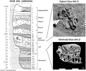
Layer sequence at Ksar Akil in the Levantine corridor, and discovery of two fossils of Homo sapiens, dated to 40,800 to 39,200 years BP for "Egbert", and 42,400–41,700 BP for "Ethelruda".

"Recent African origin," or Out of Africa II, refers to the migration of anatomically modern humans (Homo sapiens) out of Africa after their emergence at c. 300,000 to 200,000 years ago, in contrast to "Out of Africa I", which refers to the migration of archaic humans from Africa to Eurasia roughly 1.8 to 0.5 million years ago. Omo-Kibish I (Omo I) from southern Ethiopia is the oldest anatomically modern Homo sapiens skeleton currently known (196 ± 5 ka).

Since the beginning of the 21st century, the picture of "recent single-origin" migrations has become significantly more complex, not only due to the discovery of modern-archaic admixture but also due to the increasing evidence that the "recent out-of-Africa" migration took place in a number of waves spread over a long time period. As of 2010, there were two main accepted dispersal routes for the out-of-Africa migration of early anatomically modern humans: via the "Northern Route" (via Nile Valley and Sinai) and the "Southern Route" via the Bab al Mandab strait.

  • Posth et al. (2017) suggest that early Homo sapiens, or "another species in Africa closely related to us," might have first migrated out of Africa around 270,000 years ago.
  • Finds at Misliya cave, which include a partial jawbone with eight teeth, have been dated to around 185,000 years ago. Layers dating from between 250,000 and 140,000 years ago in the same cave contained tools of the Levallois type which could put the date of the first migration even earlier if the tools can be associated with the modern human jawbone finds.
  • An Eastward Dispersal from Northeast Africa to Arabia 150,000–130,000  years ago based on the finds at Jebel Faya dated to 127,000 years ago (discovered in 2011). Possibly related to this wave are the finds from Zhirendong cave, Southern China, dated to more than 100,000 years ago. Other evidence of modern human presence in China has been dated to 80,000 years ago.
  • The most significant dispersal took place around 50–70,000 years ago via the so-called Southern Route, either before or after the Toba event, which happened between 69,000 and 77,000 years ago. This dispersal followed the southern coastline of Asia, and reached Australia around 65,000-50,000 years ago, or according to some research, by 50,000 years ago at earliest. Western Asia was "re-occupied" by a different derivation from this wave around 50,000 years ago, and Europe was populated from Western Asia beginning around 43,000 years ago.
  • Wells (2003) describes an additional wave of migration after the southern coastal route, namely a northern migration into Europe at circa 45,000 years ago. However, this possibility is ruled out by Macaulay et al. (2005) and Posth et al. (2016), who argue for a single coastal dispersal, with an early offshoot into Europe.

Northern Route dispersal

Anatomically Modern Humans known archaeological remains in Europe and Africa, directly dated, calibrated carbon dates as of 2013.

Beginning 135,000 years ago, tropical Africa experienced megadroughts which drove humans from the land and towards the sea shores, and forced them to cross over to other continents.

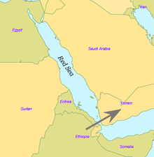
Modern humans crossed the Straits of Bab el Mandeb in the southern Red Sea, and moved along the green coastlines around Arabia, and thence to the rest of Eurasia. Fossils of early Homo sapiens were found in Qafzeh cave in Israel and have been dated 80,000 to 100,000 years ago. These humans seem to have either become extinct or retreated back to Africa 70,000 to 80,000 years ago, possibly replaced by southbound Neanderthals escaping the colder regions of ice-age Europe. Hua Liu et al. analyzed autosomal microsatellite markers dating to about 56,000 years ago. They interpret the paleontological fossil as an isolated early offshoot that retracted back to Africa.

The discovery of stone tools in the United Arab Emirates in 2011 at the Faya-1 site in Mleiha, Sharjah, indicated the presence of modern humans at least 125,000 years ago, leading to a resurgence of the "long-neglected" North African route.

In Oman, a site was discovered by Bien Joven in 2011 containing more than 100 surface scatters of stone tools belonging to the late Nubian Complex, known previously only from archaeological excavations in the Sudan. Two optically stimulated luminescence age estimates placed the Arabian Nubian Complex at approximately 106,000 years old. This provides evidence for a distinct Stone Age technocomplex in southern Arabia, around the earlier part of the Marine Isotope Stage 5.

According to Kuhlwilm and his co-authors, Neanderthals contributed genetically to modern humans then living outside of Africa around 100,000 years ago: humans which had already split off from other modern humans around 200,000 years ago, and this early wave of modern humans outside Africa also contributed genetically to the Altai Neanderthals. They found that "the ancestors of Neanderthals from the Altai Mountains and early modern humans met and interbred, possibly in the Near East, many thousands of years earlier than previously thought". According to co-author Ilan Gronau, "This actually complements archaeological evidence of the presence of early modern humans out of Africa around and before 100,000 years ago by providing the first genetic evidence of such populations." Similar genetic admixture events have been noted in other regions as well.

In China, the Liujiang man (Chinese: 柳江人) is among the earliest modern humans found in East Asia. The date most commonly attributed to the remains is 67,000 years ago. High rates of variability yielded by various dating techniques carried out by different researchers place the most widely accepted range of dates with 67,000 BP as a minimum, but do not rule out dates as old as 159,000 BP.  Liu, Martinón-Torres et al. (2015) claim that modern human teeth have been found in China dating to at least 80,000 years ago.

Southern Route dispersal

Coastal route

Red Sea crossing

By some 50-70,000 years ago, a subset of the bearers of mitochondrial haplogroup L3 migrated from East Africa into the Near East. It has been estimated that from a population of 2,000 to 5,000 individuals in Africa, only a small group, possibly as few as 150 to 1,000 people, crossed the Red Sea. The group that crossed the Red Sea travelled along the coastal route around Arabia and the Persian Plateau to India, which appears to have been the first major settling point. Wells (2003) argued for the route along the southern coastline of Asia, across about 250 kilometres (155 mi), reaching Australia by around 50,000 years ago.

Today at the Bab-el-Mandeb straits, the Red Sea is about 20 kilometres (12 mi) wide, but 50,000 years ago sea levels were 70 m (230 ft) lower (owing to glaciation) and the water was much narrower. Though the straits were never completely closed, they were narrow enough to have enabled crossing using simple rafts, and there may have been islands in between. Shell middens 125,000 years old have been found in Eritrea, indicating the diet of early humans included seafood obtained by beachcombing.

The dating of the Southern Dispersal is a matter of dispute. It may have happened either pre- or post-Toba, a catastrophic volcanic eruption that took place between 69,000 and 77,000 years ago at the site of present-day Lake Toba. Stone tools discovered below the layers of ash disposed in India may point to a pre-Toba dispersal but the source of the tools is disputed. An indication for post-Toba is haplo-group L3, that originated before the dispersal of humans out of Africa and can be dated to 60,000–70,000 years ago, "suggesting that humanity left Africa a few thousand years after Toba". Some research showing slower than expected genetic mutations in human DNA was published in 2012, indicating a revised dating for the migration to between 90,000 and 130,000 years ago. Some more recent research suggests a migration out-of-Africa of around 50,000-65,000 years ago of the ancestors of modern non-African populations, similar to most previous estimates.

Western Asia

A fossil of a modern human dated to 54,700 years ago was found in Manot Cave in Israel, named Manot 1, though the dating was questioned by Groucutt et al. (2015).

South Asia and Australia

It is thought that Australia was inhabited around 65,000–50,000 years ago. As of 2017, the earliest evidence of humans in Australia is at least 65,000 years old, while McChesney stated that

...genetic evidence suggests that a small band with the marker M168 migrated out of Africa along the coasts of the Arabian Peninsula and India, through Indonesia, and reached Australia very early, between 60,000 and 50,000 years ago. This very early migration into Australia is also supported by Rasmussen et al. (2011).

Fossils from Lake Mungo, Australia, have been dated to about 42,000 years ago. Other fossils from a site called Madjedbebe have been dated to at least 65,000 years ago, though some researchers doubt this early estimate and date the Madjedbebe fossils at about 50,000 years ago at the oldest.

East Asia

Tianyuan man from China has a probable date range between 38,000 and 42,000 years ago, while Liujiang man from the same region has a probable date range between 67,000 and 159,000 years ago. According to 2013 DNA tests, Tianyuan man is related "to many present-day Asians and Native Americans". Tianyuan is similar in morphology to Minatogawa Man, modern humans dated between 17,000 and 19,000 years ago and found on Okinawa Island, Japan.

Europe

According to Macaulay et al. (2005), an early offshoot from the southern dispersal with haplogroup N followed the Nile from East Africa, heading northwards and crossing into Asia through the Sinai. This group then branched, some moving into Europe and others heading east into Asia. This hypothesis is supported by the relatively late date of the arrival of modern humans in Europe as well as by archaeological and DNA evidence. Based on an analysis of 55 human mitochondrial genomes (mtDNAs) of hunter-gatherers, Posth et al. (2016) argue for a "rapid single dispersal of all non-Africans less than 55,000 years ago."

Genetic reconstruction

Mitochondrial haplogroups

Within Africa

Map of early diversification of modern humans according to mitochondrial population genetics (see: Haplogroup L).

The first lineage to branch off from Mitochondrial Eve was L0. This haplogroup is found in high proportions among the San of Southern Africa and the Sandawe of East Africa. It is also found among the Mbuti people.  These groups branched off early in human history and have remained relatively genetically isolated since then. Haplogroups L1, L2 and L3 are descendants of L1–L6, and are largely confined to Africa. The macro haplogroups M and N, which are the lineages of the rest of the world outside Africa, descend from L3. L3 is about 70,000 years old, while haplogroups M and N are about 65-55,000 years old. The relationship between such gene trees and demographic history is still debated when applied to dispersals.

Of all the lineages present in Africa, only the female descendants of one lineage, mtDNA haplogroup L3, are found outside Africa. If there had been several migrations, one would expect descendants of more than one lineage to be found. L3's female descendants, the M and N haplogroup lineages, are found in very low frequencies in Africa (although haplogroup M1 populations are very ancient and diversified in North and North-east Africa) and appear to be more recent arrivals. A possible explanation is that these mutations occurred in East Africa shortly before the exodus and became the dominant haplogroups thereafter by means of the founder effect. Alternatively, the mutations may have arisen shortly afterwards.

Southern Route and haplogroups M and N

Results from mtDNA collected from aboriginal Malaysians called Orang Asli indicate that the hapologroups M and N share characteristics with original African groups from approximately 85,000 years ago, and share characteristics with sub-haplogroups found in coastal south-east Asian regions, such as Australasia, the Indian subcontinent and throughout continental Asia, which had dispersed and separated from their African progenitor approximately 65,000 years ago. This southern coastal dispersal would have occurred before the dispersal through the Levant approximately 45,000 years ago. This hypothesis attempts to explain why haplogroup N is predominant in Europe and why haplogroup M is absent in Europe. Evidence of the coastal migration is thought to have been destroyed by the rise in sea levels during the Holocene epoch. Alternatively, a small European founder population that had expressed haplogroup M and N at first, could have lost haplogroup M through random genetic drift resulting from a bottleneck (i.e. a founder effect).

The group that crossed the Red Sea travelled along the coastal route around Arabia and Persia until reaching India. Haplogroup M is found in high frequencies along the southern coastal regions of Pakistan and India and it has the greatest diversity in India, indicating that it is here where the mutation may have occurred. Sixty percent of the Indian population belong to Haplogroup M. The indigenous people of the Andaman Islands also belong to the M lineage. The Andamanese are thought to be offshoots of some of the earliest inhabitants in Asia because of their long isolation from the mainland. They are evidence of the coastal route of early settlers that extends from India to Thailand and Indonesia all the way to eastern New Guinea. Since M is found in high frequencies in highlanders from New Guinea and the Andamanese and New Guineans have dark skin and Afro-textured hair, some scientists think they are all part of the same wave of migrants who departed across the Red Sea ~60,000 years ago in the Great Coastal Migration. The proportion of haplogroup M increases eastwards from Arabia to India; in eastern India, M outnumbers N by a ratio of 3:1. Crossing into Southeast Asia, haplogroup N (mostly in the form of derivatives of its R subclade) reappears as the predominant lineage. M is predominant in East Asia, but amongst Indigenous Australians, N is the more common lineage. This haphazard distribution of Haplogroup N from Europe to Australia can be explained by founder effects and population bottlenecks.

Autosomal DNA

A 2002 study of African, European and Asian populations, found greater genetic diversity among Africans than among Eurasians, and that genetic diversity among Eurasians is largely a subset of that among Africans, supporting the out of Africa model. A large study by Coop et al. (2009) found evidence for natural selection in autosomal DNA outside of Africa. The study distinguishes non-African sweeps (notably KITLG variants associated with skin color), West-Eurasian sweeps (SLC24A5) and East-Asian sweeps (MC1R, relevant to skin color). Based on this evidence, the study concluded that human populations encountered novel selective pressures as they expanded out of Africa. MC1R and its relation to skin color had already been discussed by Liu, Harding et al. (2000), p. 135. According to this study, Papua New Guineans continued to be exposed to selection for dark skin color so that, although these groups are distinct from Africans in other places, the allele for dark skin color shared by contemporary Africans, Andamanese and New Guineans is an archaism. Endicott et al. (2003) suggest convergent evolution. A 2014 study by Gurdasani et al. indicates that the higher genetic diversity in Africa was further increased in some regions by relatively recent Eurasian migrations affecting parts of Africa.

Pathogen DNA

Another promising route towards reconstructing human genetic genealogy is via the JC virus (JCV), a type of human polyomavirus which is carried by 70–90 percent of humans and which is usually transmitted vertically, from parents to offspring, suggesting codivergence with human populations. For this reason, JCV has been used as a genetic marker for human evolution and migration. This method does not appear to be reliable for the migration out of Africa, in contrast to human genetics, JCV strains associated with African populations are not basal. From this Shackelton et al. (2006) conclude that either a basal African strain of JCV has become extinct or that the original infection with JCV post-dates the migration from Africa.

Admixture of archaic and modern humans

Evidence for archaic human species (descended from Homo heidelbergensis) having interbred with modern humans outside of Africa, was discovered in the 2010s. This concerns primarily Neanderthal admixture in all modern populations except for Sub-Saharan Africans but evidence has also been presented for Denisova hominin admixture in Australasia (i.e. in Melanesians, Aboriginal Australians and some Negritos).

The rate of admixture of Neanderthal admixture to European and Asian populations as of 2017 has been estimated at between about 2–3%.

Archaic admixture in some Sub-Saharan African populations hunter-gatherer groups (Biaka Pygmies and San), derived from archaic hominins that broke away from the modern human lineage around 700,000 years, was discovered in 2011. The rate of admixture was estimated at around 2%. Admixture from archaic hominins of still earlier divergence times, estimated at 1.2 to 1.3 million years ago, was found in Pygmies, Hadza and five Sandawe in 2012. From an analysis of Mucin 7, a highly divergent haplotype that has an estimated coalescence time with other variants around 4.5 million years BP and is specific to African populations is inferred to have been derived from interbreeding between African modern and archaic humans.

Stone tools

In addition to genetic analysis, Petraglia et al. also examines the small stone tools (microlithic materials) from the Indian subcontinent and explains the expansion of population based on the reconstruction of paleoenvironment. He proposed that the stone tools could be dated to 35 ka in South Asia, and the new technology might be influenced by environmental change and population pressure.

History of the theory

Classical paleoanthropology

The frontispiece to Huxley's Evidence as to Man's Place in Nature (1863): the image compares the skeleton of a human to other apes.

The cladistic relationship of humans with the African apes was suggested by Charles Darwin after studying the behaviour of African apes, one of which was displayed at the London Zoo. The anatomist Thomas Huxley had also supported the hypothesis and suggested that African apes have a close evolutionary relationship with humans. These views were opposed by the German biologist Ernst Haeckel, who was a proponent of the Out of Asia theory. Haeckel argued that humans were more closely related to the primates of South-east Asia and rejected Darwin's African hypothesis. 

In the Descent of Man, Darwin speculated that humans had descended from apes, which still had small brains but walked upright, freeing their hands for uses which favoured intelligence; he thought such apes were African:

In each great region of the world the living mammals are closely related to the extinct species of the same region. It is, therefore, probable that Africa was formerly inhabited by extinct apes closely allied to the gorilla and chimpanzee; and as these two species are now man's nearest allies, it is somewhat more probable that our early progenitors lived on the African continent than elsewhere. But it is useless to speculate on this subject, for an ape nearly as large as a man, namely the Dryopithecus of Lartet, which was closely allied to the anthropomorphous Hylobates, existed in Europe during the Upper Miocene period; and since so remote a period the earth has certainly undergone many great revolutions, and there has been ample time for migration on the largest scale.

— Charles Darwin, Descent of Man

In 1871, there were hardly any human fossils of ancient hominins available. Almost fifty years later, Darwin's speculation was supported when anthropologists began finding fossils of ancient small-brained hominins in several areas of Africa (list of hominina fossils). The hypothesis of recent (as opposed to archaic) African origin developed in the 20th century. The "Recent African origin" of modern humans means "single origin" (monogenism) and has been used in various contexts as an antonym to polygenism. The debate in anthropology had swung in favour of monogenism by the mid-20th century. Isolated proponents of polygenism held forth in the mid-20th century, such as Carleton Coon, who thought as late as 1962 that H. sapiens arose five times from H. erectus in five places.

Multiregional origin hypothesis

The historical alternative to the recent origin model is the multiregional origin of modern humans, initially proposed by Milford Wolpoff in the 1980s. This view proposes that the derivation of anatomically modern human populations from H. erectus at the beginning of the Pleistocene 1.8 million years BP, has taken place within a continuous world population. The hypothesis necessarily rejects the assumption of an infertility barrier between ancient Eurasian and African populations of Homo. The hypothesis was controversially debated during the late 1980s and the 1990s. The now-current terminology of "recent-origin" and "Out of Africa" became current in the context of this debate in the 1990s. Originally seen as an antithetical alternative to the recent origin model, the multiregional hypothesis in its original "strong" form is obsolete, while its various modified weaker variants have become variants of a view of "recent origin" combined with archaic admixture. Stringer (2014) distinguishes the original or "classic" Multiregional model as having existed from 1984 (its formulation) until 2003, to a "weak" post-2003 variant that has "shifted close to that of the Assimilation Model".

Mitochondrial analyses

In the 1980s, Allan Wilson together with Rebecca L. Cann and Mark Stoneking worked on genetic dating of the matrilineal most recent common ancestor of modern human populations (dubbed "Mitochondrial Eve"). To identify informative genetic markers for tracking human evolutionary history, Wilson concentrated on mitochondrial DNA (mtDNA), passed from mother to child. This DNA material mutates quickly, making it easy to plot changes over relatively short times. With his discovery that human mtDNA is genetically much less diverse than chimpanzee mtDNA, Wilson concluded that modern human populations had diverged recently from a single population while older human species such as Neanderthals and Homo erectus had become extinct. With the advent of archaeogenetics in the 1990s, the dating of mitochondrial and Y-chromosomal haplogroups became possible with some confidence. By 1999, estimates ranged around 150,000 years for the mt-MRCA and 60,000 to 70,000 years for the migration out of Africa.

From 2000–2003, there was controversy about the mitochondrial DNA of "Mungo Man 3" (LM3) and its possible bearing on the multiregional hypothesis. LM3 was found to have more than the expected number of sequence differences when compared to modern human DNA (CRS). Comparison of the mitochondrial DNA with that of ancient and modern aborigines, led to the conclusion that Mungo Man fell outside the range of genetic variation seen in Aboriginal Australians and was used to support the multiregional origin hypothesis. A reanalysis on LM3 and other ancient specimens from the area published in 2016, showed it to be akin to modern Aboriginal Australian sequences, inconsistent with the results of the earlier study.

Y-Chromosome analyses

As current estimates on The male most recent common ancestor ("Y-chromosomal Adam" or Y-MRCA) converge with estimates for the age of anatomically modern humans and well predate the Out of Africa migration, geographical origin hypotheses continue to be limited to the African continent.

The most basal lineages have been detected in West, Northwest and Central Africa, suggesting plausibility for the Y-MRCA living in the general region of "Central-Northwest Africa".

another study finds a plausible placement in "the north-western quadrant of the African continent" for the emergence of the A1b haplogroup. The 2013 report of haplogroup A00 found among the Mbo people of western present-day Cameroon is also compatible with this picture.

The revision of Y-chromosomal phylogeny since 2011 has affected estimates for the likely geographical origin of Y-MRCA as well as estimates on time depth. By the same reasoning, future discovery of presently-unknown archaic haplogroups in living people would again lead to such revisions. In particular, the possible presence of between 1% and 4% Neanderthal-derived DNA in Eurasian genomes implies that the (unlikely) event of a discovery of a single living Eurasian male exhibiting a Neanderthal patrilineal line would immediately push back T-MRCA ("time to MRCA") to at least twice its current estimate. However, the discovery of a Neanderthal Y-chromosome by Mendez et al. A 2016 study suggests the extinction of Neanderthal patrilineages, as the lineage inferred from the Neanderthal sequence is outside of the range of contemporary human genetic variation. Questions of geographical origin would become part of the debate on Neanderthal evolution from Homo erectus.

Operator (computer programming)

From Wikipedia, the free encyclopedia https://en.wikipedia.org/wiki/Operator_(computer_programmin...