Search This Blog

Tuesday, March 15, 2022

Spider silk

From Wikipedia, the free encyclopedia

A garden spider spinning its web
 
A female specimen of Argiope bruennichi wraps her prey in silk.
 
Indian Summer by Józef Chełmoński (1875, National Museum in Warsaw) depicts a peasant woman with a thread of gossamer in her hand.
 
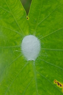
Spider cocoon

Spider silk is a protein fibre spun by spiders. Spiders use their silk to make webs or other structures, which function as sticky nets to catch other animals, or as nests or cocoons to protect their offspring, or to wrap up prey. They can also use their silk to suspend themselves, to float through the air, or to glide away from predators. Most spiders vary the thickness and stickiness of their silk for different uses.

In some cases, spiders may even use silk as a source of food. While methods have been developed to collect silk from a spider by force, it is difficult to gather silk from many spiders compared to silk-spinning organisms such as silkworms.

All spiders produce silk, and even in non-web building spiders, silk is intimately tied to courtship and mating. Silk produced by females provides a transmission channel for male vibratory courtship signals, while webs and draglines provide a substrate for female sex pheromones. Observations of male spiders producing silk during sexual interactions are also common across phylogenetically widespread taxa. However, the function of male-produced silk in mating has received very little study.

Biodiversity

Uses

All spiders produce silks, and a single spider can produce up to seven different types of silk for different uses.[4] This is in contrast to insect silks, where an individual usually only produces one type of silk. Spider silks may be used in many different ecological ways, each with properties to match the silk's function. As spiders have evolved, so has their silks' complexity and diverse uses, for example from primitive tube webs 300–400 million years ago to complex orb webs 110 million years ago.

Use Example
Prey capture The orb webs produced by the Araneidae (typical orb-weavers); tube webs; tangle webs; sheet webs; lace webs, dome webs; single thread used by the Bolas spiders for "fishing".
Prey immobilisation Silk used as "swathing bands" to wrap up prey. Often combined with immobilising prey using a venom. In species of Scytodes the silk is combined with venom and squirted from the chelicerae.
Reproduction Male spiders may produce sperm webs; spider eggs are covered in silk cocoons.
Dispersal "Ballooning" or "kiting" used by smaller spiders to float through the air, for instance for dispersal.
Source of food The kleptoparasitic Argyrodes eating the silk of host spider webs. Some daily weavers of temporary webs also eat their own unused silk daily, thus mitigating a heavy metabolic expense.
Nest lining and nest construction Tube webs used by "primitive" spiders such as the European tube web spider (Segestria florentina). Threads radiate out of nest to provide a sensory link to the outside. Silk is a component of the lids of spiders that use "trapdoors", such as members of the family Ctenizidae, and the "water" or "diving bell" spider Argyroneta aquatica builds its diving bell of silk.
Guide lines Some spiders that venture from shelter will leave a trail of silk by which to find their way home again.
Drop lines and anchor lines Many spiders, such as the Salticidae, that venture from shelter and leave a trail of silk, use that as an emergency line in case of falling from inverted or vertical surfaces. Many others, even web dwellers, will deliberately drop from a web when alarmed, using a silken thread as a drop line by which they can return in due course. Some, such as species of Paramystaria, also will hang from a drop line when feeding.
Alarm lines Some spiders that do not spin actual trap webs do lay out alarm webs that the feet of their prey (such as ants) can disturb, cueing the spider to rush out and secure the meal if it is small enough, or to avoid contact if the intruder seems too formidable.
Pheromonal trails Some wandering spiders will leave a largely continuous trail of silk impregnated with pheromones that the opposite sex can follow to find a mate.

Types

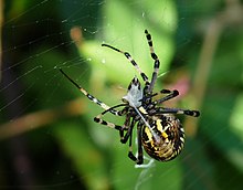
A female Argiope picta immobilizing prey by wrapping a curtain of aciniform silk around the insect for later consumption

Meeting the specification for all these ecological uses requires different types of silk suited to different broad properties, as either a fibre, a structure of fibres, or a silk-globule. These types include glues and fibres. Some types of fibres are used for structural support, others for constructing protective structures. Some can absorb energy effectively, whereas others transmit vibration efficiently. In a spider, these silk types are produced in different glands; so the silk from a particular gland can be linked to its use by the spider.

Gland Silk Use
Ampullate (major) Dragline silk – used for the web's outer rim and spokes, also for the lifeline and for ballooning.
Ampullate (minor) Used for temporary scaffolding during web construction.
Flagelliform Capture-spiral silk – used for the capturing lines of the web.
Tubuliform Egg cocoon silk – used for protective egg sacs.
Aciniform Used to wrap and secure freshly captured prey; used in the male sperm webs; used in stabilimenta.
Aggregate A silk glue of sticky globules.
Piriform Used to form bonds between separate threads for attachment points.

Properties

Mechanical properties

Each spider and each type of silk has a set of mechanical properties optimised for their biological function.

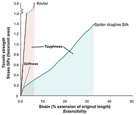
Most silks, in particular dragline silk, have exceptional mechanical properties. They exhibit a unique combination of high tensile strength and extensibility (ductility). This enables a silk fibre to absorb a large amount of energy before breaking (toughness, the area under a stress-strain curve).

An illustration of the differences between toughness, stiffness and strength

A frequent mistake made in the mainstream media is to confuse strength and toughness, when comparing silk to other materials. Weight for weight, silk is stronger than steel, but not as strong as Kevlar. Silk is, however, tougher than both.

The variability of mechanical properties of spider silk fibres may be important and it is related to their degree of molecular alignment. Mechanical properties depend strongly on the ambient conditions, i.e. humidity and temperature.

Strength

A dragline silk's tensile strength is comparable to that of high-grade alloy steel (450−2000 MPa), and about half as strong as aramid filaments, such as Twaron or Kevlar (3000 MPa).

Density

Consisting of mainly protein, silks are about a sixth of the density of steel (1.3 g/cm3). As a result, a strand long enough to circle the Earth would weigh less than 500 grams (18 oz). (Spider dragline silk has a tensile strength of roughly 1.3 GPa. The tensile strength listed for steel might be slightly higher – e.g. 1.65 GPa, but spider silk is a much less dense material, so that a given weight of spider silk is five times as strong as the same weight of steel.

Energy density

The energy density of dragline spider silk is roughly 1.2×10⁸ J/m3.

Extensibility

Silks are also extremely ductile, with some able to stretch up to five times their relaxed length without breaking.

Toughness

The combination of strength and ductility gives dragline silks a very high toughness (or work to fracture), which "equals that of commercial polyaramid (aromatic nylon) filaments, which themselves are benchmarks of modern polymer fibre technology".

Temperature

While unlikely to be relevant in nature, dragline silks can hold their strength below -40 °C (-40 °F) and up to 220 °C (428 °F). As occurs in many materials, spider silk fibres undergo a glass transition. The glass-transition temperature depends on the humidity, as water is a plasticiser for the silk.

Supercontraction

When exposed to water, dragline silks undergo supercontraction, shrinking up to 50% in length and behaving like a weak rubber under tension. Many hypotheses have been suggested as to its use in nature, with the most popular being to automatically tension webs built in the night using the morning dew.

Highest-performance

The toughest known spider silk is produced by the species Darwin's bark spider (Caerostris darwini): "The toughness of forcibly silked fibers averages 350 MJ/m3, with some samples reaching 520 MJ/m3. Thus, C. darwini silk is more than twice as tough as any previously described silk, and over 10 times tougher than Kevlar".

Adhesive properties

Silk fibre is a two-compound pyriform secretion, spun into patterns (called "attachment discs") that are employed to adhere silk threads to various surfaces using a minimum of silk substrate. The pyriform threads polymerise under ambient conditions, become functional immediately, and are usable indefinitely, remaining biodegradable, versatile and compatible with numerous other materials in the environment. The adhesive and durability properties of the attachment disc are controlled by functions within the spinnerets. Some adhesive properties of the silk resemble glue, consisting of microfibrils and lipid enclosures.

Types of silk

Many species of spiders have different glands to produce silk with different properties for different purposes, including housing, web construction, defence, capturing and detaining prey, egg protection, and mobility (fine "gossamer" thread for ballooning, or for a strand allowing the spider to drop down as silk is extruded). Different specialised silks have evolved with properties suitable for different uses. For example, Argiope argentata has five different types of silk, each used for a different purpose:

Silk Use
major-ampullate (dragline) silk Used for the web's outer rim and spokes and also for the lifeline. Can be as strong per unit weight as steel, but much tougher.
capture-spiral (flagelliform) silk Used for the capturing lines of the web. Sticky, extremely stretchy and tough. The capture spiral is sticky due to droplets of aggregate (a spider glue) that is placed on the spiral. The elasticity of flagelliform allows for enough time for the aggregate to adhere to the aerial prey flying into the web.
tubiliform (a.k.a. cylindriform) silk Used for protective egg sacs. Stiffest silk.
aciniform silk Used to wrap and secure freshly captured prey. Two to three times as tough as the other silks, including dragline.
minor-ampullate silk Used for temporary scaffolding during web construction.

Structural

Macroscopic structure down to protein hierarchy

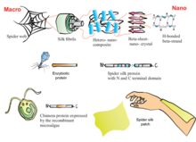
Structure of spider silk. Inside a typical fibre there are crystalline regions separated by amorphous linkages. The crystals are beta-sheets that have assembled together.

Silks, like many other biomaterials, have a hierarchical structure. The primary structure is the amino acid sequence of its proteins (spidroin), mainly consisting of highly repetitive glycine and alanine blocks, which is why silks are often referred to as a block co-polymer. On a secondary structure level, the short side chained alanine is mainly found in the crystalline domains (beta sheets) of the nanofibril, glycine is mostly found in the so-called amorphous matrix consisting of helical and beta turn structures. It is the interplay between the hard crystalline segments, and the strained elastic semi-amorphous regions, that gives spider silk its extraordinary properties. Various compounds other than protein are used to enhance the fibre's properties. Pyrrolidine has hygroscopic properties which keeps the silk moist while also warding off ant invasion. It occurs in especially high concentration in glue threads. Potassium hydrogen phosphate releases hydrogen ions in aqueous solution, resulting in a pH of about 4, making the silk acidic and thus protecting it from fungi and bacteria that would otherwise digest the protein. Potassium nitrate is believed to prevent the protein from denaturing in the acidic milieu.

This first very basic model of silk was introduced by Termonia in 1994 who suggested crystallites embedded in an amorphous matrix interlinked with hydrogen bonds. This model has refined over the years: semi-crystalline regions were found as well as a fibrillar skin core model suggested for spider silk, later visualised by AFM and TEM. Sizes of the nanofibrillar structure and the crystalline and semi-crystalline regions were revealed by neutron scattering.

It has been possible to relate microstructural information and macroscopic mechanical properties of the fibres. The results show that ordered regions (i) mainly reorient by deformation for low-stretched fibres and (ii) the fraction of ordered regions increases progressively for higher stretching of the fibres.


Biosynthesis and fibre spinning

The production of silks, including spider silk, differs in an important aspect from the production of most other fibrous biological materials: rather than being continuously grown as keratin in hair, cellulose in the cell walls of plants, or even the fibres formed from the compacted faecal matter of beetles; it is "spun" on demand from liquid silk precursor out of specialised glands.

The spinning process occurs when a fibre is pulled away from the body of a spider, whether by the spider's legs, by the spider's falling under its own weight, or by any other method including being pulled by humans. The term "spinning" is misleading because no rotation of any component occurs, but rather comes from analogy to the textile spinning wheels. Silk production is a pultrusion, similar to extrusion, with the subtlety that the force is induced by pulling at the finished fibre rather than being squeezed out of a reservoir. The unspun silk fibre is pulled through silk glands of which there may be both numerous duplicates and different types of gland on any one spider species.

Silk gland

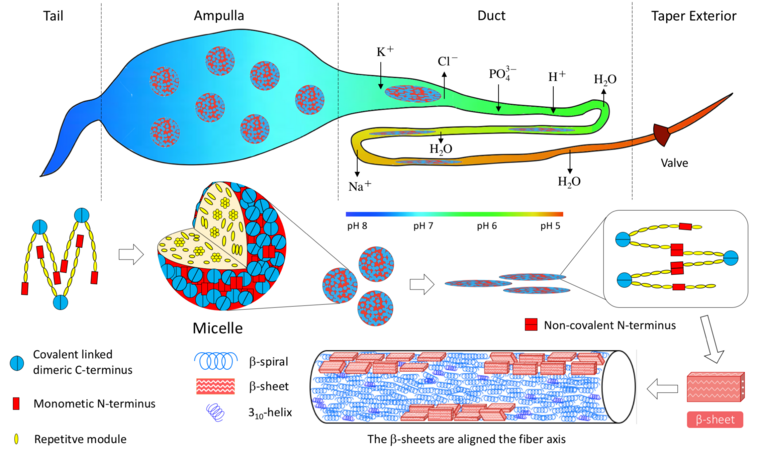
Schematic of the spiders spinning apparatus and structural hierarchy in silk assembling related to assembly into fibers. In the process of dragline production, the primary structure protein is secreted first from secretory granules in the tail. In the ampullate (neutral environment, pH = 7), the proteins form a soft micelle of several tens of nanometers by self-organization because the hydrophilic terminals are excluded. In ampullate, the concentration of the protein is very high. Then, the micelles are squeezed into the duct. The long axis direction of the molecules is aligned parallel to the duct by a mechanical frictional force and partially oriented. The continuous lowering of pH from 7.5 to 8.0 in the tail to presumably close to 5.0 occurs at the end of the duct. Ion exchange, acidification, and water removal all happen in the duct. The shear and elongational forces lead to phase separation. In the acidic bath of the duct, the molecules attain a high concentration liquid crystal state. Finally, the silk is spun from the taper exterior. The molecules become more stable helixes and β-sheets from the liquid crystal.

The gland's visible, or external, part is termed the spinneret. Depending on the complexity of the species, spiders will have two to eight spinnerets, usually in pairs. There exist highly different specialised glands in different spiders, ranging from simply a sac with an opening at one end, to the complex, multiple-section major ampullate glands of the golden silk orb-weavers.

Behind each spinneret visible on the surface of the spider lies a gland, a generalised form of which is shown in the figure to the right, "Schematic of a generalised gland".

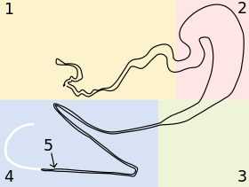
Schematic of a generalised gland of a Golden silk orb-weaver. Each differently coloured section highlights a discrete section of the gland.
Gland characteristics
  1. The first section of the gland labelled 1 on Figure 1 is the secretory or tail section of the gland. The walls of this section are lined with cells that secrete proteins Spidroin I and Spidroin II, the main components of this spider's dragline. These proteins are found in the form of droplets that gradually elongate to form long channels along the length of the final fibre, hypothesised to assist in preventing crack formation or even self-healing of the fibre.
  2. The second section is the storage sac. This stores and maintains the gel-like unspun silk dope until it is required by the spider. In addition to storing the unspun silk gel, it secretes proteins that coat the surface of the final fibre.
  3. The funnel rapidly reduces the large diameter of the storage sac to the small diameter of the tapering duct.
  4. The final length is the tapering duct, the site of most of the fibre formation. This consists of a tapering tube with several tight about turns, a valve almost at the end (mentioned in detail at point No. 5 below) ending in a spigot from which the solid silk fibre emerges. The tube here tapers hyperbolically, therefore the unspun silk is under constant elongational shear stress, which is an important factor in fibre formation. This section of the duct is lined with cells that exchange ions, reduce the dope pH from neutral to acidic, and remove water from the fibre. Collectively, the shear stress and the ion and pH changes induce the liquid silk dope to undergo a phase transition and condense into a solid protein fibre with high molecular organisation. The spigot at the end has lips that clamp around the fibre, controlling fibre diameter and further retaining water.
  5. Almost at the end of the tapering duct is a valve, approximate position marked "5" on figure 1. Though discovered some time ago, the precise purpose of this valve is still under discussion. It is believed to assist in restarting and rejoining broken fibres, acting much in the way of a helical pump, regulating the thickness of the fibre, and/or clamping the fibre as a spider falls upon it. There is some discussion of the similarity of the silk worm's silk press and the roles each of these valves play in the production of silk in these two organisms.

Throughout the process the unspun silk appears to have a nematic texture, in a similar manner to a liquid crystal, arising in part due to the extremely high protein concentration of silk dope (around 30% in terms of weight per volume). This allows the unspun silk to flow through the duct as a liquid but maintain a molecular order.

As an example of a complex spinning field, the spinneret apparatus of an adult Araneus diadematus (garden cross spider) consists of the glands shown below. Similar multiple gland architecture exists in the black widow spider.

  • 500 pyriform glands for attachment points
  • 4 ampullate glands for the web frame
  • about 300 aciniform glands for the outer lining of egg sacs, and for ensnaring prey
  • 4 tubuliform glands for egg sac silk
  • 4 aggregate glands for adhesive functions
  • 2 coronate glands for the thread of adhesion lines

Artificial synthesis

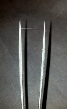
Single strand of artificial spider silk produced under laboratory conditions

To artificially synthesise spider silk into fibres, there are two broad areas that must be covered. These are synthesis of the feedstock (the unspun silk dope in spiders), and synthesis of the spinning conditions (the funnel, valve, tapering duct, and spigot). There have been a number of different approaches but few of these methods have produced silk that can efficiently be synthesised into fibres.

Feedstock

The molecular structure of unspun silk is both complex and extremely long. Though this endows the silk fibres with their desirable properties, it also makes replication of the fibre somewhat of a challenge. Various organisms have been used as a basis for attempts to replicate some components or all of some or all of the proteins involved. These proteins must then be extracted, purified and then spun before their properties can be tested.


Organism Details Average Maximum breaking stress (MPa) Average Strain (%)
Darwin's bark spider (Caerostris darwini) Malagasy spider famed for making webs with strands up to 25 m long, across rivers. "C. darwini silk is more than twice as tough as any previously described silk" 1850 ±350 33 ±0.08
Nephila clavipes Typical golden orb weaving spider 710–1200 18–27
Bombyx mori Silkworms Silkworms were genetically altered to express spider proteins and fibres measured. 660 18.5
E. coli Synthesising a large and repetitive molecule (~300 kDa) is complex, but required for the strongest silk. Here E. coli was engineered to produce a 556 kDa protein. Fibers spun from these synthetic spidroins are the first to fully replicate the mechanical performance of natural spider silk by all common metrics. 1030 ±110 18 ±6
Goats Goats were genetically modified to secrete silk proteins in their milk, which could then be purified. 285–250 30–40
Tobacco & potato plants Tobacco and potato plants were genetically modified to produce silk proteins. Patents were granted, but no fibres have yet been described in the literature. n/a n/a

Geometry

Spider silks with comparatively simple molecular structure need complex ducts to be able to spin an effective fibre. There have been a number of methods used to produce fibres, of which the main types are briefly discussed below.

Syringe and needle

Feedstock is simply forced through a hollow needle using a syringe. This method has been shown to make fibres successfully on multiple occasions.

Although very cheap and easy to produce, the shape and conditions of the gland are very loosely approximated. Fibres created using this method may need encouragement to change from liquid to solid by removing the water from the fibre with such chemicals as the environmentally undesirable methanol or acetone, and also may require post-stretching of the fibre to attain fibres with desirable properties.

Microfluidics

As the field of microfluidics matures, it is likely that more attempts to spin fibres will be made using microfluidics. These have the advantage of being very controllable and able to test spin very small volumes of unspun fibre but setup and development costs are likely to be high. A patent has been granted in this area for spinning fibres in a method mimicking the process found in nature, and fibres are successfully being continuously spun by a commercial company.

Electrospinning

Electrospinning is a very old technique whereby a fluid is held in a container in a manner such that it is able to flow out through capillary action. A conducting substrate is positioned below, and a large difference in electrical potential is applied between the fluid and the substrate. The fluid is attracted to the substrate, and tiny fibres jump almost instantly from their point of emission, the Taylor cone, to the substrate, drying as they travel. This method has been shown to create nano-scale fibres from both silk dissected from organisms and regenerated silk fibroin.

Other artificial shapes formed from silk

Silk can be formed into other shapes and sizes such as spherical capsules for drug delivery, cell scaffolds and wound healing, textiles, cosmetics, coatings, and many others. Spider silk proteins can also self-assemble on superhydrophobic surfaces to generate nanowires, as well as micron-sized circular sheets. It has recently been shown that recombinant spider silk proteins can self-assemble at the liquid air interface of a standing solution to form protein permeable, strong, and flexible nanomembranes that support cell proliferation. Suggested applications include skin transplants, and supportive membranes in organ-on-a-chip. These spider silk nanomembranes have also been used to create a static in-vitro model of a blood vessel.

Human uses

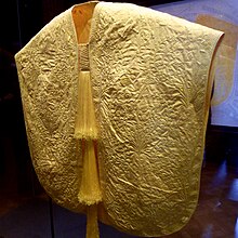
A cape made from Madagascar golden orb spider silk

Peasants in the southern Carpathian Mountains used to cut up tubes built by Atypus and cover wounds with the inner lining. It reportedly facilitated healing, and even connected with the skin. This is believed to be due to antiseptic properties of spider silk and because the silk is rich in vitamin K, which can be effective in clotting blood. Due to the difficulties in extracting and processing substantial amounts of spider silk, the largest known piece of cloth made of spider silk is an 11-by-4-foot (3.4 by 1.2 m) textile with a golden tint made in Madagascar in 2009. Eighty-two people worked for four years to collect over one million golden orb spiders and extract silk from them.

The silk of Nephila clavipes was used in research concerning mammalian neuronal regeneration.

Spider silk has been used as a thread for crosshairs in optical instruments such as telescopes, microscopes, and telescopic rifle sights. In 2011, spider silk fibres were used in the field of optics to generate very fine diffraction patterns over N-slit interferometric signals used in optical communications. In 2012, spider silk fibres were used to create a set of violin strings.

Development of methods to mass-produce spider silk has led to manufacturing of military, medical and consumer goods, such as ballistics armour, athletic footwear, personal care products, breast implant and catheter coatings, mechanical insulin pumps, fashion clothing, and outerwear.

Spider silk is used to suspend inertial confinement fusion targets during laser ignition, as it remains considerably elastic and has a high energy to break at temperatures as low as 10–20 K. In addition, it is made from "light" atomic number elements that won't emit x-rays during irradiation that could preheat the target so that the pressure differential required for fusion is not achieved.

Spider silk has been used to create biolenses that could be used in conjunction with lasers to create high-resolution images of the inside of the human body.

Attempts at producing synthetic spider silk

Proposed framework for producing artificial skin from spider silk to help patients with burns.

Replicating the complex conditions required to produce fibres that are comparable to spider silk has proven difficult in research and early-stage manufacturing. Through genetic engineering, Escherichia coli bacteria, yeasts, plants, silkworms, and animals other than silkworms have been used to produce spider silk proteins, which have different, simpler characteristics than those from a spider. Extrusion of protein fibres in an aqueous environment is known as "wet-spinning". This process has so far produced silk fibres of diameters ranging from 10 to 60 μm, compared to diameters of 2.5–4 μm for natural spider silk. Artificial spider silks have fewer and simpler proteins than natural dragline silk, and are consequently half the diameter, strength, and flexibility of natural dragline silk.

  • In March 2010, researchers from the Korea Advanced Institute of Science & Technology succeeded in making spider silk directly using the bacteria E. coli, modified with certain genes of the spider Nephila clavipes. This approach eliminates the need to milk spiders and allows the manufacture of the spider silk in a more cost-effective manner.
  • A 556 kDa spider silk protein was manufactured from 192 repeat motifs of the Nephila clavipes dragline spidroin, having similar mechanical characteristics as their natural counterparts, i.e., tensile strength (1.03 ± 0.11 GPa), modulus (13.7 ± 3.0 GPa), extensibility (18 ± 6%), and toughness (114 ± 51 MJ/m3).
  • The company AMSilk developed spidroin using bacteria, making it into an artificial spider silk.
  • The company Bolt Threads produces a recombinant spidroin using yeast, for use in apparel fibers and personal care. They produced the first commercial apparel products made of recombinant spider silk, trademarked Microsilk, demonstrated in ties and beanies. They have also partnered with vegan activist and luxury designer Stella McCartney as well as Adidas to produce Microsilk garments.
  • The company Kraig Biocraft Laboratories used research from the Universities of Wyoming and Notre Dame to create silkworms that were genetically altered to produce spider silk.
  • The now defunct Canadian biotechnology company Nexia successfully produced spider silk protein in transgenic goats that carried the gene for it; the milk produced by the goats contained significant quantities of the protein, 1–2 grams of silk proteins per litre of milk. Attempts to spin the protein into a fibre similar to natural spider silk resulted in fibres with tenacities of 2–3 grams per denier. Nexia used wet spinning and squeezed the silk protein solution through small extrusion holes in order to simulate the behavior of the spinneret, but this procedure was not sufficient to replicate the stronger properties of native spider silk.
  • The company Spiber has produced a synthetic spider silk that they are calling Q/QMONOS. In partnership with Goldwin, a ski parka made from this synthetic spider silk is currently in testing and is to be in mass production soon for less than $120,000 YEN.

Bioarchaeology

From Wikipedia, the free encyclopedia

The term bioarchaeology has been attributed to British archaeologist Grahame Clark who, in 1972, defined it as the study of animal and human bones from archaeological sites. Redefined in 1977 by Jane Buikstra, bioarchaeology in the United States now refers to the scientific study of human remains from archaeological sites, a discipline known in other countries as osteoarchaeology, osteology or palaeo-osteology. Compared to bioarchaeology, osteoarchaeology is the scientific study that solely focus on the human skeleton. The human skeleton is used to tell us about health, lifestyle, diet, mortality and physique of the past. Furthermore, palaeo-osteology is simple the study of ancient bones.

In contrast, the term bioarchaeology is used in Europe to describe the study of all biological remains from archaeological sites. Although Clark used it to describe just human remains and animal remains (zoology/archaeozoology), increasingly modern archaeologists also include botanical remains (botany/archaeobotany

Bioarchaeology was largely born from the practices of New Archaeology, which developed in the United States in the 1970s as a reaction to a mainly cultural-historical approach to understanding the past. Proponents of New Archaeology advocated using processual methods to test hypotheses about the interaction between culture and biology, or a biocultural approach. Some archaeologists advocate a more holistic approach to bioarchaeology that incorporates critical theory and is more relevant to modern descent populations.

If possible, human remains from archaeological sites are analyzed to determine sex, age, and health. which all fall under the term 'Bioarchaeology'.

Paleodemography

Paleodemography is the field that attempts to identify demographic characteristics from the past population. The information gathered is used to make interpretations. Bioarchaeologists use paleodemography sometimes and create life tables, a type of cohort analysis, to understand the demographic characteristics (such as risk of death or sex ratio) of a given age cohort within a population. Age and sex are crucial variables in the construction of a life table, although this information is often not available to bioarchaeologists. Therefore, it is often necessary to estimate the age and sex of individuals based on specific morphological characteristics of the skeleton.

Age estimation

The estimation of age in bioarchaeology and osteology actually refers to an approximation of skeletal or biological age-at-death. The primary assumption in age estimation is that an individual's skeletal age is closely associated with their chronological age. Age estimation can be based on patterns of growth and development or degenerative changes in the skeleton. Many methods tracking these types of changes have been developed using a variety of skeletal series. For instance, in children age is typically estimated by assessing their dental development, ossification and fusion of specific skeletal elements, or long bone length. For children, the different points of time at which different teeth erupt from the gums are best known for telling a child's age down to the exact year. But once the teeth are fully developed, age in hard to be determined using teeth. In adults, degenerative changes to the pubic symphysis, the auricular surface of the ilium, the sternal end of the 4th rib, and dental attrition are commonly used to estimate skeletal age.

When using bones to determine age, there might be problems that you might face. Until the age of about 30, the human bones are still growing. Different bones are fusing at different points of growth. Some bones might not follow the correct stages of growth which can mess with your analysis. Also, as you get older there is wear and tear on the humans' bones and the age estimate becomes less precise as the bone gets older. The bones then become categorized as either 'young' (20–35 years), 'middle' (35–50 years), or 'old' (50+ years).

Sex determination

Differences in male and female skeletal anatomy are used by bioarchaeologists to determine the biological sex of human skeletons. Humans are sexually dimorphic, although overlap in body shape and sexual characteristics is possible. Not all skeletons can be assigned a sex, and some may be wrongly identified as male or female. Sexing skeletons is based on the observation that biological males and biological females differ most in the skull and pelvis; bioarchaeologists focus on these parts of the body when determining sex, although other body parts can also be used. The female pelvis is generally broader than the male pelvis, and the angle between the two inferior pubic rami (the sub-pubic angle) is wider and more U-shaped, while the sub-pubic angle of the male is more V-shaped and less than 90 degrees. Phenice details numerous visual differences between the male and female pelvis.

In general, the male skeleton is more robust than the female skeleton because of the greater muscles mass of the male. Males generally have more pronounced brow ridges, nuchal crests, and mastoid processes. It should be remembered that skeletal size and robustness are influenced by nutrition and activity levels. Pelvic and cranial features are considered to be more reliable indicators of biological sex. Sexing skeletons of young people who have not completed puberty is more difficult and problematic than sexing adults, because the body has not had time to develop fully.

Bioarchaeological sexing of skeletons is not error-proof. In reviewing the sexing of Egyptian skulls from Qua and Badari, Mann found that 20.3% could be assigned to a different sex than the sex indicated in the archaeological literature. A re-evalutaion of Mann's work showed that he did not understand the tomb numbering system of the old excavation and assigned wrong tomb numbers to the skulls. The sexing of the bone material was actually quite correct. However, recording errors and re-arranging of human remains may play a part in this great incidence of misidentification.

Direct testing of bioarchaeological methods for sexing skeletons by comparing gendered names on coffin plates from the crypt at Christ Church, Spitalfields, London to the associated remains resulted in a 98 percent success rate.

Sex-based differences are not inherently a form of inequality, but become an inequality when members of one sex are given privileges based on their sex. This stems from society investing differences with cultural and social meaning. Gendered work patterns may make their marks on the bones and be identifiable in the archaeological record. Molleson found evidence of gendered work patterns by noting extremely arthritic big toes, a collapse of the last dorsal vertebrae, and muscular arms and legs among female skeletons at Abu Hureyra. She interpreted this sex-based pattern of skeletal difference as indicative of gendered work patterns. These kinds of skeletal changes could have resulted from women spending long periods of time kneeling while grinding grain with the toes curled forward. Investigation of gender from mortuary remains is of growing interest to archaeologists.

Non-specific stress indicators

Dental non-specific stress indicators

Enamel hypoplasia

Enamel hypoplasia refers to transverse furrows or pits that form in the enamel surface of teeth when the normal process of tooth growth stops, resulting in a deficit of enamel. Enamel hypoplasias generally form due to disease and/or poor nutrition. Linear furrows are commonly referred to as linear enamel hypoplasias (LEHs); LEHs can range in size from microscopic to visible to the naked eye. By examining the spacing of perikymata grooves (horizontal growth lines), the duration of the stressor can be estimated, although Mays argues that the width of the hypoplasia bears only an indirect relationship to the duration of the stressor.

Studies of dental enamel hypoplasia are used to study child health. Unlike bone, teeth are not remodeled, so they can provide a more reliable indicator of past health events as long as the enamel remains intact. Dental hypoplasias provide an indicator of health status during the time in childhood when the enamel of the tooth crown is being formed. Not all of the enamel layers are visible on the surface of the tooth because enamel layers that are formed early in crown development are buried by later layers. Hypoplasias on this part of the tooth do not show on the surface of the tooth. Because of this buried enamel, teeth record stressors form a few months after the start of the event. The proportion of enamel crown formation time represented by this buried in enamel varies from up to 50 percent in molars to 15-20 percent in anterior teeth. Surface hypoplasias record stressors occurring from about one to seven years, or up to 13 years if the third molar is included.

Skeletal non-specific stress indicators

Porotic hyperostosis/cribra orbitalia

It was long assumed that iron deficiency anemia has marked effects on the flat bones of the cranium of infants and young children. That as the body attempts to compensate for low iron levels by increasing red blood cell production in the young, sieve-like lesions develop in the cranial vaults (termed porotic hyperostosis) and/or the orbits (termed cribra orbitalia). This bone is spongy and soft.

It is however, highly unlikely that iron deficiency anemia is a cause of either porotic hyperostosis or cribra orbitalia. These are more likely the result of vascular activity in these areas and are unlikely to be pathological. The development of cribra orbitalia and porotic hyperostosis could also be attributed to other causes besides an iron deficiency in the diet, such as nutrients lost to intestinal parasites. However, dietary deficiencies are the most probable cause.

Anemia incidence may be a result of inequalities within society, and/or indicative of different work patterns and activities among different groups within society. A study of iron-deficiency among early Mongolian nomads showed that although overall rates of cribra orbitalia declined from 28.7 percent (27.8 percent of the total female population, 28.4 percent of the total male population, 75 percent of the total juvenile population) during the Bronze and Iron Ages, to 15.5 percent during the Hunnu (2209–1907 BP) period, the rate of females with cribra orbitalia remained roughly the same, while the incidence of cribra orbitalia among males and children declined (29.4 percent of the total female population, 5.3 percent of the total male population, and 25 percent of the juvenile population had cribra orbitalia). Bazarsad posits several reasons for this distribution of cribra orbitalia: adults may have lower rates of cribra orbitalia than juveniles because lesions either heal with age or lead to death. Higher rates of cribia orbitalia among females may indicate lesser health status, or greater survival of young females with cribia orbitalia into adulthood.

Harris lines

Harris lines form before adulthood, when bone growth is temporarily halted or slowed down due to some sort of stress (either disease or malnutrition). During this time, bone mineralization continues, but growth does not, or does so at very reduced levels. If and when the stressor is overcome, bone growth will resume, resulting in a line of increased mineral density that will be visible in a radiograph. If there is not recovery from the stressor, no line will be formed.

Hair

The stress hormone cortisol is deposited in hair as it grows. This has been used successfully to detect fluctuating levels of stress in the later lifespan of mummies.

Mechanical stress and activity indicators

Examining the effects that activities and workload has upon the skeleton allows the archaeologist to examine who was doing what kinds of labor, and how activities were structured within society. The division of labor within the household may be divided according to gender and age, or be based on other hierarchical social structures. Human remains can allow archaeologists to uncover patterns in the division of labor.

Living bones are subject to Wolff's law, which states that bones are physically affected and remodeled by physical activity or inactivity. Increases in mechanical stress tend to produce bones that are thicker and stronger. Disruptions in homeostasis caused by nutritional deficiency or disease or profound inactivity/disuse/disability can lead to bone loss. While the acquisition of bipedal locomotion and body mass appear to determine the size and shape of children's bones, activity during the adolescent growth period seems to exert a greater influence on the size and shape of adult bones than exercise later in life.

Muscle attachment sites (also called entheses) have been thought to be impacted in the same way causing what were once called musculoskeletal stress markers, but now widely named entheseal changes. These changes were widely used to study activity-patterns, but research has shown that processes associated with aging have a greater impact than occupational stresses. It has also been shown that geometric changes to bone structure (described above) and entheseal changes differ in their underlying cause with the latter poorly affected by occupation. Joint changes, including osteoarthritis, have also been used to infer occupations but in general these are also manifestations of the aging process.

Markers of occupational stress, which include morphological changes to the skeleton and dentition as well as joint changes at specific locations have also been widely used to infer specific (rather than general) activities. Such markers are often based on single cases described in clinical literature in the late nineteenth century. One such marker has been found to be a reliable indicator of lifestyle: the external auditory exostosis also called surfer's ear, which is a small bony protuberance in the ear canal which occurs in those working in proximity to cold water.

One example of how these changes have been used to study activities is the New York African Burial Ground in New York. This provides evidence of the brutal working conditions under which the enslaved labored; osteoarthritis of the vertebrae was very common, even among the young. The pattern of osteoarthritis combined with the early age of onset provides evidence of labor that resulted in mechanical strain to the neck. One male skeleton shows stress lesions at 37 percent of 33 muscle or ligament attachments, showing he experienced significant musculoskeletal stress. Overall, the interred show signs of significant musculoskeletal stress and heavy workloads, although workload and activities varied among different individuals. Some individuals show high levels of stress, while others do not. This references the variety of types of labor (e.g., domestic vs. carrying heavy loads) labor that enslaved individuals were forced to perform.

Injury and workload

Fractures to bones during or after excavation will appear relatively fresh, with broken surfaces appearing white and unweathered. Distinguishing between fractures around the time of death and post-depositional fractures in bone is difficult, as both types of fractures will show signs of weathering. Unless evidence of bone healing or other factors are present, researchers may choose to regard all weathered fractures as post-depositional.

Evidence of perimortal fractures (or fractures inflicted on a fresh corpse) can be distinguished in unhealed metal blade injuries to the bones. Living or freshly dead bones are somewhat resilient, so metal blade injuries to bone will generate a linear cut with relatively clean edges rather than irregular shattering. Archaeologists have tried using the microscopic parallel scratch marks on cut bones in order to estimate the trajectory of the blade that caused the injury.

Diet and dental health

Caries

Dental caries, commonly referred to as cavities or tooth decay, are caused by localized destruction of tooth enamel, as a result of acids produced by bacteria feeding upon and fermenting carbohydrates in the mouth. Subsistence based upon agriculture is strongly associated with a higher rate of caries than subsistence based upon foraging, because of the higher levels of carbohydrates in diets based upon agriculture. For example, bioarchaeologists have used caries in skeletons to correlate a diet of rice and agriculture with the disease. Females may be more vulnerable to caries compared to men, due to lower saliva flow than males, the positive correlation of estrogens with increased caries rates, and because of physiological changes associated with pregnancy, such as suppression of the immune system and a possible concomitant decrease in antimicrobial activity in the oral cavity.

Stable isotope analysis

Stable isotope analysis of carbon and nitrogen in human bone collagen allows bioarchaeologists to carry out dietary reconstruction and to make nutritional inferences. These chemical signatures reflect long-term dietary patterns, rather than a single meal or feast. Stable isotope analysis monitors the ratio of carbon 13 to carbon 12 (13C/12C), which is expressed as parts per mil (per thousand) using delta notation (δ13C). The ratio of carbon isotopes varies according to the types of plants consumed with different photosynthesis pathways. The three photosynthesis pathways are C3 carbon fixation, C4 carbon fixation and Crassulacean acid metabolism. C4 plants are mainly grasses from tropical and subtropical regions, and are adapted to higher levels of radiation than C3 plants. Corn, millet and sugar cane are some well-known C4 domesticates, while all trees and shrubs use the C3 pathway. C3 plants are more common and numerous than C4 plants. Both types of plants occur in tropical areas, but only C3 plants occur naturally in colder areas. 12C and 13C occur in a ratio of approximately 98.9 to 1.1.

The 13C and 12C ratio is either depleted (more negative) or enriched (more positive) relative to the international standard, which is set to an arbitrary zero. The different photosynthesis pathways used by C3 and C4 plants cause them to discriminate differently towards 13C The C4 and C3 plants have distinctly different ranges of 13C; C4 plants range between -9 and -16 per mil, and C3 plants range between -22 to -34 per mil. δ13C studies have been used in North America to document the transition from a C3 to a C4 (native North American plants to corn) diet. The rapid and dramatic increase in 13C after the adoption of maize agriculture attests to the change in the southeastern American diet by 1300 CE.

Isotope ratios in food, especially plant food, are directly and predictably reflected in bone chemistry, allowing researchers to partially reconstruct recent diet using stable isotopes as tracers. Nitrogen isotopes (14N and 15N) have been used to estimate the relative contributions of legumes verses nonlegumes, as well as terrestrial versus marine resources to the diet.

The increased consumption of legumes, or animals that eat them, causes 15N in the body to decrease. Nitrogen isotopes in bone collagen are ultimately derived from dietary protein, while carbon can be contributed by protein, carbohydrate, or fat in the diet. Compared to other plants, legumes have lower 14N/15N ratios because they can fix molecular nitrogen, rather than having to rely on nitrates and nitrites in the soil. Legumes have δ15N values close to 0%, while other plants, which have δ15N values that range from 2 to 6%. Nitrogen isotope ratios can be used to index the importance of animal protein in the diet. 15N increases about 3-4% with each trophic step upward. 15N values increase with meat consumption, and decrease with legume consumption. The 14N/15N ratio could be used to gauge the contribution of meat and legumes to the diet.

Skeletons excavated from the Coburn Street Burial Ground (1750 to 1827 CE) in Cape Town, South Africa, were analyzed using stable isotope data by Cox et al. in order to determine geographical histories and life histories of the interred. The people buried in this cemetery were assumed to be slaves and members of the underclass based on the informal nature of the cemetery; biomechanical stress analysis and stable isotope analysis, combined with other archaeological data, seem to support this supposition.

Based on stable isotope levels, eight Cobern Street Burial Ground individuals consumed a diet based on C4 (tropical) plants in childhood, then consumed more C3 plants, which were more common at the Cape later in their lives. Six of these individuals had dental modifications similar to those carried out by peoples inhabiting tropical areas known to be targeted by slavers who brought enslaved individuals from other parts of Africa to the colony. Based on this evidence, Cox et al. argue that these individuals represent enslaved persons from areas of Africa where C4 plants are consumed and who were brought to the Cape as laborers. Cox et al. do not assign these individuals to a specific ethnicity, but do point out that similar dental modifications are carried out by the Makua, Yao, and Marav peoples. Four individuals were buried with no grave goods, in accordance with Muslim tradition, facing Signal Hill, which is a point of significance for local Muslims. Their isotopic signatures indicate that they grew up in a temperate environment consuming mostly C3 plants, but some C4 plants. Many of the isotopic signatures of interred individuals indicate that they Cox et al. argue that these individuals were from the Indian Ocean area. They also suggest that these individuals were Muslims. Cox et al. argue that stable isotopic analysis of burials, combined with historical and archaeological data can be an effective way in of investigating the worldwide migrations forced by the African Slave Trade, as well as the emergence of the underclass and working class in the colonial Old World.

Stable isotope analysis of strontium and oxygen can also be carried out. The amounts of these isotopes vary in different geological locations. Because bone is a dynamic tissue that is remodeled over time, and because different parts of the skeleton are laid down at particular times over the course of a human life, stable isotope analysis can be used to investigate population movements in the past and indicate where people lived at various points of their lives.

Archaeological uses of DNA

aDNA analysis of past populations is used by archaeology to genetically determine the sex of individuals, determine genetic relatedness, understand marriage patterns, and investigate prehistoric population movements.

An example of Archaeologists using DNA to find evidence, in 2012 archaeologists found skeletal remains of an adult male. He was buried under a car park in England. with the use of DNA evidence, the archaeologists were able to confirm that the remains belonged to Richard III, the former king of England who died in the Battle of Bosworth.

In 2021, Canadian researchers used DNA analysis on skeletal remains found on King William Island, identifying them as belonging to Warrant Officer John Gregory, an engineer serving aboard HMS Erebus in the ill-fated 1845 Franklin Expedition. He was the first expedition member to be identified by DNA analysis.

Bioarchaeological treatments of equality and inequality

Aspects of the relationship between the physical body and socio-cultural conditions and practices can be recognized through the study of human remains. This is most often emphasized in a "biocultural bioarchaeology" model. It has often been the case that bioarchaeology has been regarded as a positivist, science-based discipline, while theories of the living body in the social sciences have been viewed as constructivist in nature. Physical anthropology and bioarchaeology have been criticized for having little to no concern for culture or history. Blakey has argued that scientific or forensic treatments of human remains from archaeological sites construct a view of the past that is neither cultural nor historic, and has suggested that a biocultural version of bioarchaeology will be able to construct a more meaningful and nuanced history that is more relevant to modern populations, especially descent populations. By biocultural, Blakey means a type of bioarchaeology that is not simply descriptive, but combines the standard forensic techniques of describing stature, sex and age with investigations of demography and epidemiology in order to verify or critique socioeconomic conditions experienced by human communities of the past. The incorporation of analysis regarding the grave goods interred with individuals may further the understanding of the daily activities experienced in life.

Currently, some bioarchaeologists are coming to view the discipline as lying at a crucial interface between the science and the humanities; as the human body is non-static, and is constantly being made and re-made by both biological and cultural factors.

Buikstra considers her work to be aligned with Blakey's biocultural version of bioarchaeology because of her emphasis on models stemming from critical theory and political economy. She acknowledges that scholars such as Larsen are productive, but points out that his is a different type of bioarchaeology that focuses on quality of life, lifestyle, behavior, biological relatedness, and population history. It does not closely link skeletal remains to their archaeological context, and is best viewed as a "skeletal biology of the past."

Inequalities exist in all human societies, even so-called “egalitarian” ones. It is important to note that bioarchaeology has helped to dispel the idea that life for foragers of the past was “nasty, brutish and short”; bioarchaeological studies have shown that foragers of the past were often quite healthy, while agricultural societies tend to have increased incidence of malnutrition and disease. However, based on a comparison of foragers from Oakhurst to agriculturalists from K2 and Mapungubwe, Steyn believes that agriculturalists from K2 and Mapungubwe were not subject to the lower nutritional levels expected for this type of subsistence system.

Danforth argues that more “complex” state-level societies display greater health differences between elites and the rest of society, with elites having the advantage, and that this disparity increases as societies become more unequal. Some status differences in society do not necessarily mean radically different nutritional levels; Powell did not find evidence of great nutritional differences between elites and commoners, but did find lower rates of anemia among elites in Moundville.

An area of increasing interest among bioarchaeologists interested in understanding inequality is the study of violence. Researchers analyzing traumatic injuries on human remains have shown that a person's social status and gender can have a significant impact on their exposure to violence. There are numerous researchers studying violence, exploring a range of different types of violent behavior among past human societies. Including intimate partner violence, child abuse, institutional abuse, torture, warfare, human sacrifice, and structural violence.

Archaeological ethics

There are ethical issues with bioarchaeology that revolve around treatment and respect for the dead. Large-scale skeletal collections were first amassed in the US in the 19th century, largely from the remains of Native Americans. No permission was ever granted from surviving family for study and display. Recently, federal laws such as NAGPRA (Native American Graves Protection and Repatriation Act) have allowed Native Americans to regain control over the skeletal remains of their ancestors and associated artifacts in order to reassert their cultural identities.

NAGPRA passed in 1990. At this time, many archaeologists underestimated the public perception of archaeologists as non-productive members of society and grave robbers. Concerns about occasional mistreatment of Native American remains are not unfounded: in a Minnesota excavation 1971, White and Native American remains were treated differently; remains of White people were reburied, while remains of Native American people were placed in cardboard boxes and placed in a natural history museum. Blakey relates the growth in African American bioarchaeology to NAGPRA and its effect of cutting physical anthropologist off from their study of Native American remains.

Bioarchaeology in Europe is not as affected by these repatriation issues as American bioarchaeology but regardless the ethical considerations associated with working with human remains are, and should, be considered. However, because much of European archaeology has been focused on classical roots, artifacts and art have been overemphasized and Roman and post-Roman skeletal remains were nearly completely neglected until the 1980s. Prehistoric archaeology in Europe is a different story, as biological remains began to be analyzed earlier than in classical archaeology.

Encephalization quotient

From Wikipedia, the free encyclopedia

Encephalization quotient (EQ), encephalization level (EL), or just encephalization is a relative brain size measure that is defined as the ratio between observed to predicted brain mass for an animal of a given size, based on nonlinear regression on a range of reference species. It has been used as a proxy for intelligence and thus as a possible way of comparing the intelligences of different species. For this purpose it is a more refined measurement than the raw brain-to-body mass ratio, as it takes into account allometric effects. Expressed as a formula, the relationship has been developed for mammals and may not yield relevant results when applied outside this group.

Perspective on intelligence measures

Encephalization quotient was developed in an attempt to provide a way of correlating an animal's physical characteristics with perceived intelligence. It improved on the previous attempt, brain-to-body mass ratio, so it has persisted. Subsequent work, notably Roth, found EQ to be flawed and suggested brain size was a better predictor, but that has problems as well.

Currently the best predictor for intelligence across all animals is forebrain neuron count. This was not seen earlier because neuron counts were previously inaccurate for most animals. For example, human brain neuron count was given as 100 billion for decades before Herculano-Houzel found a more reliable method of counting brain cells.

It could have been anticipated that EQ might be superseded because of both the number of exceptions and the growing complexity of the formulae it used. (See the rest of this article.) The simplicity of counting neurons has replaced it. The concept in EQ of comparing the brain capacity exceeding that required for body sense and motor activity may yet live on to provide an even better prediction of intelligence, but that work has not been done yet.

Variance in brain sizes

Body size accounts for 80–90% of the variance in brain size, between species, and a relationship described by an allometric equation: the regression of the logarithms of brain size on body size. The distance of a species from the regression line is a measure of its encephalization (Finlay, 2009). The scales are logarithmic, distance, or residual, is an encephalization quotient (EQ), the ratio of actual brain size to expected brain size. Encephalization is a characteristic of a species.

Rules for brain size relates to the number brain neurons have varied in evolution, then not all mammalian brains are necessarily built as larger or smaller versions of a same plan, with proportionately larger or smaller numbers of neurons. Similarly sized brains, such as a cow or chimpanzee, might in that scenario contain very different numbers of neurons, just as a very large cetacean brain might contain fewer neurons than a gorilla brain. Size comparison between the human brain and non-primate brains, larger or smaller, might simply be inadequate and uninformative – and our view of the human brain as outlier, a special oddity, may have been based on the mistaken assumption that all brains are made the same (Herculano-Houzel, 2012).

Limitations and possible improvements over EQ

There is a distinction between brain parts that are necessary for the maintenance of the body and those that are associated with improved cognitive functions. These brain parts, although functionally different, all contribute to the overall weight of the brain. Jerison (1973) for this reason, has considered 'extra neurons', neurons that contribute strictly to cognitive capacities, as more important indicators of intelligence than pure EQ. Gibson et al. (2001) reasoned that bigger brains generally contain more 'extra neurons' and thus are better predictors of cognitive abilities than pure EQ, among primates.

Factors, such as the recent evolution of the cerebral cortex and different degrees of brain folding (gyrification), which increases the surface area (and volume) of the cortex, are positively correlated to intelligence in humans.

In a meta-analysis, Deaner et al. (2007) tested ABS, cortex size, cortex-to-brain ratio, EQ, and corrected relative brain size (cRBS) against global cognitive capacities. They have found that, after normalization, only ABS and neocortex size showed significant correlation to cognitive abilities. In primates, ABS, neocortex size, and Nc (the number of cortical neurons) correlated fairly well with cognitive abilities. However, there were inconsistencies found for Nc. According to the authors, these inconsistencies were the result of the faulty assumption that Nc increases linearly with the size of the cortical surface. This notion is incorrect because the assumption does not take into account the variability in cortical thickness and cortical neuron density, which should influence Nc.

According to Cairo (2011), EQ has flaws to its design when considering individual data points rather than a species as a whole. It is inherently biased given that the cranial volume of an obese and underweight individual would be roughly similar, but their body masses would be drastically different. Another difference of this nature is a lack of accounting for sexual dimorphism. For example, the female human generally has smaller cranial volume than the male, however this does not mean that a female and male of the same body mass would have different cognitive abilities. Considering all of these flaws, EQ should be a metric for interspecies comparison only, not for intraspecies comparison.

The notion that encephalization quotient corresponds to intelligence has been disputed by Roth and Dicke (2012). They consider the absolute number of cortical neurons and neural connections as better correlates of cognitive ability. According to Roth and Dicke (2012), mammals with relatively high cortex volume and neuron packing density (NPD) are more intelligent than mammals with the same brain size. The human brain stands out from the rest of the mammalian and vertebrate taxa because of its large cortical volume and high NPD, conduction velocity, and cortical parcellation. All aspects of human intelligence are found, at least in its primitive form, in other nonhuman primates, mammals, or vertebrates, with the exception of syntactical language. Roth and Dicke consider syntactical language an "intelligence amplifier".

Brain-body size relationship

Species Simple brain-to-body
ratio (E/S)
small birds 112
human 140
mouse 140
dolphin 150
cat 1100
chimpanzee 1113
dog 1125
frog 1172
lion 1550
elephant 1560
horse 1600
shark 12496
hippopotamus 12789

Brain size usually increases with body size in animals (is positively correlated), i.e. large animals usually have larger brains than smaller animals. The relationship is not linear, however. Generally, small mammals have relatively larger brains than big ones. Mice have a direct brain/body size ratio similar to humans (140), while elephants have a comparatively small brain/body size (1560), despite being quite intelligent animals.

Several reasons for this trend are possible, one of which is that neural cells have a relative constant size. Some brain functions, like the brain pathway responsible for a basic task like drawing breath, are basically similar in a mouse and an elephant. Thus, the same amount of brain matter can govern breathing in a large or a small body. While not all control functions are independent of body size, some are, and hence large animals need comparatively less brain than small animals. This phenomenon can be described by an equation: C = E / S2/3 , where E and S are brain and body weights respectively, and C is called the cephalization factor. To determine the value of this factor, the brain- and body-weights of various mammals were plotted against each other, and the curve of E = C × S2/3 chosen as the best fit to that data.

The cephalization factor and the subsequent encephalization quotient was developed by H.J. Jerison in the late 1960s. The formula for the curve varies, but an empirical fitting of the formula to a sample of mammals gives . As this formula is based on data from mammals, it should be applied to other animals with caution. For some of the other vertebrate classes the power of 34 rather than 23 is sometimes used, and for many groups of invertebrates the formula may give no meaningful results at all.

Calculation

Snell's equation of simple allometry is:

Here "E" is the weight of the brain, "C" is the cephalization factor and "S" is body weight and "r" is the exponential constant.

The "encephalization quotient" (EQ) is the coefficient "C" in Snell's allometry equation, usually normalized with respect to a reference species. In the following table, the coefficients have been normalized with respect to the value for the cat, which is therefore attributed an EQ of 1.

Another way to calculate encephalization quotient is by dividing the actual weight of an animal's brain with its predicted weight according to Jerison's formula.

Species EQ
Human 7.4–7.8
Dog 1.2
Bottlenose dolphin 5.3
Cat 1.0
Chimpanzee 2.2–2.5
Horse 0.9
Raven 2.49
Sheep 0.8
Rhesus monkey 2.1
Mouse 0.5
African elephant 1.3
Rat 0.4
Rabbit 0.4
Opossum 0.2

This measurement of approximate intelligence is more accurate for mammals than for other classes and phyla of Animalia.

EQ and intelligence in mammals

Intelligence in animals is hard to establish, but the larger the brain is relative to the body, the more brain weight might be available for more complex cognitive tasks. The EQ formula, as opposed to the method of simply measuring raw brain weight or brain weight to body weight, makes for a ranking of animals that coincides better with observed complexity of behaviour. A primary reason for the use of EQ instead of a simple brain to body mass ratio is that smaller animals tend to have a higher proportional brain mass, but do not show the same indications of higher cognition as animals with a high EQ.

Grey floor

The driving theorization behind the development of EQ is that an animal of a certain size requires a minimum number of neurons for basic functioning- sometimes referred to as a grey floor. There is also a limit to how large an animal's brain can grow given its body size – due to limitations like gestation period, energetics, and the need to physically support the encephalized region throughout maturation. When normalizing a standard brain size for a group of animals, a slope can be determined to show what a species' expected brain to body mass ratio would be. Species with brain to body mass ratios below this standard are nearing the grey floor, and do not need extra grey matter. Species which fall above this standard have more grey matter than is necessary for basic functions. Presumably these extra neurons are used for higher cognitive processes.

Taxonomic trends

Mean EQ for mammals is around 1, with carnivorans, cetaceans and primates above 1, and insectivores and herbivores below. Large mammals tend to have the highest EQs of all animals, while small mammals and avians have similar EQs. This reflects two major trends. One is that brain matter is extremely costly in terms of energy needed to sustain it. Animals with nutrient rich diets tend to have higher EQs, which is necessary for the energetically costly tissue of brain matter. Not only is it metabolically demanding to grow throughout embryonic and postnatal development, it is costly to maintain as well.

Arguments have been made that some carnivores may have higher EQ's due to their relatively enriched diets, as well as the cognitive capacity required for effectively hunting prey. One example of this is brain size of a wolf; about 30% larger than a similarly sized domestic dog, potentially derivative of different needs in their respective way of life.

Dietary trends

It is worth noting, however, that of the animals demonstrating the highest EQ's (see associated table), many are primarily frugivores, including apes, macaques, and proboscideans. This dietary categorization is significant to inferring the pressures which drive higher EQ's. Specifically, frugivores must utilize a complex, trichromatic, map of visual space to locate and pick ripe fruits, and are able to provide for the high energetic demands of increased brain mass.

Trophic level—"height" on the food chain—is yet another factor that has been correlated with EQ in mammals. Eutheria with either high AB (absolute brain-mass) or high EQ occupy positions at high trophic levels. Eutheria low on the network of food chains can only develop a high RB (relative brain-mass) so long as they have small body masses. This presents an interesting conundrum for intelligent small animals, who have behaviors radically different from intelligent large animals.

According to Steinhausen et al.(2016):

Animals with high RB [relative brain-mass] usually have (1) a short life span, (2) reach sexual maturity early, and (3) have short and frequent gestations. Moreover, males of species with high RB also have few potential sexual partners. In contrast, animals with high EQs have (1) a high number of potential sexual partners, (2) delayed sexual maturity, and (3) rare gestations with small litter sizes.

Sociality

Another factor previously thought to have great impact on brain size is sociality and flock size. This was a long-standing theory until the correlation between frugivory and EQ was shown to be more statistically significant. While no longer the predominant inference as to selection pressure for high EQ, the social brain hypothesis still has some support. For example, dogs (a social species) have a higher EQ than cats (a mostly solitary species). Animals with very large flock size and/or complex social systems consistently score high EQ, with dolphins and orcas having the highest EQ of all cetaceans, and humans with their extremely large societies and complex social life topping the list by a good margin.

Comparisons with non-mammalian animals

Birds generally have lower EQ than mammals, but parrots and particularly the corvids show remarkable complex behaviour and high learning ability. Their brains are at the high end of the bird spectrum, but low compared to mammals. Bird cell size is on the other hand generally smaller than that of mammals, which may mean more brain cells and hence synapses per volume, allowing for more complex behaviour from a smaller brain. Both bird intelligence and brain anatomy are however very different from those of mammals, making direct comparison difficult.

Manta rays have the highest EQ among fish, and either octopuses or jumping spiders have the highest among invertebrates. Despite the jumping spider having a huge brain for its size, it is minuscule in absolute terms, and humans have a much higher EQ despite having a lower raw brain-to-body weight ratio. Mean EQs for reptiles are about one tenth of those of mammals. EQ in birds (and estimated EQ in other dinosaurs) generally also falls below that of mammals, possibly due to lower thermoregulation and/or motor control demands. Estimation of brain size in Archaeopteryx (one of the oldest known ancestors of birds), shows it had an EQ well above the reptilian range, and just below that of living birds.

Biologist Stephen Jay Gould has noted that if one looks at vertebrates with very low encephalization quotients, their brains are slightly less massive than their spinal cords. Theoretically, intelligence might correlate with the absolute amount of brain an animal has after subtracting the weight of the spinal cord from the brain. This formula is useless for invertebrates because they do not have spinal cords or, in some cases, central nervous systems.

EQ in paleoneurology

Behavioral complexity in living animals can to some degree be observed directly, making the predictive power of the encephalization quotient less relevant. It is however central in paleoneurology, where the endocast of the brain cavity and estimated body weight of an animal is all one has to work from. The behavior of extinct mammals and dinosaurs is typically investigated using EQ formulas.

Encephalization quotient is also used in estimating evolution of intelligent behavior in human ancestors. This technique can help in mapping the development of behavioral complexities during human evolution. However, this technique is only limited to when there are both cranial and post-cranial remains associated with individual fossils, to allow for brain to body size comparisons. For example, remains of one Middle Pleistocene human fossil from Jinniushan province in northern China has allowed scientists to study the relationship between brain and body size using the Encephalization Quotient. Researchers obtained an EQ of 4.150 for the Jinniushan fossil, and then compared this value with preceding Middle Pleistocene estimates of EQ at 3.7770. The difference in EQ estimates has been associated with a rapid increase in encephalization in Middle Pleistocene hominins. Paleo-neurological comparisons between Neanderthals and anatomically modern Homo sapiens (AMHS) via Encephalization quotient often rely on the use of endocasts, but there are a lot of drawbacks associated with using this method. For example, endocasts do not provide any information regarding the internal organization of the brain. Furthermore, endocasts are often unclear in terms of the preservation of their boundaries, and it becomes hard to measure where exactly a certain structure starts and ends. If endocasts themselves are not reliable, then the value for brain size used to calculate the EQ could also be unreliable. Additionally, previous studies have suggested that Neanderthals have the same encephalization quotient as modern humans, although their post-crania suggests that they weighed more than modern humans. Because EQ relies on values from both postcrania and crania, the margin for error increases in relying on this proxy in paleo-neurology because of the inherent difficulty in obtaining accurate brain and body mass measurements from the fossil record.

EQ of livestock animals

The EQ of livestock farm animals such as the domestic pig may be significantly lower than would suggest for their apparent intelligence. According to Minervini et al (2016) the brain of the domestic pig is a rather small size compared to the mass of the animal. The tremendous increase in body weight imposed by industrial farming significantly influences brain-to-body weight measures, including the EQ. The EQ of the domestic adult pig is just 0.38, yet pigs can use visual information seen in a mirror to find food, show evidence of self-recognition when presented with their reflections and there is evidence suggesting that pigs are as socially complex as many other highly intelligent animals, possibly sharing a number of cognitive capacities related to social complexity.

History

The concept of encephalization has been a key evolutionary trend throughout human evolution, and consequently an important area of study. Over the course of hominin evolution, brain size has seen an overall increase from 400 cm3 to 1400 cm3. Furthermore, the genus Homo is specifically defined by a significant increase in brain size. The earliest Homo species were larger in brain size as compared to contemporary Australopithecus counterparts, with which they co-inhabited parts of Eastern and Southern Africa.

Throughout modern history, humans have been fascinated by the large relative size of our brains, trying to connect brain sizes to overall levels of intelligence. Early brain studies were focused in the field of phrenology, which was pioneered by Franz Joseph Gall in 1796 and remained a prevalent discipline throughout the early 19th century. Specifically, phrenologists paid attention to the external morphology of the skull, trying to relate certain lumps to corresponding aspects of personality. They further measured physical brain size in order to equate larger brain sizes to greater levels of intelligence. Today, however, phrenology is considered a pseudoscience.

Among ancient Greek philosophers, Aristotle in particular believed that after the heart, the brain was the second most important organ of the body. He also focused on the size of the human brain, writing in 335 BCE that "of all the animals, man has the brain largest in proportion to his size." In 1861, French Neurologist Paul Broca tried to make a connection between brain size and intelligence. Through observational studies, he noticed that people working in what he deemed to be more complex fields had larger brains than people working in less complex fields. Also, in 1871, Charles Darwin wrote in his book The Descent of Man: "No one, I presume, doubts that the large proportion which the size of man's brain bears to his body, compared to the same proportion in the gorilla or orang, is closely connected with his mental powers." The concept of quantifying encephalization is also not a recent phenomenon. In 1889, Sir Francis Galton, through a study on college students, attempted to quantify the relationship between brain size and intelligence.

Due to Hitler's racial policies during World War II, studies on brain size and intelligence temporarily gained a negative reputation. However, with the advent of imaging techniques such as the fMRI and PET scan, several scientific studies were launched to suggest a relationship between encephalization and advanced cognitive abilities. Harry J. Jerison, who invented the formula for encephalization quotient, believed that brain size was proportional to the ability of humans to process information. With this belief, a higher level of encephalization equated to a higher ability to process information. A larger brain could mean a number of different things, including a larger cerebral cortex, a greater number of neuronal associations, or a greater number of neurons overall.

Education

From Wikipedia, the free encyclopedia https://en.wikipedia.org/wiki/Education Education is the transmissio...