Epithelium or epithelial tissue is a thin, continuous, protective layer of cells with little extracellular matrix. An example is the epidermis, the outermost layer of the skin. Epithelial (mesothelial) tissues line the outer surfaces of many internal organs, the corresponding inner surfaces of body cavities, and the inner surfaces of blood vessels. Epithelial tissue is one of the four basic types of animaltissue, along with connective tissue, muscle tissue and nervous tissue. These tissues also lack blood or lymph supply. The tissue is supplied by nerves.
There are three principal shapes of epithelial cell: squamous (scaly), columnar, and cuboidal. These can be arranged in a singular layer of cells as simple
epithelium, either simple squamous, simple columnar, or simple cuboidal,
or in layers of two or more cells deep as stratified (layered), or compound,
either squamous, columnar or cuboidal. In some tissues, a layer of
columnar cells may appear to be stratified due to the placement of the
nuclei. This sort of tissue is called pseudostratified. All glands are made up of epithelial cells. Functions of epithelial cells include diffusion, filtration, secretion, selective absorption, germination, and transcellular transport. Compound epithelium has protective functions.
Epithelial layers contain no blood vessels (avascular), so they must receive nourishment via diffusion of substances from the underlying connective tissue, through the basement membrane. Cell junctions are especially abundant in epithelial tissues.
Classification
Simple epithelium
Simple epithelium is a single layer of cells with every cell in direct contact with the basement membrane
that separates it from the underlying connective tissue. In general, it
is found where absorption and filtration occur. The thinness of the
epithelial barrier facilitates these processes.
In general, epithelial tissues are classified by the number of their layers and by the shape and function of the cells. The basic cell types are squamous, cuboidal, and columnar, classed by their shape.
Type
Description
Squamous
Squamous cells have the appearance of thin, flat plates that can look polygonal when viewed from above. Their name comes from squāma,
Latin for "scale" – as on fish or snake skin. The cells fit closely
together in tissues, providing a smooth, low-friction surface over which
fluids can move easily. The shape of the nucleus usually corresponds to
the cell form and helps to identify the type of epithelium. Squamous
cells tend to have horizontally flattened, nearly oval-shaped nuclei
because of the thin, flattened form of the cell. Squamous epithelium is
found lining surfaces such as skin or alveoli in the lung, enabling simple passive diffusion as also found in the alveolar epithelium in the lungs. Specialized squamous epithelium also forms the lining of cavities such as in blood vessels (as endothelium), in the pericardium (as mesothelium), and in other body cavities.
Cuboidal
Cuboidal epithelial cells have a cube-like shape and appear square
in cross-section. The cell nucleus is large, spherical and is in the
center of the cell. Cuboidal epithelium is commonly found in secretive
tissue such as the exocrine glands, or in absorptive tissue such as the pancreas, the lining of the kidney tubules as well as in the ducts of the glands. The germinal epithelium that covers the female ovary, and the germinal epithelium
that lines the walls of the seminferous tubules in the testes are also
of the cuboidal type. Cuboidal cells provide protection and may be
active in pumping material in or out of the lumen, or passive depending
on their location and specialisation. Simple cuboidal epithelium
commonly differentiates to form the secretory and duct portions of
glands. Stratified cuboidal epithelium protects areas such as the ducts of sweat glands, mammary glands, and salivary glands.
Columnar
Columnar epithelial cells are elongated and column-shaped and have a
height of at least four times their width. Their nuclei are elongated
and are usually located near the base of the cells. Columnar epithelium
forms the lining of the stomach and intestines. The cells here may
possess microvilli for maximizing the surface area for absorption, and these microvilli may form a brush border. Other cells may be ciliated to move mucus in the function of mucociliary clearance. Other ciliated cells are found in the fallopian tubes, the uterus and central canal of the spinal cord. Some columnar cells are specialized for sensory reception such as in the nose, ears and the taste buds. Hair cells in the inner ears have stereocilia which are similar to microvilli. Goblet cells
are modified columnar cells and are found between the columnar
epithelial cells of the duodenum. They secrete mucus, which acts as a
lubricant. Single-layered non-ciliated columnar epithelium tends to
indicate an absorptive function. Stratified columnar epithelium is rare
but is found in lobar ducts in the salivary glands, the eye, the pharynx, and sex organs.
This consists of a layer of cells resting on at least one other layer
of epithelial cells, which can be squamous, cuboidal, or columnar.
These are simple columnar epithelial cells whose nuclei appear at
different heights, giving the misleading (hence "pseudo") impression
that the epithelium is stratified when the cells are viewed in cross
section. Ciliated
pseudostratified epithelial cells have cilia. Cilia are capable of
energy-dependent pulsatile beating in a certain direction through
interaction of cytoskeletal microtubules and connecting structural
proteins and enzymes. In the respiratory tract,
the wafting effect produced causes mucus secreted locally by the goblet
cells (to lubricate and to trap pathogens and particles) to flow in
that direction (typically out of the body). Ciliated epithelium is found
in the airways (nose, bronchi), but is also found in the uterus and fallopian tubes, where the cilia propel the ovum to the uterus.
Summary showing different epithelial cells/tissues and their characteristics.
By layer, epithelium is classed as either simple epithelium, only one
cell thick (unilayered), or stratified epithelium having two or more
cells in thickness, or multi-layered – as stratified squamous epithelium, stratified cuboidal epithelium, and stratified columnar epithelium, and both types of layering can be made up of any of the cell shapes. However, when taller simple columnar epithelial cells are viewed in
cross section showing several nuclei appearing at different heights,
they can be confused with stratified epithelia. This kind of epithelium
is therefore described as pseudostratified columnar epithelium.
Transitional epithelium has cells that can change from squamous to cuboidal, depending on the amount of tension on the epithelium.
Stratified epithelium
Stratified
or compound epithelium differs from simple epithelium in that it is
multilayered. It is therefore found where body linings have to withstand
mechanical or chemical insult such that layers can be abraded and lost
without exposing subepithelial layers. Cells flatten as the layers
become more apical, though in their most basal layers, the cells can be
squamous, cuboidal, or columnar.
Stratified epithelia (of columnar, cuboidal, or squamous type) can have the following specializations:
In this particular case, the most apical layers (exterior) of cells
are dead and lose their nucleus and cytoplasm, instead contain a tough,
resistant protein called keratin. This specialization makes the
epithelium somewhat water-resistant, so is found in the mammalian skin.
The lining of the oesophagus is an example of a non-keratinized or
"moist" stratified epithelium.
Parakeratinized
In this case, the most apical layers of cells are filled with keratin, but they still retain their nuclei. These nuclei are pyknotic, meaning that they are highly condensed. Parakeratinized epithelium is sometimes found in the oral mucosa and in the upper regions of the oesophagus.
Transitional epithelia are found in tissues that stretch, and it can
appear to be stratified cuboidal when the tissue is relaxed, or
stratified squamous when the organ is distended and the tissue
stretches. It is sometimes called urothelium since it is almost exclusively found in the bladder, ureters and urethra.
Structure
Epithelial tissue cells can adopt shapes of varying complexity from polyhedral to scutoidal to punakoidal. They are tightly packed and form a continuous sheet with almost no
intercellular spaces. All epithelia is usually separated from underlying
tissues by an extracellular fibrous basement membrane.
The lining of the mouth, lung alveoli and kidney tubules are all made of
epithelial tissue. The lining of the blood and lymphatic vessels are of
a specialised form of epithelium called endothelium.
Location
Normal histology of the breast, with luminal epithelial cells annotated near bottom right.
Tissues that line the inside of the mouth, the esophagus, the vagina, and part of the rectum are composed of nonkeratinized
stratified squamous epithelium. Other surfaces that separate body
cavities from the outside environment are lined by simple squamous,
columnar, or pseudostratified epithelial cells. Other epithelial cells
line the insides of the lungs, the gastrointestinal tract, the reproductive and urinary tracts, and make up the exocrine and endocrine glands. The outer surface of the cornea is covered with fast-growing, easily regenerated epithelial cells. A specialised form of epithelium, endothelium, forms the inner lining of blood vessels and the heart, and is known as vascular endothelium, and lining lymphatic vessels as lymphatic endothelium. Another type, mesothelium, forms the walls of the pericardium, pleurae, and peritoneum.
In arthropods, the integument, or external "skin", consists of a single layer of epithelial ectoderm from which arises the cuticle, an outer covering of chitin, the rigidity of which varies as per its chemical composition.
Basement membrane
The basal surface of epithelial tissue rests on a basement membrane
and the free/apical surface faces body fluid or outside. The basement
membrane acts as a scaffolding on which epithelium can grow and
regenerate after injuries. Epithelial tissue has a nerve supply, but no blood supply
and must be nourished by substances diffusing from the blood vessels in
the underlying tissue. The basement membrane acts as a selectively
permeable membrane that determines which substances will be able to
enter the epithelium.
The basal lamina is made up of laminin (glycoproteins) secreted by epithelial cells. The reticular lamina beneath the basal lamina is made up of collagen proteins secreted by connective tissue.
Cell junctions
Cell junctions
are especially abundant in epithelial tissues. They consist of protein
complexes and provide contact between neighbouring cells, between a cell
and the extracellular matrix, or they build up the paracellular barrier of epithelia and control the paracellular transport.
Cell junctions are the contact points between plasma membrane and
tissue cells. There are mainly 5 different types of cell junctions: tight junctions, adherens junctions, desmosomes, hemidesmosomes, and gap junctions.
Tight junctions are a pair of trans-membrane protein fused on outer
plasma membrane.
Adherens junctions are a plaque (protein layer on the inside plasma
membrane) which attaches both cells' microfilaments.
Desmosomes attach to the microfilaments of cytoskeleton made up of
keratin protein.
Hemidesmosomes resemble desmosomes on a section. They are made up of the
integrin (a transmembrane protein) instead of cadherin. They attach the
epithelial cell to the basement membrane.
Gap junctions connect the cytoplasm of two cells and are made up of
proteins called connexins (six of which come together to make a connexion).
Development
Epithelial tissues are derived from all of the embryological germ layers:
However, pathologists do not consider endothelium and mesothelium
(both derived from mesoderm) to be true epithelium. This is because
such tissues present very different pathology. For that reason,
pathologists label cancers in endothelium and mesothelium sarcomas, whereas true epithelial cancers are called carcinomas.
Additionally, the filaments that support these mesoderm-derived tissues
are very distinct. Outside of the field of pathology, it is generally
accepted that the epithelium arises from all three germ layers.
Cell turnover
Epithelia
turn over at some of the fastest rates in the body. For epithelial
layers to maintain constant cell numbers essential to their functions,
the number of cells that divide must match those that die. They do this
mechanically. If there are too few of the cells, the stretch that they
experience rapidly activates cell division. Alternatively, when too many cells accumulate, crowding triggers their death by activation epithelial cell extrusion. Here, cells fated for elimination are seamlessly squeezed out by
contracting a band of actin and myosin around and below the cell,
preventing any gaps from forming that could disrupt their barriers.
Failure to do so can result in aggressive tumors and their invasion by
aberrant basal cell extrusion.
Functions
Forms of secretion in glandular tissueDifferent characteristics of glands of the body
Epithelial tissues have as their primary functions:
to protect the tissues that lie beneath from radiation, desiccation, toxins, invasion by pathogens, and physical trauma
the regulation and exchange of chemicals between the underlying tissues and a body cavity
the secretion of hormones into the circulatory system, as well as the secretion of sweat, mucus, enzymes, and other products that are delivered by ducts
to provide sensation
Absorb water and digested food in the lining of digestive canal.
Glandular tissue
Glandular tissue is the type of epithelium that forms the glands
from the infolding of epithelium and subsequent growth in the
underlying connective tissue. They may be specialized columnar or
cuboidal tissues consisting of goblet cells, which secrete mucus. There are two major classifications of glands: endocrine glands and exocrine glands:
Endocrine glands secrete their product into the extracellular space where it is rapidly taken up by the circulatory system.
Exocrine glands secrete their products into a duct that then delivers the product to the lumen of an organ or onto the free surface of the epithelium. Their secretions include tears, saliva, oil (sebum), enzyme, digestive juices, sweat, etc.
Sensing the extracellular environment
Some epithelial cells are ciliated, especially in respiratory epithelium, and they commonly exist as a sheet of polarised cells forming a tube or tubule with cilia projecting into the lumen." Primary cilia on epithelial cells provide chemosensation, thermoception, and mechanosensation
of the extracellular environment by playing "a sensory role mediating
specific signalling cues, including soluble factors in the external cell
environment, a secretory role in which a soluble protein is released to
have an effect downstream of the fluid flow, and mediation of fluid
flow if the cilia are motile.
Host immune response
Epithelial cells express many genes that encode immune mediators and proteins involved in cell-cell communication with hematopoietic immune cells. The resulting immune functions of these non-hematopoietic, structural
cells contribute to the mammalian immune system ("structural immunity"). Relevant aspects of the epithelial cell response to infections are encoded in the epigenome of these cells, which enables a rapid response to immunological challenges.
The slide shows at (1) an epithelial cell infected by Chlamydia pneumoniae; their inclusion bodies shown at (3); an uninfected cell shown at (2) and (4) showing the difference between an infected cell nucleus and an uninfected cell nucleus.
Epithelium grown in culture can be identified by examining its
morphological characteristics. Epithelial cells tend to cluster
together, and have a "characteristic tight pavement-like appearance".
But this is not always the case, such as when the cells are derived from
a tumor. In these cases, it is often necessary to use certain
biochemical markers to make a positive identification. The intermediate
filament proteins in the cytokeratin group are almost exclusively found in epithelial cells, so they are often used for this purpose.
When epithelial cells or tissues are damaged from cystic fibrosis, sweat glands are also damaged, causing a frosty coating of the skin.
Etymology and pronunciation
The word epithelium uses the Greek roots ἐπί (epi), "on" or "upon", and θηλή (thēlē),
"nipple". Epithelium is so called because the name was originally used
to describe the translucent covering of small "nipples" of tissue on the
lip. The word has both mass and count senses; the plural form is epithelia.
Additional images
Squamous epithelium 100×
Human cheek cells (nonkeratinized stratified squamous epithelium) 500×
Histology of female urethra showing transitional epithelium
Histology of sweat gland showing stratified cuboidal epithelium
An organ-on-a-chip (OOC) is a multi-channel 3D microfluidiccell culture, integrated circuit (chip) that simulates the activities, mechanics and physiological response of an entire organ or an organ system. It constitutes the subject matter of significant biomedical engineering research, more precisely in bio-MEMS. The convergence of labs-on-chips (LOCs) and cell biology has permitted the study of human physiology in an organ-specific context. By acting as a more sophisticated in vitro approximation of complex tissues than standard cell culture, they provide the potential as an alternative to animal models for drug development and toxin testing.
Although multiple publications
claim to have translated organ functions onto this interface, the
development of these microfluidic applications is still in its infancy.
Organs-on-chips vary in design and approach between different
researchers. Organs that have been simulated by microfluidic devices
include brain, lung, heart, kidney, liver, prostate, vessel (artery), skin, bone, cartilage and more.
A limitation of the early organ-on-a-chip approach is that
simulation of an isolated organ may miss significant biological
phenomena that occur in the body's complex network of physiological
processes, and that this oversimplification limits the inferences that
can be drawn. Many aspects of subsequent microphysiometry aim to address
these constraints by modeling more sophisticated physiological
responses under accurately simulated conditions via microfabrication, microelectronics and microfluidics.
The development of organ chips has enabled the study of the complex pathophysiology of human viral infections. An example is the liver chip platform that has enabled studies of viral hepatitis.
Lab-on-chip
A lab-on-a-chip
is a device that integrates one or several laboratory functions on a
single chip that deals with handling particles in hollow microfluidic
channels. It has been developed for over a decade. Advantages in
handling particles at such a small scale include lowering fluid volume
consumption (lower reagents costs, less waste), increasing portability
of the devices, increasing process control (due to quicker
thermo-chemical reactions) and decreasing fabrication costs.
Additionally, microfluidic flow is entirely laminar (i.e., no turbulence).
Consequently, there is virtually no mixing between neighboring streams
in one hollow channel. In cellular biology convergence, this rare
property in fluids has been leveraged to better study complex cell
behaviors, such as cell motility in response to chemotacticstimuli, stem cell differentiation, axon guidance, subcellular propagation of biochemical signaling and embryonic development.
Transitioning from 3D cell-culture models to OOCs
3D cell-culture models exceed 2D culture systems by promoting higher levels of cell differentiation and tissue organization. 3D culture systems are more successful because the flexibility of the ECM
gels accommodates shape changes and cell-cell connections – formerly
prohibited by rigid 2D culture substrates. Nevertheless, even the best
3D culture models fail to mimic an organ's cellular properties in many
aspects, including tissue-to-tissue interfaces (e.g., epithelium and vascular endothelium), spatiotemporal gradients of chemicals, and the mechanically active microenvironments (e.g. arteries' vasoconstriction and vasodilator
responses to temperature differentials). The application of
microfluidics in organs-on-chips enables the efficient transport and
distribution of nutrients and other soluble cues throughout the viable
3D tissue constructs. Organs-on-chips are referred to as the next wave
of 3D cell-culture models that mimic whole living organs' biological
activities, dynamic mechanical properties and biochemical
functionalities.
Brain-on-a-chip devices are devices that allow the culturing and
manipulation of brain-related tissues through microfabrication and microfluidics by: 1) improving culture viability; 2) supporting high-throughput screening for simple models; 3) modeling tissue or organ-level physiology and disease in vitro/ex vivo, and 4) adding high precision and tunability of microfluidic devices. Brain-on-a-chip devices can span multiple levels of complexity in terms
of cell culture methodology and can include brain parenchyma and/or
blood-brain barrier tissues. Devices have been made using platforms that range from traditional 2D cell culture to 3D tissues in the form of organotypic brain slices and more recently organoids.
Organotypic brain slices are an in vitro model that replicates in vivo physiology with additional throughput and optical benefits, thus pairing well with microfluidic devices. Brain slices have
advantages over primary cell culture in that tissue architecture is
preserved and multicellular interactions can still occur. There is flexibility in their use, as slices can be used acutely (less
than 6 hours after slice harvesting) or cultured for later experimental
use. Because organotypic brain slices can maintain viability for weeks,
they allow for long-term effects to be studied. Slice-based systems also provide experimental access with precise
control of extracellular environments, making it a suitable platform for
correlating disease with neuropathological outcomes. Organotypic brain slices can be extracted and cultured from multiple animal species (e.g. rats), but also from humans.
Microfluidic devices have been paired with organotypic slices to
improve culture viability. The standard procedure for culturing
organotypic brain slices (around 300 microns in thickness) uses
semi-porous membranes to create an air-medium interface, but this technique results in diffusion limitations of nutrients and dissolved gases. Because microfluidic systems introduce laminar flow of these necessary nutrients and gases, transport is improved and higher tissue viability can be achieved. In addition to keeping standard slices viable, brain-on-a-chip
platforms have allowed the successful culturing of thicker brain slices
(approximately 700 microns), despite a significant transport barrier due
to thickness. As thicker slices retain more native tissue architecture, this allows brain-on-a-chip devices to achieve more "in vivo-like"
characteristics without sacrificing cell viability. Microfluidic
devices support high-throughput screening and toxicological assessments
in both 2D and slice cultures, leading to the development of novel
therapeutics targeted for the brain. One device was able to screen the drugs pitavastatin and irinotecan combinatorically in glioblastoma multiform (the most common form of human brain cancer). These screening approaches have been combined with the modeling of the blood-brain barrier
(BBB), a significant hurdle for drugs to overcome when treating the
brain, allowing for drug efficacy across this barrier to be studied in vitro. Microfluidic probes have been used to deliver dyes with high regional
precision, making way for localized microperfusion in drug applications. Microfluidic BBB in vitro
models replicate a 3D environment for embedded cells (which provides
precise control of cellular and extracellular environment), replicate
shear stress, have more physiologically relevant morphology in
comparison to 2D models, and provide easy incorporation of different
cell types into the device. Because microfluidic devices can be designed with optical
accessibility, this also allows for the visualization of morphology and
processes in specific regions or individual cells. Brain-on-a-chip
systems can model organ-level physiology in neurological diseases, such
as Alzheimer's disease, Parkinson's disease, and multiple sclerosis more accurately than with traditional 2D and 3D cell culture techniques. The ability to model these diseases in a way that is indicative of in vivo conditions is essential for the translation of therapies and treatments.Additionally, brain-on-a-chip devices have been used for medical diagnostics, such as in biomarker detection for cancer in brain tissue slices.
Brain-on-a-chip devices can cause shear stress on cells or tissue
due to flow through small channels, which can result in cellular
damage. These small channels also introduce susceptibility to the trapping of
air bubbles that can disrupt flow and potentially cause damage to the
cells. The widespread use of PDMS (polydimethylsiloxane) in brain-on-a-chip devices has some drawbacks. Although PDMS is cheap, malleable, and transparent, proteins and small molecules can be absorbed by it and later leech at uncontrolled rates.
Despite the progress in microfluidic BBB devices, these devices
are often too technically complex, require highly specialized setups and
equipment, and are unable to detect temporal and spatial differences in
the transport kinetics of substances that migrate across cellular
barriers. Also, direct measurements of permeability in these models are
limited due to the limited perfusion and complex, poorly defined
geometry of the newly formed microvascular network.
The human gut-on-a-chip contains two microchannels that are separated by the flexible porous Extracellular Matrix (ECM)-coated membrane lined by the gut epithelial cells: Caco-2, which has been used extensively as the intestinal barrier. Caco-2 cells are cultured under spontaneous differentiation of its parental cell, a human colon adenocarcinoma, that represent the model of protective and absorptive properties of the gut. The microchannels are fabricated from polydimethylsiloxane (PDMS) polymer. In order to mimic the gut microenvironment, peristalsis-like fluid flow is designed. By inducing suction in the vacuum chambers along both sides of the main
cell channel bilayer, cyclic mechanical strain of stretching and
relaxing are developed to mimic the gut behaviors. Furthermore, cells undergo spontaneous villus morphogenesis and
differentiation, which generalizes characteristics of intestinal cells. Under the three-dimensional villi scaffold, cells not only proliferate, but metabolic activities are also enhanced. Another important player in the gut is the microbes, namely gut microbiota.
Many microbial species in the gut microbiota are strict anaerobes. In
order to co-culture these oxygen intolerant anaerobes with the oxygen
favorable intestinal cells, a polysulfone fabricated gut-on-a-chip is
designed. The system maintained the co-culture of colon epithelial cells, goblet-like cells, and bacteria Faecalibacterium prausnitzii, Eubacterium rectale, and Bacteroides thetaiotaomicron.
Oral administration
is one of the most common methods for drug administration. It allows
patients, especially out-patients, to self-serve the drugs with minimal
possibility of experiencing acute drug reactions and in most cases:
pain-free. However, the drug's action in the body can be largely
influenced by the first pass effect.
The gut, which plays an important role in the human digestive system,
determines the effectiveness of a drug by absorbing its chemical and
biological properties selectively. While it is costly and time-consuming to develop new drugs, the fact
that the gut-on-a-chip technology attains a high level of throughput has
significantly decreased research and development costs and time for new
drugs.
Even though the cause for inflammatory bowel disease (IBD) is elusive, its pathophysiology involves the gut microbiota. Current methods of inducing IBD are using inflammatory cues to activate
Caco-2. It was found that the intestinal epithelium experienced a
reduction in barrier function and increased cytokine concentrations. The gut-on-a-chip allowed for the assessment on drug transport,
absorption and toxicity as well as potential developments in studying
pathogenesis and interactions in the microenvironment overall. Immune cells are essential in mediating inflammatory processes in many
gastrointestinal disorders, a recent gut-on-a-chip system also includes
multiple immune cells, e.g., macrophages, dendritic cells, and CD4+ T
cells in the system. Additionally, the gut-on-a-chip allows the testing of anti-inflammatory effects of bacterial species.
The chip was used to model human radiation-induced injury to the intestine in vitro
as it recapitulated the injuries at both cellular and tissue levels.
Injuries include but not limited to: inhabitation of mucus production,
promotion of villus blunting, and distortion of microvilli.
Lung
Schematic drawing of a lung-on-a-chip. The membrane in the middle can be stretched by vacuum in the two side chambers.
Lung-on-a-chips are being designed in an effort to improve the physiological relevance of existing in vitro alveolar-capillary interface models. Such a multifunctional microdevice can reproduce key structural,
functional and mechanical properties of the human alveolar-capillary
interface (i.e., the fundamental functional unit of the living lung).
Dongeun Huh from Wyss Institute for Biologically Inspired
Engineering at Harvard describes their fabrication of a system
containing two closely apposed microchannels separated by a thin (10 μm)
porous flexible membrane made of PDMS. The device largely comprises three microfluidic channels, and only the
middle one holds the porous membrane. Culture cells were grown on
either side of the membrane: human alveolar epithelial cells on one
side, and human pulmonary microvascular endothelial cells on the other.
The compartmentalization of the channels facilitates not only the
flow of air as a fluid which delivers cells and nutrients to the apical surface
of the epithelium, but also allows for pressure differences to exist
between the middle and side channels. During normal inspiration in a
human's respiratory cycle, intrapleural pressure
decreases, triggering an expansion of the alveoli. As air is pulled
into the lungs, alveolar epithelium and the coupled endothelium in the
capillaries are stretched. Since a vacuum is connected to the side
channels, a decrease in pressure will cause the middle channel to
expand, thus stretching the porous membrane and subsequently, the entire
alveolar-capillary interface. The pressure-driven dynamic motion behind
the stretching of the membrane, also described as a cyclic mechanical strain
(valued at approximately 10%), significantly increases the rate of
nanoparticle translocation across the porous membrane, when compared to a
static version of this device, and to a Transwell culture system.
In order to fully validate the biological accuracy of a device,
its whole-organ responses must be evaluated. In this instance,
researchers inflicted injuries to the cells:
Pulmonary inflammation:
Pulmonary inflammatory responses entail a multistep strategy, but
alongside an increased production of epithelial cells and an early
response release of cytokines, the interface should undergo an increased number of leukocyte adhesion molecules. In Huh's experiment, the pulmonary inflammation was simulated by
introducing medium containing a potent proinflammatory mediator. Only
hours after the injury was caused, the cells in the microfluidic device
subjected to a cyclic strain reacted in accordance with the previously
mentioned biological response.
Pulmonary infection: Living E-colibacteria was used to demonstrate how the system can even mimic the innate cellular response to a bacterial pulmonary infection. The bacteria were introduced onto the apical surface of the alveolar epithelium. Within hours, neutrophils
were detected in the alveolar compartment, meaning they had
transmigrated from the vascular microchannel where the porous membrane
had phagocytized the bacteria.
Additionally, researchers believe the potential value of this
lung-on-a-chip system will aid in toxicology applications. By
investigating the pulmonary response to nanoparticles,
researchers hope to learn more about health risks in certain
environments, and correct previously oversimplified in vitro models.
Because a microfluidic lung-on-a-chip can more exactly reproduce the
mechanical properties of a living human lung, its physiological
responses will be quicker and more accurate than a Transwell culture
system. Nevertheless, published studies admit that responses of a
lung-on-a-chip do not yet fully reproduce the responses of native
alveolar epithelial cells.
Heart
Past efforts to replicate in vivo cardiac tissue environments have proven to be challenging due to difficulties when mimicking contractility and electrophysiological responses. Such features would greatly increase the accuracy of in vitro experiments.
Microfluidics has already contributed to in vitro experiments on cardiomyocytes, which generate the electrical impulses that control the heart rate. For instance, researchers have built an array of PDMS
microchambers, aligned with sensors and stimulating electrodes as a
tool that will electrochemically and optically monitor the
cardiomyocytes' metabolism. Another lab-on-a-chip similarly combined a microfluidic network in PDMS
with planar microelectrodes, this time to measure extracellular
potentials from single adult murine cardiomyocytes.
A reported design of a heart-on-a-chip claims to have built "an
efficient means of measuring structure-function relationships in
constructs that replicate the hierarchical tissue architectures of
laminar cardiac muscle." This chip determines that the alignment of the myocytes in the
contractile apparatus made of cardiac tissue and the gene expression
profile (affected by shape and cell structure deformation) contributes
to the force produced in cardiac contractility. This heart-on-a-chip is a
biohybrid construct: an engineered anisotropicventricular myocardium is an elastomericthin film.
The design and fabrication process of this particular
microfluidic device entails first covering the edges of a glass surface
with tape (or any protective film) such as to contour the substrate's
desired shape. A spin coat layer of PNIPA
is then applied. After its dissolution, the protective film is peeled
away, resulting in a self-standing body of PNIPA. The final steps
involve the spin coating of protective surface of PDMS
over the cover slip and curing. Muscular thin films (MTF) enable
cardiac muscle monolayers to be engineered on a thin flexible substrate
of PDMS. In order to properly seed the 2D cell culture, a microcontact printing technique was used to lay out a fibronectin
"brick wall" pattern on the PDMS surface. Once the ventricular myocytes
were seeded on the functionalized substrate, the fibronectin pattern
oriented them to generate an anisotropic monolayer.
After the cutting of the thin films into two rows with
rectangular teeth, and subsequent placement of the whole device in a
bath, electrodes
stimulate the contraction of the myocytes via a field-stimulation –
thus curving the strips/teeth in the MTF. Researchers have developed a
correlation between tissue stress and the radius of curvature of the MTF
strips during the contractile cycle, validating the demonstrated chip
as a "platform for quantification of stress, electrophysiology and
cellular architecture."
The microfluidic
approaches utilized for teasing apart specific mechanisms at the
single-cell level and at the tissue-level are becoming increasingly
sophisticated and so are the fabrication methods. Rapid dissemination
and availability of low cost, high resolution 3D printing
technology is revolutionizing this space and opening new possibilities
for building patient specific heart and cardiovascular systems. The
confluence of high resolution 3D printing, patient derived iPSCs with artificial intelligence is posed to make significant strides towards truly personalized heart modelling and ultimately, patient care.
Kidney
Renal cells and nephrons
have already been simulated by microfluidic devices. "Such cell
cultures can lead to new insights into cell and organ function and be
used for drug screening". A kidney-on-a-chip device has the potential to accelerate research encompassing artificial replacement for lost kidney function. Nowadays, dialysis
requires patients to go to a clinic up to three times per week. A more
transportable and accessible form of treatment would not only increase
the patient's overall health (by increasing frequency of treatment), but
the whole process would become more efficient and tolerable. Artificial kidney research is striving to bring transportability,
wearability and perhaps implantation capability to the devices through
innovative disciplines: microfluidics, miniaturization and
nanotechnology.
The nephron is the functional unit of the kidney and is composed of a glomerulus and a tubular component. Researchers at MIT claim to have designed a bioartificial device that replicates the function of the nephron's glomerulus, proximal convoluted tubule and loop of Henle.
Each part of the device has its unique design, generally
consisting of two microfabricated layers separated by a membrane. The
only inlet to the microfluidic device is designed for the entering blood
sample. In the glomerulus' section of the nephron, the membrane allows
certain blood particles through its wall of capillary cells, composed by
the endothelium, basement membrane and the epithelial podocytes. The
fluid that is filtered from the capillary blood into Bowman's space is
called filtrate or primary urine.
In the tubules, some substances are added to the filtrate as part
of the urine formation, and some substances reabsorbed out of the
filtrate and back into the blood. The first segment of these tubules is
the proximal convoluted tubule. This is where the almost complete
absorption of nutritionally important substances takes place. In the
device, this section is merely a straight channel, but blood particles
going to the filtrate have to cross the previously mentioned membrane
and a layer of renal proximal tubule cells. The second segment of the
tubules is the loop of Henle where the reabsorption of water and ions
from the urine takes place. The device's looping channels strives to
simulate the countercurrent mechanism
of the loop of Henle. Likewise, the loop of Henle requires a number of
different cell types because each cell type has distinct transport
properties and characteristics. These include the descending limb cells, thin ascending limb cells, thick ascending limb cells, corticalcollecting duct cells and medullary collecting duct cells.
One step towards validating the microfluidic device's simulation
of the full filtration and reabsorption behavior of a physiological
nephron would include demonstrating that the transport properties
between blood and filtrate are identical with regards to where they
occur and what is being let in by the membrane. For example, the large
majority of passive transport of water occurs in the proximal tubule and
the descending thin limb, or the active transport of NaCl largely occurs in the proximal tubule and the thick ascending limb. The device's design requirements would require the filtration fraction
in the glomerulus to vary between 15 and 20%, or the filtration
reabsorption in the proximal convoluted tubule to vary between 65 and
70%, and finally the urea concentration in urine (collected at one of
the two outlets of the device) to vary between 200 and 400 mM.
One recent report illustrates a biomimic nephron on hydrogel
microfluidic devices with establishing the function of passive
diffusion. The complex physiological function of nephron is achieved on the basis
of interactions between vessels and tubules (both are hollow channels). However, conventional laboratory techniques usually focus on 2D
structures, such as petri-dish that lacks capability to recapitulate
real physiology that occurs in 3D. Therefore, the authors developed a
new method to fabricate functional, cell-lining and perfusable
microchannels inside 3D hydrogel. The vessel endothelial and renal
epithelial cells are cultured inside hydrogel microchannel and form
cellular coverage to mimic vessels and tubules, respectively. They
employed confocal microscope to examine the passive diffusion of one
small organic molecule (usually drugs) between the vessels and tubules
in hydrogel. The study demonstrates the beneficial potential to mimic
renal physiology for regenerative medicine and drug screening.
Liver
Schematic of a liver-chip
Proposed positioning of the Liver-Chip within a typical pharma preclinical workflowPotential financial impact of improved preclinical testing with liver-chips according to one study
The liver is a major organ of metabolism, and it is related to glycogen storage, decomposition of red blood cells, certain protein and hormone synthesis, and detoxification. Within these functions, its detoxification response is essential for new drug development and clinical trials.
In addition, because of its multi-functions, the liver is prone to many
diseases, and liver diseases have become a global challenge.
Liver-on-a-chip devices utilize microfluidic techniques to simulate the hepatic system by imitating complex hepatic lobules
that involve liver functions. Liver-on-a-chip devices provide a good
model to help researchers work on dysfunction and pathogenesis of the
liver with relatively low cost. Researchers use primary rat hepatocytes and other nonparenchymal cells.This coculture method is extensively studied and is proved to be
beneficial for extension of hepatocytes survival time and support the
performance of liver-specific functions. Many liver-on-a-chip systems are made of poly(dimethylsiloxane) (PDMS) with multiple channels and chambers based on specific design and objective. PDMS is used and has become popular because it has relatively low price
for raw materials, and it is also easily molded for microfluidic
devices. But PDMS can absorb important signaling molecules including proteins
and hormones. Other more inert materials such as polysulfone or
polycarbonate are used in liver-chips.
A study by Emulate researchers assessed advantages of using liver-chips predicting drug-induced liver injury which could reduce the high costs and time needed in drug developmentworkflows/pipelines, sometimes described as the pharmaceutical industry's "productivity crisis". Zaher Nahle subsequently outlined 12 "reasons why micro-physiological
systems (MPS) like organ-chips are better at modeling human diseases".
One design from Kane et al. cocultures primary rat hepatocytes and 3T3-J2 fibroblasts in an 8*8 element array of microfluidic wells. Each well is separated into two chambers. The primary chamber contains
rat hepatocytes and 3T3-J2 fibroblasts and is made of glass for cells
adhesion. Each of primary chamber is connected to a microfluidic network
that supply metabolic substrate and remove metabolic byproducts. A
100 μm thick membrane of PDMS separates the primary and secondary
chamber, allowing the secondary chamber to be connected to another
microfluidic network that perfuses 37 °C room air with 10% carbon
dioxide, and producing air exchange for rat hepatocytes. The production
of urea and steady-state protein proves the viability of this device for use in high-throughput toxicity studies.
Another design from Kang et al. cocultures primary rat hepatocytes and endothelial cells. A single-channel is made first. Hepatocytes and endothelial cells are then planted on the device and are separated by a thin Matrigel
layer in between. The metabolic substrate and metabolic byproducts
share this channel to be supplied or removed. Later, a dual-channel is
made, and endothelial cells and hepatocytes cells have their own
channels to supply the substrate or remove the byproduct. The production
of urea and positive result on hepatitis B virus (HBV) replication test shows its potential to study hepatotropic viruses.
There are a few other applications on liver-on-a-chip. Lu et al.
developed a liver tumor-on-a-chip model. The decellularized liver matrix
(DLM)-gelatin methacryloyl (GelMA)-based biomimetic liver tumor-on-a-chip proved to be a suitable design for further anti-tumor studies. Zhou et al. analyzed alcohol injures on the hepatocytes and the signaling and recovery.
The liver-on-a-chip has shown its great potential for
liver-related research. Future goals for liver-on-a-chip devices focus
on recapitulating a more realistic hepatic environment, including
reagents in fluids, cell types, extending survival time, etc.
Prostate
Recreation of the prostate epithelium is motivated by evidence suggesting it to be the site of nucleation in cancer metastasis. These systems essentially serve as the next step in the development of
cells cultured from mice to two and subsequently three-dimensional human
cell culturing. PDMS developments have enabled the creation of microfluidic systems that offer the benefit of adjustable topography, gas and liquid exchange, as well as an ease of observation via conventional microscopy.
Researchers at the University of Grenoble Alpes
have outlined a methodology that utilizes such a microfluidic system in
the attempt to construct a viable Prostate epithelium model. The
approach focuses on a cylindrical microchannel configuration, mimicking
the morphology of a human secretory duct, within which the epithelium is
located. Various microchannel diameters were assessed for successful promotion
of cell cultures, and it was observed that diameters of 150-400 μm were
the most successful. Furthermore, cellular adhesion endured throughout this experimentation, despite the introduction of physical stress through variations in microfluidic currents.
The objective of these constructions is to facilitate the
collection of prostatic fluid, along with gauging cellular reactions to microenvironmental changes. Additionally, prostate-on-a-chip enables the recreation of metastasis scenarios, which allows the assessment of drug candidates and other therapeutic approaches. Scalability of this method is also attractive to researchers, as the reusable mold approach ensures a low-cost of production.
Blood vessel
Cardiovascular
diseases are often caused by changes in structure and function of small
blood vessels. For instance, self-reported rates of hypertension suggest that the rate is increasing, says a 2003 report from the National Health and Nutrition Examination Survey. A microfluidic platform simulating the biological response of an artery
could not only enable organ-based screens to occur more frequently
throughout a drug development trial, but also yield a comprehensive
understanding of the underlying mechanisms behind pathologic changes in
small arteries and develop better treatment strategies. Axel Gunther
from the University of Toronto argues that such MEMS-based devices could potentially help in the assessment of a patient's microvascular status in a clinical setting (personalized medicine).
Conventional methods used to examine intrinsic properties of isolated resistance vessels (arterioles and small arteries with diameters varying between 30 μm and 300 μm) include the pressure myography
technique. However, such methods currently require manually skilled
personnel and are not scalable. An artery-on-a-chip could overcome
several of these limitations by accommodating an artery onto a platform
which would be scalable, inexpensive and possibly automated in its
manufacturing.
An organ-based microfluidic platform has been developed as a
lab-on-a-chip onto which a fragile blood vessel can be fixed, allowing
for determinants of resistance artery malfunctions to be studied.
The artery microenvironment is characterized by surrounding temperature, transmural pressure, and luminal & abluminal drug concentrations. The multiple inputs from a microenvironment cause a wide range of mechanical or chemical stimuli on the smooth muscle cells (SMCs) and endothelial cells (ECs) that line the vessel's outer and luminal walls, respectively. Endothelial cells are responsible for releasing vasoconstriction and vasodilator factors, thus modifying tone. Vascular tone is defined as the degree of constriction inside a blood vessel relative to its maximum diameter. Pathogenic
concepts currently believe that subtle changes to this microenvironment
have pronounced effects on arterial tone and can severely alter peripheral vascular resistance. The engineers behind this design believe that a specific strength lies in its ability to control and simulate heterogeneous
spatiotemporal influences found within the microenvironment, whereas
myography protocols have, by virtue of their design, only established homogeneous microenvironments. They proved that by delivering phenylephrine through only one of the two channels providing superfusion to the outer walls, the drug-facing side constricted much more than the drug opposing side.
The artery-on-a-chip is designed for reversible implantation of
the sample. The device contains a microchannel network, an artery
loading area and a separate artery inspection area. There is a
microchannel used for loading the artery segment, and when the loading
well is sealed, it is also used as a perfusion channel, to replicate the process of nutritive delivery of arterial blood to a capillary bed in the biological tissue. Another pair of microchannels serves to fix the two ends of the
arterial segment. Finally, the last pair of microchannels is used to
provide superfusion flow rates, in order to maintain the physiological
and metabolic activity of the organ by delivering a constant sustaining
medium over the abluminal wall. A thermoelectric heater and a thermoresistor are connected to the chip and maintain physiological temperatures at the artery inspection area.
The protocol of loading and securing the tissue sample into the
inspection zone helps understand how this approach acknowledges whole
organ functions. After immersing the tissue segment into the loading
well, the loading process is driven by a syringe withdrawing a constant flow rate of buffer solution
at the far end of the loading channel. This causes the transport of the
artery towards its dedicated position. This is done with closed
fixation and superfusion in/outlet lines. After stopping the pump,
sub-atmospheric pressure is applied through one of the fixation
channels. Then after sealing the loading well shut, the second fixation
channel is subjected to a sub-atmospheric pressure. Now the artery is
symmetrically established in the inspection area, and a transmural
pressure is felt by the segment. The remaining channels are opened and
constant perfusion and superfusion are adjusted using separate syringe
pumps.
Vessel-on-chips have been applied to study many disease processes. For example, Alireza Mashaghi
and his co-workers developed a model to study viral hemorrhagic
syndrome, which involves virus induced vascular integrity loss. The
model was used to study Ebola virus disease and to study anti-Ebola drugs.[90] In 2021, the approach has been adapted to model Lassa fever and to show the therapeutic effects of peptide FX-06 for Lassa virus disease.
Skin
Human skin
is the first line of defense against many pathogens and can itself be
subject to a variety of diseases and issues, such as cancers and
inflammation. As such, skin-on-a-chip (SoC) applications include testing
of topical pharmaceuticals and cosmetics, studying the pathology of skin diseases and inflammation, and "creating noninvasive automated cellular assays" to test for the presence of antigens or antibodies that could denote the presence of a pathogen. Despite the wide variety of potential applications, relatively little
research has gone into developing a skin-on-a-chip compared to many
other organ-on-a-chips, such as lungs and kidneys. Issues such as detachment of the collagen scaffolding from microchannels, incomplete cellular differentiation, and predominant use of poly(dimethysiloxane) (PDMS) for device
fabrication, which has been shown to leach chemicals into biological
samples and cannot be mass-produced stymie standardization of a platform. One additional difficulty is the
variability of cell-culture scaffolding, or the base substance in which
to culture cells, that is used in skin-on-chip devices. In the human
body, this substance is known as the extracellular matrix.
The extracellular matrix
(ECM) is composed primarily of collagen, and various collagen-based
scaffolding has been tested in SoC models. Collagen tends to detach from
the microfluidic backbone during culturing due to the contraction of fibroblasts.
One study attempted to address this problem by comparing the qualities
of collagen scaffolding from three different animal sources: pig skin,
rat tail, and duck feet. Other studies also faced detachment issues due to contraction, which
can problematic considering that the process of full skin
differentiation can take up to several weeks. Contraction issues have been avoided by replacing collagen scaffolding with a fibrin-based dermal matrix, which did not contract. Greater differentiation
and formation of cell layers was also reported in microfluidic culture
when compared to traditional static culture, agreeing with earlier
findings of improved cell-cell and cell-matrix interactions due to
dynamic perfusion, or increased permeation through interstitial spaces
due to the pressure from continuous media flow. This improved differentiation and growth is thought to be in part a product of shear stress created by the pressure gradient along a microchannel due to fluid flow, which may also improve nutrient supply to cells not directly adjacent to the medium.
In static cultures, used in traditional skin equivalents, cells receive
nutrients in the medium only through diffusion, whereas dynamic
perfusion can improve nutrient flow through interstitial spaces, or gaps
between cells. This perfusion has also been demonstrated to improve tight junction formation of the stratum corneum, the tough outer layer of the epidermis, which is the main barrier to penetration of the surface layer of the skin.
Dynamic perfusion may also improve cell viability, demonstrated
by placing a commercial skin equivalent in a microfluidic platform that
extended the expected lifespan by several weeks. This early study also demonstrated the importance of hair follicles in
skin equivalent models. Hair follicles are the primary route into the
subcutaneous layer for topical creams and other substances applied to
the surface of the skin, a feature that more recent studies have often
not accounted for.
One study developed a SoC consisting of three layers, the epidermis, dermis, and endothelial layer, separated by porous membranes, to study edema,
swelling due to extracellular fluid accumulation, a common response to
infection or injury and an essential step for cellular repair. It was
demonstrated that pre-application of Dex, a steroidal cream with anti-inflammatory properties, reduced this swelling in the SoC.
Researchers are working towards building a multi-channel 3D microfluidic cell culture
system that compartmentalizes microenvironments in which 3D cellular
aggregates are cultured to mimic multiple organs in the body. Most organ-on-a-chip models today only culture one cell type, so even
though they may be valid models for studying whole organ functions, the
systemic effect of a drug on the human body is not verified.
In particular, an integrated cell culture analog (μCCA) was developed and included lung cells, drug-metabolizing liver and fat cells.
The cells were linked in a 2D fluidic network with culture medium
circulating as a blood surrogate, thus efficiently providing a
nutritional delivery transport system, while simultaneously removing
wastes from the cells. "The development of the μCCA laid the foundation for a realistic in vitro pharmacokinetic model and provided an integrated biomimetic
system for culturing multiple cell types with high fidelity to in vivo
situations", claim C. Zhang et al. They have developed a microfluidic
human-on-a-chip, culturing four different cell types to mimic four human
organs: liver, lung, kidney and fat. They focused on developing a standard serum-free
culture media that would be valuable to all cell types included in the
device. Optimized standard media are generally targeted to one specific
cell-type, whereas a human-on-a-chip will evidently require a common
medium (CM). In fact, they claim to have identified a cell culture CM
that, when used to perfuse all cell cultures in the microfluidic device,
maintains the cells' functional levels. Heightening the sensitivity of
the in vitro cultured cells ensures the validity of the device, or that
any drug injected into the microchannels will stimulate an identical
physiological and metabolic reaction from the sample cells as whole
organs in humans.
A human-on-a-chip design that allows tuning microfluidic
transport to multiple tissues using a single fluidic actuator was
designed and evaluated for modelling prediabetic hyperglycaemia using
liver and pancreatic tissues.
With more extensive development of these kinds of chips, pharmaceutical companies will potentially be able to measure direct effects of one organ's reaction on another. For instance, the delivery of biochemical
substances would be screened to confirm that even though it may benefit
one cell type, it does not compromise the functions of others. It is
probably already possible to print these organs with 3D printers, but
the cost is too high. Designing whole body biomimetic devices addresses a
major reservation that pharmaceutical companies have towards
organs-on-chips, namely the isolation of organs. As these devices become more and more accessible, the complexity of the
design increases exponentially. Systems will soon have to
simultaneously provide mechanical perturbation and fluid flow through a circulatory system. "Anything that requires dynamic control rather than just static control is a challenge", says Takayama from the University of Michigan. This challenge has been partially tackled by tissue engineering Linda Griffith
group from MIT. A complex multi-organ-on-a-chip was developed to have
4, 7, or 10 organs interconnected through fluidic control. The system is able to maintain the function of these organs for weeks.
Replacing animal testing
In
the early phase of drug development, animal models were the only way of
obtaining in vivo data that would predict the human pharmacokinetic
responses. However, experiments on animals are lengthy, expensive and
controversial. For example, animal models are often subjected to
mechanical or chemical techniques that simulate human injuries. There
are also concerns with regards to the validity of such animal models,
due to deficiency in cross-species extrapolation. Moreover, animal models offer very limited control of individual
variables and it can be cumbersome to harvest specific information.
Therefore, mimicking a human's physiological responses in an in
vitro model needs to be made more affordable, and needs to offer
cellular level control in biological experiments: biomimetic
microfluidic systems could replace animal testing.
The development of MEMS-based biochips that reproduce complex
organ-level pathological responses could revolutionize many fields,
including toxicology and the developmental process of pharmaceuticals
and cosmetics that rely on animal testing and clinical trials.
Recently, physiologically based perfusion in vitro systems have
been developed to provide cell culture environment close to in vivo cell
environment. A new testing platforms based on multi-compartmental
perfused systems have gained a remarkable interest in pharmacology and
toxicology. It aims to provide a cell culture environment close to the
in vivo situation to reproduce more reliably in vivo mechanisms
or ADME processes that involve its absorption, distribution, metabolism,
and elimination. Perfused in vitro systems combined with kinetic
modelling are promising tools for studying in vitro the different
processes involved in the toxicokinetics of xenobiotics.
Efforts made toward the development of micro fabricated cell
culture systems that aim to create models that replicate aspects of the
human body as closely as possible and give examples that demonstrate
their potential use in drug development, such as identifying synergistic
drug interactions as well as simulating multi-organ metabolic
interactions. Multi compartment micro fluidic-based devices,
particularly those that are physical representations of physiologically
based pharmacokinetic (PBPK)
models that represent the mass transfer of compounds in compartmental
models of the mammalian body, may contribute to improving the drug
development process. Some emerging technologies have the ability to
measure multiple biological processes in a co-culture of mixed cell
types, cells from different parts of the body, which is suggested to
provide more similarity to in Vivo models.
Mathematical pharmacokinetic (PK) models aim to estimate
concentration-time profiles within each organ on the basis of the
initial drug dose. Such mathematical models can be relatively simple,
treating the body as a single compartment in which the drug distribution
reaches a rapid equilibrium after administration. Mathematical models
can be highly accurate when all parameters involved are known. Models
that combine PK or PBPK models with PD models can predict the time-dependent pharmacological effects of a drug. We can nowadays predict with PBPK
models the PK of about any chemical in humans, almost from first
principles. These models can be either very simple, like statistical
dose-response models, or sophisticated and based on systems biology,
according to the goal pursued and the data available. All we need for
those models are good parameter values for the molecule of interest.
Microfluidic cell culture
systems such as micro cell culture analogs (μCCAs) could be used in
conjunction with PBPK models. These μCCAs scaled-down devices, termed
also body-on-a-chip devices, can simulate multi-tissue interactions
under near-physiological fluid flow conditions and with realistic
tissue-to-tissue size ratios . Data obtained with these systems may be
used to test and refine mechanistic hypotheses. Microfabricating devices
also allows us to custom-design them and scale the organs' compartments
correctly with respect to one another.
Because the device can be used with both animal and human cells,
it can facilitate cross-species extrapolation. Used in conjunction with
PBPK models, the devices permit an estimation of effective
concentrations that can be used for studies with animal models or
predict the human response. In the development of multicompartment
devices, representations of the human body such as those in used PBPK
models can be used to guide the device design with regard to the
arrangement of chambers and fluidic channel connections to augment the
drug development process, resulting in increased success in clinical
trials.


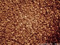 Squamous epithelium 100×
 Human cheek cells (nonkeratinized stratified squamous epithelium) 500×
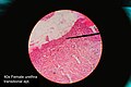 Histology of female urethra showing transitional epithelium
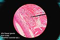 Histology of sweat gland showing stratified cuboidal epithelium


