Search This Blog

Monday, April 17, 2017

Symbiogenesis

From Wikipedia, the free encyclopedia
Internal symbiont: Mitochondrion has a matrix and membranes, like a free-living proteobacterial cell, from which it may derive.

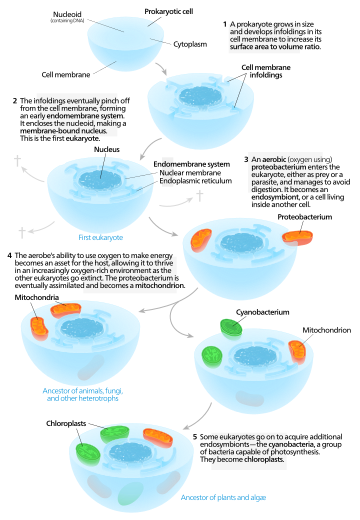
Symbiogenesis, or endosymbiotic theory, is an evolutionary theory of the origin of eukaryotic cells from prokaryotic organisms, first articulated in 1905 and 1910 by the Russian botanist Konstantin Mereschkowski, and advanced and substantiated with microbiological evidence by Lynn Margulis in 1967. It holds that the organelles distinguishing eukaryote cells evolved through symbiosis of individual single-celled prokaryotes (bacteria and archaea).

The theory holds that mitochondria, plastids such as chloroplasts, and possibly other organelles of eukaryotic cells represent formerly free-living prokaryotes taken one inside the other in endosymbiosis, around 1.5 billion years ago. In more detail, mitochondria appear to be related to Rickettsiales proteobacteria, and chloroplasts to nitrogen-fixing filamentous cyanobacteria.

Among the many lines of evidence supporting symbiogenesis are that new mitochondria and plastids are formed only through binary fission, and that cells cannot create new ones otherwise; that the transport proteins called porins are found in the outer membranes of mitochondria, chloroplasts and bacterial cell membranes; that cardiolipin is found only in the inner mitochondrial membrane and bacterial cell membranes; and that some mitochondria and plastids contain single circular DNA molecules similar to the DNA of bacteria.

History

Endosymbiotic theory
Kwang Jeon's experiment supports endosymbiosis. [I] Amoebae infected by x-bacteria [II] Many amoebae become sick and die [III] Survivors have x-bacteria living in their cytoplasm [IV] Antibiotics kill x-bacteria: host amoebae die as now dependent on x-bacteria.

The theory of symbiogenesis (Greek: σύν syn "together", βίωσις biosis "living", and γένεσις genesis "origin or birth") was first articulated by the Russian botanist Konstantin Mereschkowski in his work in 1910, The Theory of Two Plasms as the Basis of Symbiogenesis, a New Study or the Origins of Organisms, although the fundamentals of the idea already had appeared in his earlier 1905 work, The nature and origins of chromatophores in the plant kingdom.[1][2][3] Mereschkowski was familiar with work by botanist Andreas Schimper, who had observed in 1883 that the division of chloroplasts in green plants closely resembled that of free-living cyanobacteria, and who had himself tentatively proposed (in a footnote) that green plants had arisen from a symbiotic union of two organisms.[4] In 1918 the French scientist Paul Portier published Les Symbiotes in which he claimed that the mitochondria originated from a symbiosis process.[5] Ivan Wallin extended the idea of an endosymbiotic origin to mitochondria in the 1920s.[6][7] The Russian botanist Boris Kozo-Polyansky was the first to explain the theory in terms of Darwinian evolution.[8] In his 1924 book Symbiogenesis: A New Principle of Evolution he wrote, "The theory of symbiogenesis is a theory of selection relying on the phenomenon of symbiosis."[9] These theories were initially dismissed or ignored. More detailed electron microscopic comparisons between cyanobacteria and chloroplasts (for example studies by Hans Ris published in 1961[10]), combined with the discovery that plastids and mitochondria contain their own DNA[11] (which by that stage was recognized to be the hereditary material of organisms) led to a resurrection of the idea in the 1960s.

The theory was advanced and substantiated with microbiological evidence by Lynn Margulis in a 1967 paper, On the origin of mitosing cells.[12] In her 1981 work Symbiosis in Cell Evolution she argued that eukaryotic cells originated as communities of interacting entities, including endosymbiotic spirochaetes that developed into eukaryotic flagella and cilia. This last idea has not received much acceptance, because flagella lack DNA and do not show ultrastructural similarities to bacteria or archaea (see also: Evolution of flagella and Prokaryotic cytoskeleton). According to Margulis and Dorion Sagan,[13] "Life did not take over the globe by combat, but by networking" (i.e., by cooperation). The possibility that peroxisomes may have an endosymbiotic origin has also been considered, although they lack DNA. Christian de Duve proposed that they may have been the first endosymbionts, allowing cells to withstand growing amounts of free molecular oxygen in the Earth's atmosphere. However, it now appears that they may be formed de novo, contradicting the idea that they have a symbiotic origin.[14]

It is thought that over millennia these endosymbionts transferred some of their own DNA to the host cell's nucleus (called "endosymbiotic gene transfer" or "endogenosymbiosis") during the evolutionary transition from a symbiotic community to an instituted eukaryotic cell. The endosymbiotic theory is considered to be a type of saltational evolution.[15]
One model for the origin of mitochondria and plastids.

From endosymbionts to organelles

According to Keeling and Archibald,[16] the usual way to distinguish organelles from endosymbionts is by their reduced genome sizes. As an endosymbiont evolves into an organelle, most of their genes are transferred to the host cell genome. The host cell and organelle need to develop a transport mechanism that enables transfer back of the protein products needed by the organelle but now manufactured by the cell. Cyanobacteria and α-proteobacteria are the most closely related free-living organisms to plastids and mitochondria respectively.[17] Both cyanobacteria and α-proteobacteria maintain a large (>6Mb) genome encoding thousands of proteins.[17] Plastids and mitochondria exhibit a dramatic reduction in genome size when compared to their bacterial relatives.[17] Chloroplast genomes in photosynthetic organisms are normally 120-200kb[18] encoding 20-200 proteins[17] and mitochondrial genomes in humans are approximately 16kb and encode 37 genes, 13 of which are proteins.[19] Using the example of the freshwater amoeboid, however, Paulinella chromatophora, which contains chromatophores found to be evolved from cyanobacteria, Keeling and Archibald argue that this is not the only possible criterion; another is that the host cell has assumed control of the regulation of the former endosymbiont's division, thereby synchronizing it with the cell's own division.[16] Nowack and her colleagues[20] performed gene sequencing on the chromatophore (1.02 Mb) and found that only 867 proteins were encoded by these photosynthetic cells. Comparisons with their closest free living cyanobacteria of the genus Synechococcus (having a genome size 3 Mb, with 3300 genes) revealed that chromatophores underwent a drastic genome shrinkage. Chromatophores contained genes that were accountable for photosynthesis but were deficient in genes that could carry out other biosynthetic functions; this observation suggests that these endosymbiotic cells are highly dependent on their hosts for their survival and growth mechanisms. Thus, these chromatophores were found to be non-functional for organelle-specific purposes when compared to mitochondria and plastids. This distinction could have promoted the early evolution of photosynthetic organelles.

The loss of genetic autonomy, that is, the loss of many genes from endosymbionts, occurred very early in evolutionary time.[21] Taking into account the entire original endosymbiont genome, there are three main possible fates for genes over evolutionary time. The first fate involves the loss of functionally redundant genes,[21] in which genes that are already represented in the nucleus are eventually lost.The second fate involves the transfer of genes to the nucleus.[17][21][22][23][24] The loss of autonomy and integration of the endosymbiont with its host can be primarily attributed to nuclear gene transfer.[24] As organelle genomes have been greatly reduced over evolutionary time, nuclear genes have expanded and become more complex.[17] As a result, many plastid and mitochondrial processes are driven by nuclear encoded gene products.[17] In addition, many nuclear genes originating from endosymbionts have acquired novel functions unrelated to their organelles.[17][24]

The mechanisms of gene transfer are not fully known; however, multiple hypotheses exist to explain this phenomenon. The cDNA hypothesis involves the use of messenger RNA (mRNAs) to transport genes from organelles to the nucleus where they are converted to cDNA and incorporated into the genome.[17][22] The cDNA hypothesis is based on studies of the genomes of flowering plants.[17] Protein coding RNAs in mitochondria are spliced and edited using organelle-specific splice and editing sites.[17] Nuclear copies of some mitochondrial genes, however, do not contain organelle-specific splice sites, suggesting a processed mRNA intermediate.[17] The cDNA hypothesis has since been revised as edited mitochondrial cDNAs are unlikely to recombine with the nuclear genome and are more likely to recombine with their native mitochondrial genome. If the edited mitochondrial sequence recombines with the mitochondrial genome, mitochondrial splice sites would no longer exist in the mitochondrial genome.[17] Any subsequent nuclear gene transfer would therefore also lack mitochondrial splice sites.[17]

The bulk flow hypothesis is the alternative to the cDNA hypothesis, stating that escaped DNA, rather than mRNA, is the mechanism of gene transfer.[17][22] According to this hypothesis, disturbances to organelles, including autophagy (normal cell destruction), gametogenesis (the formation of gametes), and cell stress, release DNA which is imported into the nucleus and incorporated into the nuclear DNA using non-homologous end joining (repair of double stranded breaks).[22] For example, in the initial stages of endosymbiosis, due to a lack of major gene transfer, the host cell had little to no control over the endosymbiont.[21] The endosymbiont underwent cell division independently of the host cell, resulting in many "copies" of the endosymbiont within the host cell.[21] Some of the endosymbionts lysed (burst), and high levels of DNA were incorporated into the nucleus.[21] A similar mechanism is thought to occur in tobacco plants, who show a high rate of gene transfer and whose cells contain multiple chloroplasts.[21] In addition, the bulk flow hypothesis is also supported by the presence of non-random clusters of organelle genes, suggesting the simultaneous movement of multiple genes.[22]

In 2015, the biologist Roberto Cazzolla Gatti provided evidence for a variant theory,[25] endogenosymbiosis, in which not only are organelles endosymbiotic, but that pieces of genetic material from symbiotic parasites ("gene carriers" such as viruses, retroviruses and bacteriophages), are included in the host's nuclear DNA, changing the host's gene expression and contributing to the process of speciation.[26]

Molecular and biochemical evidence suggests that mitochondria are related to Rickettsiales proteobacteria (in particular, the SAR11 clade,[27][28] or close relatives), and that chloroplasts are related to nitrogen-fixing filamentous cyanobacteria.[29][30]

Organellar genomes

Plastomes and mitogenomes

The human mitochondrial genome has retained genes encoding 2 rRNAs, 22 tRNAs, and 13 redox proteins.

The third and final possible fate of endosymbiont genes is that they remain in the organelles. Plastids and mitochondria, although they have lost much of their genomes, retain genes encoding rRNAs, tRNAs, proteins involved in redox reactions, and proteins required for transcription, translation, and replication.[17][18][21] There are many hypotheses to explain why organelles retain a small portion of their genome; however no one hypothesis will apply to all organisms[21] and the topic is still quite controversial.[17] The hydrophobicity hypothesis states that highly hydrophobic (water hating) proteins (such as the membrane bound proteins involved in redox reactions) are not easily transported through the cytosol and therefore these proteins must be encoded in their respective organelles.[17][21] The code disparity hypothesis states that the limit on transfer is due to differing genetic codes and RNA editing between the organelle and the nucleus.[21] The redox control hypothesis states that genes encoding redox reaction proteins are retained in order to effectively couple the need for repair and the synthesis of these proteins.[17][18][21] For example, if one of the photosystems is lost from the plastid, the intermediate electron carriers may lose or gain too many electrons, signalling the need for repair of a photosystem.[18] The time delay involved in signalling the nucleus and transporting a cytosolic protein to the organelle results in the production of damaging reactive oxygen species.[17][18][21] The final hypothesis states that the assembly of membrane proteins, particularly those involved in redox reactions, requires coordinated synthesis and assembly of subunits; however, translation and protein transport coordination is more difficult to control in the cytoplasm.[21]

Non-photosynthetic plastid genomes

The majority of the genes in the mitochondria and plastids are related to the expression (transcription, translation and replication) of genes encoding proteins involved in either photosynthesis (in plastids) or cellular respiration (in mitochondria).[17][18][21] One might predict, that the loss of photosynthesis or cellular respiration would allow for the complete loss of the plastid genome or the mitochondrial genome respectively.[21] While there are numerous examples of mitochondrial descendants (mitosomes and hydrogenosomes) that have lost their entire organellar genome,[31] non-photosynthetic plastids tend to retain a small genome.[21] There are two main hypotheses to explain this occurrence:

The essential tRNA hypothesis notes that there have been no documented functional plastid-to-nucleus gene transfers of genes encoding RNA products (tRNAs and rRNAs).[21] As a result, plastids must make their own functional RNAs or import nuclear counterparts.[21] The genes encoding tRNA-Glu and tRNA-fmet, however, appear to be indispensable.[21] The plastid is responsible for haem biosynthesis, which requires plastid encoded tRNA-Glu (from the gene trnE) as a precursor molecule.[21] Like other genes encoding RNAs, trnE cannot be transferred to the nucleus.[21] In addition, it is unlikely trnE could be replaced by a cytosolic tRNA-Glu as trnE is highly conserved; single base changes in trnE have resulted in the loss of haem synthesis.[21] The gene for tRNA-formylmethionine (tRNA-fmet) is also encoded in the plastid genome and is required for translation initiation in both plastids and mitochondria.[21] A plastid is required to continue expressing the gene for tRNA-fmet so long as the mitochondrion is translating proteins.[21]

The limited window hypothesis offers a more general explanation for the retention of genes in non-photosynthetic plastids.[32] According to the bulk flow hypothesis, genes are transferred to the nucleus following the disturbance of organelles.[22] Disturbance was common in the early stages of endosymbiosis, however, once the host cell gained control of organelle division, eukaryotes could evolve to have only one plastid per cell.[21] Having only one plastid severely limits gene transfer[21] as the lysis of the single plastid would likely result in cell death.[21][32] Consistent with this hypothesis,[32] organisms with multiple plastids show an 80-fold increase in plastid-to-nucleus gene transfer compared to organisms with single plastids.[32]

Evidence

There are many lines of evidence that mitochondria and plastids including chloroplasts arose from bacteria.[33][34][35][36][37]
  • New mitochondria and plastids are formed only through binary fission, the form of cell division used by bacteria and archaea.[38]
  • If a cell's mitochondria or chloroplasts are removed, the cell does not have the means to create new ones.[39] For example, in some algae, such as Euglena, the plastids can be destroyed by certain chemicals or prolonged absence of light without otherwise affecting the cell. In such a case, the plastids will not regenerate.
  • Transport proteins called porins are found in the outer membranes of mitochondria and chloroplasts and are also found in bacterial cell membranes.[40][41][42]
  • A membrane lipid cardiolipin is exclusively found in the inner mitochondrial membrane and bacterial cell membranes.[43]
  • Some mitochondria and some plastids contain single circular DNA molecules that are similar to the DNA of bacteria both in size and structure.[44]
  • Genome comparisons suggest a close relationship between mitochondria and Rickettsial bacteria.[45]
  • Genome comparisons suggest a close relationship between plastids and cyanobacteria.[46]
  • Many genes in the genomes of mitochondria and chloroplasts have been lost or transferred to the nucleus of the host cell. Consequently, the chromosomes of many eukaryotes contain genes that originated from the genomes of mitochondria and plastids.[44]
  • Mitochondrial and plastid ribosomes are more similar to those of bacteria (70S) than those of eukaryotes.[47]
  • Proteins created by mitochondria and chloroplasts use N-formylmethionine as the initiating amino acid, as do proteins created by bacteria but not proteins created by eukaryotic nuclear genes or archaea.[48][49]
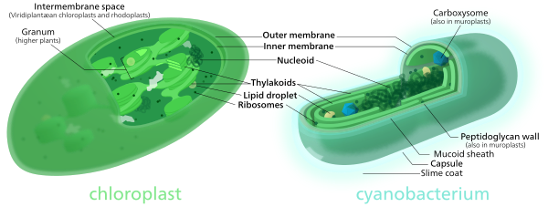
Comparison of chloroplasts and cyanobacteria showing their similarities.

Secondary endosymbiosis

Primary endosymbiosis involves the engulfment of a bacterium by another free living organism. Secondary endosymbiosis occurs when the product of primary endosymbiosis is itself engulfed and retained by another free living eukaryote. Secondary endosymbiosis has occurred several times and has given rise to extremely diverse groups of algae and other eukaryotes. Some organisms can take opportunistic advantage of a similar process, where they engulf an alga and use the products of its photosynthesis, but once the prey item dies (or is lost) the host returns to a free living state. Obligate secondary endosymbionts become dependent on their organelles and are unable to survive in their absence (for a review see McFadden 2001[50]). RedToL, the Red Algal Tree of Life Initiative funded by the National Science Foundation highlights the role red algae or Rhodophyta played in the evolution of our planet through secondary endosymbiosis.

One possible secondary endosymbiosis in process has been observed by Okamoto & Inouye (2005). The heterotrophic protist Hatena behaves like a predator until it ingests a green alga, which loses its flagella and cytoskeleton, while Hatena, now a host, switches to photosynthetic nutrition, gains the ability to move towards light and loses its feeding apparatus.[51]

The process of secondary endosymbiosis left its evolutionary signature within the unique topography of plastid membranes. Secondary plastids are surrounded by three (in euglenophytes and some dinoflagellates) or four membranes (in haptophytes, heterokonts, cryptophytes, and chlorarachniophytes). The two additional membranes are thought to correspond to the plasma membrane of the engulfed alga and the phagosomal membrane of the host cell. The endosymbiotic acquisition of a eukaryote cell is represented in the cryptophytes; where the remnant nucleus of the red algal symbiont (the nucleomorph) is present between the two inner and two outer plastid membranes.[citation needed]

Despite the diversity of organisms containing plastids, the morphology, biochemistry, genomic organisation, and molecular phylogeny of plastid RNAs and proteins suggest a single origin of all extant plastids – although this theory is still debated.[52][53]

Some species including Pediculus humanus (lice) have multiple chromosomes in the mitochondrion. This and the phylogenetics of the genes encoded within the mitochondrion suggest that mitochondria have multiple ancestors, that these were acquired by endosymbiosis on several occasions rather than just once, and that there have been extensive mergers and rearrangements of genes on the several original mitochondrial chromosomes.[54]

Symbiosis

From Wikipedia, the free encyclopedia
 
In a symbiotic mutualistic relationship, the clownfish feeds on small invertebrates that otherwise have potential to harm the sea anemone, and the fecal matter from the clownfish provides nutrients to the sea anemone. The clownfish is additionally protected from predators by the anemone's stinging cells, to which the clownfish is immune. The clownfish also emits a high pitched sound that deters butterfly fish, which would otherwise eat the anemone.[1]

Symbiosis (from Greek συμβίωσις "living together", from σύν "together" and βίωσις "living")[2] is any type of a close and long-term biological interaction between two different species, be it mutualistic, commensalistic, or parasitic. In 1879, Heinrich Anton de Bary defined it as "the living together of unlike organisms."

Symbiosis can be obligatory, which means that one or both of the symbionts entirely depend on each other for survival, or facultative (optional) when they can generally live independently.

Symbiosis is also classified by physical attachment; symbiosis in which the organisms have bodily union is called conjunctive symbiosis, and symbiosis in which they are not in union is called disjunctive symbiosis.[3] When one organism lives on another such as mistletoe, it is called ectosymbiosis, or endosymbiosis when one partner lives inside the tissues of another, as in Symbiodinium in corals.[4][5]

Definition

In 1877, Albert Bernhard Frank used the term symbiosis which previously had been used to depict people living together in community to describe the mutualistic relationship in lichens.[6] In 1879, the German mycologist Heinrich Anton de Bary defined it as "the living together of unlike organisms."[7][8] The definition has varied among scientists with some advocating that it should only refer to persistent mutualisms, while others thought it should apply to any type of persistent biological interaction in other words mutualistic, commensalistic, or parasitic.[9]
After 130 years of debate,[10] current biology and ecology textbooks use the latter "de Bary" definition or an even broader definition where symbiosis means all species interactions, and the restrictive definition where symbiosis means only mutualism is no longer used.[11]

Obligate versus facultative

Symbiotic relationships can be obligate, meaning that one or both of the symbionts entirely depend on each other for survival. For example, in lichens, which consist of fungal and photosynthetic symbionts, the fungal partners cannot live on their own.[7][12][13][14] The algal or cyanobacterial symbionts in lichens, such as Trentepohlia, can generally live independently, and their symbiosis is, therefore, facultative (optional).[citation needed]

Physical interaction

Alder tree root nodule

Endosymbiosis is any symbiotic relationship in which one symbiont lives within the tissues of the other, either within the cells or extracellularly.[5][15] Examples include diverse microbiomes, rhizobia, nitrogen-fixing bacteria that live in root nodules on legume roots; actinomycete nitrogen-fixing bacteria called Frankia, which live in alder root nodules; single-celled algae inside reef-building corals; and bacterial endosymbionts that provide essential nutrients to about 10%–15% of insects.[citation needed]

Ectosymbiosis, also referred to as exosymbiosis, is any symbiotic relationship in which the symbiont lives on the body surface of the host, including the inner surface of the digestive tract or the ducts of exocrine glands.[5][16] Examples of this include ectoparasites such as lice, commensal ectosymbionts such as the barnacles which attach themselves to the jaw of baleen whales, and mutualist ectosymbionts such as cleaner fish.

Mutualism

Hermit crab, Calcinus laevimanus, with sea anemone.

Mutualism or interspecies reciprocal altruism is a relationship between individuals of different species where both individuals benefit.[17] In general, only lifelong interactions involving close physical and biochemical contact can properly be considered symbiotic. Mutualistic relationships may be either obligate for both species, obligate for one but facultative for the other, or facultative for both.
Bryoliths document a mutualistic symbiosis between a hermit crab and encrusting bryozoans; Banc d'Arguin, Mauritania

A large percentage of herbivores have mutualistic gut flora to help them digest plant matter, which is more difficult to digest than animal prey.[4] This gut flora is made up of cellulose-digesting protozoans or bacteria living in the herbivores' intestines.[18] Coral reefs are the result of mutualisms between coral organisms and various types of algae which live inside them.[19] Most land plants and land ecosystems rely on mutualisms between the plants, which fix carbon from the air, and mycorrhyzal fungi, which help in extracting water and minerals from the ground.[20]

An example of mutual symbiosis is the relationship between the ocellaris clownfish that dwell among the tentacles of Ritteri sea anemones. The territorial fish protects the anemone from anemone-eating fish, and in turn the stinging tentacles of the anemone protect the clownfish from its predators. A special mucus on the clownfish protects it from the stinging tentacles.[21]

A further example is the goby fish, which sometimes lives together with a shrimp. The shrimp digs and cleans up a burrow in the sand in which both the shrimp and the goby fish live. The shrimp is almost blind, leaving it vulnerable to predators when outside its burrow. In case of danger the goby fish touches the shrimp with its tail to warn it. When that happens both the shrimp and goby fish quickly retreat into the burrow.[22] Different species of gobies (Elacatinus spp.) also exhibit mutualistic behavior through cleaning up ectoparasites in other fish.[23]

Another non-obligate symbiosis is known from encrusting bryozoans and hermit crabs. The bryozoan colony (Acanthodesia commensale) develops a cirumrotatory growth and offers the crab (Pseudopagurus granulimanus) a helicospiral-tubular extension of its living chamber that initially was situated within a gastropod shell.[24]

A spectacular examples of obligate mutualism is between the siboglinid tube worms and symbiotic bacteria that live at hydrothermal vents and cold seeps. The worm has no digestive tract and is wholly reliant on its internal symbionts for nutrition. The bacteria oxidize either hydrogen sulfide or methane, which the host supplies to them. These worms were discovered in the late 1980s at the hydrothermal vents near the Galapagos Islands and have since been found at deep-sea hydrothermal vents and cold seeps in all of the world's oceans.[25]

There are many types of tropical and sub-tropical ants that have evolved very complex relationships with certain tree species.[26]

Mutualism and endosymbiosis

During mutualistic symbioses, the host cell lacks some of the nutrients which the endosymbiont provides. As a result, the host favors endosymbiont's growth processes within itself by producing some specialized cells. These cells affect the genetic composition of the host in order to regulate the increasing population of the endosymbionts and ensure that these genetic changes are passed onto the offspring via vertical transmission (heredity).[27]

As the endosymbiont adapts to the host's lifestyle the endosymbiont changes dramatically. There is a drastic reduction in its genome size, as many genes are lost during the process of metabolism, and DNA repair and recombination, while important genes participating in the DNA to RNA transcription, protein translation and DNA/RNA replication are retained. The decrease in genome size is due to loss of protein coding genes and not due to lessening of inter-genic regions or open reading frame (ORF) size. Species that are naturally evolving and contain reduced sizes of genes can be accounted for an increased number of noticeable differences between them, thereby leading to changes in their evolutionary rates. When endosymbiotic bacteria related with insects are passed on to the offspring strictly via vertical genetic transmission, intracellular bacteria go across many hurdles during the process, resulting in the decrease in effective population sizes, as compared to the free living bacteria. The incapability of the endosymbiotic bacteria to reinstate their wild type phenotype via a recombination process is called Muller's ratchet phenomenon. Muller's ratchet phenomenon together with less effective population sizes leads to an accretion of deleterious mutations in the non-essential genes of the intracellular bacteria.[28] This can be due to lack of selection mechanisms prevailing in the relatively "rich" host environment.[29][30]

Commensalism

Phoretic mites on a fly (Pseudolynchia canariensis).

Commensalism describes a relationship between two living organisms where one benefits and the other is not significantly harmed or helped. It is derived from the English word commensal, which is used of human social interaction. The word derives from the medieval Latin word, formed from com- and mensa, meaning "sharing a table."[17][31]

Commensal relationships may involve one organism using another for transportation (phoresy) or for housing (inquilinism), or it may also involve one organism using something another created, after its death (metabiosis). Examples of metabiosis are hermit crabs using gastropod shells to protect their bodies and spiders building their webs on plants.

Parasitism

Flea bites on a human is an example of parasitism.

A parasitic relationship is one in which one member of the association benefits while the other is harmed.[32] This is also known as antagonistic or antipathetic symbiosis.[3] Parasitic symbioses take many forms, from endoparasites that live within the host's body to ectoparasites that live on its surface. In addition, parasites may be necrotrophic, which is to say they kill their host, or biotrophic, meaning they rely on their host's surviving. Biotrophic parasitism is an extremely successful mode of life. Depending on the definition used, as many as half of all animals have at least one parasitic phase in their life cycles, and it is also frequent in plants and fungi. Moreover, almost all free-living animals are host to one or more parasite taxa. An example of a biotrophic relationship would be a tick feeding on the blood of its host.

Amensalism

Amensalism is the type of relationship that exists where one species is inhibited or completely obliterated and one is unaffected by the other. There are two types of amensalism, competition and antibiosis. Competition is where a larger or stronger organism deprives a smaller or weaker one from a resource. Antibiosis occurs when one organism is damaged or killed by another through a chemical secretion. An example of competition is a sapling growing under the shadow of a mature tree. The mature tree can rob the sapling of necessary sunlight and, if the mature tree is very large, it can take up rainwater and deplete soil nutrients. Throughout the process, the mature tree is unaffected by the sapling. Indeed, if the sapling dies, the mature tree gains nutrients from the decaying sapling. Note that these nutrients become available because of the sapling's decomposition, rather than from the living sapling, which would be a case of parasitism.[citation needed] An example of antibiosis is Juglans nigra (black walnut), secreting juglone, a substance which destroys many herbaceous plants within its root zone.[33]
Amensalism is an interaction where an organism inflicts harm to another organism without any costs or benefits to the perpetrator.[34] A clear case of amensalism is where sheep or cattle trample grass. Whilst the presence of the grass causes negligible detrimental effects to the animal's hoof, the grass suffers from being crushed. Amensalism is often used to describe strongly asymmetrical competitive interactions, such as has been observed between the Spanish ibex and weevils of the genus Timarcha which feed upon the same type of shrub. Whilst the presence of the weevil has almost no influence on food availability, the presence of ibex has an enormous detrimental effect on weevil numbers, as they consume significant quantities of plant matter and incidentally ingest the weevils upon it.[35]

Synnecrosis

Synnecrosis is a rare type of symbiosis in which the interaction between species is detrimental to both organisms involved.[3] It is a short-lived condition, as the interaction eventually causes death. Because of this, evolution selects against synnecrosis and it is uncommon in nature. An example of this is the relationship between some species of bees and victims of the bee sting. Species of bees who die after stinging their prey inflict pain on themselves (albeit to protect the hive) as well as on the victim. This term is rarely used.[36]

Evolution

Leafhoppers protected by meat ants

While historically, symbiosis has received less attention than other interactions such as predation or competition,[37] it is increasingly recognized as an important selective force behind evolution,[4][38] with many species having a long history of interdependent co-evolution.[39] In fact, the evolution of all eukaryotes (plants, animals, fungi, and protists) is believed under the endosymbiotic theory to have resulted from a symbiosis between various sorts of bacteria.[4][40][41] This theory is supported by certain organelles dividing independently of the cell, and the observation that some organelles seem to have their own nucleic acid.[42]

Vascular plants

About 80% of vascular plants worldwide form symbiotic relationships with fungi, for example, in arbuscular mycorrhizas.[43]

Symbiogenesis

The biologist Lynn Margulis, famous for her work on endosymbiosis, contends that symbiosis is a major driving force behind evolution. She considers Darwin's notion of evolution, driven by competition, to be incomplete and claims that evolution is strongly based on co-operation, interaction, and mutual dependence among organisms. According to Margulis and Dorion Sagan, "Life did not take over the globe by combat, but by networking."[44]

Co-evolution

Symbiosis played a major role in the co-evolution of flowering plants and the animals that pollinate them. Many plants that are pollinated by insects, bats, or birds have highly specialized flowers modified to promote pollination by a specific pollinator that is also correspondingly adapted. The first flowering plants in the fossil record had relatively simple flowers. Adaptive speciation quickly gave rise to many diverse groups of plants, and, at the same time, corresponding speciation occurred in certain insect groups. Some groups of plants developed nectar and large sticky pollen, while insects evolved more specialized morphologies to access and collect these rich food sources. In some taxa of plants and insects the relationship has become dependent,[45] where the plant species can only be pollinated by one species of insect.[46]

Sunday, April 16, 2017

Geological history of oxygen

From Wikipedia, the free encyclopedia
 
O2 build-up in the Earth's atmosphere. Red and green lines represent the range of the estimates while time is measured in billions of years ago (Ga).
Stage 1 (3.85–2.45 Ga): Practically no O2 in the atmosphere.
Stage 2 (2.45–1.85 Ga): O2 produced, but absorbed in oceans and seabed rock.
Stage 3 (1.85–0.85 Ga): O2 starts to gas out of the oceans, but is absorbed by land surfaces and formation of ozone layer.
Stages 4 and 5 (0.85 Ga–present): O2 sinks filled, the gas accumulates.[1]

Before photosynthesis evolved, Earth's atmosphere had no free oxygen (O2).[2] Photosynthetic prokaryotic organisms that produced O2 as a waste product lived long before the first build-up of free oxygen in the atmosphere,[3] perhaps as early as 3.5 billion years ago. The oxygen they produced would have been rapidly removed from the atmosphere by weathering of reducing minerals, most notably iron. This "mass rusting" led to the deposition of iron oxide on the ocean floor, forming banded iron formations. Oxygen only began to persist in the atmosphere in small quantities about 50 million years before the start of the Great Oxygenation Event.[4] This mass oxygenation of the atmosphere resulted in rapid buildup of free oxygen. At current rates of primary production, today's concentration of oxygen could be produced by photosynthetic organisms in 2,000 years.[5] In the absence of plants, the rate of oxygen production by photosynthesis was slower in the Precambrian, and the concentrations of O2 attained were less than 10% of today's and probably fluctuated greatly; oxygen may even have disappeared from the atmosphere again around 1.9 billion years ago.[6] These fluctuations in oxygen concentration had little direct effect on life,[citation needed] with mass extinctions not observed until the appearance of complex life around the start of the Cambrian period, 541 million years ago.[7] The presence of O
2
provided life with new opportunities. Aerobic metabolism is more efficient than anaerobic pathways, and the presence of oxygen undoubtedly created new possibilities for life to explore.[8]:214, 586[9] Since the start of the Cambrian period, atmospheric oxygen concentrations have fluctuated between 15% and 35% of atmospheric volume.[10] The maximum of 35% was reached towards the end of the Carboniferous period (about 300 million years ago), a peak which may have contributed to the large size of insects and amphibians at that time.[9] Whilst human activities, such as the burning of fossil fuels, affect relative carbon dioxide concentrations, their effect on the much larger concentration of oxygen is less significant.[11]

Effects on life

The concentration of oxygen in the atmosphere is often cited as a possible contributor to large-scale evolutionary phenomena, such as the origin of the multicellular Ediacara biota, the Cambrian explosion, trends in animal body size, and other extinction and diversification events.[9]

The large size of insects and amphibians in the Carboniferous period, when the oxygen concentration in the atmosphere reached 35%, has been attributed to the limiting role of diffusion in these organisms' metabolism.[citation needed] But Haldane's essay[12] points out that it would only apply to insects. However, the biological basis for this correlation is not firm, and many lines of evidence show that oxygen concentration is not size-limiting in modern insects.[9] There is no significant correlation between atmospheric oxygen and maximum body size elsewhere in the geological record.[9] Ecological constraints can better explain the diminutive size of post-Carboniferous dragonflies - for instance, the appearance of flying competitors such as pterosaurs, birds and bats.[9]

Rising oxygen concentrations have been cited as a driver for evolutionary diversification, although the physiological arguments behind such arguments are questionable, and a consistent pattern between oxygen concentrations and the rate of evolution is not clearly evident.[9] The most celebrated link between oxygen and evolution occurs at the end of the last of the Snowball glaciations, where complex multicellular life is first found in the fossil record. Under low oxygen concentrations and before the evolution of nitrogen fixation, biologically-available nitrogen compounds were in limited supply [13] and periodic "nitrogen crises" could render the ocean inhospitable to life.[9] Significant concentrations of oxygen were just one of the prerequisites for the evolution of complex life.[9] Models based on uniformitarian principles (i.e. extrapolating present-day ocean dynamics into deep time) suggest that such a concentration was only reached immediately before metazoa first appeared in the fossil record.[9] Further, anoxic or otherwise chemically "nasty" oceanic conditions that resemble those supposed to inhibit macroscopic life re-occur at intervals through the early Cambrian, and also in the late Cretaceous – with no apparent effect on lifeforms at these times.[9] This might suggest that the geochemical signatures found in ocean sediments reflect the atmosphere in a different way before the Cambrian - perhaps as a result of the fundamentally different mode of nutrient cycling in the absence of planktivory.[7][9]

Operator (computer programming)

From Wikipedia, the free encyclopedia https://en.wikipedia.org/wiki/Operator_(computer_programmin...