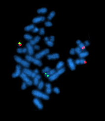
Image: example of karyotyping showing a total of 46 chromosomes in the genome.
Molecular cytogenetics combines two disciplines, molecular biology and cytogenetics,
and involves the analyzation of chromosome structure to help
distinguish normal and cancer-causing cells. Human cytogenetics began in
1956 when it was discovered that normal human cells contain 46
chromosomes. However, the first microscopic observations of chromosomes
were reported by Arnold, Flemming, and Hansemann
in the late 1800's. Their work was ignored for decades until the actual
chromosome number in humans was discovered as 46. In 1879, Arnold
examined sarcoma and carcinoma cells having very large nuclei. Today,
the study of molecular cytogenetics can be useful in diagnosing and
treating various malignancies such as hematological malignancies, brain
tumors, and other precursors of cancer. The field is overall focused on
studying the evolution of chromosomes, more specifically the number,
structure, function, and origin of chromosome abnormalities. It includes a series of techniques referred to as fluorescence in situ hybridization, or FISH, in which DNA
probes are labeled with different colored fluorescent tags to visualize
one or more specific regions of the genome. Introduced in the 1980's,
FISH uses probes with complimentary base sequences to locate the
presence or absence of the specific DNA regions you are looking for.
FISH can either be performed as a direct approach to metaphase
chromosomes or interphase nuclei. Alternatively, an indirect approach
can be taken in which the entire genome can be assessed for copy number
changes using virtual karyotyping. Virtual karyotypes are generated from arrays made of thousands to millions of probes, and computational tools are used to recreate the genome in silico.
Common techniques
Fluorescence in situ hybridization (FISH)
FISH
images of chromosomes from dividing orangutan (left) and human (right)
cells. Yellow probe shows 4 copies of a region in the orangutan genome
and only 2 copies in human.
Fluorescence In Situ Hybridization
maps out single copy or repetitive DNA sequences through localization
labeling of specific nucleic acids. The technique utilizes different DNA
probes labeled with fluorescent tags that bind to one or more specific
regions of the genome.
It labels all individual chromosomes at every stage of cell division to
display structural and numerical abnormalities that may arise
throughout the cycle. This is done with a probe that can be locus
specific, centromeric, telomeric, and whole-chromosomal. This technique
is typically preformed on interphase cells and paraffin block tissues.
FISH maps out single copy or repetitive DNA sequences through
localization labeling of specific nucleic acids. The technique utilizes
different DNA probes labeled with fluorescent tags that bind to one or
more specific regions of the genome. Signals from the fluorescent tags
can be seen with microscopy,
and mutations can be seen by comparing these signals to healthy
cells.For this to work, DNA must be denatured using heat or chemicals to
break the hydrogen bonds; this allows hybridization to occur once two
samples are mixed. The florescent probes create new hydrogen bonds, thus
repairing DNA with their complimentary bases, which can be detected
through microscopy. FISH allows one to visualize different parts of the
chromosome at different stages of the cell cycle. FISH can either be
performed as a direct approach to metaphase chromosomes or interphase
nuclei. Alternatively, an indirect approach can be taken in which the
entire genome can be assessed for copy number changes using virtual
karyotyping. Virtual karyotypes
are generated from microarrays made of thousands to millions of probes,
and computational tools are used to recreate the genome in silico.
Comparative genomic hybridization (CGH)
Comparative
genomic hybridization (CGH), derived from FISH, is used to compare
variations in copy number between a biological sample and a reference.
CGH was originally developed to observe chromosomal aberrations in
tumour cells. This method uses two genomes, a sample and a control,
which are labeled fluorescently to distinguish them.
In CGH, DNA is isolated from a tumour sample and biotin is attached.
Another labelling protein, digoxigenin, is attached to the reference DNA
sample.
The labelled DNA samples are co-hybridized to probes during cell
division, which is the most informative time for observing copy number
variation.
CGH uses creates a map that shows the relative abundance of DNA and
chromosome number. By comparing the fluorescence in a sample compared to
a reference, CGH can point to gains or losses of chromosomal regions.
CGH differs from FISH because it does not require a specific target or
previous knowledge of the genetic region being analyzed. CGH can also
scan an entire genome relatively quickly for various chromosome
imbalances, and this is helpful in patients with underlying genetic
issues and when an official diagnosis is not known. This often occurs
with hematological cancers.
Array comparative genomic hybridization (aCGH)
Array
comparative genomic hybridization (aCGH) allows CGH to be performed
without cell culture and isolation. Instead, it is performed on glass
slides containing small DNA fragments.
Removing the cell culture and isolation step dramatically simplifies
and expedites the process. Using similar principles to CGH, the sample
DNA is isolated and fluorescently labelled, then co-hybridized to single
stranded probes to generate signals. Thousands of these signals can be
detected for at once, and this process is referred to as parallel
screening.
Fluorescence ratios between the sample and reference signals are
measured, representing the average difference between the amount of
each. This will show if there is more or less sample DNA than is
expected by reference.
Applications
A
cell containing a rearrangement of the bcr/abl chromosomal regions
(upper left red and green chromosome). This rearrangement is associated
with chronic myelogenous leukemia, and was detected using FISH.
FISH chromosome in-situ hybridization allows the study cytogenetics in pre- and postnatal samples and is also widely used in cytogenetic testing
for cancer. While cytogenetics is the study of chromosomes and their
structure, cytogenetic testing involves the analysis of cells in the
blood, tissue, bone marrow, or fluid to identify changes in chromosomes
of an individual. This was often done through karyotyping, and is now
done with FISH. This method is commonly used to detect chromosomal
deletions or translocations often associated with cancer. FISH is also
used for melanocytic lesions, distinguishing atypical melanocytic or
malignant melanoma.
Cancer cells often accumulate complex chromosomal structural
changes such as loss, duplication, inversion or movement of a segment.
When using FISH, any changes to a chromosome will be made visible
through discrepancies between fluorescent-labelled cancer chromosomes
and healthy chromosomes.
The findings of these cytogenetic experiments can shed light on the
genetic causes for the cancer and can locate potential therapeutic
targets.
Molecular cytogenetics can also be used as a diagnostic tool for congenital syndromes in which the underlying genetic causes of the disease are unknown.
Analysis of a patient's chromosome structure can reveal causative
changes. New molecular biology methods developed in the past two decades
such as next generation sequencing and RNA-seq
have largely replaced molecular cytogenetics in diagnostics, but
recently the use of derivatives of FISH such as multicolour FISH and
multicolour banding (mBAND) has been growing in medical applications.
Cancer projects
One of the current projects involving Molecular Cytogenetics involves genomic research on rare cancers, called the Cancer Genome Characterization Initiative (CGCI).
The CGCI is a group interested in describing the genetic abnormalities
of some rare cancers, by employing advanced sequencing of genomes,
exomes, and transcriptomes, which may ultimately play a role in cancer
pathogenesis. Currently, the CGCI has elucidated some previously undetermined genetic alterations in medulloblastoma and B-cell non-Hodgkin lymphoma. The next steps for the CGCI is to identify genomic alternations in HIV+ tumors and in Burkitt's Lymphoma.
Some high-throughput sequencing techniques that are used by the CGCI include: whole genome sequencing, transcriptome sequencing, ChIP-sequencing, and Illumina Infinum MethylationEPIC BeadCHIP.


