Search This Blog

Thursday, September 20, 2018

Artificial cell

From Wikipedia, the free encyclopedia
 
Two types of artificial cells, one with contents meant to stay inside, the other for drug delivery and diffusing contents.
Standard artificial cell (top) and drug delivery artificial cell (bottom).

An artificial cell or minimal cell is an engineered particle that mimics one or many functions of a biological cell. The term does not refer to a specific physical entity, but rather to the idea that certain functions or structures of biological cells can be replaced or supplemented with a synthetic entity. Often, artificial cells are biological or polymeric membranes which enclose biologically active materials. As such, nanoparticles, liposomes, polymersomes, microcapsules and a number of other particles have qualified as artificial cells. Micro-encapsulation allows for metabolism within the membrane, exchange of small molecules and prevention of passage of large substances across it. The main advantages of encapsulation include improved mimicry in the body, increased solubility of the cargo and decreased immune responses. Notably, artificial cells have been clinically successful in hemoperfusion.

In the area of synthetic biology, a "living" artificial cell has been defined as a completely synthetically made cell that can capture energy, maintain ion gradients, contain macromolecules as well as store information and have the ability to mutate. Such a cell is not technically feasible yet, but a variation of an artificial cell has been created in which a completely synthetic genome was introduced to genomically emptied host cells. Although not completely artificial because the cytoplasmic components as well as the membrane from the host cell are kept, the engineered cell is under control of a synthetic genome and is able to replicate.

History

The first artificial cells were developed by Thomas Chang at McGill University in the 1960s. These cells consisted of ultrathin membranes of nylon, collodion or crosslinked protein whose semipermeable properties allowed diffusion of small molecules in and out of the cell. These cells were micron-sized and contained cell, enzymes, hemoglobin, magnetic materials, adsorbents and proteins.

Later artificial cells have ranged from hundred-micrometer to nanometer dimensions and can carry microorganisms, vaccines, genes, drugs, hormones and peptides. The first clinical use of artificial cells was in hemoperfusion by the encapsulation of activated charcoal.

In the 1970s, researchers were able to introduce enzymes, proteins and hormones to biodegradable microcapsules, later leading to clinical use in diseases such as Lesch-Nyhan syndrome. Although Chang's initial research focused on artificial red blood cells, only in the mid-1990s were biodegradable artificial red blood cells developed. Artificial cells in biological cell encapsulation were first used in the clinic in 1994 for treatment in a diabetic patient and since then other types of cells such as hepatocytes, adult stem cells and genetically engineered cells have been encapsulated and are under study for use in tissue regeneration.

On December 29, 2011, chemists at Harvard University reported the creation of an artificial cell membrane.

By 2014, self-replicating, synthetic bacterial cells with cell walls and synthetic DNA had been produced. In January of that year researchers produced an artificial eukaryotic cell capable of undertaking multiple chemical reactions through working organelles.

In September 2018, researchers at the University of California developed artificial cells that can kill bacteria. The cells were engineered from the bottom-up — like Lego blocks — to destroy bacteria.

Materials

Different types of artificial cell membranes.
Representative types of artificial cell membranes.

Membranes for artificial cells be made of simple polymers, crosslinked proteins, lipid membranes or polymer-lipid complexes. Further, membranes can be engineered to present surface proteins such as albumin, antigens, Na/K-ATPase carriers, or pores such as ion channels. Commonly used materials for the production of membranes include hydrogel polymers such as alginate, cellulose and thermoplastic polymers such as hydroxyethyl methacrylate-methyl methacrylate (HEMA- MMA), polyacrylonitrile-polyvinyl chloride (PAN-PVC), as well as variations of the above-mentioned. The material used determines the permeability of the cell membrane, which for polymer depends on the molecular weight cut off (MWCO). The MWCO is the maximum molecular weight of a molecule that may freely pass through the pores and is important in determining adequate diffusion of nutrients, waste and other critical molecules. Hydrophilic polymers have the potential to be biocompatible and can be fabricated into a variety of forms which include polymer micelles, sol-gel mixtures, physical blends and crosslinked particles and nanoparticles. Of special interest are stimuli-responsive polymers that respond to pH or temperature changes for the use in targeted delivery. These polymers may be administered in the liquid form through a macroscopic injection and solidify or gel in situ because of the difference in pH or temperature. Nanoparticle and liposome preparations are also routinely used for material encapsulation and delivery. A major advantage of liposomes is their ability to fuse to cell and organelle membranes.

Preparation

Many variations for artificial cell preparation and encapsulation have been developed. Typically, vesicles such as a nanoparticle, polymersome or liposome are synthesized. An emulsion is typically made through the use of high pressure equipment such as a high pressure homogenizer or a Microfluidizer. Two micro-encapsulation methods for nitrocellulose are also described below.

High-pressure homogenization

In a high-pressure homogenizer, two liquids in oil/liquid suspension are forced through a small orifice under very high pressure. This process shears the products and allows the creation of extremely fine particles, as small as 1 nm.

Microfluidization

This technique uses a patented Microfluidizer to obtain a greater amount of homogenous suspensions that can create smaller particles than homogenizers. A homogenizer is first used to create a coarse suspension which is then pumped into the microfluidizer under high pressure. The flow is then split into two streams which will react at very high velocities in an interaction chamber until desired particle size is obtained. This technique allows for large scale production of phospholipid liposomes and subsequent material nanoencapsulations.

Drop method

In this method, a cell solution is incorporated dropwise into a collodion solution of cellulose nitrate. As the drop travels through the collodion, it is coated with a membrane thanks to the interfacial polymerization properties of the collodion. The cell later settles into paraffin where the membrane sets and is finally suspended a saline solution. The drop method is used for the creation of large artificial cells which encapsulate biological cells, stem cells and genetically engineered stem cells.

Emulsion method

The emulsion method differs in that the material to be encapsulated is usually smaller and is placed in the bottom of a reaction chamber where the collodion is added on top and centrifuged, or otherwise disturbed in order to create an emulsion. The encapsulated material is then dispersed and suspended in saline solution.

Clinical relevance

Drug release and delivery

Artificial cells used for drug delivery differ from other artificial cells since their contents are intended to diffuse out of the membrane, or be engulfed and digested by a host target cell. Often used are submicron, lipid membrane artificial cells that may be referred to as nanocapsules, nanoparticles, polymersomes, or other variations of the term.

Enzyme therapy

Enzyme therapy is being actively studied for genetic metabolic diseases where an enzyme is over-expressed, under-expressed, defective, or not at all there. In the case of under-expression or expression of a defective enzyme, an active form of the enzyme is introduced in the body to compensate for the deficit. On the other hand, an enzymatic over-expression may be counteracted by introduction of a competing non-functional enzyme; that is, an enzyme which metabolizes the substrate into non-active products. When placed within an artificial cell, enzymes can carry out their function for a much longer period compared to free enzymes and can be further optimized by polymer conjugation.

The first enzyme studied under artificial cell encapsulation was asparaginase for the treatment of lymphosarcoma in mice. This treatment delayed the onset and growth of the tumor. These initial findings led to further research in the use of artificial cells for enzyme delivery in tyrosine dependent melanomas. These tumors have a higher dependency on tyrosine than normal cells for growth, and research has shown that lowering systemic levels of tyrosine in mice can inhibit growth of melanomas. The use of artificial cells in the delivery of tyrosinase; and enzyme that digests tyrosine, allows for better enzyme stability and is shown effective in the removal of tyrosine without the severe side-effects associated with tyrosine depravation in the diet.

Artificial cell enzyme therapy is also of interest for the activation of prodrugs such as ifosfamide in certain cancers. Artificial cells encapsulating the cytochrome p450 enzyme which converts this prodrug into the active drug can be tailored to accumulate in the pancreatic carcinoma or implanting the artificial cells close to the tumor site. Here, the local concentration of the activated ifosfamide will be much higher than in the rest of the body thus preventing systemic toxicity. The treatment was successful in animals and showed a doubling in median survivals amongst patients with advanced-stage pancreatic cancer in phase I/II clinical trials, and a tripling in one-year survival rate.

Gene therapy

In treatment of genetic diseases, gene therapy aims to insert, alter or remove genes within an afflicted individual's cells. The technology relies heavily on viral vectors which raises concerns about insertional mutagenesis and systemic immune response that have led to human deaths and development of leukemia in clinical trials. Circumventing the need for vectors by using naked or plasmid DNA as its own delivery system also encounters problems such as low transduction efficiency and poor tissue targeting when given systemically.

Artificial cells have been proposed as a non-viral vector by which genetically modified non-autologous cells are encapsulated and implanted to deliver recombinant proteins in vivo. This type of immuno-isolation has been proven efficient in mice through delivery of artificial cells containing mouse growth hormone which rescued a growth-retardation in mutant mice. A few strategies have advanced to human clinical trials for the treatment of pancreatic cancer, lateral sclerosis and pain control.

Hemoperfusion

The first clinical use of artificial cells was in hemoperfusion by the encapsulation of activated charcoal. Activated charcoal has the capability of adsorbing many large molecules and has for a long time been known for its ability to remove toxic substances from the blood in accidental poisoning or overdose. However, perfusion through direct charcoal administration is toxic as it leads to embolisms and damage of blood cells followed by removal by platelets. Artificial cells allow toxins to diffuse into the cell while keeping the dangerous cargo within their ultrathin membrane.

Artificial cell hemoperfusion has been proposed as a less costly and more efficient detoxifying option than hemodialysis, in which blood filtering takes place only through size separation by a physical membrane. In hemoperfusion, thousands of adsorbent artificial cells are retained inside a small container through the use of two screens on either end through which patient blood perfuses. As the blood circulates, toxins or drugs diffuse into the cells and are retained by the absorbing material. The membranes of artificial cells are much thinner those used in dialysis and their small size means that they have a high membrane surface area. This means that a portion of cell can have a theoretical mass transfer that is a hundredfold higher than that of a whole artificial kidney machine. The device has been established as a routine clinical method for patients treated for accidental or suicidal poisoning but has also been introduced as therapy in liver failure and kidney failure by carrying out part of the function of these organs. Artificial cell hemoperfusion has also been proposed for use in immunoadsorption through which antibodies can be removed from the body by attaching an immunoadsorbing material such as albumin on the surface of the artificial cells. This principle has been used to remove blood group antibodies from plasma for bone marrow transplantation and for the treatment of hypercholesterolemia through monoclonal antibodies to remove low-density lipoproteins. Hemoperfusion is especially useful in countries with a weak hemodialysis manufacturing industry as the devices tend to be cheaper there and used in kidney failure patients.

Encapsulated cells

Schematic of cells encapsulated within an artificial membrane.
Schematic representation of encapsulated cells within artificial membrane.

The most common method of preparation of artificial cells is through cell encapsulation. Encapsulated cells are typically achieved through the generation of controlled-size droplets from a liquid cell suspension which are then rapidly solidified or gelated to provide added stability. The stabilization may be achieved through a change in temperature or via material crosslinking. The microenvironment that a cell sees changes upon encapsulation. It typically goes from being on a monolayer to a suspension in a polymer scaffold within a polymeric membrane. A drawback of the technique is that encapsulating a cell decreases its viability and ability to proliferate and differentiate. Further, after some time within the microcapsule, cells form clusters that inhibit the exchange of oxygen and metabolic waste, leading to apoptosis and necrosis thus limiting the efficacy of the cells and activating the host's immune system. Artificial cells have been successful for transplanting a number of cells including islets of Langerhans for diabetes treatment, parathyroid cells and adrenal cortex cells.

Encapsulated hepatocytes

Shortage of organ donors make artificial cells key players in alternative therapies for liver failure. The use of artificial cells for hepatocyte transplantation has demonstrated feasibility and efficacy in providing liver function in models of animal liver disease and bioartificial liver devices. Research stemmed off experiments in which the hepatocytes were attached to the surface of a micro-carriers and has evolved into hepatocytes which are encapsulated in a three-dimensional matrix in alginate microdroplets covered by an outer skin of polylysine. A key advantage to this delivery method is the circumvention of immunosuppression therapy for the duration of the treatment. Hepatocyte encapsulations have been proposed for use in a bioartifical liver. The device consists of a cylindrical chamber imbedded with isolated hepatocytes through which patient plasma is circulated extra-corporeally in a type of hemoperfusion. Because microcapsules have a high surface area to volume ratio, they provide large surface for substrate diffusion and can accommodate a large number of hepatocytes. Treatment to induced liver failure mice showed a significant increase in the rate of survival. Artificial liver systems are still in early development but show potential for patients waiting for organ transplant or while a patient's own liver regenerates sufficiently to resume normal function. So far, clinical trials using artificial liver systems and hepatocyte transplantation in end-stage liver diseases have shown improvement of health markers but have not yet improved survival. The short longevity and aggregation of artificial hepatocytes after transplantation are the main obstacles encountered. Hepatocytes co-encapsulated with stem cells show greater viability in culture and after implantation and implantation of artificial stem cells alone have also shown liver regeneration. As such interest has arisen in the use of stem cells for encapsulation in regenerative medicine.

Encapsulated bacterial cells

The oral ingestion of live bacterial cell colonies has been proposed and is currently in therapy for the modulation of intestinal microflora, prevention of diarrheal diseases, treatment of H. Pylori infections, atopic inflammations, lactose intolerance and immune modulation, amongst others. The proposed mechanism of action is not fully understood but is believed to have two main effects. The first is the nutritional effect, in which the bacteria compete with toxin producing bacteria. The second is the sanitary effect, which stimulates resistance to colonization and stimulates immune response. The oral delivery of bacterial cultures is often a problem because they are targeted by the immune system and often destroyed when taken orally. Artificial cells help address these issues by providing mimicry into the body and selective or long term release thus increasing the viability of bacteria reaching the gastrointestinal system. In addition, live bacterial cell encapsulation can be engineered to allow diffusion of small molecules including peptides into the body for therapeutic purposes. Membranes that have proven successful for bacterial delivery include cellulose acetate and variants of alginate. Additional uses that have arosen from encapsulation of bacterial cells include protection against challenge from M. Tuberculosis and upregulation of Ig secreting cells from the immune system. The technology is limited by the risk of systemic infections, adverse metabolic activities and the risk of gene transfer. However, the greater challenge remains the delivery of sufficient viable bacteria to the site of interest.

Artificial blood cell

Oxygen carriers

Nano sized oxygen carriers are used as a type of red blood cell substitutes, although they lack other components of red blood cells. They are composed of a synthetic polymersome or an artificial membrane surrounding purified animal, human or recombinant hemoglobin. Overall, hemoglobin delivery continues to be a challenge because it is highly toxic when delivered without any modifications. In some clinical trials, vasopressor effects have been observed for first generation hemoglobin blood substitutes.

Red blood cells

Research interest in the use of artificial cells for blood arose after the AIDS scare of the 1980s. Besides bypassing the potential for disease transmission, artificial red blood cells are desired because they eliminate drawbacks associated with allogenic blood transfusions such as blood typing, immune reactions and its short storage life of 42 days. A hemoglobin substitute may be stored at room temperature and not under refrigeration for more than a year. Attempts have been made to develop a complete working red blood cell which comprises carbonic not only an oxygen carrier but also the enzymes associated with the cell. The first attempt was made in 1957 by replacing the red blood cell membrane by an ultrathin polymeric membrane which was followed by encapsulation through a lipid membrane and more recently a biodegradable polymeric membrane. A biological red blood cell membrane including lipids and associated proteins can also be used to encapsulate nanoparticles and increase residence time in vivo by bypassing macrophage uptake and systemic clearance.

Leuko-polymersome

A leuko-polymersome is a polymersome engineered to have the adhesive properties of a leukocyte. Polymersomes are vesicles composed of a bilayer sheet that can encapsulate many active molecules such as drugs or enzymes. By adding the adhesive properties of a leukocyte to their membranes, they can be made to slow down, or roll along epithelial walls within the quickly flowing circulatory system.

Synthetic cells

The minimal cell

The German pathologist Rudolf Virchow brought forward the idea that not only does life arise from cells, but every cell comes from another cell; "Omnis cellula e cellula". Until now, most attempts to create an artificial cell have only created a package that can mimic certain tasks of the cell. Advances in cell-free transcription and translation reactions allow the expression of many genes, but these efforts are far from producing a fully operational cell.

The future is in the creation of a protocell, or a cell which has all the minimum requirements for life. Members from the J. Craig Venter Institute have used a top-down computational approach to knock out genes in a living organism to a minimum set of genes. In 2010, the team succeeded in creating a replicating strain of Mycoplasma mycoides (Mycoplasma laboratorium) using synthetically created DNA deemed to be the minimum requirement for life which was inserted into a genomically empty bacterium. It is hoped that the process of top-down biosynthesis will enable the insertion of new genes that would perform profitable functions such as generation of hydrogen for fuel or capturing excess carbon dioxide in the atmosphere. the myriad regulatory, metabolic, and signaling networks are not completely characterized. These top-down approaches have limitations for the understanding of fundamental molecular regulation, since the host organisms have a complex and incompletely defined molecular composition.

A bottom-up approach to build an artificial cell would involve creating a protocell de novo, entirely from non-living materials. It is proposed to create a phospholipid bilayer vesicle with DNA capable of self-reproducing using synthetic genetic information. The three primary elements of such artificial cells are the formation of a lipid membrane, DNA and RNA replication through a template process and the harvesting of chemical energy for active transport across the membrane. The main hurdles foreseen and encountered with this proposed protocell are the creation of a minimal synthetic DNA that holds all sufficient information for life, and the reproduction of non-genetic components that are integral in cell development such as molecular self-organization. However, it is hoped that this kind of bottom-up approach would provide insight into the fundamental questions of organizations at the cellular level and the origins of biological life. So far, no completely artificial cell capable of self-reproduction has been synthesized using the molecules of life, and this objective is still in a distant future although various groups are currently working towards this goal.

Another method proposed to create a protocell more closely resembles the conditions believed to have been present during evolution known as the primordial soup. Various RNA polymers could be encapsulated in vesicles and in such small boundary conditions, chemical reactions would be tested for.

Heavy investing in biology has been done by large companies such as ExxonMobil, who has partnered with Synthetic Genomics Inc; Craig Venter's own biosynthetics company in the development of fuel from algae.

Electronic artificial cell

The concept of an Electronic Artificial Cell has been expanded in a series of 3 EU projects coordinated by John McCaskill from 2004-2015.

The European Commission sponsored the development of the "Programmable Artificial Cell Evolution" (PACE) program from 2004-2008 whose goal was to lay the foundation for the creation of "microscopic self-organizing, self-replicating, and evolvable autonomous entities built from simple organic and inorganic substances that can be genetically programmed to perform specific functions" for the eventual integration into information systems. The PACE project developed the first Omega Machine, a microfluidic life support system for artificial cells that could complement chemically missing functionalities (as originally proposed by Norman Packard, Steen Rasmussen, Mark Beadau and John McCaskill). The ultimate aim was to attain an evolvable hybrid cell in a complex microscale programmable environment. The functions of the Omega Machine could then be removed stepwise, posing a series of solvable evolution challenges to the artificial cell chemistry. The project achieved chemical integration up to the level of pairs of the three core functions of artificial cells (a genetic subsystem, a containment system and a metabolic system), and generated novel spatially resolved programmable microfluidic environments for the integration of containment and genetic amplification."Programmable Artificial Cell Evolution" (PACE) The project led to the creation of the  European center for living technology] which is now continuing similar research.

Following this research, in 2007, John McCaskill proposed to concentrate on an electronically complemented artificial cell, called the Electronic Chemical Cell. The key idea was to use a massively parallel array of electrodes coupled to locally dedicated electronic circuitry, in a two-dimensional thin film, to complement emerging chemical cellular functionality. Local electronic information defining the electrode switching and sensing circuits could serve as an electronic genome, complementing the molecular sequential information in the emerging protocols. A research proposal was successful with the European Commission and an international team of scientists partially overlapping with the PACE consortium commenced work 2008-2012 on the project Electronic Chemical Cells. The project demonstrated among other things that electronically controlled local transport of specific sequences could be used as an artificial spatial control system for the genetic proliferation of future artificial cells, and that core processes of metabolism could be delivered by suitably coated electrode arrays.

The major limitation of this approach, apart from the initial difficulties in mastering microscale electrochemistry and electrokinetics, is that the electronic system is interconnected as a rigid non-autonomous piece of macroscopic hardware. In 2011, McCaskill proposed to invert the geometry of electronics and chemistry : instead of placing chemicals in an active electronic medium, to place microscopic autonomous electronics in a chemical medium. He organized a project to tackle a third generation of Electronic Artificial Cells at the 100 µm scale that could self-assemble from two half-cells "lablets" to enclose an internal chemical space, and function with the aid of active electronics powered by the medium they are immersed in. Such cells can copy both their electronic and chemical contents and will be capable of evolution within the constraints provided by their special pre-synthesized microscopic building blocks. In Sep 2012 work commenced on this project Microscale Chemically Reactive Electronic Agents.

Ethics and controversy

Protocell research has created controversy and opposing opinions, including critics of the vague definition of "artificial life". The creation of a basic unit of life is the most pressing ethical concern, although the most widespread worry about protocells is their potential threat to human health and the environment through uncontrolled replication.

Protocell

From Wikipedia, the free encyclopedia

A protocell (or protobiont) is a self-organized, endogenously ordered, spherical collection of lipids proposed as a stepping-stone to the origin of life. A central question in evolution is how simple protocells first arose and how they could differ in reproductive output, thus enabling the accumulation of novel biological emergences over time, i.e. biological evolution. Although a functional protocell has not yet been achieved in a laboratory setting, the goal to understand the process appears well within reach.

Overview

Compartmentalization was important in the origins of life. Membranes form enclosed compartments that are separate from the external environment, thus providing the cell with functionally specialized aqueous spaces. Because lipid bilayer of membranes is impermeable to most hydrophilic molecules (dissolved by water), cells have membrane transport systems that achive the import of nutritive molecules as well as the export of waste. It is very challenging to construct protocells from molecular assemblies. An important step in this challenge is the achievement of vesicle dynamics that are relevant to cellular functions, such as membrane trafficking and self-reproduction, using amphiphilic molecules. On the primitive Earth, numerous chemical reactions of organic compounds produced the ingredients of life. Of these substances, amphiphilic molecules might be the first player in the evolution from molecular assembly to cellular life. A step from vesicle toward protocell might be to develop self-reproducing vesicles coupled with the metabolic system.

Selectivity for compartmentalization

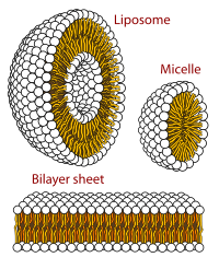
The three main structures phospholipids form in solution; the liposome (a closed bilayer), the micelle and the bilayer.

Self-assembled vesicles are essential components of primitive cells. The second law of thermodynamics requires that the universe move in a direction in which disorder (or entropy) increases, yet life is distinguished by its great degree of organization. Therefore, a boundary is needed to separate life processes from non-living matter. The cell membrane is the only cellular structure that is found in all of the cells of all of the organisms on Earth.

Researchers Irene A. Chen and Jack W. Szostak (Nobel Prize in Physiology or Medicine 2009) amongst others, demonstrated that simple physicochemical properties of elementary protocells can give rise to essential cellular behaviors, including primitive forms of Darwinian competition and energy storage. Such cooperative interactions between the membrane and encapsulated contents could greatly simplify the transition from replicating molecules to true cells. Competition for membrane molecules would favor stabilized membranes, suggesting a selective advantage for the evolution of cross-linked fatty acids and even the phospholipids of today. This micro-encapsulation allowed for metabolism within the membrane, exchange of small molecules and prevention of passage of large substances across it. The main advantages of encapsulation include increased solubility of the cargo and creating energy in the form of chemical gradient. Energy is thus often said to be stored by cells in the structures of molecules of substances such as carbohydrates (including sugars), lipids, and proteins, which release energy when chemically combined with oxygen during cellular respiration.

Energy gradient

A March 2014 study by NASA's Jet Propulsion Laboratory demonstrated a unique way to study the origins of life: fuel cells. Fuel cells are similar to biological cells in that electrons are also transferred to and from molecules. In both cases, this results in electricity and power. The study states that one important factor was that the Earth provides electrical energy at the seafloor. "This energy could have kick-started life and could have sustained life after it arose. Now, we have a way of testing different materials and environments that could have helped life arise not just on Earth, but possibly on Mars, Europa and other places in the Solar System."

Vesicles and micelles

Scheme of a micelle spontaneously formed by phospholipids in an aqueous solution

When phospholipids are placed in water, the molecules spontaneously arrange such that the tails are shielded from the water, resulting in the formation of membrane structures such as bilayers, vesicles, and micelles. In modern cells, vesicles are involved in metabolism, transport, buoyancy control, and enzyme storage. They can also act as natural chemical reaction chambers. A typical vesicle or micelle in aqueous solution forms an aggregate with the hydrophilic "head" regions in contact with surrounding solvent, sequestering the hydrophobic single-tail regions in the micelle centre. This phase is caused by the packing behavior of single-tail lipids in a bilayer. Although the protocellular self-assembly process that spontaneously form lipid monolayer vesicles and micelles in nature resemble the kinds of primordial vesicles or protocells that might have existed at the beginning of evolution, they are not as sophisticated as the bilayer membranes of today's living organisms.

Rather than being made up of phospholipids, however, early membranes may have formed from monolayers or bilayers of fatty acids, which may have formed more readily in a prebiotic environment. Fatty acids have been synthesized in laboratories under a variety of prebiotic conditions and have been found on meteorites, suggesting their natural synthesis in nature.

Oleic acid vesicles represent good models of membrane protocells that could have existed in prebiotic times.

Electrostatic interactions induced by short, positively charged, hydrophobic peptides containing 7 amino acids in length or fewer, can attach RNA to a vesicle membrane, the basic cell membrane.

Geothermal ponds and clay

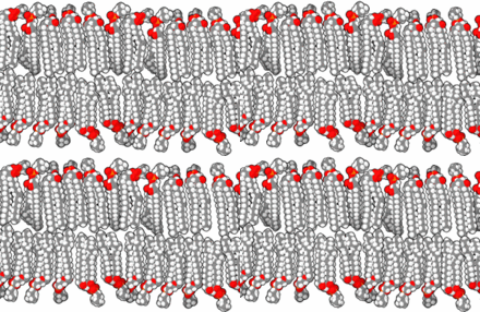
This fluid lipid bilayer cross section is made up entirely of phosphatidylcholine.

Scientists have come to conclude that life began in hydrothermal vents in the deep sea, but a 2012 study suggests that inland pools of condensed and cooled geothermal vapor have the ideal characteristics for the origin of life. The conclusion is based mainly on the chemistry of modern cells, where the cytoplasm is rich in potassium, zinc, manganese, and phosphate ions, which are not widespread in marine environments. Such conditions, the researchers argue, are found only where hot hydrothermal fluid brings the ions to the surface—places such as geysers, mud pots, fumaroles and other geothermal features. Within these fuming and bubbling basins, water laden with zinc and manganese ions could have collected, cooled and condensed in shallow pools.

Another study in the 1990s showed that montmorillonite clay can help create RNA chains of as many as 50 nucleotides joined together spontaneously into a single RNA molecule. Later, in 2002, it was discovered that by adding montmorillonite to a solution of fatty acid micelles (lipid spheres), the clay sped up the rate of vesicle formation 100-fold.

Research has shown that some minerals can catalyze the stepwise formation of hydrocarbon tails of fatty acids from hydrogen and carbon monoxide gases—gases that may have been released from hydrothermal vents or geysers. Fatty acids of various lengths are eventually released into the surrounding water, but vesicle formation requires a higher concentration of fatty acids, so it is suggested that protocell formation started at land-bound hydrothermal vents such as geysers, mud pots, fumaroles and other geothermal features where water evaporates and concentrates the solute.

Montmorillonite bubbles

Another group suggests that primitive cells might have formed inside inorganic clay microcompartments, which can provide an ideal container for the synthesis and compartmentalization of complex organic molecules. Clay-armored bubbles form naturally when particles of montmorillonite clay collect on the outer surface of air bubbles under water. This creates a semi permeable vesicle from materials that are readily available in the environment. The authors remark that montmorillonite is known to serve as a chemical catalyst, encouraging lipids to form membranes and single nucleotides to join into strands of RNA. Primitive reproduction can be envisioned when the clay bubbles burst, releasing the lipid membrane-bound product into the surrounding medium.

Membrane transport

Schematic showing two possible conformations of the lipids at the edge of a pore. In the top image the lipids have not rearranged, so the pore wall is hydrophobic. In the bottom image some of the lipid heads have bent over, so the pore wall is hydrophilic.

For cellular organisms, the transport of specific molecules across compartmentalizing membrane barriers is essential in order to exchange content with their environment and with other individuals. For example, content exchange between individuals enables horizontal gene transfer, an important factor in the evolution of cellular life. While modern cells can rely on complicated protein machineries to catalyze these crucial processes, protocells must have accomplished this using more simple mechanisms.

Protocells composed of fatty acids would have been able to easily exchange small molecules and ions with their environment. Membranes consisting of fatty acids have a relatively high permeability to molecules such as nucleoside monophosphate (NMP), nucleoside diphosphate (NDP), and nucleoside triphosphate (NTP), and may withstand millimolar concentrations of Mg2+. Osmotic pressure can also play a significant role regarding this passive membrane transport.

Environmental effects have been suggested to trigger conditions under which a transport of larger molecules, such as DNA and RNA, across the membranes of protocells is possible. For example, it has been proposed that electroporation resulting from lightning strikes could enable such transport. Electroporation is the rapid increase in bilayer permeability induced by the application of a large artificial electric field across the membrane. During electroporation in laboratory procedures, the lipid molecules are not chemically altered but simply shift position, opening up a pore (hole) that acts as the conductive pathway through the bilayer as it is filled with water. Experimentally, electroporation is used to introduce hydrophilic molecules into cells. It is a particularly useful technique for large highly charged molecules such as DNA and RNA, which would never passively diffuse across the hydrophobic bilayer core. Because of this, electroporation is one of the key methods of transfection as well as bacterial transformation.

A similar transfer of content across protocells and with the surrounding solution can also be caused by freezing and subsequent thawing. For example, this could occur in an environment in which day and night cycles cause recurrent freezing. Laboratory experiments have shown that such conditions allow an exchange of genetic information between populations of protocells. This can be explained by the fact that membranes are highly permeable at temperatures slightly below their phase transition temperature. If this point is reached during the freeze-thaw cycle, even large and highly charged molecules like nucleic acids can temporarily pass the protocell membrane.

Some molecules or particles are too large or too hydrophilic to pass through a lipid bilayer even under these conditions, but can be moved across the membrane through fusion or budding of vesicles, events which have also been observed for freeze-thaw cycles. This may eventually have led to mechanisms that facilitate movement of molecules to the inside of the protocell (endocytosis) or to release its contents into the extracellular space (exocytosis).

Artificial models

Langmuir-Blodgett deposition

Starting with a technique commonly used to deposit molecules on a solid surface, Langmuir–Blodgett deposition, scientists are able to assemble phospholipid membranes of arbitrary complexity layer by layer. These artificial phospholipid membranes support functional insertion both of purified and of in situ expressed membrane proteins. The technique could help astrobiologists understand how the first living cells originated.

Jeewanu protocells

Surfactant molecules arranged on an air – water interface

Jeewanu protocells are synthetic chemical particles that possess cell-like structure and seem to have some functional living properties. First synthesized in 1963 from simple minerals and basic organics while exposed to sunlight, it is still reported to have some metabolic capabilities, the presence of semipermeable membrane, amino acids, phospholipids, carbohydrates and RNA-like molecules. However, the nature and properties of the Jeewanu remains to be clarified.

In a similar synthesis experiment a frozen mixture of water, methanol, ammonia and carbon monoxide was exposed to ultraviolet (UV) radiation. This combination yielded large amounts of organic material that self-organised to form globules or vesicles when immersed in water. The investigating scientist considered these globules to resemble cell membranes that enclose and concentrate the chemistry of life, separating their interior from the outside world. The globules were between 10 to 40 micrometres (0.00039 to 0.00157 in), or about the size of red blood cells. Remarkably, the globules fluoresced, or glowed, when exposed to UV light. Absorbing UV and converting it into visible light in this way was considered one possible way of providing energy to a primitive cell. If such globules played a role in the origin of life, the fluorescence could have been a precursor to primitive photosynthesis. Such fluorescence also provides the benefit of acting as a sunscreen, diffusing any damage that otherwise would be inflicted by UV radiation. Such a protective function would have been vital for life on the early Earth, since the ozone layer, which blocks out the sun's most destructive UV rays, did not form until after photosynthetic life began to produce oxygen.

Ethics and controversy

Protocell research has created controversy and opposing opinions, including critics of the vague definition of "artificial life". The creation of a basic unit of life is the most pressing ethical concern, although the most widespread worry about protocells is their potential threat to human health and the environment through uncontrolled replication.

Cyanobacteria

From Wikipedia, the free encyclopedia

Cyanobacteria
Temporal range: 3500–0Ma
Tolypothrix (Cyanobacteria).JPG
Tolypothrix sp.
Scientific classification
Domain: Bacteria
Kingdom: Eubacteria
Superphylum: Terrabacteria
Phylum: Cyanobacteria
Stanier, 1973
Orders
As of 2014 the taxonomy was under revision
Ozarkcollenia

Synonyms
  • Myxophyceae Wallroth, 1833
  • Phycochromaceae Rabenhorst, 1865
  • Cyanophyceae Sachs, 1874
  • Schizophyceae Cohn, 1879
  • Cyanophyta Steinecke, 1931
  • Oxyphotobacteria Gibbons & Murray, 1978
Cyanobacteria /sˌænbækˈtɪəriə/, also known as Cyanophyta, are a phylum of bacteria that obtain their energy through photosynthesis, and are the only photosynthetic prokaryotes able to produce oxygen. The name "cyanobacteria" comes from the color of the bacteria (Greek: κυανός, translit. kyanós, lit. 'blue'). Cyanobacteria, which are prokaryotes, are also called "blue-green algae", though the term "algae" in modern usage is restricted to eukaryotes.

Unlike heterotrophic prokaryotes, cyanobacteria have internal membranes. These are flattened sacs called thylakoids where photosynthesis is performed.

Phototrophic eukaryotes perform photosynthesis by plastids that may have their ancestry in cyanobacteria, acquired long ago via a process called endosymbiosis. These endosymbiotic cyanobacteria in eukaryotes may have evolved or differentiated into specialized organelles such as chloroplasts, etioplasts and leucoplasts.

By producing and releasing oxygen (as a byproduct of photosynthesis), cyanobacteria are thought to have converted the early oxygen-poor, reducing atmosphere into an oxidizing one, causing the Great Oxygenation Event and the "rusting of the Earth", which dramatically changed the composition of the Earth's life forms and led to the near-extinction of anaerobic organisms.

Description

Cyanobacteria are a group of photosynthetic bacteria, some of which are nitrogen-fixing, that live in a wide variety of moist soils and water either freely or in a symbiotic relationship with plants or lichen-forming fungi (as in the lichen genus Peltigera). They range from unicellular to filamentous and include colonial species. Colonies may form filaments, sheets, or even hollow spheres. Some filamentous species can differentiate into several different cell types: vegetative cells – the normal, photosynthetic cells that are formed under favorable growing conditions; akinetes – climate-resistant spores that may form when environmental conditions become harsh; and thick-walled heterocysts – which contain the enzyme nitrogenase, vital for nitrogen fixation in an anaerobic environment due to its sensitivity to oxygen.

Nitrogen fixation

Some cyanobacteria can fix atmospheric nitrogen in anaerobic conditions by means of specialized cells called heterocysts. Heterocysts may also form under the appropriate environmental conditions (anoxic) when fixed nitrogen is scarce. Heterocyst-forming species are specialized for nitrogen fixation and are able to fix nitrogen gas into ammonia (NH3), nitrites (NO
2
) or nitrates (NO
3
), which can be absorbed by plants and converted to protein and nucleic acids (atmospheric nitrogen is not bioavailable to plants, except for those having endosymbiotic nitrogen-fixing bacteria, especially the Fabaceae family, among others).

Free-living cyanobacteria are present in the water of rice paddies, and cyanobacteria can be found growing as epiphytes on the surfaces of the green alga, Chara, where they may fix nitrogen. Cyanobacteria such as Anabaena (a symbiont of the aquatic fern Azolla), can provide rice plantations with biofertilizer.

Morphology

Colonies of Nostoc pruniforme
 

Many cyanobacteria form motile filaments of cells, called hormogonia, that travel away from the main biomass to bud and form new colonies elsewhere. The cells in a hormogonium are often thinner than in the vegetative state, and the cells on either end of the motile chain may be tapered. To break away from the parent colony, a hormogonium often must tear apart a weaker cell in a filament, called a necridium.

Each individual cell (each single cyanobacterium) typically has a thick, gelatinous cell wall. They lack flagella, but hormogonia of some species can move about by gliding along surfaces. Many of the multicellular filamentous forms of Oscillatoria are capable of a waving motion; the filament oscillates back and forth. In water columns, some cyanobacteria float by forming gas vesicles, as in archaea. These vesicles are not organelles as such. They are not bounded by lipid membranes, but by a protein sheath.

Ecology

Cyanobacterial bloom near Fiji

Cyanobacteria can be found in almost every terrestrial and aquatic habitatoceans, fresh water, damp soil, temporarily moistened rocks in deserts, bare rock and soil, and even Antarctic rocks. They can occur as planktonic cells or form phototrophic biofilms. They are found in almost every endolithic ecosystem. A few are endosymbionts in lichens, plants, various protists, or sponges and provide energy for the host. Some live in the fur of sloths, providing a form of camouflage.

Aquatic cyanobacteria are known for their extensive and highly visible blooms that can form in both freshwater and marine environments. The blooms can have the appearance of blue-green paint or scum. These blooms can be toxic, and frequently lead to the closure of recreational waters when spotted. Marine bacteriophages are significant parasites of unicellular marine cyanobacteria.

Cyanobacteria growth is favoured in ponds and lakes where waters are calm and have less turbulent mixing. Their life cycles are disrupted when the water naturally or artificially mixes from churning currents caused by the flowing water of streams or the churning water of fountains. For this reason blooms of cyanobacteria seldom occur in rivers unless the water is flowing slowly. Growth is also favored at higher temperatures, making increasing water temperature as a result of global warming more problematic. At higher temperatures Microcystis species are able to outcompete diatoms and green algae. This is a concern because of the production of toxins produced by Microcystis.

Based on environmental trends, models and observations suggest cyanobacteria will likely increase their dominance in aquatic environments. This can lead to serious consequences, particularly the contamination of sources of drinking water. Cyanobacteria can interfere with water treatment in various ways, primarily by plugging filters (often large beds of sand and similar media), and by producing cyanotoxins which have the potential to cause serious illness if consumed. Consequences may also lie within fisheries and waste management practices. Anthropogenic eutrophication, rising temperatures, vertical stratification and increased atmospheric carbon dioxide are contributors to cyanobacteria increasing dominance of aquatic ecosystems.

Cyanobacteria have been found to play an important role in terrestrial habitats. It has been widely reported that cyanobacteria soil crusts help to stabilize soil to prevent erosion and retain water. An example of a cyanobacterial species that does so is Microcoleus vaginatus. M. vaginatus stabilizes soil using a polysaccharide sheath that binds to sand particles and absorbs water.

Some of these organisms contribute significantly to global ecology and the oxygen cycle. The tiny marine cyanobacterium Prochlorococcus was discovered in 1986 and accounts for more than half of the photosynthesis of the open ocean. Many cyanobacteria even display the circadian rhythms that were once thought to exist only in eukaryotic cells.
"Cyanobacteria are arguably the most successful group of microorganisms on earth. They are the most genetically diverse; they occupy a broad range of habitats across all latitudes, widespread in freshwater, marine, and terrestrial ecosystems, and they are found in the most extreme niches such as hot springs, salt works, and hypersaline bays. Photoautotrophic, oxygen-producing cyanobacteria created the conditions in the planet's early atmosphere that directed the evolution of aerobic metabolism and eukaryotic photosynthesis. Cyanobacteria fulfill vital ecological functions in the world's oceans, being important contributors to global carbon and nitrogen budgets." – Stewart and Falconer

Photosynthesis

While contemporary cyanobacteria are linked to the plant kingdom as descendants of the endosymbiotic progenitor of the chloroplast, there are several features which are unique to this group.

Carbon fixation

Cyanobacteria use the energy of sunlight to drive photosynthesis, a process where the energy of light is used to synthesize organic compounds from carbon dioxide. Because they are aquatic organisms, they typically employ several strategies which are collectively known as a "carbon concentrating mechanism" to aid in the acquisition of inorganic carbon (CO2 or bicarbonate). Among the more specific strategies is the widespread prevalence of the bacterial microcompartments known as carboxysomes. These icosahedral structures are composed of hexameric shell proteins that assemble into cage-like structures that can be several hundreds of nanometers in diameter. It is believed that these structures tether the CO2-fixing enzyme, RuBisCO, to the interior of the shell, as well as the enzyme carbonic anhydrase, using the paradigm of metabolic channeling to enhance the local CO2 concentrations and thus increase the efficiency of the RuBisCO enzyme.

Electron transport

In contrast to purple bacteria and other bacteria performing anoxygenic photosynthesis, thylakoid membranes of cyanobacteria are not continuous with the plasma membrane but are separate compartments. The photosynthetic machinery is embedded in the thylakoid membranes, with phycobilisomes acting as light-harvesting antennae attached to the membrane, giving the green pigmentation observed (with wavelengths from 450 nm to 660 nm) in most cyanobacteria.

Diagram of a typical cyanobacterial cell

While most of the high-energy electrons derived from water are used by the cyanobacterial cells for their own needs, a fraction of these electrons may be donated to the external environment via electrogenic activity.

Respiration

Respiration in cyanobacteria can occur in the thylakoid membrane alongside photosynthesis, with their photosynthetic electron transport sharing the same compartment as the components of respiratory electron transport. While the goal of photosynthesis is to store energy by building carbohydrates from CO2, respiration is the reverse of this, with carbohydrates turned back into CO2 accompanying energy release.

Cyanobacteria appear to separate these two processes with their plasma membrane containing only components of the respiratory chain, while the thylakoid membrane hosts an interlinked respiratory and photosynthetic electron transport chain. Cyanobacteria use electrons from succinate dehydrogenase rather than from NADPH for respiration.

Electron transport chain

Many cyanobacteria are able to reduce nitrogen and carbon dioxide under aerobic conditions, a fact that may be responsible for their evolutionary and ecological success. The water-oxidizing photosynthesis is accomplished by coupling the activity of photosystem (PS) II and I (Z-scheme). In contrast to green sulfur bacteria which only use one photosystem, the use of water as an electron donor is energetically demanding, requiring two photosystems.

Attached to the thylakoid membrane, phycobilisomes act as light-harvesting antennae for the photosystems. The phycobilisome components (phycobiliproteins) are responsible for the blue-green pigmentation of most cyanobacteria. The variations on this theme are due mainly to carotenoids and phycoerythrins that give the cells their red-brownish coloration. In some cyanobacteria, the color of light influences the composition of the phycobilisomes. In green light, the cells accumulate more phycoerythrin, whereas in red light they produce more phycocyanin. Thus, the bacteria appear green in red light and red in green light. This process of complementary chromatic adaptation is a way for the cells to maximize the use of available light for photosynthesis.

A few genera lack phycobilisomes and have chlorophyll b instead (Prochloron, Prochlorococcus, Prochlorothrix). These were originally grouped together as the prochlorophytes or chloroxybacteria, but appear to have developed in several different lines of cyanobacteria. For this reason, they are now considered as part of the cyanobacterial group.

Metabolism

In general, photosynthesis in cyanobacteria uses water as an electron donor and produces oxygen as a byproduct, though some may also use hydrogen sulfide a process which occurs among other photosynthetic bacteria such as the purple sulfur bacteria.

Carbon dioxide is reduced to form carbohydrates via the Calvin cycle. The large amounts of oxygen in the atmosphere are considered to have been first created by the activities of ancient cyanobacteria. They are often found as symbionts with a number of other groups of organisms such as fungi (lichens), corals, pteridophytes (Azolla), angiosperms (Gunnera), etc.

There are some groups capable of heterotrophic growth, while others are parasitic, causing diseases in invertebrates or algae (e.g., the black band disease).

Relationship to chloroplasts

Primary chloroplasts are cell organelles found in some eukaryotic lineages, where they are specialized in performing the photosynthesis. They are known to have evolved from cyanobacteria through endosymbiosis, i.e. after the engulfment of a cell by another. In this case, a photosynthesizing cyanobacteria that was engulfed in some ancient eukaryotic cell. After some years of debate, it is now generally accepted that the three major groups of primary endosymbiotic eukaryotes (i.e. green plants, red algae and glaucophytes) form one large monophyletic group called Archaeplastida, which evolved after one unique endosymbiotic event.

The morphological similarity between chloroplasts and cyanobacteria was first reported by German botanist Andreas Schimper in the 19th century Chloroplasts are only found in plants and algae, thus paving the way for Russian biologist Konstantin Mereschkowski to suggest the symbiogenic origin of the plastid in 1905. Lynn Margulis brought this hypothesis back to attention more than 60 years later but it was not until supplementary data started to accumulate that the idea became fully accepted. The cyanobacterial origin of plastids is now supported by various pieces of phylogenetic, genomic, biochemical and structural evidence. The fact that another independent and more recent primary endosymbiosis event has been described between a cyanobacterium and a separate eukaryote lineage (the rhizarian Paulinella chromatophora) also gives credibility to the endosymbiotic origin of the plastid.

In addition to this primary endosymbiosis, many eukaryotic lineages have been subject to secondary or even tertiary endosymbiotic events, that is the "Matryoshka-like" engulfment by a eukaryote of another plastid-bearing eukaryote.

Within this evolutionary context, it is noteworthy that, as far as we can tell, oxygenic photosynthesis only evolved once (in cyanobacteria), and all other photosynthetic eukaryotes (including all plants and algae) have acquired this ability from them. In other words, all the oxygen that makes the atmosphere breathable for aerobic organisms originally comes from cyanobacteria or their later descendants.

DNA repair

Cyanobacteria are challenged by environmental stresses and internally generated reactive oxygen species that cause DNA damages. Cyanobacteria possess numerous E. coli-like DNA repair genes.  Several DNA repair genes are highly conserved in cyanobacteria, even in small genomes, suggesting that core DNA repair processes such as recombinational repair, nucleotide excision repair and methyl-directed DNA mismatch repair are common among cyanobacteria.

Natural genetic transformation

Cyanobacteria are capable of natural genetic transformation. Natural genetic transformation is the genetic alteration of a cell resulting from the direct uptake and incorporation of exogenous DNA from its surroundings. For bacterial transformation to take place, the recipient bacteria must be in a state of competence, which may occur in nature as a response to conditions such as starvation, high cell density or exposure to DNA damaging agents. In chromosomal transformation, homologous transforming DNA can be integrated into the recipient genome by homologous recombination, and this process appears to be an adaptation for repairing DNA damage.

Classification

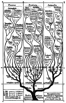
Tree of Life in Generelle Morphologie der Organismen (1866). Note the location of the genus Nostoc with algae and not with bacteria (kingdom "Monera")

Historically, bacteria were first classified as plants constituting the class Schizomycetes, which along with the Schizophyceae (blue-green algae/Cyanobacteria) formed the phylum Schizophyta, then in the phylum Monera in the kingdom Protista by Haeckel in 1866, comprising Protogens, Protamaeba, Vampyrella, Protomonae, and Vibrio, but not Nostoc and other cyanobacteria, which were classified with algae, later reclassified as the Prokaryotes by Chatton.

The cyanobacteria were traditionally classified by morphology into five sections, referred to by the numerals I–V. The first three – Chroococcales, Pleurocapsales, and Oscillatoriales – are not supported by phylogenetic studies. The latter two – Nostocales and Stigonematales – are monophyletic, and make up the heterocystous cyanobacteria.

The members of Chroococales are unicellular and usually aggregate in colonies. The classic taxonomic criterion has been the cell morphology and the plane of cell division. In Pleurocapsales, the cells have the ability to form internal spores (baeocytes). The rest of the sections include filamentous species. In Oscillatoriales, the cells are uniseriately arranged and do not form specialized cells (akinetes and heterocysts). In Nostocales and Stigonematales, the cells have the ability to develop heterocysts in certain conditions. Stigonematales, unlike Nostocales, include species with truly branched trichomes.

Most taxa included in the phylum or division Cyanobacteria have not yet been validly published under the Bacteriological Code, except:
The remainder are validly published under the International Code of Nomenclature for algae, fungi, and plants.

Formerly, some bacteria, like Beggiatoa, were thought to be colorless Cyanobacteria.

Earth history

Stromatolites are layered biochemical accretionary structures formed in shallow water by the trapping, binding, and cementation of sedimentary grains by biofilms (microbial mats) of microorganisms, especially cyanobacteria.

Stromatolites left behind by cyanobacteria are the oldest known fossils of life on Earth. This one-billion-year-old fossil is from Glacier National Park in Montana.

During the Precambrian, stromatolite communities of microorganisms grew in most marine and non-marine environments in the photic zone. After the Cambrian explosion of marine animals, grazing on the stromatolite mats by herbivores greatly reduced the occurrence of the stromatolites in marine environments. Since then, they are found mostly in hypersaline conditions where grazing invertebrates cannot live (e.g. Shark Bay, Western Australia). Stromatolites provide ancient records of life on Earth by fossil remains which might date from more than 3.5 Ga ago, but this is disputed. As of 2010 the oldest undisputed evidence of cyanobacteria is from 2.1 Ga ago, but there is some evidence for them as far back as 2.7 Ga ago. Oxygen concentrations in the atmosphere remained around or below 1% of today's level until 2.4 Ga ago. The rise in oxygen may have caused a fall in the concentration of atmospheric methane, and triggered the Huronian glaciation from around 2.4 to 2.1 Ga ago. In this way, cyanobacteria may have killed off much of the other bacteria of the time.

Oncolites are sedimentary structures composed of oncoids, which are layered structures formed by cyanobacterial growth. Oncolites are similar to stromatolites, but instead of forming columns, they form approximately spherical structures that were not attached to the underlying substrate as they formed. The oncoids often form around a central nucleus, such as a shell fragment, and a calcium carbonate structure is deposited by encrusting microbes. Oncolites are indicators of warm waters in the photic zone, but are also known in contemporary freshwater environments. These structures rarely exceed 10 cm in diameter.

Biotechnology and applications

The unicellular cyanobacterium Synechocystis sp. PCC6803 was the third prokaryote and first photosynthetic organism whose genome was completely sequenced. It continues to be an important model organism. Cyanothece ATCC 51142 is an important diazotrophic model organism. The smallest genomes have been found in Prochlorococcus spp. (1.7 Mb) and the largest in Nostoc punctiforme (9 Mb). Those of Calothrix spp. are estimated at 12–15 Mb, as large as yeast.


Recent research has suggested the potential application of cyanobacteria to the generation of renewable energy by converting sunlight into electricity. Internal photosynthetic pathways can be coupled to chemical mediators that transfer electrons to external electrodes. Currently, efforts are underway to commercialize algae-based fuels such as diesel, gasoline, and jet fuel.

Cyanobacteria cultured in specific media: Cyanobacteria can be helpful in agriculture as they have the ability to fix atmospheric nitrogen in soil.

Researchers from a company called Algenol have cultured genetically modified cyanobacteria in sea water inside a clear plastic enclosure so they first make sugar (pyruvate) from CO2 and the water via photosynthesis. Then, the bacteria secrete ethanol from the cell into the salt water. As the day progresses, and the solar radiation intensifies, ethanol concentrations build up and the ethanol itself evaporates onto the roof of the enclosure. As the sun recedes, evaporated ethanol and water condense into droplets, which run along the plastic walls and into ethanol collectors, from where it is extracted from the enclosure with the water and ethanol separated outside the enclosure. As of March 2013, Algenol was claiming to have tested its technology in Florida and to have achieved yields of 9,000 US gallons per acre per year. This could potentially meet US demands for ethanol in gasoline in 2025, assuming a B30 blend, from an area of around half the size of California’s San Bernardino County, requiring less than one-tenth of the area than ethanol from other biomass, such as corn, and only very limited amounts of fresh water.

Cyanobacteria may possess the ability to produce substances that could one day serve as anti-inflammatory agents and combat bacterial infections in humans.

Spirulina's extracted blue color is used as a natural food coloring in gum and candy.

Researchers from several space agencies argue that cyanobacteria could be used for producing goods for human consumption in future manned outposts on Mars, by transforming materials available on this planet.

Health risks

Cyanobacteria can produce neurotoxins, cytotoxins, endotoxins, and hepatotoxins (e.g., the microcystin-producing bacteria genus microcystis), which are collectively known as cyanotoxins.

Specific toxins include, anatoxin-a, anatoxin-as, aplysiatoxin, cyanopeptolin, cylindrospermopsin, domoic acid, nodularin R (from Nodularia), neosaxitoxin, and saxitoxin. Cyanobacteria reproduce explosively under certain conditions. This results in algal blooms, which can become harmful to other species, and pose a danger to humans and animals, if the cyanobacteria involved produce toxins. Several cases of human poisoning have been documented, but a lack of knowledge prevents an accurate assessment of the risks.

Recent studies suggest that significant exposure to high levels of cyanobacteria producing toxins such as BMAA can cause amyotrophic lateral sclerosis (ALS). People living within half a mile of cyanobacterially contaminated lakes have had a 2.3 times greater risk of developing ALS than the rest of the population; people around New Hampshire's Lake Mascoma had an up to 25 times greater risk of ALS than the expected incidence. BMAA from desert crusts found throughout Qatar might have contributed to higher rates of ALS in Gulf War veterans.

Chemical control

Several chemicals can eliminate cyanobacterial blooms from water-based systems. They include: calcium hypochlorite, copper sulphate, cupricide, and simazine. The calcium hypochlorite amount needed varies depending on the cyanobacteria bloom, and treatment is needed periodically. According to the Department of Agriculture Australia, a rate of 12 g of 70% material in 1000 l of water is often effective to treat a bloom. Copper sulfate is also used commonly, but no longer recommended by the Australian Department of Agriculture, as it kills livestock, crustaceans, and fish. Culpricide is a chelated copper product that eliminates blooms with lower toxicity risks than copper sulfate. Dosage recommendations vary from 190 ml to 4.8 l per 1000 m2. Ferric alum treatments at the rate of 50 mg/l will reduce algae blooms. Simazine, which is also a herbicide, will continue to kill blooms for several days after an application. Simazine is marketed at different strengths (25, 50, and 90%), the recommended amount needed for one cubic meter of water per product is 25% product 8 ml; 50% product 4 ml; or 90% product 2.2 ml.

Dietary supplementation

Spirulina tablets

Some cyanobacteria are sold as food, notably Aphanizomenon flos-aquae and Arthrospira platensis (Spirulina).

Despite the associated toxins which many of the members of this phylum produce, some microalgae also contain substances of high biological value, such as polyunsaturated fatty acids, amino acids, proteins, pigments, antioxidants, vitamins, and minerals. Edible blue-green algae reduce the production of pro-inflammatory cytokines by inhibiting NF-κB pathway in macrophages and splenocytes. Sulfate polysaccharides exhibit immunomodulatory, antitumor, antithrombotic, anticoagulant, anti-mutagenic, anti-inflammatory, antimicrobial, and even antiviral activity against HIV, herpes, and hepatitis.

Lie group

From Wikipedia, the free encyclopedia https://en.wikipedia.org/wiki/Lie_group In mathematics , a Lie gro...