Keratin (/ˈkɛrətɪn/) is one of a family of structural fibrous proteins also known as scleroproteins. Alpha-keratin (α-keratin) is a type of keratin found in vertebrates. It is the key structural material making up scales, hair, nails, feathers, horns, claws, hooves, and the outer layer of skin among vertebrates. Keratin also protects epithelial cells from damage or stress. Keratin is extremely insoluble in water and organic solvents. Keratin monomers assemble into bundles to form intermediate filaments, which are tough and form strong unmineralized epidermal appendages found in reptiles, birds, amphibians, and mammals. Excessive keratinization participate in fortification of certain tissues such as in horns of cattle and rhinos, and armadillos' osteoderm. The only other biological matter known to approximate the toughness of keratinized tissue is chitin.
Keratin comes in two types, the primitive, softer forms found in all vertebrates and harder, derived forms found only among sauropsids (reptiles and birds).
Spider silk is classified as keratin, although production of the protein may have evolved independently of the process in vertebrates.
Examples of occurrence
The horns of the impala are made of keratin covering a core of bone.
Alpha-keratins (α-keratins) are found in all vertebrates. They form the hair (including wool), the outer layer of skin, horns, nails, claws and hooves of mammals, and the slime threads of hagfish. The baleen plates of filter-feeding whales are also made of keratin. Keratin filaments are abundant in keratinocytes in the hornified layer of the epidermis; these are proteins which have undergone keratinization. They are also present in epithelial cells in general. For example, mouse thymic epithelial cells react with antibodies for keratin 5, keratin 8, and keratin 14. These antibodies are used as fluorescent markers to distinguish subsets of mouse thymic epithelial cells in genetic studies of the thymus.
The harder beta-keratins (β-keratins) are found only in the sauropsids, that is all living reptiles and birds. They are found in the nails, scales, and claws of reptiles, in some reptile shells (Testudines, such as tortoise, turtle, terrapin), and in the feathers, beaks, and claws of birds. These keratins are formed primarily in beta sheets. However, beta sheets are also found in α-keratins.
Recent scholarship has shown that sauropsid β-keratins are fundamentally
different from α-keratins at a genetic and structural level. The new
term corneous beta protein (CBP) has been proposed to avoid confusion with α-keratins.
The neutral–basic keratins are encoded on chromosome 12 (12q13.13).The acidic keratins are encoded on chromosome 17 (17q21.2).
The human genome encodes 54 functional keratin genes, located in two clusters on chromosomes 12 and 17. This suggests that they originated from a series of gene duplications on these chromosomes.
Table of keratin genes and biological processes (GeneCards)
Protein
sequence alignment of human keratin 1, 2A, 3,4, 5, 6A, 7, and 8 (KRT1 –
KRT8). Only the first rod domain is shown above. Alignment was created
using Clustal Omega.
Protein structure
The first sequences of keratins were determined by Israel Hanukoglu and Elaine Fuchs (1982, 1983).
These sequences revealed that there are two distinct but homologous
keratin families, which were named type I and type II keratins.
By analysis of the primary structures of these keratins and other
intermediate filament proteins, Hanukoglu and Fuchs suggested a model in
which keratins and intermediate filament proteins contain a central
~310 residue domain with four segments in α-helical conformation that
are separated by three short linker segments predicted to be in
beta-turn conformation. This model has been confirmed by the determination of the crystal structure of a helical domain of keratins.
Type 1 and 2 Keratins
The human genome has 54 functional annotated Keratin genes, 28 are in the Keratin type 1 family, and 26 are in the Keratin type 2 family.
Keratin (high molecular weight) in bile duct cell and oval cells of horseliver.
Fibrous keratin molecules supercoil to form a very stable, left-handed superhelical motif to multimerise, forming filaments consisting of multiple copies of the keratin monomer.
The major force that keeps the coiled-coil structure is hydrophobic interactions between apolar residues along the keratins helical segments.
Limited interior space is the reason why the triple helix of the (unrelated) structural protein collagen, found in skin, cartilage and bone, likewise has a high percentage of glycine. The connective tissue protein elastin also has a high percentage of both glycine and alanine. Silkfibroin, considered a β-keratin, can have these two as 75–80% of the total, with 10–15% serine, with the rest having bulky side groups. The chains are antiparallel, with an alternating C → N orientation. A preponderance of amino acids with small, nonreactive side groups is characteristic of structural proteins, for which H-bonded close packing is more important than chemical specificity.
Disulfide bridges
In addition to intra- and intermolecular hydrogen bonds, the distinguishing feature of keratins is the presence of large amounts of the sulfur-containing amino acid cysteine, required for the disulfide bridges that confer additional strength and rigidity by permanent, thermally stable crosslinking—in much the same way that non-protein sulfur bridges stabilize vulcanizedrubber. Human hair is approximately 14% cysteine. The pungent
smells of burning hair and skin are due to the volatile sulfur
compounds formed. Extensive disulfide bonding contributes to the insolubility of keratins, except in a small number of solvents such as dissociating or reducing agents.
A human toe nail that fell off after a small trauma. Three small punctures were made on it, while it was still attached.
The more flexible and elastic keratins of hair have fewer interchain disulfide bridges than the keratins in mammalianfingernails, hooves and claws (homologous structures), which are harder and more like their analogs in other vertebrate classes. Hair and other α-keratins consist of α-helically coiled single protein strands (with regular intra-chain H-bonding), which are then further twisted into superhelical ropes
that may be further coiled. The β-keratins of reptiles and birds have
β-pleated sheets twisted together, then stabilized and hardened by
disulfide bridges.
Thiolated polymers (=thiomers) can form disulfide bridges with cysteine substructures of keratins getting covalently attached to these proteins. Thiomers exhibit therefore high binding properties to keratins found in hair, on skin and on the surface of many cell types.
Filament formation
It has been proposed that keratins can be divided into 'hard' and 'soft' forms, or 'cytokeratins' and 'other keratins'. That model is now understood to be correct. A new nuclear addition in 2006 to describe keratins takes this into account.
Keratin filaments are intermediate filaments.
Like all intermediate filaments, keratin proteins form filamentous
polymers in a series of assembly steps beginning with dimerization;
dimers assemble into tetramers and octamers and eventually, if the
current hypothesis holds, into unit-length-filaments (ULF) capable of annealing end-to-end into long filaments.
Cornification is the process of forming an epidermal barrier in
stratified squamous epithelial tissue. At the cellular level,
cornification is characterised by:
production of keratin
production of small proline-rich (SPRR) proteins and transglutaminase which eventually form a cornified cell envelope beneath the plasma membrane
loss of nuclei and organelles, in the final stages of cornification
Metabolism ceases, and the cells are almost completely filled by
keratin. During the process of epithelial differentiation, cells become
cornified as keratin protein is incorporated into longer keratin
intermediate filaments. Eventually the nucleus and cytoplasmic
organelles disappear, metabolism ceases and cells undergo a programmed death
as they become fully keratinized. In many other cell types, such as
cells of the dermis, keratin filaments and other intermediate filaments
function as part of the cytoskeleton to mechanically stabilize the cell
against physical stress. It does this through connections to desmosomes,
cell–cell junctional plaques, and hemidesmosomes, cell-basement
membrane adhesive structures.
Cells in the epidermis contain a structural matrix of keratin,
which makes this outermost layer of the skin almost waterproof, and
along with collagen and elastin gives skin its strength. Rubbing and
pressure cause thickening of the outer, cornified layer of the epidermis
and form protective calluses, which are useful for athletes and on the
fingertips of musicians who play stringed instruments. Keratinized
epidermal cells are constantly shed and replaced.
These hard, integumentary structures are formed by intercellular
cementing of fibers formed from the dead, cornified cells generated by
specialized beds deep within the skin. Hair grows continuously and
feathers molt and regenerate. The constituent proteins may be
phylogenetically homologous but differ somewhat in chemical structure
and supermolecular organization. The evolutionary relationships are
complex and only partially known. Multiple genes have been identified
for the β-keratins in feathers, and this is probably characteristic of
all keratins.
Silk
The silkfibroins produced by insects and spiders are often classified as keratins, though it is unclear whether they are phylogenetically related to vertebrate keratins.
Silk found in insect pupae, and in spider webs
and egg casings, also has twisted β-pleated sheets incorporated into
fibers wound into larger supermolecular aggregates. The structure of the
spinnerets on spiders' tails, and the contributions of their interior glands, provide remarkable control of fast extrusion.
Spider silk is typically about 1 to 2 micrometers (μm) thick, compared
with about 60 μm for human hair, and more for some mammals. The biologically and commercially useful properties of silk fibers depend on the organization of multiple adjacent protein chains into hard, crystalline regions of varying size, alternating with flexible, amorphous regions where the chains are randomly coiled. A somewhat analogous situation occurs with syntheticpolymers such as nylon, developed as a silk substitute. Silk from the hornetcocoon
contains doublets about 10 μm across, with cores and coating, and may
be arranged in up to 10 layers, also in plaques of variable shape. Adult
hornets also use silk as a glue, as do spiders.
Glue
Glues made from partially-hydrolysed keratin include hoof glue and horn glue.
Keratin is highly resistant to digestive acids if ingested. Cats regularly ingest hair as part of their grooming behavior, leading to the gradual formation of hairballs that may be expelled orally or excreted. In humans, trichophagia may lead to Rapunzel syndrome, an extremely rare but potentially fatal intestinal condition.
https://en.wikipedia.org/wiki/Thermoregulation Thermoregulation is the ability of an organism to keep its body temperature
within certain boundaries, even when the surrounding temperature is
very different. A thermoconforming organism, by contrast, simply adopts
the surrounding temperature as its own body temperature, thus avoiding
the need for internal thermoregulation. The internal thermoregulation
process is one aspect of homeostasis: a state of dynamic stability in an organism's internal conditions, maintained far from thermal equilibrium with its environment (the study of such processes in zoology has been called physiological ecology). If the body is unable to maintain a normal temperature and it increases significantly above normal, a condition known as hyperthermia occurs. Humans may also experience lethal hyperthermia when the wet bulb temperature is sustained above 35 °C (95 °F) for six hours.
Work in 2022 established by experiment that a wet-bulb
temperature exceeding 30.55°C caused uncompensable heat stress in young,
healthy adult humans. The opposite condition, when body temperature
decreases below normal levels, is known as hypothermia.
It results when the homeostatic control mechanisms of heat within the
body malfunction, causing the body to lose heat faster than producing
it. Normal body temperature is around 37°C (98.6°F), and hypothermia
sets in when the core body temperature gets lower than 35 °C (95 °F).
Usually caused by prolonged exposure to cold temperatures, hypothermia
is usually treated by methods that attempt to raise the body temperature
back to a normal range.
It was not until the introduction of thermometers
that any exact data on the temperature of animals could be obtained. It
was then found that local differences were present, since heat
production and heat loss vary considerably in different parts of the
body, although the circulation of the blood tends to bring about a mean
temperature of the internal parts. Hence it is important to identify the
parts of the body that most closely reflect the temperature of the internal organs. Also, for such results to be comparable, the measurements must be conducted under comparable conditions. The rectum
has traditionally been considered to reflect most accurately the
temperature of internal parts, or in some cases of sex or species, the vagina, uterus or bladder. Some animals undergo one of various forms of dormancy
where the thermoregulation process temporarily allows the body
temperature to drop, thereby conserving energy. Examples include hibernatingbears and torpor in bats.
Classification of animals by thermal characteristics
Endothermy vs. ectothermy
Thermoregulation in organisms runs along a spectrum from endothermy to ectothermy. Endotherms create most of their heat via metabolic processes and are colloquially referred to as warm-blooded.
When the surrounding temperatures are cold, endotherms increase
metabolic heat production to keep their body temperature constant, thus
making the internal body temperature of an endotherm more or less
independent of the temperature of the environment.
Endotherms possess a larger number of mitochondria per cell than
ectotherms, enabling them to generate more heat by increasing the rate
at which they metabolize fats and sugars.
Ectotherms use external sources of temperature to regulate their body
temperatures. They are colloquially referred to as cold-blooded despite
the fact that body temperatures often stay within the same temperature
ranges as warm-blooded animals. Ectotherms are the opposite of
endotherms when it comes to regulating internal temperatures. In
ectotherms, the internal physiological sources of heat are of negligible
importance; the biggest factor that enables them to maintain adequate
body temperatures is due to environmental influences. Living in areas
that maintain a constant temperature throughout the year, like the
tropics or the ocean, has enabled ectotherms to develop behavioral
mechanisms that respond to external temperatures, such as sun-bathing to
increase body temperature, or seeking the cover of shade to lower body
temperature.
Increasing blood flow to body surfaces to maximize heat transfer across the advective gradient.
Conduction:
Losing heat by being in contact with a colder surface. For instance:
Lying on cool ground.
Staying wet in a river, lake or sea.
Covering in cool mud.
Radiation:
Releasing heat by radiating it away from the body.
Ectothermic heating (or minimizing heat loss)
The red line represents the air temperature. The purple line represents the body temperature of the lizard. The green line represents the base temperature of the burrow.
Lizards are ectotherms and use behavioral adaptations to control their
temperature. They regulate their behavior based on the temperature
outside, if it is warm they will go outside up to a point and return to
their burrow as necessary.
Convection:
Climbing to higher ground up trees, ridges, rocks.
Entering a warm water or air current.
Building an insulated nest or burrow.
Conduction:
Lying on a hot surface.
Radiation:
Lying in the sun (heating this way is affected by the body's angle in relation to the sun).
To cope with low temperatures, some fish have developed the ability to remain functional even when the water temperature is below freezing; some use natural antifreeze or antifreeze proteins to resist ice crystal formation in their tissues.[7]Amphibians and reptiles
cope with heat gain by evaporative cooling and behavioral adaptations.
An example of behavioral adaptation is that of a lizard lying in the sun
on a hot rock in order to heat through radiation and conduction.
An endotherm is an animal that regulates its own body temperature,
typically by keeping it at a constant level. To regulate body
temperature, an organism may need to prevent heat gains in arid
environments. Evaporation of water, either across respiratory surfaces
or across the skin in those animals possessing sweat glands,
helps in cooling body temperature to within the organism's tolerance
range. Animals with a body covered by fur have limited ability to sweat,
relying heavily on panting to increase evaporation of water
across the moist surfaces of the lungs and the tongue and mouth. Mammals
like cats, dogs and pigs, rely on panting or other means for thermal
regulation and have sweat glands only in foot pads and snout. The sweat
produced on pads of paws and on palms and soles mostly serves to
increase friction and enhance grip. Birds also counteract overheating by gular fluttering, or rapid vibrations of the gular (throat) skin.[8]Down feathers
trap warm air acting as excellent insulators just as hair in mammals
acts as a good insulator. Mammalian skin is much thicker than that of
birds and often has a continuous layer of insulating fat beneath the
dermis. In marine mammals, such as whales, or animals that live in very
cold regions, such as the polar bears, this is called blubber. Dense coats found in desert endotherms also aid in preventing heat gain such as in the case of the camels.[citation needed]
A cold weather strategy is to temporarily decrease metabolic
rate, decreasing the temperature difference between the animal and the
air and thereby minimizing heat loss. Furthermore, having a lower
metabolic rate is less energetically expensive. Many animals survive
cold frosty nights through torpor,
a short-term temporary drop in body temperature. Organisms, when
presented with the problem of regulating body temperature, have not only
behavioural, physiological, and structural adaptations but also a
feedback system to trigger these adaptations to regulate temperature
accordingly. The main features of this system are stimulus, receptor, modulator, effector and then the feedback of the newly adjusted temperature to the stimulus. This cyclical process aids in homeostasis.[citation needed]
Homeothermy compared with poikilothermy
Homeothermy and poikilothermy refer to how stable an organism's deep-body temperature is. Most endothermic organisms are homeothermic, like mammals. However, animals with facultative endothermy are often poikilothermic, meaning their temperature can vary considerably. Most fish are ectotherms, as most of their heat comes from the surrounding water. However, almost all fish are poikilothermic.[citation needed]
Beetles
The physiology of the Dendroctonus micans
beetle encompasses a suite of adaptations crucial for its survival and
reproduction. Flight capabilities enable them to disperse and locate new
host trees, while sensory organs aid in detecting environmental cues
and food sources. Of particular importance is their ability to
thermoregulate, ensuring optimal body temperature in fluctuating forest
conditions. This physiological mechanism, coupled with thermosensation,
allows them to thrive across diverse environments. Overall, these
adaptations underscore the beetle's remarkable resilience and highlight
the significance of understanding their physiology for effective
management and conservation efforts.[9]
Vertebrates
By numerous observations upon humans and other animals, John Hunter showed that the essential difference between the so-called warm-blooded and cold-blooded
animals lies in observed constancy of the temperature of the former,
and the observed variability of the temperature of the latter. Almost
all birds and mammals have a high temperature almost constant and
independent of that of the surrounding air (homeothermy). Almost all other animals display a variation of body temperature, dependent on their surroundings (poikilothermy).[10]
In cold environments, birds and mammals employ the following adaptations and strategies to minimize heat loss:
Using small smooth muscles (arrector pili
in mammals), which are attached to feather or hair shafts; this
distorts the surface of the skin making feather/hair shaft stand erect
(called goose bumps or goose pimples) which slows the movement of air across the skin and minimizes heat loss.
Increasing body size to more easily maintain core body temperature
(warm-blooded animals in cold climates tend to be larger than similar
species in warmer climates (see Bergmann's rule))
Having the ability to store energy as fat for metabolism
Have shortened extremities
Have countercurrent blood flow
in extremities – this is where the warm arterial blood travelling to
the limb passes the cooler venous blood from the limb and heat is
exchanged warming the venous blood and cooling the arterial (e.g., Arctic wolf or penguins)
In warm environments, birds and mammals employ the following adaptations and strategies to maximize heat loss:
Behavioural adaptations like living in burrows during the day and being nocturnal
Evaporative cooling by perspiration and panting
Storing fat reserves in one place (e.g., camel's hump) to avoid its insulating effect
Elongated, often vascularized extremities to conduct body heat to the air
As in other mammals, thermoregulation is an important aspect of human homeostasis.
Most body heat is generated in the deep organs, especially the liver,
brain, and heart, and in contraction of skeletal muscles.
Humans have been able to adapt to a great diversity of climates,
including hot humid and hot arid. High temperatures pose serious
stresses for the human body, placing it in great danger of injury or
even death. For example, one of the most common reactions to hot
temperatures is heat exhaustion, which is an illness that could happen
if one is exposed to high temperatures, resulting in some symptoms such
as dizziness, fainting, or a rapid heartbeat. For humans, adaptation to varying climatic conditions includes both physiological mechanisms resulting from evolution and behavioural mechanisms resulting from conscious cultural adaptations.
The physiological control of the body's core temperature takes place
primarily through the hypothalamus, which assumes the role as the body's
"thermostat".
This organ possesses control mechanisms as well as key temperature
sensors, which are connected to nerve cells called thermoreceptors.
Thermoreceptors come in two subcategories; ones that respond to cold
temperatures and ones that respond to warm temperatures. Scattered
throughout the body in both peripheral and central nervous systems,
these nerve cells are sensitive to changes in temperature and are able
to provide useful information to the hypothalamus through the process of
negative feedback, thus maintaining a constant core temperature.
There are four avenues of heat loss: evaporation, convection,
conduction, and radiation. If skin temperature is greater than that of
the surrounding air temperature, the body can lose heat by convection
and conduction. However, if air temperature of the surroundings is
greater than that of the skin, the body gains heat by convection
and conduction. In such conditions, the only means by which the body can
rid itself of heat is by evaporation. So, when the surrounding
temperature is higher than the skin temperature, anything that prevents
adequate evaporation will cause the internal body temperature to rise. During intense physical activity (e.g. sports), evaporation becomes the main avenue of heat loss. Humidity affects thermoregulation by limiting sweat evaporation and thus heat loss.
In reptiles
Thermoregulation is also an integral part of a reptile's life, specifically lizards such as Microlophus occipitalis and Ctenophorus decresii who must change microhabitats to keep a constant body temperature.
By moving to cooler areas when it is too hot and to warmer areas when
it is cold, they can thermoregulate their temperature to stay within
their necessary bounds.
In plants
Thermogenesis occurs in the flowers of many plants in the family Araceae as well as in cycad cones. In addition, the sacred lotus (Nelumbo nucifera) is able to thermoregulate itself,
remaining on average 20 °C (36 °F) above air temperature while
flowering. Heat is produced by breaking down the starch that was stored
in their roots, which requires the consumption of oxygen at a rate approaching that of a flying hummingbird.
One possible explanation for plant thermoregulation is to provide
protection against cold temperature. For example, the skunk cabbage is
not frost-resistant, yet it begins to grow and flower when there is
still snow on the ground.
Another theory is that thermogenicity helps attract pollinators, which
is borne out by observations that heat production is accompanied by the
arrival of beetles or flies.
Some plants are known to protect themselves against colder temperatures using antifreeze proteins. This occurs in wheat (Triticum aestivum),potatoes (Solanum tuberosum) and several other angiosperm species.
Behavioral temperature regulation
Animals
other than humans regulate and maintain their body temperature with
physiological adjustments and behavior. Desert lizards are ectotherms,
and therefore are unable to regulate their internal temperature
themselves. To regulate their internal temperature, many lizards
relocate themselves to a more environmentally favorable location. They
may do this in the morning only by raising their head from its burrow
and then exposing their entire body. By basking
in the sun, the lizard absorbs solar heat. It may also absorb heat by
conduction from heated rocks that have stored radiant solar energy. To
lower their temperature, lizards exhibit varied behaviors. Sand seas, or
ergs, produce up to 57.7 °C (135.9 °F), and the sand lizard
will hold its feet up in the air to cool down, seek cooler objects with
which to contact, find shade, or return to its burrow. They also go to
their burrows to avoid cooling when the temperature falls. Aquatic
animals can also regulate their temperature behaviorally by changing
their position in the thermal gradient. Sprawling prone in a cool shady spot, "splooting," has been observed in squirrels on hot days.
During cold weather, many animals increase their thermal inertia by huddling.
Animals also engage in kleptothermy
in which they share or steal each other's body warmth. Kleptothermy is
observed, particularly amongst juveniles, in endotherms such as bats and birds (such as the mousebird and emperor penguin). This allows the individuals to increase their thermal inertia (as with gigantothermy) and so reduce heat loss. Some ectotherms share burrows of ectotherms. Other animals exploit termite mounds.
Some animals living in cold environments maintain their body
temperature by preventing heat loss. Their fur grows more densely to
increase the amount of insulation. Some animals are regionally heterothermic
and are able to allow their less insulated extremities to cool to
temperatures much lower than their core temperature—nearly to 0 °C
(32 °F). This minimizes heat loss through less insulated body parts,
like the legs, feet (or hooves), and nose.
Different species of Drosophila found in the Sonoran Desert will exploit different species of cacti based on the thermotolerance differences between species and hosts. For example, Drosophila mettleri is found in cacti like the saguaro and senita;
these two cacti remain cool by storing water. Over time, the genes
selecting for higher heat tolerance were reduced in the population due
to the cooler host climate the fly is able to exploit.
Some flies, such as Lucilia sericata,
lay their eggs en masse. The resulting group of larvae, depending on
its size, is able to thermoregulate and keep itself at the optimum
temperature for development.
An ostrich
can keep its body temperature relatively constant, even though the
environment can be very hot during the day and cold at night.
Koalas
also can behaviorally thermoregulate by seeking out cooler portions of
trees on hot days. They preferentially wrap themselves around the
coolest portions of trees, typically near the bottom, to increase their
passive radiation of internal body heat.
Hibernation, estivation and daily torpor
To cope with limited food resources and low temperatures, some mammals hibernate during cold periods. To remain in "stasis" for long periods, these animals build up brown fat
reserves and slow all body functions. True hibernators (e.g.,
groundhogs) keep their body temperatures low throughout hibernation
whereas the core temperature
of false hibernators (e.g., bears) varies; occasionally the animal may
emerge from its den for brief periods. Some bats are true hibernators
and rely upon a rapid, non-shivering thermogenesis of their brown fat
deposit to bring them out of hibernation.
Previously, average oral temperature for healthy adults had been
considered 37.0 °C (98.6 °F), while normal ranges are 36.1 to 37.8 °C
(97.0 to 100.0 °F). In Poland and Russia, the temperature had been
measured axillarily
(under the arm). 36.6 °C (97.9 °F) was considered "ideal" temperature
in these countries, while normal ranges are 36.0 to 36.9 °C (96.8 to
98.4 °F).
Recent studies suggest that the average temperature for healthy
adults is 36.8 °C (98.2 °F) (same result in three different studies).
Variations (one standard deviation) from three other studies are:
36.4–37.1 °C (97.5–98.8 °F)
36.3–37.1 °C (97.3–98.8 °F) for males, 36.5–37.3 °C (97.7–99.1 °F) for females
36.6–37.3 °C (97.9–99.1 °F)
Measured temperature varies according to thermometer placement, with
rectal temperature being 0.3–0.6 °C (0.5–1.1 °F) higher than oral
temperature, while axillary temperature is 0.3–0.6 °C (0.5–1.1 °F) lower
than oral temperature. The average difference between oral and axillary temperatures of Indian children aged 6–12 was found to be only 0.1 °C (standard deviation 0.2 °C), and the mean difference in Maltese
children aged 4–14 between oral and axillary temperature was 0.56 °C,
while the mean difference between rectal and axillary temperature for
children under 4 years old was 0.38 °C.
Variations due to circadian rhythms
In
humans, a diurnal variation has been observed dependent on the periods
of rest and activity, lowest at 11 p.m. to 3 a.m. and peaking at 10 a.m.
to 6 p.m. Monkeys
also have a well-marked and regular diurnal variation of body
temperature that follows periods of rest and activity, and is not
dependent on the incidence of day and night; nocturnal
monkeys reach their highest body temperature at night and lowest during
the day. Sutherland Simpson and J.J. Galbraith observed that all
nocturnal animals and birds – whose periods of rest and activity are
naturally reversed through habit and not from outside interference –
experience their highest temperature during the natural period of
activity (night) and lowest during the period of rest (day). Those diurnal temperatures can be reversed by reversing their daily routine.
In essence, the temperature curve of diurnal
birds is similar to that of humans and other homeothermic animals,
except that the maximum occurs earlier in the afternoon and the minimum
earlier in the morning. Also, the curves obtained from rabbits, guinea pigs, and dogs were quite similar to those from humans. These observations indicate that body temperature is partially regulated by circadian rhythms.
Variations due to human menstrual cycles
During the follicular phase (which lasts from the first day of menstruation until the day of ovulation), the average basal body temperature
in women ranges from 36.45 to 36.7 °C (97.61 to 98.06 °F). Within 24
hours of ovulation, women experience an elevation of 0.15–0.45 °C
(0.27–0.81 °F) due to the increased metabolic rate caused by sharply
elevated levels of progesterone. The basal body temperature ranges between 36.7–37.3 °C (98.1–99.1 °F) throughout the luteal phase, and drops down to pre-ovulatory levels within a few days of menstruation. Women can chart this phenomenon to determine whether and when they are ovulating, so as to aid conception or contraception.
Variations due to fever
Fever is a regulated elevation of the set point of core temperature in the hypothalamus,
caused by circulating pyrogens produced by the immune system. To the
subject, a rise in core temperature due to fever may result in feeling
cold in an environment where people without fever do not.
Variations due to biofeedback
Some monks are known to practice Tummo, biofeedbackmeditation techniques, that allow them to raise their body temperatures substantially.
Effect on lifespan
The effects of such a genetic change in body temperature on longevity is difficult to study in humans.
Limits compatible with life
There are limits both of heat and cold that an endothermic animal can bear and other far wider limits that an ectothermic animal may endure and yet live. The effect of too extreme a cold is to decrease metabolism, and hence to lessen the production of heat. Both catabolic and anabolic
pathways share in this metabolic depression, and, though less energy is
used up, still less energy is generated. The effects of this diminished
metabolism become telling on the central nervous system first, especially the brain and those parts concerning consciousness; both heart rate and respiration rate
decrease; judgment becomes impaired as drowsiness supervenes, becoming
steadily deeper until the individual loses consciousness; without
medical intervention, death by hypothermia quickly follows. Occasionally, however, convulsions may set in towards the end, and death is caused by asphyxia.
In experiments on cats performed by Sutherland Simpson and Percy
T. Herring, the animals were unable to survive when rectal temperature
fell below 16 °C (61 °F).
At this low temperature, respiration became increasingly feeble;
heart-impulse usually continued after respiration had ceased, the beats
becoming very irregular, appearing to cease, then beginning again. Death
appeared to be mainly due to asphyxia, and the only certain sign that it had taken place was the loss of knee-jerks.
However, too high a temperature speeds up the metabolism of
different tissues to such a rate that their metabolic capital is soon
exhausted. Blood that is too warm produces dyspnea by exhausting the metabolic capital of the respiratory centre;
heart rate is increased; the beats then become arrhythmic and
eventually cease. The central nervous system is also profoundly affected
by hyperthermia and delirium, and convulsions may set in. Consciousness may also be lost, propelling the person into a comatose condition. These changes can sometimes also be observed in patients experiencing an acute fever.[citation needed]Mammalian muscle becomes rigid with heat rigor at about 50 °C, with the sudden rigidity of the whole body rendering life impossible.
H.M. Vernon performed work on the death temperature and paralysis
temperature (temperature of heat rigor) of various animals. He found
that species of the same class showed very similar temperature values, those from the Amphibia examined being 38.5 °C, fish 39 °C, reptiles 45 °C, and various molluscs 46 °C. Also, in the case of pelagic
animals, he showed a relation between death temperature and the
quantity of solid constituents of the body. In higher animals, however,
his experiments tend to show that there is greater variation in both the
chemical and physical characteristics of the protoplasm and, hence, greater variation in the extreme temperature compatible with life.
A 2022 study on the effect of heat on young people found that the
critical wet-bulb temperature at which heat stress can no longer be
compensated, Twb,crit, in young, healthy adults performing
tasks at modest metabolic rates mimicking basic activities of daily life
was much lower than the 35°C usually assumed, at about 30.55°C in
36–40°C humid environments, but progressively decreased in hotter, dry
ambient environments.
Arthropoda
The maximum temperatures tolerated by certain thermophilic arthropods exceeds the lethal temperatures for most vertebrates.
The most heat-resistant insects are three genera of desert ants
recorded from three different parts of the world. The ants have
developed a lifestyle of scavenging for short durations during the
hottest hours of the day, in excess of 50 °C (122 °F), for the carcasses
of insects and other forms of life which have died from heat stress.
In April 2014, the South Californian mite Paratarsotomus macropalpis
has been recorded as the world's fastest land animal relative to body
length, at a speed of 322 body lengths per second. Besides the unusually
great speed of the mites, the researchers were surprised to find the
mites running at such speeds on concrete at temperatures up to 60 °C
(140 °F), which is significant because this temperature is well above
the lethal limit for the majority of animal species. In addition, the
mites are able to stop and change direction very quickly.
Spiders like Nephila pilipes exhibits active thermal regulation behavior.
During high temperature sunny days, it aligns its body with the
direction of sunlight to reduce the body area under direct sunlight.
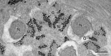
Schematic two-dimensional cross-sectional view of glycogen: A core protein of glycogenin is surrounded by branches of glucose units. The entire globular granule may contain around 30,000 glucose units.A view of the atomic structure of a single branched strand of glucose units in a glycogen molecule.Glycogen (black granules) in spermatozoa of a flatworm; transmission electron microscopy, scale: 0.3 μm
Glycogen is a multibranched polysaccharide of glucose that serves as a form of energy storage in animals, fungi, and bacteria. It is the main storage form of glucose in the human body.
Glycogen functions as one of three regularly used forms of energy reserves, creatine phosphate being for very short-term, glycogen being for short-term and the triglyceride stores in adipose tissue
(i.e., body fat) being for long-term storage. Protein, broken down into
amino acids, is seldom used as a main energy source except during
starvation and glycolytic crisis (see bioenergetic systems).
In humans, glycogen is made and stored primarily in the cells of the liver and skeletal muscle.
In the liver, glycogen can make up 5–6% of the organ's fresh weight:
the liver of an adult, weighing 1.5 kg, can store roughly 100–120 grams
of glycogen. In skeletal muscle, glycogen is found in a low concentration (1–2% of the muscle mass): the skeletal muscle of an adult weighing 70 kg stores roughly 400 grams of glycogen. Small amounts of glycogen are also found in other tissues and cells, including the kidneys, red blood cells, white blood cells, and glial cells in the brain. The uterus also stores glycogen during pregnancy to nourish the embryo.
The amount of glycogen stored in the body mostly depends on oxidative type 1 fibres, physical training, basal metabolic rate, and eating habits.
Different levels of resting muscle glycogen are reached by changing the
number of glycogen particles, rather than increasing the size of
existing particles though most glycogen particles at rest are smaller than their theoretical maximum.
Approximately 4 grams of glucose are present in the blood of humans at all times; in fasting individuals, blood glucose
is maintained constant at this level at the expense of glycogen stores,
primarily from the liver (glycogen in skeletal muscle is mainly used as
an immediate source of energy for that muscle rather than being used to
maintain physiological blood glucose levels). Glycogen stores in skeletal muscle serve as a form of energy storage for the muscle itself;
however, the breakdown of muscle glycogen impedes muscle glucose uptake
from the blood, thereby increasing the amount of blood glucose
available for use in other tissues. Liver glycogen stores serve as a store of glucose for use throughout the body, particularly the central nervous system. The human brain consumes approximately 60% of blood glucose in fasted, sedentary individuals.
Glycogen is an analogue of starch, a glucose polymer that functions as energy storage in plants. It has a structure similar to amylopectin (a component of starch), but is more extensively branched and compact than starch. Both are white powders in their dry state. Glycogen is found in the form of granules in the cytosol/cytoplasm in many cell types, and plays an important role in the glucose cycle. Glycogen forms an energy
reserve that can be quickly mobilized to meet a sudden need for
glucose, but one that is less compact than the energy reserves of triglycerides (lipids). As such it is also found as storage reserve in many parasitic protozoa.
Structure
α(1→4)-glycosidic linkages in the glycogen oligomerα(1→4)-glycosidic and α(1→6)-glycosidic linkages in the glycogen oligomer
Glycogen is a branched biopolymer consisting of linear chains of glucoseresidues with an average chain length of approximately 8–12 glucose units and 2,000-60,000 residues per one molecule of glycogen.
Like amylopectin, glucose units are linked together linearly by α(1→4) glycosidic bonds
from one glucose to the next. Branches are linked to the chains from
which they are branching off by α(1→6) glycosidic bonds between the
first glucose of the new branch and a glucose on the stem chain.
Each glycogen is essentially a ball of glucose trees, with around 12 layers, centered on a glycogenin
protein, with three kinds of glucose chains: A, B, and C. There is only
one C-chain, attached to the glycogenin. This C-chain is formed by the
self-glucosylation of the glycogenin, forming a short primer chain. From
the C-chain grows out B-chains, and from B-chains branch out B- and
A-chains. The B-chains have on average 2 branch points, while the
A-chains are terminal, thus unbranched. On average, each chain has
length 12, tightly constrained to be between 11 and 15. All A-chains
reach the spherical surface of the glycogen.
Glycogen in muscle, liver, and fat cells is stored in a hydrated
form, composed of three or four parts of water per part of glycogen
associated with 0.45 millimoles (18 mg) of potassium per gram of glycogen.
Glucose is an osmotic molecule, and can have profound effects on
osmotic pressure in high concentrations possibly leading to cell damage
or death if stored in the cell without being modified.
Glycogen is a non-osmotic molecule, so it can be used as a solution to
storing glucose in the cell without disrupting osmotic pressure.
Functions
Liver
As a meal containing carbohydrates or protein is eaten and digested, blood glucose levels rise, and the pancreas secretes insulin. Blood glucose from the portal vein enters liver cells (hepatocytes). Insulin acts on the hepatocytes to stimulate the action of several enzymes, including glycogen synthase. Glucose molecules are added to the chains of glycogen as long as both insulin and glucose remain plentiful. In this postprandial or "fed" state, the liver takes in more glucose from the blood than it releases.
After a meal has been digested and glucose levels begin to fall,
insulin secretion is reduced, and glycogen synthesis stops. When it is
needed for energy, glycogen is broken down and converted again to glucose. Glycogen phosphorylase
is the primary enzyme of glycogen breakdown. For the next 8–12 hours,
glucose derived from liver glycogen is the primary source of blood
glucose used by the rest of the body for fuel.
Glucagon,
another hormone produced by the pancreas, in many respects serves as a
countersignal to insulin. In response to insulin levels being below
normal (when blood levels of glucose begin to fall below the normal
range), glucagon is secreted in increasing amounts and stimulates both glycogenolysis (the breakdown of glycogen) and gluconeogenesis (the production of glucose from other sources).
Muscle
Metabolism of common monosaccharides
Muscle glycogen appears to function as a reserve of quickly available phosphorylated glucose, in the form of glucose-1-phosphate, for muscle cells. Glycogen contained within skeletal muscle cells are primarily in the form of β particles. Other cells that contain small amounts use it locally as well. As muscle cells lack glucose-6-phosphatase,
which is required to pass glucose into the blood, the glycogen they
store is available solely for internal use and is not shared with other
cells. This is in contrast to liver cells, which, on demand, readily do
break down their stored glycogen into glucose and send it through the
blood stream as fuel for other organs.
Skeletal muscle needs ATP (provides energy) for muscle contraction and relaxation, in what is known as the sliding filament theory. Skeletal muscle relies predominantly on glycogenolysis
for the first few minutes as it transitions from rest to activity, as
well as throughout high-intensity aerobic activity and all anaerobic
activity. During anaerobic activity, such as weightlifting and isometric exercise, the phosphagen system (ATP-PCr) and muscle glycogen are the only substrates used as they do not require oxygen nor blood flow.
Different bioenergetic systems produce ATP at different speeds, with ATP produced from muscle glycogen being much faster than fatty acid oxidation. The level of exercise intensity
determines how much of which substrate (fuel) is used for ATP synthesis
also. Muscle glycogen can supply a much higher rate of substrate for
ATP synthesis than blood glucose. During maximum intensity exercise,
muscle glycogen can supply 40 mmol glucose/kg wet weight/minute, whereas blood glucose can supply 4 - 5 mmol. Due to its high supply rate and quick ATP synthesis, during
high-intensity aerobic activity (such as brisk walking, jogging, or
running), the higher the exercise intensity, the more the muscle cell
produces ATP from muscle glycogen.
This reliance on muscle glycogen is not only to provide the muscle with
enough ATP during high-intensity exercise, but also to maintain blood
glucose homeostasis (that is, to not become hypoglycaemic by the muscles
needing to extract far more glucose from the blood than the liver can
provide). A deficit of muscle glycogen leads to muscle fatigue known as "hitting the wall" or "the bonk" (see below under glycogen depletion).
Structure Type
In
1999, Meléndez et al claimed that the structure of glycogen is optimal
under a particular metabolic constraint model, where the structure was
suggested to be "fractal" in nature. However, research by Besford et al
used small angle X-ray scattering experiments accompanied by branching
theory models to show that glycogen is a randomly hyperbranched polymer
nanoparticle. Glycogen is not fractal in nature. This has been
subsequently verified by others who have performed Monte Carlo simulations
of glycogen particle growth, and shown that the molecular density
reaches a maximum near the centre of the nanoparticle structure, not at
the periphery (contradicting a fractal structure that would have greater
density at the periphery).
History
Glycogen was discovered by Claude Bernard.
His experiments showed that the liver contained a substance that could
give rise to reducing sugar by the action of a "ferment" in the liver.
By 1857, he described the isolation of a substance he called "la matière glycogène",
or "sugar-forming substance". Soon after the discovery of glycogen in
the liver, M.A. Sanson found that muscular tissue also contains
glycogen. The empirical formula for glycogen of (C 6H 10O 5)n was established by August Kekulé in 1858.
Sanson, M. A. "Note sur la formation physiologique du sucre dans
l’economie animale." Comptes rendus des seances de l’Academie des
Sciences 44 (1857): 1323-5.
Glycogen synthesis is, unlike its breakdown, endergonic—it requires the input of energy. Energy for glycogen synthesis comes from uridine triphosphate (UTP), which reacts with glucose-1-phosphate, forming UDP-glucose, in a reaction catalysed by UTP—glucose-1-phosphate uridylyltransferase. Glycogen is synthesized from monomers of UDP-glucose initially by the protein glycogenin, which has two tyrosine
anchors for the reducing end of glycogen, since glycogenin is a
homodimer. After about eight glucose molecules have been added to a
tyrosine residue, the enzyme glycogen synthase
progressively lengthens the glycogen chain using UDP-glucose, adding
α(1→4)-bonded glucose to the nonreducing end of the glycogen chain.
The glycogen branching enzyme
catalyzes the transfer of a terminal fragment of six or seven glucose
residues from a nonreducing end to the C-6 hydroxyl group of a glucose
residue deeper into the interior of the glycogen molecule. The branching
enzyme can act upon only a branch having at least 11 residues, and the
enzyme may transfer to the same glucose chain or adjacent glucose
chains.
Glycogen is cleaved from the nonreducing ends of the chain by the enzyme glycogen phosphorylase to produce monomers of glucose-1-phosphate:
In vivo, phosphorolysis proceeds in the direction of glycogen
breakdown because the ratio of phosphate and glucose-1-phosphate is
usually greater than 100. Glucose-1-phosphate is then converted to glucose 6 phosphate (G6P) by phosphoglucomutase. A special debranching enzyme
is needed to remove the α(1→6) branches in branched glycogen and
reshape the chain into a linear polymer. The G6P monomers produced have
three possible fates:
G6P can continue on the glycolysis pathway and be used as fuel.
In the liver and kidney, G6P can be dephosphorylated back to glucose by the enzyme glucose 6-phosphatase. This is the final step in the gluconeogenesis pathway.
Clinical relevance
Disorders of glycogen metabolism
The most common disease in which glycogen metabolism becomes abnormal is diabetes,
in which, because of abnormal amounts of insulin, liver glycogen can be
abnormally accumulated or depleted. Restoration of normal glucose
metabolism usually normalizes glycogen metabolism, as well.
In hypoglycemia caused by excessive insulin, liver glycogen levels are high, but the high insulin levels prevent the glycogenolysis necessary to maintain normal blood sugar levels. Glucagon is a common treatment for this type of hypoglycemia.
Long-distance athletes, such as marathon runners, cross-country skiers, and cyclists,
often experience glycogen depletion, where almost all of the athlete's
glycogen stores are depleted after long periods of exertion without
sufficient carbohydrate consumption. This phenomenon is referred to as "hitting the wall" in running and "bonking" in cycling.
Glycogen depletion can be forestalled in three possible ways:
First, during exercise, carbohydrates with the highest possible rate of conversion to blood glucose (high glycemic index)
are ingested continuously. The best possible outcome of this strategy
replaces about 35% of glucose consumed at heart rates above about 80% of
maximum.
Second, through endurance training adaptations and specialized
regimens (e.g. fasting, low-intensity endurance training), the body can
condition type I muscle fibers to improve both fuel use efficiency and workload capacity to increase the percentage of fatty acids used as fuel, sparing carbohydrate use from all sources.
Third, by consuming large quantities of carbohydrates after
depleting glycogen stores as a result of exercise or diet, the body can
increase storage capacity of intramuscular glycogen stores.This process is known as carbohydrate loading.
In general, glycemic index of carbohydrate source does not matter since
muscular insulin sensitivity is increased as a result of temporary
glycogen depletion.
When athletes ingest both carbohydrate and caffeine following exhaustive exercise, their glycogen stores tend to be replenished more rapidly; however, the minimum dose of caffeine at which there is a clinically significant effect on glycogen repletion has not been established.