Search This Blog

Sunday, April 29, 2018

Limbic system

From Wikipedia, the free encyclopedia
Limbic system
Back Cover, STRESS R US.jpg
Cross section of the human brain showing parts of the limbic system from below.
Traité d'Anatomie et de Physiologie (1786)
1511 The Limbic Lobe.jpg
The limbic system largely consists of what was previously known as the limbic lobe.
Details
Identifiers
Latin Systema limbicum
NeuroNames 2055
FMA 242000

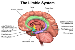
The limbic system is a set of brain structures located on both sides of the thalamus, immediately beneath the cerebrum.[1] It has also been referred to as the paleomammalian cortex. It is not a separate system but a collection of structures from the telencephalon, diencephalon, and mesencephalon.[2] It includes the olfactory bulbs, hippocampus, hypothalamus, amygdala, anterior thalamic nuclei, fornix, columns of fornix, mammillary body, septum pellucidum, habenular commissure, cingulate gyrus, parahippocampal gyrus, entorhinal cortex, and limbic midbrain areas.[3]

The limbic system supports a variety of functions including emotion, behavior, motivation, long-term memory, and olfaction.[4] Emotional life is largely housed in the limbic system, and it has a great deal to do with the formation of memories.

Although the term only originated in the 1940s, some neuroscientists, including Joseph LeDoux, have suggested that the concept of a functionally unified limbic system should be abandoned as obsolete because it is grounded mainly in historical concepts of brain anatomy that are no longer accepted as accurate.[5]

Structure


Anatomical components of the limbic system

The limbic system was originally defined by Paul Broca as a series of cortical structures surrounding the limit between the cerebral hemispheres and the brainstem: the border, or limbus, of the brain. These structures were known together as the limbic lobe.[6] Further studies began to associate these areas with emotional and motivational processes and linked them to subcortical components that were grouped into the limbic system.[7] The existence of such a system as an isolated entity responsible for the neurological regulation of emotion has gone into disuse and currently it is considered as one of the many parts of the brain that regulate visceral, autonomic processes.[8]

Therefore, the definition of anatomical structures considered part of the limbic system is a controversial subject. The following structures are, or have been considered, part of the limbic system:[9][10]

Function

The structures of the limbic system are involved in motivation, emotion, learning, and memory. The limbic system is where the subcortical structures meet the cerebral cortex.[1] The limbic system operates by influencing the endocrine system and the autonomic nervous system. It is highly interconnected with the nucleus accumbens, which plays a role in sexual arousal and the "high" derived from certain recreational drugs. These responses are heavily modulated by dopaminergic projections from the limbic system. In 1954, Olds and Milner found that rats with metal electrodes implanted into their nucleus accumbens, as well as their septal nuclei, repeatedly pressed a lever activating this region, and did so in preference to eating and drinking, eventually dying of exhaustion.[11] The limbic system also includes the basal ganglia. The basal ganglia are a set of subcortical structures that direct intentional movements. The basal ganglia are located near the thalamus and hypothalamus. They receive input from the cerebral cortex, which sends outputs to the motor centers in the brain stem. A part of the basal ganglia called the striatum controls posture and movement. Recent studies indicate that, if there is an inadequate supply of dopamine, the striatum is affected, which can lead to visible behavioral symptoms of Parkinson's disease.[1]

The limbic system is also tightly connected to the prefrontal cortex. Some scientists contend that this connection is related to the pleasure obtained from solving problems. To cure severe emotional disorders, this connection was sometimes surgically severed, a procedure of psychosurgery, called a prefrontal lobotomy (this is actually a misnomer). Patients having undergone this procedure often became passive and lacked all motivation.

The limbic system is often classified as a “cerebral structure”. This structure is closely linked to olfaction, emotions, drives, autonomic regulation, memory, and pathologically to encephalopathy, epilepsy, psychotic symptoms, cognitive defects.[12] The functional relevance of the limbic system has proven to serve many different functions such as affects/emotions, memory, sensory processing, time perception, attention, consciousness, instincts, autonomic/vegetative control, and actions/motor behavior. Some of the disorders associated with the limbic system are epilepsy and schizophrenia.[13]

Hippocampus

Various processes of cognition involve the hippocampus.

Spatial memory

The first and most widely researched area concerns memory, spatial memory in particular. Spatial memory was found to have many sub-regions in the hippocampus, such as the dentate gyrus (DG) in the dorsal hippocampus, the left hippocampus, and the parahippocampal region. The dorsal hippocampus was found to be an important component for the generation of new neurons, called adult-born granules (GC), in adolescence and adulthood.[14] These new neurons contribute to pattern separation in spatial memory, increasing the firing in cell networks, and overall causing stronger memory formations.

While the dorsal hippocampus is involved in spatial memory formation, the left hippocampus is a participant in the recall of these spatial memories. Eichenbaum[15] and his team found, when studying the hippocampal lesions in rats, that the left hippocampus is “critical for effectively combining the ‘what, ‘when,’ and ‘where’ qualities of each experience to compose the retrieved memory.” This makes the left hippocampus a key component in the retrieval of spatial memory. However, Spreng[16] found that the left hippocampus is, in fact, a general concentrated region for binding together bits and pieces of memory composed not only by the hippocampus, but also by other areas of the brain to be recalled at a later time. Eichenbaum’s research in 2007 also demonstrates that the parahippocampal area of the hippocampus is another specialized region for the retrieval of memories just like the left hippocampus.

Learning

The hippocampus, over the decades, has also been found to have a huge impact in learning. Curlik and Shors[17] examined the effects of neurogenesis in the hippocampus and its effects on learning. This researcher and his team employed many different types of mental and physical training on their subjects, and found that the hippocampus is highly responsive to these latter tasks. Thus, they discovered an upsurge of new neurons and neural circuits in the hippocampus as a result of the training, causing an overall improvement in the learning of the task. This neurogenesis contributes to the creation of adult-born granules cells (GC), cells also described by Eichenbaum[15] in his own research on neurogenesis and its contributions to learning. The creation of these cells exhibited "enhanced excitability" in the dentate gyrus (DG) of the dorsal hippocampus, impacting the hippocampus and its contribution to the learning process.[15]

Hippocampus damage

Damage related to the hippocampal region of the brain has reported vast effects on overall cognitive functioning, particularly memory such as spatial memory. As previously mentioned, spatial memory is a cognitive function greatly intertwined with the hippocampus. While damage to the hippocampus may be a result of a brain injury or other injuries of that sort, researchers particularly investigated the effects that high emotional arousal and certain types of drugs had on the recall ability in this specific memory type. In particular, in a study performed by Parkard,[18] rats were given the task of correctly making their way through a maze. In the first condition, rats were stressed by shock or restraint which caused a high emotional arousal. When completing the maze task, these rats had an impaired effect on their hippocampal-dependent memory when compared to the control group. Then, in a second condition, a group of rats were injected with anxiogenic drugs. Like the former these results reported similar outcomes, in that hippocampal-memory was also impaired. Studies such as these reinforce the impact that the hippocampus has on memory processing, in particular the recall function of spatial memory. Furthermore, impairment to the hippocampus can occur from prolonged exposure to stress hormones such as Glucocorticoids (GCs), which target the hippocampus and cause disruption in explicit memory.[19]

In an attempt to curtail life-threatening epileptic seizures, 27-year-old Henry Gustav Molaison underwent bilateral removal of almost all of his hippocampus in 1953. Over the course of fifty years he participated in thousands of tests and research projects that provided specific information on exactly what he had lost. Semantic and episodic events faded within minutes, having never reached his long term memory, yet emotions, unconnected from the details of causation, were often retained. Dr. Suzanne Corkin, who worked with him for 46 years until his death, described the contribution of this tragic "experiment" in her 2013 book.[20]

Amygdala

Episodic-autobiographical memory (EAM) networks

Another integrative part of the limbic system, the amygdala is involved in many cognitive processes. Like the hippocampus, processes in the amygdala seem to impact memory; however, it is not spatial memory as in the hippocampus but episodic-autobiographical memory (EAM) networks.

Markowitsch's[21] amygdala research shows it encodes, stores, and retrieves EAM memories. To delve deeper into these types of processes by the amygdala, Markowitsch[21] and his team provided extensive evidence through investigations that the "amygdala's main function is to charge cues so that mnemonic events of a specific emotional significance can be successfully searched within the appropriate neural nets and re-activated." These cues for emotional events created by the amygdala encompass the EAM networks previously mentioned.

Attentional and emotional processes

Besides memory, the amygdala also seems to be an important brain region involved in attentional and emotional processes. First, to define attention in cognitive terms, attention is the ability to home in on some stimuli while ignoring others. Thus, the amygdala seems to be an important structure in this ability. Foremost, however, this structure was historically thought to be linked to fear, allowing the individual to take action in response to that fear. However, as time has gone by, researchers such as Pessoa,[22] generalized this concept with help from evidence of EEG recordings, and concluded that the amygdala helps an organism to define a stimulus and therefore respond accordingly. However, when the amygdala was initially thought to be linked to fear, this gave way for research in the amygdala for emotional processes. Kheirbek[14] demonstrated research that the amygdala is involved in emotional processes, in particular the ventral hippocampus. He described the ventral hippocampus as having a role in neurogenesis and the creation of adult-born granule cells (GC). These cells not only were a crucial part of neurogenesis and the strengthening of spatial memory and learning in the hippocampus but also appear to be an essential component in the amygdala. A deficit of these cells, as Pessoa (2009) predicted in his studies, would result in low emotional functioning, leading to high retention rate of mental diseases, such as anxiety disorders.

Social processing

Social processing, specifically the evaluation of faces in social processing, is an area of cognition specific to the amygdala. In a study done by Todorov,[23] fMRI tasks were performed with participants to evaluate whether the amygdala was involved in the general evaluation of faces. After the study, Todorov concluded from his fMRI results that the amygdala did indeed play a key role in the general evaluation of faces. However, in a study performed by researchers Koscik[24] and his team, the trait of trustworthiness was particularly examined in the evaluation of faces. Koscik and his team demonstrated that the amygdala was involved in evaluating the trustworthiness of an individual. They investigated how brain damage to the amygdala played a role in trustworthiness, and found that individuals that suffered damage tended to confuse trust and betrayal, and thus placed trust in those having done them wrong. Furthermore, Rule,[25] along with his colleagues, expanded on the idea of the amygdala in its critique of trustworthiness in others by performing a study in 2009 in which he examined the amygdala's role in evaluating general first impressions and relating them to real-world outcomes. Their study involved first impressions of CEOs. Rule demonstrated that while the amygdala did play a role in the evaluation of trustworthiness, as observed by Koscik in his own research two years later in 2011, the amygdala also played a generalized role in the overall evaluation of first impression of faces. This latter conclusion, along with Todorov's study on the amygdala’s role in general evaluations of faces and Koscik’s research on trustworthiness and the amygdala, further solidified evidence that the amygdala plays a role in overall social processing.

Evolution

Paul D. MacLean, as part of his triune brain theory, hypothesized that the limbic system is older than other parts of the forebrain, and that it developed to manage circuitry attributed to the fight or flight first identified by Hans Selye [26] in his report of the General Adaptation Syndrome in 1936. It may be considered a part of survival adaptation, leading to what describes evolution adaptation throughout the history of species differentiation in reptiles as well as mammals (including humans). MacLean postulated that the human brain has evolved three components, that evolved successively, with more recent components developing at the top/front. These components are, respectively:
  1. The archipallium or primitive ("reptilian") brain, comprising the structures of the brain stem – medulla, pons, cerebellum, mesencephalon, the oldest basal nuclei – the globus pallidus and the olfactory bulbs.
  2. The paleopallium or intermediate ("old mammalian") brain, comprising the structures of the limbic system.
  3. The neopallium, also known as the superior or rational ("new mammalian") brain, comprises almost the whole of the hemispheres (made up of a more recent type of cortex, called neocortex) and some subcortical neuronal groups. It corresponds to the brain of the superior mammals, thus including the primates and, as a consequence, the human species. Similar development of the neocortex in mammalian species unrelated to humans and primates has also occurred, for example in cetaceans and elephants; thus the designation of "superior mammals" is not an evolutionary one, as it has occurred independently in different species. The evolution of higher degrees of intelligence is an example of convergent evolution, and is also seen in non-mammals such as birds.
According to Maclean, each of the components, although connected with the others, retained "their peculiar types of intelligence, subjectivity, sense of time and space, memory, mobility and other less specific functions".

However, while the categorization into structures is reasonable, the recent studies of the limbic system of tetrapods, both living and extinct, have challenged several aspects of this hypothesis, notably the accuracy of the terms "reptilian" and "old mammalian". The common ancestors of reptiles and mammals had a well-developed limbic system in which the basic subdivisions and connections of the amygdalar nuclei were established.[27] Further, birds, which evolved from the dinosaurs, which in turn evolved separately but around the same time as the mammals, have a well-developed limbic system. While the anatomic structures of the limbic system are different in birds and mammals, there are functional equivalents.

Clinical significance

Damage to the structures of limbic system results in conditions like Alzheimer's disease, anterograde amnesia, retrograde amnesia, and Klüver-Bucy syndrome.[citation needed]

Society and culture

Etymology and history

The term limbic comes from the Latin limbus, for "border" or "edge", or, particularly in medical terminology, a border of an anatomical component. Paul Broca coined the term based on its physical location in the brain, sandwiched between two functionally different components.

The limbic system is a term that was introduced in 1949 by the American physician and neuroscientist, Paul D. MacLean.[28][29] The French physician Paul Broca first called this part of the brain le grand lobe limbique in 1878.[6] He examined the differentiation between deeply recessed cortical tissue and underlying, subcortical nuclei.[30] However, most of its putative role in emotion was developed only in 1937 when the American physician James Papez described his anatomical model of emotion, the Papez circuit.[31]

The first evidence that the limbic system was responsible for the cortical representation of emotions was discovered in 1939, by Heinrich Kluver and Paul Bucy. Kluver and Bucy, after much research, demonstrated that the bilateral removal of the temporal lobes in monkeys created an extreme behavioral syndrome. After performing a temporal lobectomy, the monkeys showed a decrease in aggression. The animals revealed a reduced threshold to visual stimuli, and were thus unable to recognize objects that were once familiar.[32] MacLean expanded these ideas to include additional structures in a more dispersed "limbic system", more on the lines of the system described above.[29] MacLean developed the intriguing theory of the "triune brain" to explain its evolution and to try to reconcile rational human behavior with its more primal and violent side. He became interested in the brain's control of emotion and behavior. After initial studies of brain activity in epileptic patients, he turned to cats, monkeys, and other models, using electrodes to stimulate different parts of the brain in conscious animals recording their responses.[33] In the 1950s, he began to trace individual behaviors like aggression and sexual arousal to their physiological sources. He analyzed the brain's center of emotions, the limbic system, and described an area that includes structures called the hippocampus and amygdala. Developing observations made by Papez, he determined that the limbic system had evolved in early mammals to control fight-or-flight responses and react to both emotionally pleasurable and painful sensations. The concept is now broadly accepted in neuroscience.[34] Additionally, MacLean said that the idea of the limbic system leads to a recognition that its presence "represents the history of the evolution of mammals and their distinctive family way of life." In the 1960s, Dr. MacLean enlarged his theory to address the human brain's overall structure and divided its evolution into three parts, an idea that he termed the triune brain. In addition to identifying the limbic system, he pointed to a more primitive brain called the R-complex, related to reptiles, which controls basic functions like muscle movement and breathing. The third part, the neocortex, controls speech and reasoning and is the most recent evolutionary arrival.[35] The concept of the limbic system has since been further expanded and developed by Walle Nauta, Lennart Heimer and others.
Academic dispute

There is controversy over the use of the term limbic system, with scientists such as LeDoux arguing that the term be considered obsolete and abandoned.[36] Originally, the limbic system was believed to be the emotional center of the brain, with cognition being the business of the neocortex. However, cognition depends on acquisition and retention of memories, in which the hippocampus, a primary limbic structure, is involved: hippocampus damage causes severe cognitive (memory) deficits. More important, the "boundaries" of the limbic system have been repeatedly redefined because of advances in neuroscience. Therefore, while it is true that limbic structures are more closely related to emotion, the brain can be thought of as an integrated whole.

Cranial nerves

From Wikipedia, the free encyclopedia

Cranial nerves
Skull brain human normal.svg
Right View of the human brain from below, showing origins of cranial nerves.
Left Juxtaposed skull base with foramina in which many nerves exit the skull.
Skull and brainstem inner ear.svg
Cranial nerves as they pass through the skull base to the brain.
Details
Identifiers
Latin nervus cranialis
(pl: nervi craniales)
MeSH D003391
TA A14.2.01.001
A14.2.00.038
FMA 5865

Cranial nerves are the nerves that emerge directly from the brain (including the brainstem), in contrast to spinal nerves (which emerge from segments of the spinal cord).[1] 10 of 12 of the cranial nerves originate in the brainstem. Cranial nerves relay information between the brain and parts of the body, primarily to and from regions of the head and neck.[2]

Spinal nerves emerge sequentially from the spinal cord with the spinal nerve closest to the head (C1) emerging in the space above the first cervical vertebra. The cranial nerves, however, emerge from the central nervous system above this level.[3] Each cranial nerve is paired and is present on both sides. Depending on definition in humans there are twelve or thirteen cranial nerves pairs, which are assigned Roman numerals I–XII, sometimes also including cranial nerve zero. The numbering of the cranial nerves is based on the order in which they emerge from the brain, front to back (brainstem).[1]

The terminal nerves, olfactory nerves (I) and optic nerves (II) emerge from the cerebrum or forebrain, and the remaining ten pairs arise from the brainstem, which is the lower part of the brain.[1]

The cranial nerves are considered components of the peripheral nervous system (PNS),[1] although on a structural level the olfactory, optic and trigeminal nerves are more accurately considered part of the central nervous system (CNS).[4]

Anatomy


View of the human brain from below showing the cranial nerves on an autopsy specimen
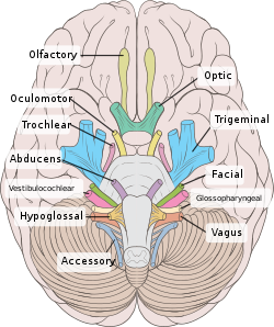
View from below of the brain and brainstem showing the cranial nerves, numbered from olfactory to hypoglossal after the order in which they emerge
The brainstem, with deeper cranial nerve nuclei and tracts inside the brain-stem shaded red.

Most typically, humans are considered to have twelve pairs of cranial nerves (I–XII). They are: the olfactory nerve (I), the optic nerve (II), oculomotor nerve (III), trochlear nerve (IV), trigeminal nerve (V), abducens nerve (VI), facial nerve (VII), vestibulocochlear nerve (VIII), glossopharyngeal nerve (IX), vagus nerve (X), accessory nerve (XI), and hypoglossal nerve (XII). (There may be a thirteenth cranial nerve, the terminal nerve (nerve O or N), which is very small and may or may not be functional in humans[1][3])

Terminology

Cranial nerves are generally named according to their structure or function. For example, the olfactory nerve (I) supplies smell, and the facial nerve (VII) supplies motor innervation to the face. Because Latin was the lingua franca (common language) of the study of anatomy when the nerves were first documented, recorded, and discussed, many nerves maintain Latin or Greek names, including the trochlear nerve (IV), named according to its structure, as it supplies a muscle that attaches to a pulley (Greek: trochlea). The trigeminal nerve (V) is named in accordance with its three components (Latin: trigeminus meaning triplets),[5] and the vagus nerve (X) is named for its wandering course (Latin: vagus).[6]

Cranial nerves are numbered based on their rostral-caudal (front-back) position,[1] when viewing the brain. If the brain is carefully removed from the skull the nerves are typically visible in their numeric order, with the exception of the last, CN XII, which appears to emerge rostrally to (above) CN XI.[7]

Cranial nerves have paths within and outside the skull. The paths within the skull are called "intracranial" and the paths outside the skull are called "extracranial". There are many holes in the skull called "foramina" by which the nerves can exit the skull. All cranial nerves are paired, which means that they occur on both the right and left sides of the body. The muscle, skin, or additional function supplied by a nerve on the same side of the body as the side it originates from, is referred to an ipsilateral function. If the function is on the opposite side to the origin of the nerve, this is known as a contralateral function.[8]

Intracranial course

Nuclei

The cell bodies of many of the neurons of most of the cranial nerves are contained in one or more nuclei in the brainstem. These nuclei are important relative to cranial nerve dysfunction because damage to these nuclei such as from a stroke or trauma can mimic damage to one or more branches of a cranial nerve. In terms of specific cranial nerve nuclei, the midbrain of the brainstem has the nuclei of the oculomotor nerve (III) and trochlear nerve (IV); the pons has the nuclei of the trigeminal nerve (V), abducens nerve (VI), facial nerve (VII) and vestibulocochlear nerve (VIII); and the medulla has the nuclei of the glossopharyngeal nerve (IX), vagus nerve (X), accessory nerve (XI) and hypoglossal nerve (XII). The fibers of these cranial nerves exit the brainstem from these nuclei.[1]

Ganglia

Some of the cranial nerves have sensory or parasympathetic ganglia (collections of cell bodies) of neurons, which are located outside the brain (but can be inside or outside the skull).[1]

The sensory ganglia are directly correspondent to dorsal root ganglia of spinal nerves and are known as cranial sensory ganglia.[7] Sensory ganglia exist for nerves with sensory function: V, VII, VIII, IX, X.[3] There are also parasympathetic ganglia, which are part of the autonomic nervous system for cranial nerves III, VII, IX and X.

Exiting the skull and extracranial course

Exits of cranial nerves from the skull.[1][9]
Location Nerve
cribriform plate Olfactory nerve (I)
optic foramen Optic nerve (II)
superior orbital fissure Oculomotor (III)
Trochlear (IV)
Abducens (VI)
Trigeminal V1
(ophthalmic)
Foramen rotundum Trigeminal V2
(maxillary)
Foramen ovale Trigeminal V3
(mandibular)
internal auditory canal Facial (VII)
Vestibulocochlear (VIII)
jugular foramen Glossopharyngeal (IX)
Vagus (X)
Accessory (XI)
hypoglossal canal Hypoglossal (XII)
After emerging from the brain, the cranial nerves travel within the skull, and some must leave this bony compartment in order to reach their destinations. Often the nerves pass through holes in the skull, called foramina, as they travel to their destinations. Other nerves pass through bony canals, longer pathways enclosed by bone. These foramina and canals may contain more than one cranial nerve, and may also contain blood vessels.[9]
  • The olfactory nerve (I), actually composed of many small separate nerve fibers, passes through perforations in the cribiform plate part of the ethmoid bone. These fibers terminate in the upper part of the nasal cavity and function to convey impulses containing information about odors to the brain.
  • The optic nerve (II) passes through the optic foramen in the sphenoid bone as it travels to the eye. It conveys visual information to the brain.
  • The oculomotor nerve (III), trochlear nerve (IV), abducens nerve (VI) and the ophthalmic branch of the trigeminal nerve (V1) travel through the cavernous sinus into the superior orbital fissure, passing out of the skull into the orbit. These nerves control the small muscles that move the eye and also provide sensory innervation to the eye and orbit.
  • The maxillary division of the trigeminal nerve (V2) passes through foramen rotundum in the sphenoid bone to supply the skin of the middle of the face.
  • The mandibular division of the trigeminal nerve (V3) passes through foramen ovale of the sphenoid bone to supply the lower face with sensory innervation. This nerve also sends branches to almost all of the muscles that control chewing.
  • The facial nerve (VII) and vestibulocochlear nerve (VIII) both enter the internal auditory canal in the temporal bone. The facial nerve then reaches the side of the face by using the stylomastoid foramen, also in the temporal bone. Its fibers then spread out to reach and control all of the muscles of facial expression. The vestibulocochlear nerve reaches the organs that control balance and hearing in the temporal bone, and therefore does not reach the external surface of the skull.
  • The glossopharyngeal (IX), vagus (X) and accessory nerve (XI) all leave the skull via the jugular foramen to enter the neck. The glossopharyngeal nerve provides innervation to the upper throat and the back of the tongue, the vagus provides innervation to the muscles in the voicebox, and continues downward to supply parasympathetic innervation to the chest and abdomen. The accessory nerve controls the trapezius and sternocleidomastoid muscles in the neck and shoulder.
  • The hypoglossal nerve (XII) exits the skull using the hypoglossal canal in the occipital bone and reaches the tongue to control almost all of the muscles involved in movements of this organ.[1]

Function

The cranial nerves provide motor and sensory innervation mainly to the structures within the head and neck. The sensory innervation includes both "general" sensation such as temperature and touch, and "special" innervation such as taste, vision, smell, balance and hearing[1][10]

The vagus nerve (X) provides sensory and autonomic (parasympathetic) motor innervation to structures in the neck and also to most of the organs in the chest and abdomen.[1][3]

Smell (I)

The olfactory nerve (I) conveys the sense of smell.

Damage to the olfactory nerve (I) can cause an inability to smell (anosmia), a distortion in the sense of smell (parosmia), or a distortion or lack of taste. If there is suspicion of a change in the sense of smell, each nostril is tested with substances of known odors such as coffee or soap. Intensely smelling substances, for example ammonia, may lead to the activation of pain receptors (nociceptors) of the trigeminal nerve that are located in the nasal cavity and this can confound olfactory testing.[1][11]

Vision (II)

The optic nerve (II) transmits visual information.[3][10]

Damage to the optic nerve (II) affects specific aspects of vision that depend on the location of the lesion. A person may not be able to see objects on their left or right sides (homonymous hemianopsia), or may have difficulty seeing objects on their outer visual fields (bitemporal hemianopsia) if the optic chiasm is involved.[12] Vision may be tested by examining the visual field, or by examining the retina with an ophthalmoscope, using a process known as funduscopy. Visual field testing may be used to pin-point structural lesions in the optic nerve, or further along the visual pathways.[11]

Eye movement (III, IV, VI)


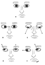
Various deviations of the eyes due to abnormal function of the targets of the cranial nerves

The oculomotor nerve (III), trochlear nerve (IV) and abducens nerve (VI) coordinate eye movement.

Damage to nerves III, IV, or VI may affect the movement of the eyeball (globe). Both or one eye may be affected; in either case double vision (diplopia) will likely occur because the movements of the eyes are no longer synchronized. Nerves III, IV and VI are tested by observing how the eye follows an object in different directions. This object may be a finger or a pin, and may be moved at different directions to test for pursuit velocity.[11] If the eyes do not work together, the most likely cause is damage to a specific cranial nerve or its nuclei.[11]

Damage to the oculomotor nerve (III) can cause double vision (diplopia) and inability to coordinate the movements of both eyes (strabismus), also eyelid drooping (ptosis) and pupil dilation (mydriasis).[12][12] Lesions may also lead to inability to open the eye due to paralysis of the levator palpebrae muscle. Individuals suffering from a lesion to the oculomotor nerve may compensate by tilting their heads to alleviate symptoms due to paralysis of one or more of the eye muscles it controls.[11]

Damage to the trochlear nerve (IV) can also cause diplopia with the eye adducted and elevated.[12] The result will be an eye which can not move downwards properly (especially downwards when in an inward position). This is due to impairment in the superior oblique muscle, which is innervated by the trochlear nerve.[11]

Damage to the abducens nerve (VI) can also result in diplopia.[12] This is due to impairment in the lateral rectus muscle, which is innervated by the abducens nerve.[11]

Trigeminal nerve (V)

The trigeminal nerve (V) comprises three distinct parts: The Ophthalmic (V1), the Maxillary (V2), and the Mandibular (V3) nerves. Combined, these nerves provide sensation to the skin of the face and also controls the muscles of mastication (chewing).[1] Conditions affecting the trigeminal nerve (V) include trigeminal neuralgia,[1] cluster headache,[13] and trigeminal zoster.[1] Trigeminal neuralgia occurs later in life, from middle age onwards, most often after age 60, and is a condition typically associated with very strong pain distributed over the area innervated by the maxillary or mandibular nerve divisions of the trigeminal nerve (V2 and V3).[14]


Facial expression (VII)

Lesions of the facial nerve (VII) may manifest as facial palsy. This is where a person is unable to move the muscles on one or both sides of their face. A very common and generally temporary facial palsy is known as Bell's palsy. Bell's Palsy is the result of an idiopathic (unknown cause), unilateral lower motor neuron lesion of the facial nerve and is characterized by an inability to move the ipsilateral muscles of facial expression, including elevation of the eyebrow and furrowing of the forehead. Patients with Bell's palsy often have a drooping mouth on the affected side and often have trouble chewing because the buccinator muscle is affected.[1] Bell's palsy occurs very rarely, affecting around 40,000 Americans annually. There are studies in mice and humans suggesting members of the family Herpesviridae are capable of producing Bell's palsy. Facial paralysis may be caused by other conditions including stroke, and similar conditions to Bell's Palsy are occasionally misdiagnosed as Bell's Palsy.[15] Bell's Palsy is a temporary condition usually lasting 2-6 months, but can have life-changing effects and can reoccur. Strokes typically also affect the seventh cranial nerve by cutting off blood supply to nerves in the brain that signal this nerve and so can present with similar symptoms.

Hearing and balance (VIII)

The vestibulocochlear nerve (VIII) splits into the vestibular and cochlear nerve. The vestibular part is responsible for innervating the vestibules and semicircular canal of the inner ear; this structure transmits information about balance, and is an important component of the vestibuloocular reflex, which keeps the head stable and allows the eyes to track moving objects. The cochlear nerve transmits information from the cochlea, allowing sound to be heard.[3]

When damaged, the vestibular nerve may give rise to the sensation of spinning and dizziness. Function of the vestibular nerve may be tested by putting cold and warm water in the ears and watching eye movements caloric stimulation.[1][11] Damage to the vestibulocochlear nerve can also present as repetitive and involuntary eye movements (nystagmus), particularly when looking in a horizontal plane.[11] Damage to the cochlear nerve will cause partial or complete deafness in the affected ear.[11]

Oral sensation, taste, and salivation (IX)


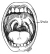
Deviating uvula due to cranial nerve IX lesion

The glossopharyngeal nerve (IX) innervates the stylopharyngeus muscle and provides sensory innervation to the oropharynx and back of the tongue.[1][16] The glossopharyngeal nerve also provides parasympathetic innervation to the parotid gland.[1] Unilateral absence of a gag reflex suggests a lesion of the glossopharyngeal nerve (IX), and perhaps the vagus nerve (X).[17]

Vagus nerve (X)

Loss of function of the vagus nerve (X) will lead to a loss of parasympathetic innervation to a very large number of structures. Major effects of damage to the vagus nerve may include a rise in blood pressure and heart rate. Isolated dysfunction of only the vagus nerve is rare, but can be diagnosed by a hoarse voice, due to dysfunction of one of its branches, the recurrent laryngeal nerve.[1]

Damage to this nerve may result in difficulties swallowing.[11]

Shoulder elevation and head-turning (XI)


Winged scapula may occur due to lesion of the spinal accessory.

Damage to the accessory nerve (XI) will lead to ipsilateral weakness in the trapezius muscle. This can be tested by asking the subject to raise their shoulders or shrug, upon which the shoulder blade (scapula) will protrude into a winged position.[1] Additionally, if the nerve is damaged, weakness or an inability to elevate the scapula may be present because the levator scapulae muscle is now solely able to provide this function.[14] Depending on the location of the lesion there may also be weakness present in the sternocleidomastoid muscle, which acts to turn the head so that the face points to the opposite side.[1]

Tongue movement (XII)

A damaged hypoglossal nerve will result in an inability to stick the tongue out straight.
A case with unilateral hypoglossal nerve injury in branchial cyst surgery. [18]


The hypoglossal nerve (XII) is unique in that it is innervated from the motor cortices of both hemispheres of the brain. Damage to the nerve at lower motor neuron level may lead to fasciculations or atrophy of the muscles of the tongue. The fasciculations of the tongue are sometimes said to look like a "bag of worms". Upper motor neuron damage will not lead to atrophy or fasciculations, but only weakness of the innervated muscles.[11]

When the nerve is damaged, it will lead to weakness of tongue movement on one side. When damaged and extended, the tongue will move towards the weaker or damaged side, as shown in the image.[11]

Clinical significance

Examination

Physicians, neurologists, and other medical professionals may conduct a cranial nerve examination as part of a neurological examination to examine the functionality of the cranial nerves. This is a highly formalized series of tests that assess the status of each nerve.[19] A cranial nerve exam begins with observation of the patient because some cranial nerve lesions may affect the symmetry of the eyes or face. The visual fields are tested for nerve lesions or nystagmus via an analysis of specific eye movements. The sensation of the face is tested, and patients are asked to perform different facial movements, such as puffing out of the cheeks. Hearing is checked by voice and tuning forks. The position of the patient's uvula is examined because asymmetry in the position could indicate a lesion of the glossopharyngeal nerve. After the ability of the patient to use their shoulder to assess the accessory nerve (XI), and the patient's tongue function is assessed by observing various tongue movements.[1][19]

Damage

Compression

Nerves may be compressed because of increased intracranial pressure, a mass effect of an intracerebral haemorrhage, or tumour that presses against the nerves and interferes with the transmission of impulses along the nerve.[20] A loss of functionality of a single cranial nerve may sometimes be the first symptom of an intracranial or skull base cancer.[21]

An increase in intracranial pressure may lead to impairment of the optic nerves (II) due to compression of the surrounding veins and capillaries, causing swelling of the eyeball (papilloedema).[22] A cancer, such as an optic glioma, may also impact the optic nerve (II). A pituitary tumour may compress the optic tracts or the optic chiasm of the optic nerve (II), leading to visual field loss. A pituitary tumour may also extend into the cavernous sinus, compressing the oculuomotor nerve (III), trochlear nerve (IV) and abducens nerve (VI), leading to double-vision and strabismus. These nerves may also be affected by herniation of the temporal lobes of the brain through the falx cerebri.[20]

The cause of trigeminal neuralgia, in which one side of the face is exquisitely painful, is thought to be compression of the nerve by an artery as the nerve emerges from the brain stem.[20] An acoustic neuroma, particularly at the junction between the pons and medulla, may compress the facial nerve (VII) and vestibulocochlear nerve (VIII), leading to hearing and sensory loss on the affected side.[20][23]

Stroke

Occlusion of blood vessels that supply the nerves or their nuclei, an ischemic stroke, may cause specific signs and symptoms that can localise where the occlusion occurred. A clot in a blood vessel draining the cavernous sinus (cavernous sinus thrombosis) affects the oculomotor (III), trochlear (IV), opthalamic branch of the trigeminal nerve (V1) and the abducens nerve (VI).[23]

Inflammation

Inflammation resulting from infection may impair the function of any of the cranial nerves. Inflammation of the facial nerve (VII) may result in Bell's palsy.[24]

Multiple sclerosis, an inflammatory process that may produce a loss of the myelin sheathes which surround the cranial nerves, may cause a variety of shifting symptoms affecting multiple cranial nerves.[24]

Other

Trauma to the skull, disease of bone such as Paget's disease, and injury to nerves during neurosurgery (such as tumor removal) are other possible causes of cranial nerve damage.[23]

History

The Graeco-Roman anatomist Galen (AD 129–210) named seven pairs of cranial nerves.[25] Much later, in 1664, English anatomist Sir Thomas Willis suggested that there were actually 9 pairs of nerves. Finally, in 1778, German anatomist Samuel Soemmering named the 12 pairs of nerves that are generally accepted today.[25] However, because many of the nerves emerge from the brain stem as rootlets, there is continual debate as to how many nerves there actually are, and how they should be grouped.[25] There is reason to consider both the olfactory (I) and Optic (II) nerves to be brain tracts, rather than cranial nerves.[25] Further, the very small terminal nerve (nerve N or O) exists in humans but may not be functional. In other animals, it appears to be important to sexual receptivity based on perceptions of phermones[1][26]

Other animals

Dog-fish brain in two projections.
top; ventral bottom; lateral
The accessory nerve (XI) and hypoglossal nerve (XII) cannot be seen, as they are not always present in all vertebrates.

Cranial nerves are also present in other vertebrates. Other amniotes (non-amphibian tetrapods) have cranial nerves similar to those of humans. In anamniotes (fishes and amphibians), the accessory nerve (XI) and hypoglossal nerve (XII) do not exist, with the accessory nerve (XI) being an integral part of the vagus nerve (X); the hypoglossal nerve (XII) is represented by a variable number of spinal nerves emerging from vertebral segments fused into the occiput. These two nerves only became discrete nerves in the ancestors of amniotes (non-amphibian tetrapods).[27]

Operator (computer programming)

From Wikipedia, the free encyclopedia https://en.wikipedia.org/wiki/Operator_(computer_programmin...