Search This Blog

Friday, April 23, 2021

Cable theory

From Wikipedia, the free encyclopedia
 
Schematic of resistance and capacitance in an abstract neuronal fiber
Figure. 1: Cable theory's simplified view of a neuronal fiber

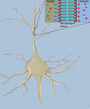
Classical cable theory uses mathematical models to calculate the electric current (and accompanying voltage) along passive neurites, particularly the dendrites that receive synaptic inputs at different sites and times. Estimates are made by modeling dendrites and axons as cylinders composed of segments with capacitances and resistances combined in parallel (see Fig. 1). The capacitance of a neuronal fiber comes about because electrostatic forces are acting through the very thin lipid bilayer (see Figure 2). The resistance in series along the fiber is due to the axoplasm's significant resistance to movement of electric charge.

Capacitance in a neuron fiber
Figure. 2: Fiber capacitance

History

Cable theory in computational neuroscience has roots leading back to the 1850s, when Professor William Thomson (later known as Lord Kelvin) began developing mathematical models of signal decay in submarine (underwater) telegraphic cables. The models resembled the partial differential equations used by Fourier to describe heat conduction in a wire.

The 1870s saw the first attempts by Hermann to model neuronal electrotonic potentials also by focusing on analogies with heat conduction. However, it was Hoorweg who first discovered the analogies with Kelvin's undersea cables in 1898 and then Hermann and Cremer who independently developed the cable theory for neuronal fibers in the early 20th century. Further mathematical theories of nerve fiber conduction based on cable theory were developed by Cole and Hodgkin (1920s–1930s), Offner et al. (1940), and Rushton (1951).

Experimental evidence for the importance of cable theory in modelling the behavior of axons began surfacing in the 1930s from work done by Cole, Curtis, Hodgkin, Sir Bernard Katz, Rushton, Tasaki and others. Two key papers from this era are those of Davis and Lorente de Nó (1947) and Hodgkin and Rushton (1946).

The 1950s saw improvements in techniques for measuring the electric activity of individual neurons. Thus cable theory became important for analyzing data collected from intracellular microelectrode recordings and for analyzing the electrical properties of neuronal dendrites. Scientists like Coombs, Eccles, Fatt, Frank, Fuortes and others now relied heavily on cable theory to obtain functional insights of neurons and for guiding them in the design of new experiments.

Later, cable theory with its mathematical derivatives allowed ever more sophisticated neuron models to be explored by workers such as Jack, Rall, Redman, Rinzel, Idan Segev, Tuckwell, Bell, and Iannella. More recently, cable theory has been applied to model electrical activity in bundled neurons in the white matter of the brain.

Deriving the cable equation

Note, various conventions of rm exist. Here rm and cm, as introduced above, are measured per membrane-length unit (per meter (m)). Thus rm is measured in ohm·meters (Ω·m) and cm in farads per meter (F/m). This is in contrast to Rm (in Ω·m2) and Cm (in F/m2), which represent the specific resistance and capacitance respectively of one unit area of membrane (in m2). Thus, if the radius, a, of the axon is known, then its circumference is 2πa, and its rm, and its cm values can be calculated as:


(1)


(2)

These relationships make sense intuitively, because the greater the circumference of the axon, the greater the area for charge to escape through its membrane, and therefore the lower the membrane resistance (dividing Rm by 2πa); and the more membrane available to store charge (multiplying Cm by 2πa). The specific electrical resistance, ρl, of the axoplasm allows one to calculate the longitudinal intracellular resistance per unit length, rl, (in Ω·m−1) by the equation:


(3)

The greater the cross sectional area of the axon, πa2, the greater the number of paths for the charge to flow through its axoplasm, and the lower the axoplasmic resistance.

Several important avenues of extending classical cable theory have recently seen the introduction of endogenous structures in order to analyze the effects of protein polarization within dendrites and different synaptic input distributions over the dendritic surface of a neuron.

To better understand how the cable equation is derived, first simplify the theoretical neuron even further and pretend it has a perfectly sealed membrane (rm=∞) with no loss of current to the outside, and no capacitance (cm = 0). A current injected into the fiber  at position x = 0 would move along the inside of the fiber unchanged. Moving away from the point of injection and by using Ohm's law (V = IR) we can calculate the voltage change as:


(4)

where the negative is because current flows down the potential gradient.

Letting Δx go towards zero and having infinitely small increments of x, one can write (4) as:


(5)

or


(6)

Bringing rm back into the picture is like making holes in a garden hose. The more holes, the faster the water will escape from the hose, and the less water will travel all the way from the beginning of the hose to the end. Similarly, in an axon, some of the current traveling longitudinally through the axoplasm will escape through the membrane.

If im is the current escaping through the membrane per length unit, m, then the total current escaping along y units must be y·im. Thus, the change of current in the axoplasm, Δil, at distance, Δx, from position x=0 can be written as:


(7)

or, using continuous, infinitesimally small increments:


(8)

can be expressed with yet another formula, by including the capacitance. The capacitance will cause a flow of charge (a current) towards the membrane on the side of the cytoplasm. This current is usually referred to as displacement current (here denoted .) The flow will only take place as long as the membrane's storage capacity has not been reached. can then be expressed as:


(9)

where is the membrane's capacitance and is the change in voltage over time. The current that passes the membrane () can be expressed as:


(10)

and because the following equation for can be derived if no additional current is added from an electrode:


(11)

where represents the change per unit length of the longitudinal current.

Combining equations (6) and (11) gives a first version of a cable equation:


(12)

which is a second-order partial differential equation (PDE).

By a simple rearrangement of equation (12) (see later) it is possible to make two important terms appear, namely the length constant (sometimes referred to as the space constant) denoted and the time constant denoted . The following sections focus on these terms.

Length constant

The length constant, (lambda), is a parameter that indicates how far a stationary current will influence the voltage along the cable. The larger the value of , the farther the charge will flow. The length constant can be expressed as:


(13)

The larger the membrane resistance, rm, the greater the value of , and the more current will remain inside the axoplasm to travel longitudinally through the axon. The higher the axoplasmic resistance, , the smaller the value of , the harder it will be for current to travel through the axoplasm, and the shorter the current will be able to travel. It is possible to solve equation and arrive at the following equation (which is valid in steady-state conditions, i.e. when time approaches infinity):


(14)

Where is the depolarization at (point of current injection), e is the exponential constant (approximate value 2.71828) and is the voltage at a given distance x from x=0. When then


(15)

and


(16)

which means that when we measure at distance from we get


(17)

Thus is always 36.8 percent of .

Time constant

Neuroscientists are often interested in knowing how fast the membrane potential, , of an axon changes in response to changes in the current injected into the axoplasm. The time constant, , is an index that provides information about that value. can be calculated as:


(18)

The larger the membrane capacitance, , the more current it takes to charge and discharge a patch of membrane and the longer this process will take. The larger the membrane resistance , the harder it is for a current to induce a change in membrane potential. So the higher the the slower the nerve impulse can travel. That means, membrane potential (voltage across the membrane) lags more behind current injections. Response times vary from 1–2 milliseconds in neurons that are processing information that needs high temporal precision to 100 milliseconds or longer. A typical response time is around 20 milliseconds.

Generic form and mathematical structure

If one multiplies equation (12) by on both sides of the equal sign we get:


(19)

and recognize on the left side and on the right side. The cable equation can now be written in its perhaps best known form:


(20)

This is a 1D Heat equation or Diffusion Equation for which many solution methods, such as Green's functions and Fourier methods, have been developed.

It is also a special degenerate case of the Telegrapher's equation, where the inductance vanishes and the signal propagation speed is infinite.

Phospholipid

From Wikipedia, the free encyclopedia
 
Phospholipid arrangement in cell membranes.
 
Phosphatidylcholine is the major component of lecithin. It is also a source for choline in the synthesis of acetylcholine in cholinergic neurons.

Phospholipids, also known as phosphatides, are a class of lipids whose molecule has a hydrophilic "head" containing a phosphate group, and two hydrophobic "tails" derived from fatty acids, joined by a glycerol molecule. The phosphate group can be modified with simple organic molecules such as choline, ethanolamine or serine.

Phospholipids are a key component of all cell membranes. They can form lipid bilayers because of their amphiphilic characteristic. In eukaryotes, cell membranes also contain another class of lipid, sterol, interspersed among the phospholipids. The combination provides fluidity in two dimensions combined with mechanical strength against rupture. Purified phospholipids are produced commercially and have found applications in nanotechnology and materials science.

The first phospholipid identified in 1847 as such in biological tissues was lecithin, or phosphatidylcholine, in the egg yolk of chickens by the French chemist and pharmacist Theodore Nicolas Gobley.

Phospholipids in biological membranes

Arrangement

The phospholipids are amphiphilic. The hydrophilic end usually contains a negatively charged phosphate group, and the hydrophobic end usually consists of two "tails" that are long fatty acid residues.

In aqueous solutions, phospholipids are driven by hydrophobic interactions that result in the fatty acid tails aggregating to minimize interactions with water molecules. The result is often a phospholipid bilayer: a membrane that consists of two layers of oppositely oriented phospholipid molecules, with their heads exposed to the liquid on both sides, and with the tails directed into the membrane. That is the dominant structural motif of the membranes of all cells and of some other biological structures, such as vescicles or virus coatings.

Phospholipid bilayers are the main structural component of cell membranes.

In biological membranes, the phospholipids often occur with other molecules (e.g., proteins, glycolipids, sterols) in a bilayer such as a cell membrane. Lipid bilayers occur when hydrophobic tails line up against one another, forming a membrane of hydrophilic heads on both sides facing the water.

Dynamics

These specific properties allow phospholipids to play an important role in the cell membrane. Their movement can be described by the fluid mosaic model, that describes the membrane as a mosaic of lipid molecules that act as a solvent for all the substances and proteins within it, so proteins and lipid molecules are then free to diffuse laterally through the lipid matrix and migrate over the membrane. Sterols contribute to membrane fluidity by hindering the packing together of phospholipids. However, this model has now been superseded, as through the study of lipid polymorphism it is now known that the behaviour of lipids under physiological (and other) conditions is not simple.[citation needed]

Main phospholipids

Diacylglyceride structures

Phosphosphingolipids

Applications

Phospholipids have been widely used to prepare liposomal, ethosomal and other nanoformulations of topical, oral and parenteral drugs for differing reasons like improved bio-availability, reduced toxicity and increased permeability across membranes. Liposomes are often composed of phosphatidylcholine-enriched phospholipids and may also contain mixed phospholipid chains with surfactant properties. The ethosomal formulation of ketoconazole using phospholipids is a promising option for transdermal delivery in fungal infections.

Simulations

Computational simulations of phospholipids are often performed using molecular dynamics with force fields such as GROMOS, CHARMM, or AMBER.

Characterization

Phospholipids are optically highly birefringent, i.e. their refractive index is different along their axis as opposed to perpendicular to it. Measurement of birefringence can be achieved using cross polarisers in a microscope to obtain an image of e.g. vesicle walls or using techniques such as dual polarisation interferometry to quantify lipid order or disruption in supported bilayers.

Analysis

There are no simple methods available for analysis of phospholipids since the close range of polarity between different phospholipid species makes detection difficult. Oil chemists often use spectroscopy to determine total Phosphorus abundance and then calculate approximate mass of phospholipids based on molecular weight of expected fatty acid species. Modern lipid profiling employs more absolute methods of analysis, with nuclear magnetic resonance spectroscopy (NMR spectroscopy), particularly 31P-NMR, while HPLC-ELSD provides relative values.

Phospholipid synthesis

Phospholipid synthesis occurs in the cytosolic side of ER membrane  that is studded with proteins that act in synthesis (GPAT and LPAAT acyl transferases, phosphatase and choline phosphotransferase) and allocation (flippase and floppase). Eventually a vesicle will bud off from the ER containing phospholipids destined for the cytoplasmic cellular membrane on its exterior leaflet and phospholipids destined for the exoplasmic cellular membrane on its inner leaflet.

Sources

Common sources of industrially produced phospholipids are soya, rapeseed, sunflower, chicken eggs, bovine milk, fish eggs etc. Each source has a unique profile of individual phospholipid species as well as fatty acids and consequently differing applications in food, nutrition, pharmaceuticals, cosmetics and drug delivery.

In signal transduction

Some types of phospholipid can be split to produce products that function as second messengers in signal transduction. Examples include phosphatidylinositol (4,5)-bisphosphate (PIP2), that can be split by the enzyme Phospholipase C into inositol triphosphate (IP3) and diacylglycerol (DAG), which both carry out the functions of the Gq type of G protein in response to various stimuli and intervene in various processes from long term depression in neurons to leukocyte signal pathways started by chemokine receptors.

Phospholipids also intervene in prostaglandin signal pathways as the raw material used by lipase enzymes to produce the prostaglandin precursors. In plants they serve as the raw material to produce Jasmonic acid, a plant hormone similar in structure to prostaglandins that mediates defensive responses against pathogens.

Food technology

Phospholipids can act as emulsifiers, enabling oils to form a colloid with water. Phospholipids are one of the components of lecithin which is found in egg-yolks, as well as being extracted from soybeans, and is used as a food additive in many products, and can be purchased as a dietary supplement. Lysolecithins are typically used for water-oil emulsions like margarine, due to their higher HLB ratio.

Phospholipid derivatives

See table below for an extensive list.

Abbreviations used and chemical information of glycerophospholipids

Abbreviation CAS Name Type
DDPC 3436-44-0 1,2-Didecanoyl-sn-glycero-3-phosphocholine Phosphatidylcholine
DEPA-NA 80724-31-8 1,2-Dierucoyl-sn-glycero-3-phosphate (Sodium Salt) Phosphatidic acid
DEPC 56649-39-9 1,2-Dierucoyl-sn-glycero-3-phosphocholine Phosphatidylcholine
DEPE 988-07-2 1,2-Dierucoyl-sn-glycero-3-phosphoethanolamine Phosphatidylethanolamine
DEPG-NA
1,2-Dierucoyl-sn-glycero-3[Phospho-rac-(1-glycerol...) (Sodium Salt) Phosphatidylglycerol
DLOPC 998-06-1 1,2-Dilinoleoyl-sn-glycero-3-phosphocholine Phosphatidylcholine
DLPA-NA
1,2-Dilauroyl-sn-glycero-3-phosphate (Sodium Salt) Phosphatidic acid
DLPC 18194-25-7 1,2-Dilauroyl-sn-glycero-3-phosphocholine Phosphatidylcholine
DLPE
1,2-Dilauroyl-sn-glycero-3-phosphoethanolamine Phosphatidylethanolamine
DLPG-NA
1,2-Dilauroyl-sn-glycero-3[Phospho-rac-(1-glycerol...) (Sodium Salt) Phosphatidylglycerol
DLPG-NH4
1,2-Dilauroyl-sn-glycero-3[Phospho-rac-(1-glycerol...) (Ammonium Salt) Phosphatidylglycerol
DLPS-NA
1,2-Dilauroyl-sn-glycero-3-phosphoserine (Sodium Salt) Phosphatidylserine
DMPA-NA 80724-3 1,2-Dimyristoyl-sn-glycero-3-phosphate (Sodium Salt) Phosphatidic acid
DMPC 18194-24-6 1,2-Dimyristoyl-sn-glycero-3-phosphocholine Phosphatidylcholine
DMPE 988-07-2 1,2-Dimyristoyl-sn-glycero-3-phosphoethanolamine Phosphatidylethanolamine
DMPG-NA 67232-80-8 1,2-Dimyristoyl-sn-glycero-3[Phospho-rac-(1-glycerol...) (Sodium Salt) Phosphatidylglycerol
DMPG-NH4
1,2-Dimyristoyl-sn-glycero-3[Phospho-rac-(1-glycerol...) (Ammonium Salt) Phosphatidylglycerol
DMPG-NH4/NA
1,2-Dimyristoyl-sn-glycero-3[Phospho-rac-(1-glycerol...) (Sodium/Ammonium Salt) Phosphatidylglycerol
DMPS-NA
1,2-Dimyristoyl-sn-glycero-3-phosphoserine (Sodium Salt) Phosphatidylserine
DOPA-NA
1,2-Dioleoyl-sn-glycero-3-phosphate (Sodium Salt) Phosphatidic acid
DOPC 4235-95-4 1,2-Dioleoyl-sn-glycero-3-phosphocholine Phosphatidylcholine
DOPE 4004-5-1- 1,2-Dioleoyl-sn-glycero-3-phosphoethanolamine Phosphatidylethanolamine
DOPG-NA 62700-69-0 1,2-Dioleoyl-sn-glycero-3[Phospho-rac-(1-glycerol...) (Sodium Salt) Phosphatidylglycerol
DOPS-NA 70614-14-1 1,2-Dioleoyl-sn-glycero-3-phosphoserine (Sodium Salt) Phosphatidylserine
DPPA-NA 71065-87-7 1,2-Dipalmitoyl-sn-glycero-3-phosphate (Sodium Salt) Phosphatidic acid
DPPC 63-89-8 1,2-Dipalmitoyl-sn-glycero-3-phosphocholine Phosphatidylcholine
DPPE 923-61-5 1,2-Dipalmitoyl-sn-glycero-3-phosphoethanolamine Phosphatidylethanolamine
DPPG-NA 67232-81-9 1,2-Dipalmitoyl-sn-glycero-3[Phospho-rac-(1-glycerol...) (Sodium Salt) Phosphatidylglycerol
DPPG-NH4 73548-70-6 1,2-Dipalmitoyl-sn-glycero-3[Phospho-rac-(1-glycerol...) (Ammonium Salt) Phosphatidylglycerol
DPPS-NA
1,2-Dipalmitoyl-sn-glycero-3-phosphoserine (Sodium Salt) Phosphatidylserine
DSPA-NA 108321-18-2 1,2-Distearoyl-sn-glycero-3-phosphate (Sodium Salt) Phosphatidic acid
DSPC 816-94-4 1,2-Distearoyl-sn-glycero-3-phosphocholine Phosphatidylcholine
DSPE 1069-79-0 1,2-Distearoyl-sn-glycero-3-phosphoethanolamine Phosphatidylethanolamine
DSPG-NA 67232-82-0 1,2-Distearoyl-sn-glycero-3[Phospho-rac-(1-glycerol...) (Sodium Salt) Phosphatidylglycerol
DSPG-NH4 108347-80-4 1,2-Distearoyl-sn-glycero-3[Phospho-rac-(1-glycerol...) (Ammonium Salt) Phosphatidylglycerol
DSPS-NA
1,2-Distearoyl-sn-glycero-3-phosphoserine (Sodium Salt) Phosphatidylserine
EPC
Egg-PC Phosphatidylcholine
HEPC
Hydrogenated Egg PC Phosphatidylcholine
HSPC
Hydrogenated Soy PC Phosphatidylcholine
LYSOPC MYRISTIC 18194-24-6 1-Myristoyl-sn-glycero-3-phosphocholine Lysophosphatidylcholine
LYSOPC PALMITIC 17364-16-8 1-Palmitoyl-sn-glycero-3-phosphocholine Lysophosphatidylcholine
LYSOPC STEARIC 19420-57-6 1-Stearoyl-sn-glycero-3-phosphocholine Lysophosphatidylcholine
Milk Sphingomyelin MPPC
1-Myristoyl-2-palmitoyl-sn-glycero 3-phosphocholine Phosphatidylcholine
MSPC
1-Myristoyl-2-stearoyl-sn-glycero-3–phosphocholine Phosphatidylcholine
PMPC
1-Palmitoyl-2-myristoyl-sn-glycero-3–phosphocholine Phosphatidylcholine
POPC 26853-31-6 1-Palmitoyl-2-oleoyl-sn-glycero-3-phosphocholine Phosphatidylcholine
POPE
1-Palmitoyl-2-oleoyl-sn-glycero-3-phosphoethanolamine Phosphatidylethanolamine
POPG-NA 81490-05-3 1-Palmitoyl-2-oleoyl-sn-glycero-3[Phospho-rac-(1-glycerol)...] (Sodium Salt) Phosphatidylglycerol
PSPC
1-Palmitoyl-2-stearoyl-sn-glycero-3–phosphocholine Phosphatidylcholine
SMPC
1-Stearoyl-2-myristoyl-sn-glycero-3–phosphocholine Phosphatidylcholine
SOPC
1-Stearoyl-2-oleoyl-sn-glycero-3-phosphocholine Phosphatidylcholine
SPPC
1-Stearoyl-2-palmitoyl-sn-glycero-3-phosphocholine Phosphatidylcholine


Entropy (information theory)

From Wikipedia, the free encyclopedia https://en.wikipedia.org/wiki/Entropy_(information_theory) In info...