Search This Blog

Friday, April 23, 2021

Cell signaling

From Wikipedia, the free encyclopedia
https://en.wikipedia.org/wiki/Cell_signaling

In biology, cell signaling (cell signalling in British English), or cell-cell communication, governs the basic activities of cells and coordinates multiple-cell actions. A signal is an entity that codes or conveys information. Biological processes are complex molecular interactions that involve many signals. The ability of cells to perceive and correctly respond to their microenvironment is the basis of development, tissue repair, and immunity, as well as normal tissue homeostasis. Errors in signaling interactions and cellular information processing may cause diseases such as cancer, autoimmunity, and diabetes. By understanding cell signaling, clinicians may treat diseases more effectively and, theoretically, researchers may develop artificial tissues.

All cells receive and respond to signals from their surroundings. This is accomplished by a variety of signal molecules that are secreted or expressed on the surface of one cell and bind to a receptor expressed by the other cells, thereby integrating and coordinating the function of the many individual cells that make up organisms. Each cell is programmed to respond to specific extracellular signal molecules. Extracellular signaling usually entails the following steps:

  1. Synthesis and release of the signaling molecule by the signaling cell;
  2. Transport of the signal to the target cell;
  3. Binding of the signal by a specific receptor leading to its activation;
  4. Initiation of signal-transduction pathways.

Signaling agents could be physical agents like mechanical pressure, voltage, temperature, and light or chemical agents like peptides, steroids, terpenoids, etc. It may be food material or pathogen-associated patterns, or it may be oxygen or carbon dioxide levels or specially biosynthesised signaling molecules like hormones and ferromones (ektohormones). Signaling molecules vary greatly in their physio-chemical properties such as solubility (hydrophobic or hydrophillic). Some of the signaling molecules are gaseous, such as nitric oxide. Additionally, proteins on the surface of neighboring cells could also be signals.

Synthesis involves various biosynthetic pathways, and happens in specific time and place. Signal molecules may be released from the cell and sometimes they are not released at all, such as cellular localization signals and DNA damage signals. Such intracellular signaling networks work within the cell. Signal molecules that can be released through various ways like membrane-diffusion, exocytosis or cell damage. In some cases the signal molecules remain attached with cell-surface, a mode that helps in juxtacrine signaling (discussed below). Sometimes signal molecules require activation, such as through proteolytic cleavage or covalent modifications.

The ultimate path of the signal may be intracellular or intercellular. The intercellular signaling is also called cell-to-cell communication. It can be short or long distance. Based on nature of this path of signal molecule from source to target cell; the signaling pathways are classified into autocrine, juxtacrine, intracrine, paracrine and endocrine (discussed below)

Receptors play a key role in cell signaling. Receptors help in recognising the signal molecule (ligand). However some receptor molecules respond to physical agents (voltage, light, etc). Receptor molecules are generally proteins. Receptors may be located at cell surface, or interior of the cell such as cytosol, the organelles and nucleus (especially the transcription factors). Usually the cell surface receptors bind membrane-impermeable signal molecules, but sometimes they also interact with membrane permeable signal molecules. A key step in signaling is removal and degradation of the signal molecule. Sometimes the receptor is also degraded. Neurotransmitter reuptake is a mechanism of signaling molecule removal that is commonly seen in nervous system, and is a target of some class of prescription psychiatric medications.

Binding with the ligand causes conformational change in the receptor, which leads to further transmission of signaling. Due to conformational change, the receptor may either show an enzymic activity (called enzymic receptor), or a ion channel opening or closing activity (called a channel receptor). Sometimes the receptors themselves do not contain enzymatic or channel-like domains but they are linked with enzyme or transporter. Some receptors (like the nuclear-cytoplasmic superfamily) have a different mechanism. Once they bind with signal, they change their DNA binding properties and cellular localisation to the nucleus.

Result of enzymatic activity of the receptor usually leads to recruitment of additional molecular changes thus causing a signal transduction "cascade". These intermediates often forms a second messenger system. Within the signal transduction cascade there may be enzymes and transporters which work similar way as receptors. Enzymatic activities include covalent modifications like proteolytic cleavage, phosphorylation/dephosphorylation, methylation/demethylation, ubiquitinylation/deubiquitinylation etc. These changes help regulate the propagation of the signal through the cell. An important phenomena that happens in the intracellular portion of signaling is signal amplification. During signal amplification, a few number of receptors are initially activated. The intracellular response results in multiple secondary messengers to be activated, thereby amplifying the initial signal.

Systems biology studies the underlying structure of cell-signaling networks and how changes in these networks may affect the transmission and flow of information (signal transduction). Such networks are complex systems in their organization and may exhibit a number of emergent properties, including bistability and ultrasensitivity. Analysis of cell-signaling networks requires a combination of experimental and theoretical approaches, including the development and analysis of simulations and modeling. Long-range allostery is often a significant component of cell-signaling events.

Between individual organisms of same species

Figure 1. Example of signaling between bacteria. Salmonella enteritidis uses N-Acyl homoserine lactone for Quorum sensing

Cell signaling has been most extensively studied in the context of human diseases and signaling between cells of a single organism. However, cell signaling may also occur between the cells of two different individuals of the same species. In many mammals, early embryo cells exchange signals with cells of the uterus. In the human gastrointestinal tract, bacteria exchange signals with each other and with human epithelial and immune system cells. For the yeast Saccharomyces cerevisiae during mating, some cells send a peptide signal (mating factor pheromones) into their environment. The mating factor peptide may bind to a cell surface receptor on other yeast cells and induce them to prepare for mating.

Classification

Cell signaling can be classified as either mechanical or biochemical based on the type of the signal. Mechanical signals are the forces exerted on the cell and the forces produced by the cell. These forces can both be sensed and responded to by the cells. Biochemical signals are biochemical molecules such as proteins, lipids, ions, and gases. These signals can be categorized based on the distance between signaling and responder cells. Signaling within, between, and amongst cells is subdivided into the following classifications:

  • Intracrine signals are produced by the target cell that stay within the target cell.
  • Autocrine signals are produced by the target cell, are secreted, and affect the target cell itself via receptors. Sometimes autocrine cells can target cells close by if they are the same type of cell as the emitting cell. An example of this are immune cells.
  • Juxtacrine signals target adjacent (touching) cells. These signals are transmitted along cell membranes via protein or lipid components integral to the membrane and are capable of affecting either the emitting cell or cells immediately adjacent.
  • Paracrine signals target cells in the vicinity of the emitting cell. Neurotransmitters represent an example.
  • Endocrine signals target distant cells. Endocrine cells produce hormones that travel through the blood to reach all parts of the body.
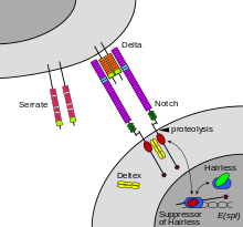
Figure 2. Notch-mediated juxtacrine signal between adjacent cells.

Cells communicate with each other via direct contact (juxtacrine signaling), over short distances (paracrine signaling), or over large distances and/or scales (endocrine signaling).

Some cell–cell communication requires direct cell–cell contact. Some cells can form gap junctions that connect their cytoplasm to the cytoplasm of adjacent cells. In cardiac muscle, gap junctions between adjacent cells allow for action potential propagation from the cardiac pacemaker region of the heart to spread and coordinate the contraction of the heart.

The notch signaling mechanism is an example of juxtacrine signaling (also known as contact-dependent signaling) in which two adjacent cells must make physical contact in order to communicate. This requirement for direct contact allows for very precise control of cell differentiation during embryonic development. In the worm Caenorhabditis elegans, two cells of the developing gonad each have an equal chance of terminally differentiating or becoming a uterine precursor cell that continues to divide. The choice of which cell continues to divide is controlled by competition of cell surface signals. One cell will happen to produce more of a cell surface protein that activates the Notch receptor on the adjacent cell. This activates a feedback loop or system that reduces Notch expression in the cell that will differentiate and that increases Notch on the surface of the cell that continues as a stem cell.

Many cell signals are carried by molecules that are released by one cell and move to make contact with another cell. Endocrine signals are called hormones. Hormones are produced by endocrine cells and they travel through the blood to reach all parts of the body. Specificity of signaling can be controlled if only some cells can respond to a particular hormone. Paracrine signals such as retinoic acid target only cells in the vicinity of the emitting cell. Neurotransmitters represent another example of a paracrine signal. Some signaling molecules can function as both a hormone and a neurotransmitter. For example, epinephrine and norepinephrine can function as hormones when released from the adrenal gland and are transported to the heart by way of the blood stream. Norepinephrine can also be produced by neurons to function as a neurotransmitter within the brain. Estrogen can be released by the ovary and function as a hormone or act locally via paracrine or autocrine signaling. Active species of oxygen and nitric oxide can also act as cellular messengers. This process is dubbed redox signaling.

In multicellular organisms

In a multicellular organism, signaling between cells occurs either through release into the extracellular space, divided in paracrine signaling (over short distances) and endocrine signaling (over long distances), or by direct contact, known as juxtacrine signaling. Autocrine signaling is a special case of paracrine signaling where the secreting cell has the ability to respond to the secreted signaling molecule. Synaptic signaling is a special case of paracrine signaling (for chemical synapses) or juxtacrine signaling (for electrical synapses) between neurons and target cells. Signaling molecules interact with a target cell as a ligand to cell surface receptors, and/or by entering into the cell through its membrane or endocytosis for intracrine signaling. This generally results in the activation of second messengers, leading to various physiological effects.

A particular molecule is generally used in diverse modes of signaling, and therefore a classification by mode of signaling is not possible. At least three important classes of signaling molecules are widely recognized, although non-exhaustive and with imprecise boundaries, as such membership is non-exclusive and depends on the context:

Signaling molecules can belong to several chemical classes: lipids, phospholipids, amino acids, monoamines, proteins, glycoproteins, or gases. Signaling molecules binding surface receptors are generally large and hydrophilic (e.g. TRH, Vasopressin, Acetylcholine), while those entering the cell are generally small and hydrophobic (e.g. glucocorticoids, thyroid hormones, cholecalciferol, retinoic acid), but important exceptions to both are numerous, and a same molecule can act both via surface receptor or in an intracrine manner to different effects. In intracrine signaling, once inside the cell, a signaling molecule can bind to intracellular receptors, other elements, or stimulate enzyme activity (e.g. gasses). The intracrine action of peptide hormones remains a subject of debate.

Hydrogen sulfide is produced in small amounts by some cells of the human body and has a number of biological signaling functions. Only two other such gases are currently known to act as signaling molecules in the human body: nitric oxide and carbon monoxide.

In plants

Signaling in plants happen through plant hormones, Phytochromes, Cryptochromes etc.

Important families of plant hormones are Auxin, cytokinin, gibberelline, ethyline, jasmonic acid, salicylic acid, strigolactones, polyamines, nitric oxide, peptide hormones etc. Translocation of RNA also has been reported.

Signaling receptors


Transmembrane receptor working principle

Cells receive information from their neighbors through a class of proteins known as receptors. Receptors may bind with some molecules (ligands) or may interact with physical agents like light, mechanical temperature, pressure, etc. Some receptors are membrane bound and some receptors are cytosolic. A large number of cytosolic receptors belong to nuclear-cytoplasmic-superfamily.

Some important transmembrane receptors are Voltage gated ion channels , Ligand gated ion channels, Seven helix receptors or GPCRs, Two component receptors, Cytokine receptors, Receptor tyrosine kinase, Tyrosine kinase linked receptor, Receptor Serine threonine kinase, Receptor tyrosine phosphatase, Receptor guanylyl cyclase, Sphingomylinase linked receptor, Integrin, selectin, Cadherin, etc.

Notch is a cell surface protein that functions as a receptor. Animals have a small set of genes that code for signaling proteins that interact specifically with Notch receptors and stimulate a response in cells that express Notch on their surface. Molecules that activate (or, in some cases, inhibit) receptors can be classified as hormones, neurotransmitters, cytokines, and growth factors, in general called receptor ligands. Ligand receptor interactions such as that of the Notch receptor interaction, are known to be the main interactions responsible for cell signaling mechanisms and communication.

As shown in Figure 2 (above; left), notch acts as a receptor for ligands that are expressed on adjacent cells. While some receptors are cell-surface proteins, others are found inside cells. For example, estrogen is a hydrophobic molecule that can pass through the lipid bilayer of the membranes. As part of the endocrine system, intracellular estrogen receptors from a variety of cell types can be activated by estrogen produced in the ovaries.

A number of transmembrane receptors for small molecules and peptide hormones, as well as intracellular receptors for steroid hormones exist, giving cells the ability to respond to a great number of hormonal and pharmacological stimuli. In diseases, often, proteins that interact with receptors are aberrantly activated, resulting in constitutively activated downstream signals.

For several types of intercellular signaling molecules that are unable to permeate the hydrophobic cell membrane due to their hydrophilic nature, the target receptor is expressed on the membrane. When such a signaling molecule activates its receptor, the signal is carried into the cell usually by means of a second messenger such as cAMP.

The receptor-ligand interaction can be classified as:

  • Agonism: it is when a ligand increases the activity of a ligand. Agonism is demonstrated in absence of any other competing ligand for the same receptor.
  • Inverse agonism: When a receptor is constitutively active, and the constitutive activity is suppressed or inhibited by the ligand
  • Antagonism: In presence of the agonist ligand, the antagonist molecule hinder the activation of the receptor by the ligand.
  • Partial agonism: it is when a ligand shows agonism, but in spite of increasing dosage of the ligand, the receptor activation does not reach the full activation state.
  • Partial inverse agonism: When a receptor is constitutively active, and in spite of increasing dosage of the ligand, the receptor activity decreases but does not become fully inactive.
  • Protean agonism: Protean agonists can act both as an agonist or an inverse agonist based on whether the receptor is already inactive (quiescent) or already active.
  • Biased agonism : when a receptor acts on more than one variants of next molecule in transduction chain; and binding with one agonist favours only one of the possible transduction pathway.

Signaling pathways

Brief overview of some signaling pathways (based on receptor families)
Receptor Family Example of Ligands/ activators (Bracket: receptor for it) Example of effectors Further downstream effects
Ligand Gated Ion Channels Acetylcholine
(Such as Nicotinic acetylcholine receptor),
Changes in membrane permeability Change in membrane potential
Seven Helix Receptor Light(Rhodopsin),
Dopamine (Dopamine receptor),
GABA (GABA receptor),
Prostaglandin (prostaglandin receptor) etc.
Trimeric G protein Adenylate Cyclase,
cGMP phosphodiesterase,
G-protein gated ion channel, etc.
Frizzled (Special type of 7Helix receptor) Wnt Dishevelled, axin - APC, GSK3-beta - Beta catenin Gene expression
Two Component Diverse activators Histidine Kinase Response Regulator - flagellar movement, Gene expression
Receptor Tyrosine Kinase Insulin (insulin receptor),
EGF (EGF receptor),
FGF-Alpha, FGF-Beta, etc (FGF-receptors)
Ras, MAP-kinases, PLC, PI3-Kinase Gene expression change
Cytokine receptors Erythropoietin,
Growth Hormone (Growth Hormone Receptor),
IFN-Gamma (IFN-Gamma receptor) etc
JAK kinase STAT transcription factor - Gene expression
Tyrosine kinase Linked- receptors MHC-peptide complex - TCR, Antigens - BCR Cytoplasmic Tyrosine Kinase Gene expression
Receptor Serine/Threonine Kinase Activin(activin receptor),
Inhibin,
Bone-morphogenetic protein(BMP Receptor),
TGF-beta
Smad transcription factors Control of gene expression
Membrane Guanylyl Cyclase Atrial natriuretic peptide,
Sea urching egg peptide etc.
cGMP Regulation of Kinases and channels- Diverse actions
Cytoplasmic Guanylyl cyclase Nitric Oxide(Nitric oxide receptor) cGMP Regulation of cGMP Gated channels, Kinases
Sphingomyelinase linked receptors IL-1(IL-1 receptor),
TNF (TNF-receptors)
Ceramide activated kinases Gene expression
Integrins Fibronectins, other extracellular matrix proteins Nonreceptor tyrosine kinase Diverse response
Cytoplasmic Steroid receptors Steroid hormones,
Thyroid hormones,
Retinoic acid etc
Work as/ interact with transcription factors Gene expression


Overview of signal transduction pathways
 
Figure 3. Key components of a signal transduction pathway (MAPK/ERK pathway shown)

In some cases, receptor activation caused by ligand binding to a receptor is directly coupled to the cell's response to the ligand. For example, the neurotransmitter GABA can activate a cell surface receptor that is part of an ion channel. GABA binding to a GABAA receptor on a neuron opens a chloride-selective ion channel that is part of the receptor. GABAA receptor activation allows negatively charged chloride ions to move into the neuron, which inhibits the ability of the neuron to produce action potentials.  However, for many cell surface receptors, ligand-receptor interactions are not directly linked to the cell's response. The activated receptor must first interact with other proteins inside the cell before the ultimate physiological effect of the ligand on the cell's behavior is produced. Often, the behavior of a chain of several interacting cell proteins is altered following receptor activation. The entire set of cell changes induced by receptor activation is called a signal transduction mechanism or pathway.

In the case of Notch-mediated signaling, the signal transduction mechanism can be relatively simple. As shown in Figure 2, the activation of Notch can cause the Notch protein to be altered by a protease. Part of the Notch protein is released from the cell surface membrane and takes part in gene regulation. Cell signaling research involves studying the spatial and temporal dynamics of both receptors and the components of signaling pathways that are activated by receptors in various cell types. Emerging methods for single-cell mass-spectrometry analysis promise to enable studying signal transduction with single-cell resolution.

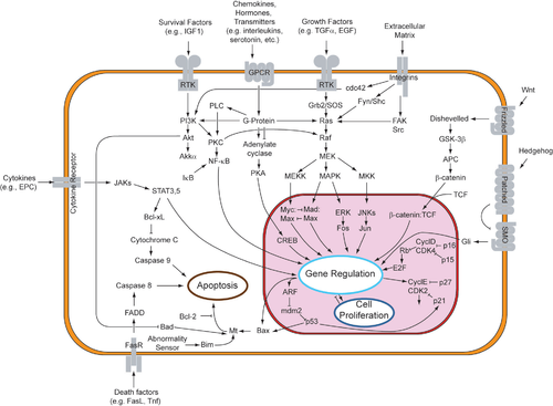
A more complex signal transduction pathway is shown in Figure 3. This pathway involves changes of protein–protein interactions inside the cell, induced by an external signal. Many growth factors bind to receptors at the cell surface and stimulate cells to progress through the cell cycle and divide. Several of these receptors are kinases that start to phosphorylate themselves and other proteins when binding to a ligand. This phosphorylation can generate a binding site for a different protein and thus induce protein–protein interaction. In Figure 3, the ligand (called epidermal growth factor, or EGF) binds to the receptor (called EGFR). This activates the receptor to phosphorylate itself. The phosphorylated receptor binds to an adaptor protein (GRB2), which couples the signal to further downstream signaling processes. For example, one of the signal transduction pathways that are activated is called the mitogen-activated protein kinase (MAPK) pathway. The signal transduction component labeled as "MAPK" in the pathway was originally called "ERK," so the pathway is called the MAPK/ERK pathway. The MAPK protein is an enzyme, a protein kinase that can attach phosphate to target proteins such as the transcription factor MYC and, thus, alter gene transcription and, ultimately, cell cycle progression. Many cellular proteins are activated downstream of the growth factor receptors (such as EGFR) that initiate this signal transduction pathway.

Some signaling transduction pathways respond differently, depending on the amount of signaling received by the cell. For instance, the hedgehog protein activates different genes, depending on the amount of hedgehog protein present.

Complex multi-component signal transduction pathways provide opportunities for feedback, signal amplification, and interactions inside one cell between multiple signals and signaling pathways.

Intra- and inter-species signaling

Molecular signaling can occur between different organisms, whether unicellular or multicellular. The emitting organism produces the signaling molecule, secretes it into the environment, where it diffuses, and it is sensed or internalized by the receiving organism. In some cases of interspecies signaling, the emitting organism can actually be a host of the receiving organism, or vice versa.

Intraspecies signaling occurs especially in bacteria, yeast, social insects, but also many vertebrates. The signaling molecules used by multicellular organisms are often called pheromones. They can have such purposes as alerting against danger, indicating food supply, or assisting in reproduction. In unicellular organisms such as bacteria, signaling can be used to 'activate' peers from a dormant state, enhance virulence, defend against bacteriophages, etc. In quorum sensing, which is also found in social insects, the multiplicity of individual signals has the potentiality to create a positive feedback loop, generating coordinated response. In this context, the signaling molecules are called autoinducers. This signaling mechanism may have been involved in evolution from unicellular to multicellular organisms. Bacteria also use contact-dependent signaling, notably to limit their growth.

Molecular signaling can also occur between individuals of different species. This has been particularly studied in bacteria. Different bacterial species can coordinate to colonize a host and participate in common quorum sensing. Therapeutic strategies to disrupt this phenomenon are being investigated. Interactions mediated through signaling molecules are also thought to occur between the gut flora and their host, as part of their commensal or symbiotic relationship. Gram negative microbes deploy bacterial outer membrane vesicles for intra- and inter-species signaling in natural environments and at the host-pathogen interface.

Additionally, interspecies signaling occurs between multicellular organisms. In Vespa mandarinia, individuals release a scent that directs the colony to a food source.

In plants, inter-species signaling is particularly important in mycorrhizal symbiosis and root nodule symbiosis. In both symbioses, Receptor-like kinase (RLK), G-proteins, MAP-kinases and Ca2+ plays a very important role 

Computational models

Recent approaches to better understand elements of pathway crosstalk, complex ligand-receptor binding, and signaling network dynamics have been aided by the use of systems biology approaches. Computational models often take aim at compiling information from published literature to generate a coherent set of signaling components and their associated interactions. The development of computational models allows for a more in-depth probing of cell signaling pathways at a global level by manipulating different variables and systemically evaluating the resulting response. The use of analytical models for the study of signal transduction has been heavily applied in the fields of pharmacology and drug discovery to assess receptor-ligand interactions and pharmacokinetics as well as the flow of metabolites in large networks. A commonly applied strategy to model cell signaling mechanisms is through the use of ordinary differential equation (ODE) models by expressing the time-dependent concentration of a signaling molecule as a function of other molecules downstream and/or upstream within the pathway. ODE models have already been applied for dynamic analysis of the Mitogen-activated protein kinase, Estrogen receptor alpha, and MTOR signaling pathways among numerous others.

Crystallography

From Wikipedia, the free encyclopedia

A crystalline solid: atomic resolution image of strontium titanate. Brighter atoms are strontium and darker ones are titanium.

Crystallography is the experimental science of determining the arrangement of atoms in crystalline solids. The word "crystallography" is derived from the Greek words crystallon "cold drop, frozen drop", with its meaning extending to all solids with some degree of transparency, and graphein "to write". In July 2012, the United Nations recognised the importance of the science of crystallography by proclaiming that 2014 would be the International Year of Crystallography.

Before the development of X-ray diffraction crystallography (see below), the study of crystals was based on physical measurements of their geometry using a goniometer. This involved measuring the angles of crystal faces relative to each other and to theoretical reference axes (crystallographic axes), and establishing the symmetry of the crystal in question. The position in 3D space of each crystal face is plotted on a stereographic net such as a Wulff net or Lambert net. The pole to each face is plotted on the net. Each point is labelled with its Miller index. The final plot allows the symmetry of the crystal to be established.

Crystallographic methods now depend on analysis of the diffraction patterns of a sample targeted by a beam of some type. X-rays are most commonly used; other beams used include electrons or neutrons. Crystallographers often explicitly state the type of beam used, as in the terms X-ray crystallography, neutron diffraction and electron diffraction. These three types of radiation interact with the specimen in different ways.

Because of these different forms of interaction, the three types of radiation are suitable for different crystallographic studies.

Theory

With conventional imaging techniques such as optical microscopy, obtaining an image of a small object requires collecting light with a magnifying lens. The resolution of any optical system is limited by the diffraction-limit of light, which depends on its wavelength. Thus, the overall clarity of resulting crystallographic electron density maps is highly dependent upon the resolution of the diffraction data, which can be categorized as: low, medium, high and atomic. For example, visible light has a wavelength of about 4000 to 7000 ångström, which is three orders of magnitude longer than the length of typical atomic bonds and atoms themselves (about 1 to 2 Å). Therefore, a conventional optical microscope cannot resolve the spatial arrangement of atoms in a crystal. To do so, we would need radiation with much shorter wavelengths, such as X-ray or neutron beams.

Unfortunately, focusing X-rays with conventional optical lens can be a challenge. Scientists have had some success focusing X-rays with microscopic Fresnel zone plates made from gold, and by critical-angle reflection inside long tapered capillaries. Diffracted X-ray or neutron beams cannot be focused to produce images, so the sample structure must be reconstructed from the diffraction pattern.

Diffraction patterns arise from the constructive interference of incident radiation (x-rays, electrons, neutrons), scattered by the periodic, repeating features of the sample. Because of their highly ordered and repetitive atomic structure (Bravais lattice), crystals diffract x-rays in a coherent manner, also referred to as Bragg's reflection.

Notation

  • Coordinates in square brackets such as [100] denote a direction vector (in real space).
  • Coordinates in angle brackets or chevrons such as <100> denote a family of directions which are related by symmetry operations. In the cubic crystal system for example, <100> would mean [100], [010], [001] or the negative of any of those directions.
  • Miller indices in parentheses such as (100) denote a plane of the crystal structure, and regular repetitions of that plane with a particular spacing. In the cubic system, the normal to the (hkl) plane is the direction [hkl], but in lower-symmetry cases, the normal to (hkl) is not parallel to [hkl].
  • Indices in curly brackets or braces such as {100} denote a family of planes and their normals. In cubic materials the symmetry makes them equivalent, just as the way angle brackets denote a family of directions. In non-cubic materials, <hkl> is not necessarily perpendicular to {hkl}.

Techniques

Some materials that have been analyzed crystallographically, such as proteins, do not occur naturally as crystals. Typically, such molecules are placed in solution and allowed to slowly crystallize through vapor diffusion. A drop of solution containing the molecule, buffer, and precipitants is sealed in a container with a reservoir containing a hygroscopic solution. Water in the drop diffuses to the reservoir, slowly increasing the concentration and allowing a crystal to form. If the concentration were to rise more quickly, the molecule would simply precipitate out of solution, resulting in disorderly granules rather than an orderly and hence usable crystal.

Once a crystal is obtained, data can be collected using a beam of radiation. Although many universities that engage in crystallographic research have their own X-ray producing equipment, synchrotrons are often used as X-ray sources, because of the purer and more complete patterns such sources can generate. Synchrotron sources also have a much higher intensity of X-ray beams, so data collection takes a fraction of the time normally necessary at weaker sources. Complementary neutron crystallography techniques are used to identify the positions of hydrogen atoms, since X-rays only interact very weakly with light elements such as hydrogen.

Producing an image from a diffraction pattern requires sophisticated mathematics and often an iterative process of modelling and refinement. In this process, the mathematically predicted diffraction patterns of a hypothesized or "model" structure are compared to the actual pattern generated by the crystalline sample. Ideally, researchers make several initial guesses, which through refinement all converge on the same answer. Models are refined until their predicted patterns match to as great a degree as can be achieved without radical revision of the model. This is a painstaking process, made much easier today by computers.

The mathematical methods for the analysis of diffraction data only apply to patterns, which in turn result only when waves diffract from orderly arrays. Hence crystallography applies for the most part only to crystals, or to molecules which can be coaxed to crystallize for the sake of measurement. In spite of this, a certain amount of molecular information can be deduced from patterns that are generated by fibers and powders, which while not as perfect as a solid crystal, may exhibit a degree of order. This level of order can be sufficient to deduce the structure of simple molecules, or to determine the coarse features of more complicated molecules. For example, the double-helical structure of DNA was deduced from an X-ray diffraction pattern that had been generated by a fibrous sample.

Materials science

Crystallography is used by materials scientists to characterize different materials. In single crystals, the effects of the crystalline arrangement of atoms is often easy to see macroscopically, because the natural shapes of crystals reflect the atomic structure. In addition, physical properties are often controlled by crystalline defects. The understanding of crystal structures is an important prerequisite for understanding crystallographic defects. Mostly, materials do not occur as a single crystal, but in poly-crystalline form (i.e., as an aggregate of small crystals with different orientations). Because of this, the powder diffraction method, which takes diffraction patterns of polycrystalline samples with a large number of crystals, plays an important role in structural determination.

Other physical properties are also linked to crystallography. For example, the minerals in clay form small, flat, platelike structures. Clay can be easily deformed because the platelike particles can slip along each other in the plane of the plates, yet remain strongly connected in the direction perpendicular to the plates. Such mechanisms can be studied by crystallographic texture measurements.

In another example, iron transforms from a body-centered cubic (bcc) structure to a face-centered cubic (fcc) structure called austenite when it is heated. The fcc structure is a close-packed structure unlike the bcc structure; thus the volume of the iron decreases when this transformation occurs.

Crystallography is useful in phase identification. When manufacturing or using a material, it is generally desirable to know what compounds and what phases are present in the material, as their composition, structure and proportions will influence the material's properties. Each phase has a characteristic arrangement of atoms. X-ray or neutron diffraction can be used to identify which patterns are present in the material, and thus which compounds are present. Crystallography covers the enumeration of the symmetry patterns which can be formed by atoms in a crystal and for this reason is related to group theory and geometry.

Biology

X-ray crystallography is the primary method for determining the molecular conformations of biological macromolecules, particularly protein and nucleic acids such as DNA and RNA. In fact, the double-helical structure of DNA was deduced from crystallographic data. The first crystal structure of a macromolecule was solved in 1958, a three-dimensional model of the myoglobin molecule obtained by X-ray analysis. The Protein Data Bank (PDB) is a freely accessible repository for the structures of proteins and other biological macromolecules. Computer programs such as RasMol, Pymol or VMD can be used to visualize biological molecular structures. Neutron crystallography is often used to help refine structures obtained by X-ray methods or to solve a specific bond; the methods are often viewed as complementary, as X-rays are sensitive to electron positions and scatter most strongly off heavy atoms, while neutrons are sensitive to nucleus positions and scatter strongly even off many light isotopes, including hydrogen and deuterium. Electron crystallography has been used to determine some protein structures, most notably membrane proteins and viral capsids.

Reference literature

The International Tables for Crystallography is an eight-book series that outlines the standard notations for formatting, describing and testing crystals. The series contains books that covers analysis methods and the mathematical procedures for determining organic structure though x-ray crystallography, electron diffraction, and neutron diffraction. The International tables are focused on procedures, techniques and descriptions and do not list the physical properties of individual crystals themselves. Each book is about 1000 pages and the titles of the books are:

Vol A - Space Group Symmetry,
Vol A1 - Symmetry Relations Between Space Groups,
Vol B - Reciprocal Space,
Vol C - Mathematical, Physical, and Chemical Tables,
Vol D - Physical Properties of Crystals,
Vol E - Subperiodic Groups,
Vol F - Crystallography of Biological Macromolecues, and
Vol G - Definition and Exchange of Crystallographic Data.

Steroid

From Wikipedia, the free encyclopedia

Complex chemical diagram
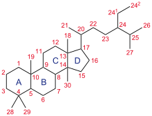
Structure of cholestane, a steroid with 27 carbon atoms. Its core ring system (ABCD), composed of 17 carbon atoms, is shown with IUPAC-approved ring lettering and atom numbering.

A steroid is a biologically active organic compound with four rings arranged in a specific molecular configuration. Steroids have two principal biological functions: as important components of cell membranes which alter membrane fluidity; and as signaling molecules. Hundreds of steroids are found in plants, animals and fungi. All steroids are manufactured in cells from the sterols lanosterol (opisthokonts) or cycloartenol (plants). Lanosterol and cycloartenol are derived from the cyclization of the triterpene squalene.

The steroid core structure is typically composed of seventeen carbon atoms, bonded in four "fused" rings: three six-member cyclohexane rings (rings A, B and C in the first illustration) and one five-member cyclopentane ring (the D ring). Steroids vary by the functional groups attached to this four-ring core and by the oxidation state of the rings. Sterols are forms of steroids with a hydroxy group at position three and a skeleton derived from cholestane. Steroids can also be more radically modified, such as by changes to the ring structure, for example, cutting one of the rings. Cutting Ring B produces secosteroids one of which is vitamin D3.

Examples include the lipid cholesterol, the sex hormones estradiol and testosterone, and the anti-inflammatory drug dexamethasone.

Filled-in diagram of a steroid
Space-filling representation
 
Ball-and-stick diagram of the same steroid
Ball-and-stick representation
 
5α-dihydroprogesterone (5α-DHP), a steroid. The shape of the four rings of most steroids is illustrated (carbon atoms in black, oxygens in red and hydrogens in grey). The nonpolar "slab" of hydrocarbon in the middle (grey, black) and the polar groups at opposing ends (red) are common features of natural steroids. 5α-DHP is an endogenous steroid hormone and a biosynthetic intermediate.

Nomenclature

Chemical diagram
Gonane, the simplest steroid, consisting only of the common steroid nucleus
 
Chemical diagram
Steroid 5α and 5β stereoisomers

Gonane, also known as steran or cyclopentanoperhydrophenanthrene, the simplest steroid and the nucleus of all steroids and sterols, is composed of seventeen carbon atoms in carbon-carbon bonds forming four fused rings in a three-dimensional shape. The three cyclohexane rings (A, B, and C in the first illustration) form the skeleton of a perhydro derivative of phenanthrene. The D ring has a cyclopentane structure. When the two methyl groups and eight carbon side chains (at C-17, as shown for cholesterol) are present, the steroid is said to have a cholestane framework. The two common 5α and 5β stereoisomeric forms of steroids exist because of differences in the side of the largely planar ring system where the hydrogen (H) atom at carbon-5 is attached, which results in a change in steroid A-ring conformation. Isomerisation at the C-21 side chain produces a parallel series of compounds, referred to as isosteroids.

Examples of steroid structures are:

In addition to the ring scissions (cleavages), expansions and contractions (cleavage and reclosing to a larger or smaller rings)—all variations in the carbon-carbon bond framework—steroids can also vary:

  • in the bond orders within the rings,
  • in the number of methyl groups attached to the ring (and, when present, on the prominent side chain at C17),
  • in the functional groups attached to the rings and side chain, and
  • in the configuration of groups attached to the rings and chain.

For instance, sterols such as cholesterol and lanosterol have a hydroxyl group attached at position C-3, while testosterone and progesterone have a carbonyl (oxo substituent) at C-3; of these, lanosterol alone has two methyl groups at C-4 and cholesterol (with a C-5 to C-6 double bond) differs from testosterone and progesterone (which have a C-4 to C-5 double bond).

Chemical diagram
Cholesterol, a prototypical animal sterol. This structural lipid and key steroid biosynthetic precursor.
Chemical diagram
5α-cholestane, a common steroid core

Species distribution and function

In eukaryotes, steroids are found in fungi, animals, and plants.

Fungal steroids

Fungal steroids include the ergosterols, which are involved in maintaining the integrity of the fungal cellular membrane. Various antifungal drugs, such as amphotericin B and azole antifungals, utilize this information to kill pathogenic fungi. Fungi can alter their ergosterol content (e.g. through loss of function mutations in the enzymes ERG3 or ERG6, inducing depletion of ergosterol, or mutations that decrease the ergosterol content) to develop resistance to drugs that target ergosterol. Ergosterol is analogous to the cholesterol found in the cellular membranes of animals (including humans), or the phytosterols found in the cellular membranes of plants. All mushrooms contain large quantities of ergosterol, in the range of tens to hundreds of milligrams per 100 grams of dry weight. Oxygen is necessary for the synthesis of ergosterol in fungi. Ergosterol is responsible for the vitamin D content found in mushrooms; ergosterol is chemically converted into provitamin D2 by exposure to ultraviolet light. Provitamin D2 spontaneously forms vitamin D2. However, not all fungi utilize ergosterol in their cellular membranes; for example, the pathogenic fungal species Pneumocystis jirovecii does not, which has important clinical implications (given the mechanism of action of many antifungal drugs). Using the fungus Saccharomyces cerevisiae as an example, other major steroids include ergosta‐5,7,22,24(28)‐tetraen‐3β‐ol, zymosterol, and lanosterol. S. cerevisiae utilizes 5,6‐dihydroergosterol in place of ergosterol in its cell membrane.

Animal steroids

Animal steroids include compounds of vertebrate and insect origin, the latter including ecdysteroids such as ecdysterone (controlling molting in some species). Vertebrate examples include the steroid hormones and cholesterol; the latter is a structural component of cell membranes which helps determine the fluidity of cell membranes and is a principal constituent of plaque (implicated in atherosclerosis). Steroid hormones include:

Plant steroids

Plant steroids include steroidal alkaloids found in Solanaceae and Melanthiaceae (specially the genus Veratrum), cardiac glycosides, the phytosterols and the brassinosteroids (which include several plant hormones).

Prokaryotes

In prokaryotes, biosynthetic pathways exist for the tetracyclic steroid framework (e.g. in mycobacteria) – where its origin from eukaryotes is conjectured – and the more-common pentacyclic triterpinoid hopanoid framework.

Types

By function

The major classes of steroid hormones, with prominent members and examples of related functions, are:

Additional classes of steroids include:

As well as the following class of secosteroids (open-ring steroids):

By structure

Intact ring system

Steroids can be classified based on their chemical composition. One example of how MeSH performs this classification is available at the Wikipedia MeSH catalog. Examples of this classification include:

Chemical diagram
Cholecalciferol (vitamin D3), an example of a 9,10-secosteroid
 
Chemical diagram
Cyclopamine, an example of a complex C-nor-D-homosteroid
 
Class Example Number of carbon atoms
Cholestanes Cholesterol 27
Cholanes Cholic acid 24
Pregnanes Progesterone 21
Androstanes Testosterone 19
Estranes Estradiol 18

The gonane (steroid nucleus) is the parent 17-carbon tetracyclic hydrocarbon molecule with no alkyl sidechains.

Cleaved, contracted, and expanded rings

Secosteroids (Latin seco, "to cut") are a subclass of steroidal compounds resulting, biosynthetically or conceptually, from scission (cleavage) of parent steroid rings (generally one of the four). Major secosteroid subclasses are defined by the steroid carbon atoms where this scission has taken place. For instance, the prototypical secosteroid cholecalciferol, vitamin D3 (shown), is in the 9,10-secosteroid subclass and derives from the cleavage of carbon atoms C-9 and C-10 of the steroid B-ring; 5,6-secosteroids and 13,14-steroids are similar.

Norsteroids (nor-, L. norma; "normal" in chemistry, indicating carbon removal) and homosteroids (homo-, Greek homos; "same", indicating carbon addition) are structural subclasses of steroids formed from biosynthetic steps. The former involves enzymic ring expansion-contraction reactions, and the latter is accomplished (biomimetically) or (more frequently) through ring closures of acyclic precursors with more (or fewer) ring atoms than the parent steroid framework.

Combinations of these ring alterations are known in nature. For instance, ewes who graze on corn lily ingest cyclopamine (shown) and veratramine, two of a sub-family of steroids where the C- and D-rings are contracted and expanded respectively via a biosynthetic migration of the original C-13 atom. Ingestion of these C-nor-D-homosteroids results in birth defects in lambs: cyclopia from cyclopamine and leg deformity from veratramine. A further C-nor-D-homosteroid (nakiterpiosin) is excreted by Okinawan cyanobacteriosponges. e.g., Terpios hoshinota, leading to coral mortality from black coral disease. Nakiterpiosin-type steroids are active against the signaling pathway involving the smoothened and hedgehog proteins, a pathway which is hyperactive in a number of cancers.

Biological significance

Steroids and their metabolites often function as signalling molecules (the most notable examples are steroid hormones), and steroids and phospholipids are components of cell membranes. Steroids such as cholesterol decrease membrane fluidity. Similar to lipids, steroids are highly concentrated energy stores. However, they are not typically sources of energy; in mammals, they are normally metabolized and excreted.

Steroids play critical roles in a number of disorders, including malignancies like prostate cancer, where steroid production inside and outside the tumour promotes cancer cell aggressiveness.

Biosynthesis and metabolism

Chemical-diagram flow chart
Simplification of the end of the steroid synthesis pathway, where the intermediates isopentenyl pyrophosphate (PP or IPP) and dimethylallyl pyrophosphate (DMAPP) form geranyl pyrophosphate (GPP), squalene and lanosterol (the first steroid in the pathway)

The hundreds of steroids found in animals, fungi, and plants are made from lanosterol (in animals and fungi; see examples above) or cycloartenol (in plants). Lanosterol and cycloartenol derive from cyclization of the triterpenoid squalene.

Steroid biosynthesis is an anabolic pathway which produces steroids from simple precursors. A unique biosynthetic pathway is followed in animals (compared to many other organisms), making the pathway a common target for antibiotics and other anti-infection drugs. Steroid metabolism in humans is also the target of cholesterol-lowering drugs, such as statins.

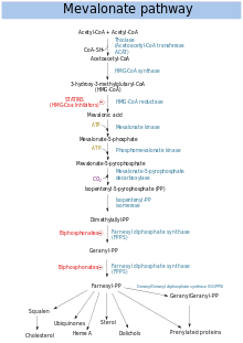
In humans and other animals the biosynthesis of steroids follows the mevalonate pathway, which uses acetyl-CoA as building blocks for dimethylallyl pyrophosphate (DMAPP) and isopentenyl pyrophosphate (IPP). In subsequent steps DMAPP and IPP join to form geranyl pyrophosphate (GPP), which synthesizes the steroid lanosterol. Modifications of lanosterol into other steroids are classified as steroidogenesis transformations.

Mevalonate pathway

Chemical flow chart
Mevalonate pathway

The mevalonate pathway (also called HMG-CoA reductase pathway) begins with acetyl-CoA and ends with dimethylallyl pyrophosphate (DMAPP) and isopentenyl pyrophosphate (IPP).

DMAPP and IPP donate isoprene units, which are assembled and modified to form terpenes and isoprenoids (a large class of lipids, which include the carotenoids and form the largest class of plant natural products. Here, the isoprene units are joined to make squalene and folded into a set of rings to make lanosterol. Lanosterol can then be converted into other steroids, such as cholesterol and ergosterol.

Two classes of drugs target the mevalonate pathway: statins (like rosuvastatin), which are used to reduce elevated cholesterol levels, and bisphosphonates (like zoledronate), which are used to treat a number of bone-degenerative diseases.

Steroidogenesis

Chemical-diagram flow chart
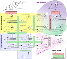
Human steroidogenesis, with the major classes of steroid hormones, individual steroids and enzymatic pathways. Changes in molecular structure from a precursor are highlighted in white.

Steroidogenesis is the biological process by which steroids are generated from cholesterol and changed into other steroids. The pathways of steroidogenesis differ among species. The major classes of steroid hormones, as noted above (with their prominent members and functions), are the Progestogen, Corticosteroids (corticoids), Androgens, and Estrogens. Human steroidogenesis of these classes occurs in a number of locations:

  • Progestogens are the precursors of all other human steroids, and all human tissues which produce steroids must first convert cholesterol to pregnenolone. This conversion is the rate-limiting step of steroid synthesis, which occurs inside the mitochondrion of the respective tissue.
  • Cortisol, corticosterone, aldosterone, and testosterone are produced in the adrenal cortex.
  • Estradiol, estrone and progesterone are made primarily in the ovary, estriol in placenta during pregnancy, and testosterone primarily in the testes (some testosterone is also produced in the adrenal cortex).
  • Estradiol is converted from testosterone directly (in males), or via the primary pathway DHEA - androstenedione - estrone and secondarily via testosterone (in females).
  • Stromal cells have been shown to produce steroids in response to signaling produced by androgen-starved prostate cancer cells.
  • Some neurons and glia in the central nervous system (CNS) express the enzymes required for the local synthesis of pregnenolone, progesterone, DHEA and DHEAS, de novo or from peripheral sources.

Alternative pathways

In plants and bacteria, the non-mevalonate pathway uses pyruvate and glyceraldehyde 3-phosphate as substrates.

During diseases pathways otherwise not significant in healthy humans can become utilized. For example, in one form of congenital adrenal hyperplasia a deficiency in the 21-hydroxylase enzymatic pathway leads to an excess of 17α-Hydroxyprogesterone (17-OHP) – this pathological excess of 17-OHP in turn may be converted to dihydrotestosterone (DHT, a potent androgen) through among others 17,20 Lyase (a member of the cytochrome P450 family of enzymes), 5α-Reductase and 3α-Hydroxysteroid dehydrogenase.

Catabolism and excretion

Steroids are primarily oxidized by cytochrome P450 oxidase enzymes, such as CYP3A4. These reactions introduce oxygen into the steroid ring, allowing the cholesterol to be broken up by other enzymes into bile acids. These acids can then be eliminated by secretion from the liver in bile. The expression of the oxidase gene can be upregulated by the steroid sensor PXR when there is a high blood concentration of steroids. Steroid hormones, lacking the side chain of cholesterol and bile acids, are typically hydroxylated at various ring positions or oxidized at the 17 position, conjugated with sulfate or glucuronic acid and excreted in the urine.

Isolation, structure determination, and methods of analysis

Steroid isolation, depending on context, is the isolation of chemical matter required for chemical structure elucidation, derivitzation or degradation chemistry, biological testing, and other research needs (generally milligrams to grams, but often more or the isolation of "analytical quantities" of the substance of interest (where the focus is on identifying and quantifying the substance (for example, in biological tissue or fluid). The amount isolated depends on the analytical method, but is generally less than one microgram. The methods of isolation to achieve the two scales of product are distinct, but include extraction, precipitation, adsorption, chromatography, and crystallization. In both cases, the isolated substance is purified to chemical homogeneity; combined separation and analytical methods, such as LC-MS, are chosen to be "orthogonal"—achieving their separations based on distinct modes of interaction between substance and isolating matrix—to detect a single species in the pure sample. Structure determination refers to the methods to determine the chemical structure of an isolated pure steroid, using an evolving array of chemical and physical methods which have included NMR and small-molecule crystallography. Methods of analysis overlap both of the above areas, emphasizing analytical methods to determining if a steroid is present in a mixture and determining its quantity.

Chemical synthesis

Microbial catabolism of phytosterol side chains yields C-19 steroids, C-22 steroids, and 17-ketosteroids (i.e. precursors to adrenocortical hormones and contraceptives). The addition and modification of functional groups is key when producing the wide variety of medications available within this chemical classification. These modifications are performed using conventional organic synthesis and/or biotransformation techniques.

Precursors

Semisynthesis

The semisynthesis of steroids often begins from precursors such as cholesterol, phytosterols, or sapogenins. The efforts of Syntex, a company involved in the Mexican barbasco trade, used Dioscorea mexicana to produce the sapogenin diosgenin in the early days of the synthetic steroid pharmaceutical industry.

Total synthesis

Some steroidal hormones are economically obtained only by total synthesis from petrochemicals (e.g. 13-alkyl steroids). For example, the pharmaceutical Norgestrel begins from Methoxy-1-tetralone, a petrochemical derived from phenol.

Research awards

A number of Nobel Prizes have been awarded for steroid research, including:

Introduction to entropy

From Wikipedia, the free encyclopedia https://en.wikipedia.org/wiki/Introduct...