Search This Blog

Thursday, September 5, 2024

Intracranial aneurysm

From Wikipedia, the free encyclopedia
Intracranial aneurysm
Other namesCerebral aneurysm, brain aneurysm
Aneurysm of the basilar artery and the vertebral arteries
SpecialtyInterventional neuroradiology, neurosurgery, neurology 
SymptomsNone, severe headache, visual problems, nausea and vomiting, confusion
Usual onset30–60 years old
CausesHypertension, infection, head trauma
Risk factorsold age, family history, smoking, alcoholism, cocaine use
Diagnostic methodAngiography, CT scan
TreatmentEndovascular coiling, surgical clipping, cerebral bypass surgery, pipeline embolization

An intracranial aneurysm, also known as a cerebral aneurysm, is a cerebrovascular disorder characterized by a localized dilation or ballooning of a blood vessel in the brain due to a weakness in the vessel wall. These aneurysms can occur in any part of the brain but are most commonly found in the arteries of the circle of Willis. The risk of rupture varies with the size and location of the aneurysm, with those in the posterior circulation being more prone to rupture.

Cerebral aneurysms are classified by size into small, large, giant , and super-giant, and by shape into saccular (berry), fusiform, and microaneurysms. Saccular aneurysms are the most common type and can result from various risk factors, including genetic conditions, hypertension, smoking, and drug abuse.

Symptoms of an unruptured aneurysm are often minimal, but a ruptured aneurysm can cause severe headaches, nausea, vision impairment, and loss of consciousness, leading to a subarachnoid hemorrhage. Treatment options include surgical clipping and endovascular coiling, both aimed at preventing further bleeding.

Diagnosis typically involves imaging techniques such as CT or MR angiography and lumbar puncture to detect subarachnoid hemorrhage. Prognosis depends on factors like the size and location of the aneurysm and the patient’s age and health, with larger aneurysms having a higher risk of rupture and poorer outcomes.

Advances in medical imaging have led to increased detection of unruptured aneurysms, prompting ongoing research into their management and the development of predictive tools for rupture risk.

Classification

Diagram of cerebral aneurysm.

Cerebral aneurysms are classified both by size and shape. Small aneurysms have a diameter of less than 15 mm. Larger aneurysms include those classified as large (15 to 25 mm), giant (25 to 50 mm) (0.98 inches to 1.97 inches), and super-giant (over 50 mm).

Berry (saccular) aneurysms

Saccular aneurysms, also known as berry aneurysms, appear as a round outpouching and are the most common form of cerebral aneurysm. Causes include connective tissue disorders, polycystic kidney disease, arteriovenous malformations, untreated hypertension, tobacco smoking, cocaine and amphetamines, intravenous drug abuse (can cause infectious mycotic aneurysms), alcoholism, heavy caffeine intake, head trauma, and infection in the arterial wall from bacteremia (mycotic aneurysms).

Fusiform aneurysms

Fusiform dolichoectatic aneurysms represent a widening of a segment of an artery around the entire blood vessel, rather than just arising from a side of an artery's wall. They have an estimated annual risk of rupture between 1.6 and 1.9 percent.

Microaneurysms

Microaneurysms, also known as Charcot–Bouchard aneurysms, typically occur in small blood vessels (less than 300 micrometre diameter), most often the lenticulostriate vessels of the basal ganglia, and are associated with chronic hypertension. Charcot–Bouchard aneurysms are a common cause of intracranial hemorrhage.

Signs and symptoms

A small, unchanging aneurysm will produce few, if any, symptoms. Before a larger aneurysm ruptures, the individual may experience such symptoms as a sudden and unusually severe headache, nausea, vision impairment, vomiting, and loss of consciousness, or no symptoms at all.

Subarachnoid bleed

If an aneurysm ruptures, blood leaks into the space around the brain. This is called a subarachnoid hemorrhage. Onset is usually sudden without prodrome, classically presenting as a "thunderclap headache" worse than previous headaches. Symptoms of a subarachnoid hemorrhage differ depending on the site and size of the aneurysm. Symptoms of a ruptured aneurysm can include:

  • a sudden severe headache that can last from several hours to days
  • nausea and vomiting
  • drowsiness, confusion and/or loss of consciousness
  • visual abnormalities
  • meningism
  • dizziness

Almost all aneurysms rupture at their apex. This leads to hemorrhage in the subarachnoid space and sometimes in brain parenchyma. Minor leakage from aneurysm may precede rupture, causing warning headaches. About 60% of patients die immediately after rupture. Larger aneurysms have a greater tendency to rupture, though most ruptured aneurysms are less than 10 mm in diameter.

Microaneurysms

A ruptured microaneurysm may cause an intracerebral hemorrhage, presenting as a focal neurological deficit.

Rebleeding, hydrocephalus (the excessive accumulation of cerebrospinal fluid), vasospasm (spasm, or narrowing, of the blood vessels), or multiple aneurysms may also occur. The risk of rupture from a cerebral aneurysm varies according to the size of an aneurysm, with the risk rising as the aneurysm size increases.

Vasospasm

Vasospasm, referring to blood vessel constriction, can occur secondary to subarachnoid hemorrhage following a ruptured aneurysm. This is most likely to occur within 21 days and is seen radiologically within 60% of such patients. The vasospasm is thought to be secondary to the apoptosis of inflammatory cells such as macrophages and neutrophils that become trapped in the subarachnoid space. These cells initially invade the subarachnoid space from the circulation in order to phagocytose the hemorrhaged red blood cells. Following apoptosis, it is thought there is a massive degranulation of vasoconstrictors, including endothelins and free radicals, that cause the vasospasm.

Risk factors

Intracranial aneurysms may result from diseases acquired during life, or from genetic conditions. Hypertension, smoking, alcoholism, and obesity are associated with the development of brain aneurysms. Cocaine use has also been associated with the development of intracranial aneurysms.

Other acquired associations with intracranial aneurysms include head trauma and infections.

Genetic associations

Coarctation of the aorta is also a known risk factor, as is arteriovenous malformation. Genetic conditions associated with connective tissue disease may also be associated with the development of aneurysms. This includes:

Specific genes have also had reported association with the development of intracranial aneurysms, including perlecan, elastin, collagen type 1 A2, endothelial nitric oxide synthase, endothelin receptor A and cyclin dependent kinase inhibitor. Recently, several genetic loci have been identified as relevant to the development of intracranial aneurysms. These include 1p34–36, 2p14–15, 7q11, 11q25, and 19q13.1–13.3.

Pathophysiology

Aneurysm means an outpouching of a blood vessel wall that is filled with blood. Aneurysms occur at a point of weakness in the vessel wall. This can be because of acquired disease or hereditary factors. The repeated trauma of blood flow against the vessel wall presses against the point of weakness and causes the aneurysm to enlarge. As described by the law of Young-Laplace, the increasing area increases tension against the aneurysmal walls, leading to enlargement. In addition, a combination of computational fluid dynamics and morphological indices have been proposed as reliable predictors of cerebral aneurysm rupture.

Both high and low wall shear stress of flowing blood can cause aneurysm and rupture. However, the mechanism of action is still unknown. It is speculated that low shear stress causes growth and rupture of large aneurysms through inflammatory response while high shear stress causes growth and rupture of small aneurysm through mural response (response from the blood vessel wall). Other risk factors that contributes to the formation of aneurysm are: cigarette smoking, hypertension, female gender, family history of cerebral aneurysm, infection, and trauma. Damage to structural integrity of the arterial wall by shear stress causes an inflammatory response with the recruitment of T cells, macrophages, and mast cells. The inflammatory mediators are: interleukin 1 beta, interleukin 6, tumor necrosis factor alpha (TNF alpha), MMP1, MMP2, MMP9, prostaglandin E2, complement system, reactive oxygen species (ROS), and angiotensin II. However, smooth muscle cells from the tunica media layer of the artery moved into the tunica intima, where the function of the smooth muscle cells changed from contractile function into pro-inflammatory function. This causes the fibrosis of the arterial wall, with reduction of number of smooth muscle cells, abnormal collagen synthesis, resulting in a thinning of the arterial wall and the formation of aneurysm and rupture. No specific gene loci has been identified to be associated with cerebral aneurysms.

Generally, aneurysms larger than 7 mm in diameter should be treated because they are prone for rupture. Meanwhile, aneurysms less than 7 mm arise from the anterior and posterior communicating artery and are more easily ruptured when compared to aneurysms arising from other locations.

Saccular aneurysms

The most common sites of intracranial saccular aneurysms

Saccular aneurysms are almost always the result of hereditary weaknesses in blood vessels and typically occur within the arteries of the circle of Willis, in order of frequency affecting the following arteries:

Saccular aneurysms tend to have a lack of tunica media and elastic lamina around their dilated locations (congenital), with a wall of sac made up of thickened hyalinized intima and adventitia. In addition, some parts of the brain vasculature are inherently weak—particularly areas along the circle of Willis, where small communicating vessels link the main cerebral vessels. These areas are particularly susceptible to saccular aneurysms. Approximately 25% of patients have multiple aneurysms, predominantly when there is a familial pattern.

Diagnosis

CT angiography showing aneurysm measuring 2.6 mm in diameter at the ACOM (anterior communicating artery).

Once suspected, intracranial aneurysms can be diagnosed radiologically using magnetic resonance or CT angiography. But these methods have limited sensitivity for diagnosis of small aneurysms, and often cannot be used to specifically distinguish them from infundibular dilations without performing a formal angiogram. The determination of whether an aneurysm is ruptured is critical to diagnosis. Lumbar puncture (LP) is the gold standard technique for determining aneurysm rupture (subarachnoid hemorrhage). Once an LP is performed, the CSF is evaluated for RBC count, and presence or absence of xanthochromia.

Treatment

A selection of Mayfield and Drake aneurysm clips ready for implantation.

Emergency treatment for individuals with a ruptured cerebral aneurysm generally includes restoring deteriorating respiration and reducing intracranial pressure. Currently there are two treatment options for securing intracranial aneurysms: surgical clipping or endovascular coiling. If possible, either surgical clipping or endovascular coiling is typically performed within the first 24 hours after bleeding to occlude the ruptured aneurysm and reduce the risk of recurrent hemorrhage.

While a large meta-analysis found the outcomes and risks of surgical clipping and endovascular coiling to be statistically similar, no consensus has been reached. In particular, the large randomised control trial International Subarachnoid Aneurysm Trial appears to indicate a higher rate of recurrence when intracerebral aneurysms are treated using endovascular coiling. Analysis of data from this trial has indicated a 7% lower eight-year mortality rate with coiling, a high rate of aneurysm recurrence in aneurysms treated with coiling—from 28.6 to 33.6% within a year, a 6.9 times greater rate of late retreatment for coiled aneurysms, and a rate of rebleeding 8 times higher than surgically clipped aneurysms.

Surgical clipping

Aneurysms can be treated by clipping the base of the aneurysm with a specially-designed clip. Whilst this is typically carried out by craniotomy, a new endoscopic endonasal approach is being trialled. Surgical clipping was introduced by Walter Dandy of the Johns Hopkins Hospital in 1937. After clipping, a catheter angiogram or CTA can be performed to confirm complete clipping.

Endovascular coiling

Endovascular coiling refers to the insertion of platinum coils into the aneurysm. A catheter is inserted into a blood vessel, typically the femoral artery, and passed through blood vessels into the cerebral circulation and the aneurysm. Coils are pushed into the aneurysm, or released into the blood stream ahead of the aneurysm. Upon depositing within the aneurysm, the coils expand and initiate a thrombotic reaction within the aneurysm. If successful, this prevents further bleeding from the aneurysm. In the case of broad-based aneurysms, a stent may be passed first into the parent artery to serve as a scaffold for the coils.

Cerebral bypass surgery

Cerebral bypass surgery was developed in the 1960s in Switzerland by Gazi Yaşargil. When a patient has an aneurysm involving a blood vessel or a tumor at the base of the skull wrapping around a blood vessel, surgeons eliminate the problem vessel by replacing it with an artery from another part of the body.

Prognosis

Outcomes depend on the size of the aneurysm. Small aneurysms (less than 7 mm) have a low risk of rupture and increase in size slowly. The risk of rupture is less than one percent for aneurysms of this size.

The prognosis for a ruptured cerebral aneurysm depends on the extent and location of the aneurysm, the person's age, general health, and neurological condition. Some individuals with a ruptured cerebral aneurysm die from the initial bleeding. Other individuals with cerebral aneurysm recover with little or no neurological deficit. The most significant factors in determining outcome are the Hunt and Hess grade, and age. Generally patients with Hunt and Hess grade I and II hemorrhage on admission to the emergency room and patients who are younger within the typical age range of vulnerability can anticipate a good outcome, without death or permanent disability. Older patients and those with poorer Hunt and Hess grades on admission have a poor prognosis. Generally, about two-thirds of patients have a poor outcome, death, or permanent disability.

Increased availability and greater access to medical imaging has caused a rising number of asymptomatic, unruptured cerebral aneurysms to be discovered incidentally during medical imaging investigations. Unruptured aneurysms may be managed by endovascular clipping or stenting. For those subjects that underwent follow-up for the unruptured aneurysm, computed tomography angiography (CTA) or magnetic resonance angiography (MRA) of the brain can be done yearly. Recently, an increasing number of aneurysm features have been evaluated in their ability to predict aneurysm rupture status, including aneurysm height, aspect ratio, height-to-width ratio, inflow angle, deviations from ideal spherical or elliptical forms, and radiomics morphological features.

Epidemiology

The prevalence of intracranial aneurysm is about 1–5% (10 million to 12 million persons in the United States) and the incidence is 1 per 10,000 persons per year in the United States (approximately 27,000), with 30- to 60-year-olds being the age group most affected. Intracranial aneurysms occur more in women, by a ratio of 3 to 2, and are rarely seen in pediatric populations.

Aneurysm

From Wikipedia, the free encyclopedia
 

An aneurysm is an outward bulging, likened to a bubble or balloon, caused by a localized, abnormal, weak spot on a blood vessel wall. Aneurysms may be a result of a hereditary condition or an acquired disease. Aneurysms can also be a nidus (starting point) for clot formation (thrombosis) and embolization. As an aneurysm increases in size, the risk of rupture, which leads to uncontrolled bleeding, increases. Although they may occur in any blood vessel, particularly lethal examples include aneurysms of the circle of Willis in the brain, aortic aneurysms affecting the thoracic aorta, and abdominal aortic aneurysms. Aneurysms can arise in the heart itself following a heart attack, including both ventricular and atrial septal aneurysms. There are congenital atrial septal aneurysms, a rare heart defect.

Etymology

The word is from Greek: ἀνεύρυσμα, aneurysma, "dilation", from ἀνευρύνειν, aneurynein, "to dilate".

Classification

Aneurysms are classified by type, morphology, or location.

True and false aneurysms

A true aneurysm is one that involves all three layers of the wall of an artery (intima, media and adventitia). True aneurysms include atherosclerotic, syphilitic, and congenital aneurysms, as well as ventricular aneurysms that follow transmural myocardial infarctions (aneurysms that involve all layers of the attenuated wall of the heart are also considered true aneurysms).

A false aneurysm, or pseudoaneurysm, is a collection of blood leaking completely out of an artery or vein but confined next to the vessel by the surrounding tissue. This blood-filled cavity will eventually either thrombose (clot) enough to seal the leak or rupture out of the surrounding tissue.

Pseudoaneurysms can be caused by trauma that punctures the artery, such as knife and bullet wounds, as a result of percutaneous surgical procedures such as coronary angiography or arterial grafting, or use of an artery for injection.

Morphology

Cross-section of an arterial aneurysm, showing most of the area consisting of organized mural thrombus (tan-brown area)

Aneurysms can also be classified by their macroscopic shapes and sizes and are described as either saccular or fusiform. The shape of an aneurysm is not specific for a specific disease. The size of the base or neck is useful in determining the chance of for example endovascular coiling.

Saccular aneurysms, or "berry" aneurysms, are spherical in shape and involve only a portion of the vessel wall; they usually range from 5 to 20 cm (2.0 to 7.9 in) in diameter, and are often filled, either partially or fully, by a thrombus.Saccular aneurysms have a "neck" that connects the aneurysm to its main ("parent") artery, a larger, rounded area, called the dome.

Fusiform aneurysms ("spindle-shaped" aneurysms) are variable in both their diameter and length; their diameters can extend up to 20 cm (7.9 in). They often involve large portions of the ascending and transverse aortic arch, the abdominal aorta, or, less frequently, the iliac arteries.

Location

Aneurysms can also be classified by their location:

Ultrasonography of an aneurysm of the great saphenous vein due to venous valve insufficiency.

Cerebral aneurysms, also known as intracranial or brain aneurysms, occur most commonly in the anterior cerebral artery, which is part of the circle of Willis. This can cause severe strokes leading to death. The next most common sites of cerebral aneurysm occurrence are in the internal carotid artery.

Size

Abdominal aorta size classification
Ectatic or
mild dilatation
>2.0 cm and <3.0 cm
Moderate 3.0–5.0 cm
Large or severe >5.0 or 5.5 cm

Abdominal aortic aneurysms are commonly divided according to their size and symptomatology. An aneurysm is usually defined as an outer aortic diameter over 3 cm (normal diameter of the aorta is around 2 cm), or more than 50% of normal diameter that of a healthy individual of the same sex and age. If the outer diameter exceeds 5.5 cm, the aneurysm is considered to be large.

The common iliac artery is classified as:

Normal Diameter ≤12 mm
Ectatic Diameter 12 to 18 mm
Aneurysm Diameter ≥18 mm

Signs and symptoms

Aneurysm presentation may range from life-threatening complications of hypovolemic shock to being found incidentally on X-ray. Symptoms will differ by the site of the aneurysm and can include:

Cerebral aneurysm

Symptoms can occur when the aneurysm pushes on a structure in the brain. Symptoms will depend on whether an aneurysm has ruptured or not. There may be no symptoms present at all until the aneurysm ruptures. For an aneurysm that has not ruptured the following symptoms can occur:

For a ruptured aneurysm, symptoms of a subarachnoid hemorrhage may present:

  • Severe headaches
  • Loss of vision
  • Double vision
  • Neck pain or stiffness
  • Pain above or behind the eyes

Abdominal aneurysm

Illustration depicting location of abdominal aneurysm
3D model of aortic aneurism

Abdominal aortic aneurysm involves a regional dilation of the aorta and is diagnosed using ultrasonography, computed tomography, or magnetic resonance imaging. A segment of the aorta that is found to be greater than 50% larger than that of a healthy individual of the same sex and age is considered aneurysmal. Abdominal aneurysms are usually asymptomatic but in rare cases can cause lower back pain or lower limb ischemia.

Renal (kidney) aneurysm

  • Flank pain and tenderness
  • Hypertension
  • Haematuria
  • Signs of hypovolemic shock

Risk factors

Risk factors for an aneurysm include diabetes, obesity, hypertension, tobacco use, alcoholism, high cholesterol, copper deficiency, increasing age, and tertiary syphilis infection. Connective tissue disorders such as Loeys-Dietz syndrome, Marfan syndrome, and certain forms of Ehlers-Danlos syndrome are also associated with aneurysms. Aneurysms, dissections, and ruptures in individuals under 40 years of age are a major diagnostic criteria of the vascular form of Ehlers-Danlos syndrome (vEDS).

Specific infective causes associated with aneurysm include:

A minority of aneurysms are associated with genetic factors. Examples include:

Pathophysiology

Aneurysms form for a variety of interacting reasons. Multiple factors, including factors affecting a blood vessel wall and the blood through the vessel, contribute.

The pressure of blood within the expanding aneurysm may also injure the blood vessels supplying the artery itself, further weakening the vessel wall. Without treatment, these aneurysms will ultimately progress and rupture.

Infection. A mycotic aneurysm is an aneurysm that results from an infectious process that involves the arterial wall. A person with a mycotic aneurysm has a bacterial infection in the wall of an artery, resulting in the formation of an aneurysm. One of the causes of mycotic aneurysms is infective endocarditis. The most common locations include arteries in the abdomen, thigh, neck, and arm. A mycotic aneurysm can result in sepsis, or life-threatening bleeding if the aneurysm ruptures. Less than 3% of abdominal aortic aneurysms are mycotic aneurysms.

Syphilis. The third stage of syphilis also manifests as aneurysm of the aorta, which is due to loss of the vasa vasorum in the tunica adventitia.

Copper deficiency. A minority of aneurysms are caused by copper deficiency, which results in a decreased activity of the lysyl oxidase enzyme, affecting elastin, a key component in vessel walls. Copper deficiency results in vessel wall thinning, and thus has been noted as a cause of death in copper-deficient humans, chickens, and turkeys.

Mechanics

Aneurysmal blood vessels are prone to rupture under normal blood pressure and flow due to the special mechanical properties that make them weaker. To better understand this phenomenon, we can first look at healthy arterial vessels which exhibit a J-shaped stress-strain curve with high strength and high toughness (for a biomaterial in vivo). Unlike crystalline materials whose linear elastic region follows Hooke's Law under uniaxial loading, many biomaterials exhibit a J-shaped stress-strain curve which is non-linear and concave up. The blood vessel can be under large strain, or the amount of stretch the blood vessel can undergo, for a range of low applied stress before fracture, as shown by the lower part of the curve. The area under the curve up to a given strain is much lower than that for the equivalent Hookean curve, which is correlated to toughness. Toughness is defined as the amount of energy per unit volume material can absorb before rupturing. Because the amount of energy released is proportional to the amount of crack propagation, the blood vessel wall can withstand pressure and is "tough". Thus, healthy blood vessels with the mechanical properties of the J-shaped stress-strain curve have greater stability against aneurysms than materials with linear elasticity.

Blood vessels with aneurysms, on the other hand, are under the influence of an S-shaped stress-strain curve. As a visual aid, aneurysms can be understood as a long, cylindrical balloon. Because it's a tight balloon under pressure, it can pop at any time stress beyond a certain force threshold is applied. In the same vein, an unhealthy blood vessel has elastic instabilities that lead to rupture. Initially, for a given radius and pressure, stiffness of the material increases linearly. At a certain point, the stiffness of the arterial wall starts to decrease with increasing load. At higher strain values, the area under the curve increases, thus increasing the impact on the material that would promote crack propagation. The differences in the mechanical properties of the aneurysmal blood vessels and the healthy blood vessels stem from the compositional differences of the vessels. Compared to normal aortas, aneurysmal aortas have a much higher volume fraction of collagen and ground substance (54.8% vs. 95.6%) and a much lower volume fraction of elastin (22.7% vs. 2.4%) and smooth muscles (22.6% vs. 2.2%), which contribute to higher initial stiffness. It was also found that the ultimate tensile strength, or the strength to withstand rupture, of aneurysmal vessel wall is 50% lower than that of normal aortas. The wall strength of ruptured aneurysmal aortic wall was also found to be 54.2 N/cm2, which is much lower than that of a repaired aorta wall, 82.3 N/cm2. Due to the change in composition of the arterial wall, aneurysms overall have much lower strength to resist rupture. Predicting the risk of rupture is difficult due to the regional anisotropy the hardened blood vessels exhibit, meaning that the stress and strength values vary depending on the region and the direction of the vessel they are measured along.

Diagnosis

Ruptured 7 mm left vertebral artery aneurysm resulting in a subarachnoid hemorrhage as seen on a CT scan with contrast

Diagnosis of a ruptured cerebral aneurysm is commonly made by finding signs of subarachnoid hemorrhage on a computed tomography (CT) scan. If the CT scan is negative but a ruptured aneurysm is still suspected based on clinical findings, a lumbar puncture can be performed to detect blood in the cerebrospinal fluid. Computed tomography angiography (CTA) is an alternative to traditional angiography and can be performed without the need for arterial catheterization. This test combines a regular CT scan with a contrast dye injected into a vein. Once the dye is injected into a vein, it travels to the cerebral arteries, and images are created using a CT scan. These images show exactly how blood flows into the brain arteries.

Treatment

Historically, the treatment of arterial aneurysms has been limited to either surgical intervention or watchful waiting in combination with control of blood pressure. At least, in the case of abdominal aortic aneurysm (AAA), the decision does not come without significant risk and cost, hence, there is a great interest in identifying more advanced decision-making approaches that are not solely based on the AAA diameter, but involve other geometrical and mechanical nuances such as local thickness and wall stress. In recent years, endovascular or minimally invasive techniques have been developed for many types of aneurysms. Aneurysm clips are used for surgical procedure i.e. clipping of aneurysms.

Intracranial

There are currently two treatment options for brain aneurysms: surgical clipping or endovascular coiling. There is currently debate in the medical literature about which treatment is most appropriate given particular situations.

Surgical clipping was introduced by Walter Dandy of the Johns Hopkins Hospital in 1937. It consists of a craniotomy to expose the aneurysm and closing the base or neck of the aneurysm with a clip. The surgical technique has been modified and improved over the years.

Endovascular coiling was introduced by Italian neurosurgeon Guido Guglielmi at UCLA in 1989. It consists of passing a catheter into the femoral artery in the groin, through the aorta, into the brain arteries, and finally into the aneurysm itself. Platinum coils initiate a clotting reaction within the aneurysm that, if successful, fills the aneurysm dome and prevents its rupture. A flow diverter can be used, but risks complications.

Aortic and peripheral

Endovascular stent and endovascular coil

For aneurysms in the aorta, arms, legs, or head, the weakened section of the vessel may be replaced by a bypass graft that is sutured at the vascular stumps. Instead of sewing, the graft tube ends, made rigid and expandable by nitinol wireframe, can be easily inserted in its reduced diameter into the vascular stumps and then expanded up to the most appropriate diameter and permanently fixed there by external ligature. New devices were recently developed to substitute the external ligature by expandable ring allowing use in acute ascending aorta dissection, providing airtight (i.e. not dependent on the coagulation integrity), easy and quick anastomosis extended to the arch concavity Less invasive endovascular techniques allow covered metallic stent grafts to be inserted through the arteries of the leg and deployed across the aneurysm.

Renal

Renal aneurysms are very rare consisting of only 0.1–0.09% while rupture is even more rare. Conservative treatment with control of concomitant hypertension being the primary option with aneurysms smaller than 3 cm. If symptoms occur, or enlargement of the aneurysm, then endovascular or open repair should be considered. Pregnant women (due to high rupture risk of up to 80%) should be treated surgically.

Epidemiology

Incidence rates of cranial aneurysms are estimated at between 0.4% and 3.6%. Those without risk factors have expected prevalence of 2–3%. In adults, females are more likely to have aneurysms. They are most prevalent in people ages 35 – 60 but can occur in children as well. Aneurysms are rare in children with a reported prevalence of .5% to 4.6%. The most common incidence is among 50-year-olds, and there are typically no warning signs. Most aneurysms develop after the age of 40. 

Pediatric aneurysms

Pediatric aneurysms have different incidences and features than adult aneurysms. Intracranial aneurysms are rare in childhood, with over 95% of all aneurysms occurring in adults.

Risk factors

Incidence rates are two to three times higher in males, while there are more large and giant aneurysms and fewer multiple aneurysms. Intracranial hemorrhages are 1.6 times more likely to be due to aneurysms than cerebral arteriovenous malformations in whites, but four times less in certain Asian populations.

Most patients, particularly infants, present with subarachnoid hemorrhage and corresponding headaches or neurological deficits. The mortality rate for pediatric aneurysms is lower than in adults.

Modeling

Vortex formation inside an aneurysm. 1- Blood flow inlet. 2- Vortex formation inside aneurysm. Velocity at center is near zero. 3- Blood flow exit

Modeling of aneurysms consists of creating a 3D model that mimics a particular aneurysm. Using patient data for the blood velocity, and blood pressure, along with the geometry of the aneurysm, researchers can apply computational fluid dynamics (CFD) to predict whether an aneurysm is benign or if it is at risk of complication. One risk is rupture. Analyzing the velocity and pressure profiles of the blood flow leads to obtaining the resulting wall shear stress on the vessel and aneurysm wall. The neck of the aneurysm is the most at risk due to the combination of a small wall thickness and high wall shear stress. When the wall shear stress reaches its limit, the aneurysm ruptures, leading to intracranial hemorrhage. Conversely, another risk of aneurysms is the creation of clots. Aneurysms create a pocket which diverts blood flow. This diverted blood flow creates a vortex inside of the aneurysm. This vortex can lead to areas inside of the aneurysm where the blood flow is stagnant, which promotes formations of clots. Blood clots can dislodge from the aneurysm, which can then lead to an embolism when the clot gets stuck and disrupts blood flow. Model analysis allows these risky aneurysms to be identified and treated.

In the past, aneurysms were modeled as rigid spheres with linear inlets and outlets. As technology advances, the ability to detect and analyze aneurysms becomes easier. Researchers are able to CT scan a patient's body to create a 3D computer model that possesses the correct geometry. Aneurysms can now be modeled with their distinctive "balloon" shape. Nowadays researchers are optimizing the parameters required to accurately model a patient's aneurysm that will lead to a successful intervention. Current modeling is not able to take into account all variables though. For example, blood is considered to be a non-Newtonian fluid. Some researchers treat blood as a Newtonian fluid instead, as it sometimes has negligible effects to the analysis in large vessels. When analyzing small vessels though, such as those present in intracranial aneurysms. Similarly, sometimes it is difficult to model the varying wall thickness in small vessels, so researchers treat wall thickness as constant. Researchers make these assumptions to reduce computational time. Nonetheless, making erroneous assumptions could lead to a misdiagnosis that could put a patient's life at risk.

Bleeding

From Wikipedia, the free encyclopedia
https://en.wikipedia.org/wiki/Bleeding

Bleeding
Other namesHemorrhaging, haemorrhaging, blood loss
A bleeding wound in the finger

Bleeding, hemorrhage, haemorrhage or blood loss is blood escaping from the circulatory system from damaged blood vessels. Bleeding can occur internally, or externally either through a natural opening such as the mouth, nose, ear, urethra, vagina or anus, or through a puncture in the skin. Hypovolemia is a massive decrease in blood volume, and death by excessive loss of blood is referred to as exsanguination. Typically, a healthy person can endure a loss of 10–15% of the total blood volume without serious medical difficulties (by comparison, blood donation typically takes 8–10% of the donor's blood volume). The stopping or controlling of bleeding is called hemostasis and is an important part of both first aid and surgery.

Types

Causes

Bleeding arises due to either traumatic injury, underlying medical condition, or a combination.

Traumatic injury

Traumatic bleeding is caused by some type of injury. There are different types of wounds which may cause traumatic bleeding. These include:

  • Abrasion — Also called a graze, this is caused by transverse action of a foreign object against the skin, and usually does not penetrate below the epidermis.
  • Excoriation — In common with Abrasion, this is caused by mechanical destruction of the skin, although it usually has an underlying medical cause.
  • Hematoma — Caused by damage to a blood vessel that in turn causes blood to collect in an enclosed area.
  • Laceration — Irregular wound caused by blunt impact to soft tissue overlying hard tissue or tearing such as in childbirth. In some instances, this can also be used to describe an incision.
  • Incision — A cut into a body tissue or organ, such as by a scalpel, made during surgery.
  • Puncture Wound — Caused by an object that penetrated the skin and underlying layers, such as a nail, needle or knife.
  • Contusion — Also known as a bruise, this is a blunt trauma damaging tissue under the surface of the skin.
  • Crushing Injuries — Caused by a great or extreme amount of force applied over a period of time. The extent of a crushing injury may not immediately present itself.
  • Ballistic Trauma — Caused by a projectile weapon such as a firearm. This may include two external wounds (entry and exit) and a contiguous wound between the two.

The pattern of injury, evaluation and treatment will vary with the mechanism of the injury. Blunt trauma causes injury via a shock effect; delivering energy over an area. Wounds are often not straight and unbroken skin may hide significant injury. Penetrating trauma follows the course of the injurious device. As the energy is applied in a more focused fashion, it requires less energy to cause significant injury. Any body organ, including bone and brain, can be injured and bleed. Bleeding may not be readily apparent; internal organs such as the liver, kidney and spleen may bleed into the abdominal cavity. The only apparent signs may come with blood loss. Bleeding from a bodily orifice, such as the rectum, nose, or ears may signal internal bleeding, but cannot be relied upon. Bleeding from a medical procedure also falls into this category.

Medical condition

"Medical bleeding" denotes hemorrhage as a result of an underlying medical condition (i.e. causes of bleeding that are not directly due to trauma). Blood can escape from blood vessels as a result of 3 basic patterns of injury:

The underlying scientific basis for blood clotting and hemostasis is discussed in detail in the articles, coagulation, hemostasis and related articles. The discussion here is limited to the common practical aspects of blood clot formation which manifest as bleeding.

Some medical conditions can also make patients susceptible to bleeding. These are conditions that affect the normal hemostatic (bleeding-control) functions of the body. Such conditions either are, or cause, bleeding diatheses. Hemostasis involves several components. The main components of the hemostatic system include platelets and the coagulation system.

Platelets are small blood components that form a plug in the blood vessel wall that stops bleeding. Platelets also produce a variety of substances that stimulate the production of a blood clot. One of the most common causes of increased bleeding risk is exposure to nonsteroidal anti-inflammatory drugs (NSAIDs). The prototype for these drugs is aspirin, which inhibits the production of thromboxane. NSAIDs (for example Ibuprofen) inhibit the activation of platelets, and thereby increase the risk of bleeding. The effect of aspirin is irreversible; therefore, the inhibitory effect of aspirin is present until the platelets have been replaced (about ten days). Other NSAIDs, such as "ibuprofen" (Motrin) and related drugs, are reversible and therefore, the effect on platelets is not as long-lived.

There are several named coagulation factors that interact in a complex way to form blood clots, as discussed in the article on coagulation. Deficiencies of coagulation factors are associated with clinical bleeding. For instance, deficiency of Factor VIII causes classic hemophilia A while deficiencies of Factor IX cause "Christmas disease"(hemophilia B). Antibodies to Factor VIII can also inactivate the Factor VII and precipitate bleeding that is very difficult to control. This is a rare condition that is most likely to occur in older patients and in those with autoimmune diseases. Another common bleeding disorder is Von Willebrand disease. It is caused by a deficiency or abnormal function of the "Von Willebrand" factor, which is involved in platelet activation. Deficiencies in other factors, such as factor XIII or factor VII are occasionally seen, but may not be associated with severe bleeding and are not as commonly diagnosed.

In addition to NSAID-related bleeding, another common cause of bleeding is that related to the medication, warfarin ("Coumadin" and others). This medication needs to be closely monitored as the bleeding risk can be markedly increased by interactions with other medications. Warfarin acts by inhibiting the production of Vitamin K in the gut. Vitamin K is required for the production of the clotting factors, II, VII, IX, and X in the liver. One of the most common causes of warfarin-related bleeding is taking antibiotics. The gut bacteria make vitamin K and are killed by antibiotics. This decreases vitamin K levels and therefore the production of these clotting factors.

Deficiencies of platelet function may require platelet transfusion while deficiencies of clotting factors may require transfusion of either fresh frozen plasma or specific clotting factors, such as Factor VIII for patients with hemophilia.

Infection

Infectious diseases such as Ebola, Marburg virus disease and yellow fever can cause bleeding.

Diagnosis/Imaging

Dioxaborolane chemistry enables radioactive fluoride (18F) labeling of red blood cells, which allows for positron emission tomography (PET) imaging of intracerebral hemorrhages.

Classification

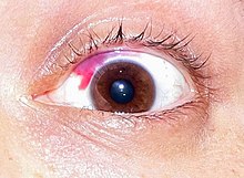
A subconjunctival hemorrhage is a common and relatively minor post-LASIK complication.
Micrograph showing abundant hemosiderin-laden alveolar macrophages (dark brown), as seen in a pulmonary hemorrhage. H&E stain.

Blood loss

Hemorrhaging is broken down into four classes by the American College of Surgeons' advanced trauma life support (ATLS).

  • Class I Hemorrhage involves up to 15% of blood volume. There is typically no change in vital signs and fluid resuscitation is not usually necessary.
  • Class II Hemorrhage involves 15–30% of total blood volume. A patient is often tachycardic (rapid heart beat) with a reduction in the difference between the systolic and diastolic blood pressures. The body attempts to compensate with peripheral vasoconstriction. Skin may start to look pale and be cool to the touch. The patient may exhibit slight changes in behavior. Volume resuscitation with crystalloids (Saline solution or Lactated Ringer's solution) is all that is typically required. Blood transfusion is not usually required.
  • Class III Hemorrhage involves loss of 30–40% of circulating blood volume. The patient's blood pressure drops, the heart rate increases, peripheral hypoperfusion (shock) with diminished capillary refill occurs, and the mental status worsens. Fluid resuscitation with crystalloid and blood transfusion are usually necessary.
  • Class IV Hemorrhage involves loss of >40% of circulating blood volume. The limit of the body's compensation is reached and aggressive resuscitation is required to prevent death.

This system is basically the same as used in the staging of hypovolemic shock.

Individuals in excellent physical and cardiovascular shape may have more effective compensatory mechanisms before experiencing cardiovascular collapse. These patients may look deceptively stable, with minimal derangements in vital signs, while having poor peripheral perfusion. Elderly patients or those with chronic medical conditions may have less tolerance to blood loss, less ability to compensate, and may take medications such as betablockers that can potentially blunt the cardiovascular response. Care must be taken in the assessment.

Massive hemorrhage

Although there is no universally accepted definition of massive hemorrhage, the following can be used to identify the condition: "(i) blood loss exceeding circulating blood volume within a 24-hour period, (ii) blood loss of 50% of circulating blood volume within a 3-hour period, (iii) blood loss exceeding 150 ml/min, or (iv) blood loss that necessitates plasma and platelet transfusion."

World Health Organization

The World Health Organization made a standardized grading scale to measure the severity of bleeding.

Grade 0 no bleeding;
Grade 1 petechial bleeding;
Grade 2 mild blood loss (clinically significant);
Grade 3 gross blood loss, requires transfusion (severe);
Grade 4 debilitating blood loss, retinal or cerebral associated with fatality

Management

Acute bleeding from an injury to the skin is often treated by the application of direct pressure. For severely injured patients, tourniquets are helpful in preventing complications of shock. Anticoagulant medications may need to be discontinued and possibly reversed in patients with clinically significant bleeding. Patients that have lost excessive amounts of blood may require a blood transfusion.

The use of cyanoacrylate glue to prevent bleeding and seal battle wounds was designed and first used in the Vietnam War. Skin glue, a medical version of "super glue", is sometimes used instead of using traditional stitches used for small wounds that need to be closed at the skin level.

Etymology

The word "Haemorrhage" (or hæmorrhage; using the æ ligature) comes from Latin haemorrhagia, from Ancient Greek αἱμορραγία (haimorrhagía, "a violent bleeding"), from αἱμορραγής (haimorrhagḗs, "bleeding violently"), from αἷμα (haîma, "blood") + -ραγία (-ragía), from ῥηγνύναι (rhēgnúnai, "to break, burst").

Maitrī

From Wikipedia, the free encyclopedia https://en.wikipedia.org/wiki/Maitr%C4%...