Search This Blog

Friday, August 22, 2025

Molecular machine

From Wikipedia, the free encyclopedia
 
Molecular machines are a class of molecules typically described as an assembly of a discrete number of molecular components intended to produce mechanical movements in response to specific stimuli, mimicking macromolecular devices such as switches and motors. Naturally occurring or biological molecular machines are responsible for vital living processes such as DNA replication and ATP synthesis. Kinesins and ribosomes are examples of molecular machines, and they often take the form of multi-protein complexes. For the last several decades, scientists have attempted, with varying degrees of success, to miniaturize machines found in the macroscopic world. The first example of an artificial molecular machine (AMM) was reported in 1994, featuring a rotaxane with a ring and two different possible binding sites. In 2016 the Nobel Prize in Chemistry was awarded to Jean-Pierre Sauvage, Sir J. Fraser Stoddart, and Bernard L. Feringa for the design and synthesis of molecular machines.
Kinesin walking on a microtubule is a molecular biological machine using protein domain dynamics on nanoscales. Such motions can now be seen by neutron spin echo spectroscopy

AMMs have diversified rapidly over the past few decades. A major point is to exploit existing motion in proteins, such as rotation about single bonds or cis-trans isomerization. Different AMMs are produced by introducing various functionalities, such as the introduction of bistability to create switches. A broad range of AMMs has been designed, featuring different properties and applications; some of these include molecular motors, switches, and logic gates. A wide range of applications have been demonstrated for AMMs, including those integrated into polymeric, liquid crystal, and crystalline systems for varied functions (such as materials research, homogenous catalysis and surface chemistry).

Terminology

Several definitions describe a "molecular machine" as a class of molecules typically described as an assembly of a discrete number of molecular components intended to produce mechanical movements in response to specific stimuli. The expression is often more generally applied to molecules that simply mimic functions that occur at the macroscopic level. A few prime requirements for a molecule to be considered a "molecular machine" are: the presence of moving parts, the ability to consume energy, and the ability to perform a task. Molecular machines differ from other stimuli-responsive compounds that can produce motion (such as cis-trans isomers) in their relatively larger amplitude of movement (potentially due to chemical reactions) and the presence of a clear external stimulus to regulate the movements (as compared to random thermal motion). Piezoelectric, magnetostrictive, and other materials that produce a movement due to external stimuli on a macro-scale are generally not included, since despite the molecular origin of the motion the effects are not useable on the molecular scale.

This definition generally applies to synthetic molecular machines, which have historically gained inspiration from the naturally occurring biological molecular machines (also referred to as "nanomachines"). Biological machines are considered to be nanoscale devices (such as molecular proteins) in a living system that convert various forms of energy to mechanical work in order to drive crucial biological processes such as intracellular transport, muscle contractions, ATP generation and cell division.

History

What would be the utility of such machines? Who knows? I cannot see exactly what would happen, but I can hardly doubt that when we have some control of the arrangement of things on a molecular scale we will get an enormously greater range of possible properties that substances can have, and of the different things we can do.

Biological molecular machines have been known and studied for decades given their vital role in sustaining life, and have served as inspiration for synthetically designed systems with similar useful functionality. The advent of conformational analysis, or the study of conformers to analyze complex chemical structures, in the 1950s gave rise to the idea of understanding and controlling relative motion within molecular components for further applications. This led to the design of "proto-molecular machines" featuring conformational changes such as cog-wheeling of the aromatic rings in triptycenes. By 1980, scientists could achieve desired conformations using external stimuli and utilize this for different applications. A major example is the design of a photoresponsive crown ether containing an azobenzene unit, which could switch between cis and trans isomers on exposure to light and hence tune the cation-binding properties of the ether. In his seminal 1959 lecture There's Plenty of Room at the Bottom, Richard Feynman alluded to the idea and applications of molecular devices designed artificially by manipulating matter at the atomic level. This was further substantiated by Eric Drexler during the 1970s, who developed ideas based on molecular nanotechnology such as nanoscale "assemblers", though their feasibility was disputed.

The first example of an artificial molecular machine (a switchable molecular shuttle). The positively charged ring (blue) is initially positioned over the benzidine unit (green), but shifts to the biphenol unit (red) when the benzidine gets protonated (purple) as a result of electrochemical oxidation or lowering of the pH.

Though these events served as inspiration for the field, the actual breakthrough in practical approaches to synthesize artificial molecular machines (AMMs) took place in 1991 with the invention of a "molecular shuttle" by Sir Fraser Stoddart. Building upon the assembly of mechanically linked molecules such as catenanes and rotaxanes as developed by Jean-Pierre Sauvage in the early 1980s, this shuttle features a rotaxane with a ring that can move across an "axle" between two ends or possible binding sites (hydroquinone units). This design realized the well-defined motion of a molecular unit across the length of the molecule for the first time. In 1994, an improved design allowed control over the motion of the ring by pH variation or electrochemical methods, making it the first example of an AMM. Here the two binding sites are a benzidine and a biphenol unit; the cationic ring typically prefers staying over the benzidine ring, but moves over to the biphenol group when the benzidine gets protonated at low pH or if it gets electrochemically oxidized. In 1998, a study could capture the rotary motion of a decacyclene molecule on a copper-base metallic surface using a scanning tunneling microscope. Over the following decade, a broad variety of AMMs responding to various stimuli were invented for different applications. In 2016, the Nobel Prize in Chemistry was awarded to Sauvage, Stoddart, and Bernard L. Feringa for the design and synthesis of molecular machines.

Artificial molecular machines

Over the past few decades, AMMs have diversified rapidly and their design principles, properties, and characterization methods have been outlined more clearly. A major starting point for the design of AMMs is to exploit the existing modes of motion in molecules. For instance, single bonds can be visualized as axes of rotation, as can be metallocene complexes. Bending or V-like shapes can be achieved by incorporating double bonds, that can undergo cis-trans isomerization in response to certain stimuli (typically irradiation with a suitable wavelength), as seen in numerous designs consisting of stilbene and azobenzene units. Similarly, ring-opening and -closing reactions such as those seen for spiropyran and diarylethene can also produce curved shapes. Another common mode of movement is the circumrotation of rings relative to one another as observed in mechanically interlocked molecules (primarily catenanes). While this type of rotation can not be accessed beyond the molecule itself (because the rings are confined within one another), rotaxanes can overcome this as the rings can undergo translational movements along a dumbbell-like axis. Another line of AMMs consists of biomolecules such as DNA and proteins as part of their design, making use of phenomena like protein folding and unfolding.
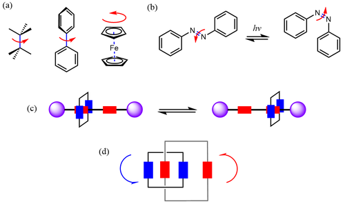
Some common types of motion seen in some simple components of artificial molecular machines. a) Rotation around single bonds and in sandwich-like metallocenes. b) Bending due to cis-trans isomerization. c) Translational motion of a ring along the dumbbell-like rotaxane axis. d) Rotation of interlocked rings in a catenane
Some common types of motion seen in some simple components of artificial molecular machines. a) Rotation around single bonds and in sandwich-like metallocenes. b) Bending due to cis-trans isomerization. c) Translational motion of a ring (blue) between two possible binding sites (red) along the dumbbell-like rotaxane axis (purple). d) Rotation of interlocked rings (depicted as blue and red rectangles) in a catenane.

AMM designs have diversified significantly since the early days of the field. A major route is the introduction of bistability to produce molecular switches, featuring two distinct configurations for the molecule to convert between. This has been perceived as a step forward from the original molecular shuttle which consisted of two identical sites for the ring to move between without any preference, in a manner analogous to the ring flip in an unsubstituted cyclohexane. If these two sites are different from each other in terms of features like electron density, this can give rise to weak or strong recognition sites as in biological systems — such AMMs have found applications in catalysis and drug delivery. This switching behavior has been further optimized to acquire useful work that gets lost when a typical switch returns to its original state. Inspired by the use of kinetic control to produce work in natural processes, molecular motors are designed to have a continuous energy influx to keep them away from equilibrium to deliver work.

Various energy sources are employed to drive molecular machines today, but this was not the case during the early years of AMM development. Though the movements in AMMs were regulated relative to the random thermal motion generally seen in molecules, they could not be controlled or manipulated as desired. This led to the addition of stimuli-responsive moieties in AMM design, so that externally applied non-thermal sources of energy could drive molecular motion and hence allow control over the properties. Chemical energy (or "chemical fuels") was an attractive option at the beginning, given the broad array of reversible chemical reactions (heavily based on acid-base chemistry) to switch molecules between different states. However, this comes with the issue of practically regulating the delivery of the chemical fuel and the removal of waste generated to maintain the efficiency of the machine as in biological systems. Though some AMMs have found ways to circumvent this, more recently waste-free reactions such based on electron transfers or isomerization have gained attention (such as redox-responsive viologens). Eventually, several different forms of energy (electric, magnetic, optical and so on) have become the primary energy sources used to power AMMs, even producing autonomous systems such as light-driven motors.

Types

Various AMMs are tabulated below along with indicative images:

Type Details Image
Molecular balance A molecule that can interconvert between two or more conformational or configurational states in response to the dynamic of multiple intra- and intermolecular driving forces, such as hydrogen bonding, solvophobic or hydrophobic effects, π interactions, and steric and dispersion interactions. The distinct conformers of a molecular balance can show different interactions with the same molecule, such that analyzing the ratio of the conformers and the energies for these interactions can enable quantification of different properties (such as CH-π or arene-arene interactions, see image). An example of a molecular balance
Molecular hinge A molecular hinge is a molecule that can typically rotate in a crank-like motion around a rigid axis, such as a double bond or aromatic ring, to switch between reversible configurations. Such configurations must have distinguishable geometries; for instance, azobenzene groups in a linear molecule may undergo cis-trans isomerization when irradiated with ultraviolet light, triggering a reversible transition to a bent or V-shaped conformation (see image). Molecular hinges have been adapted for applications such as nucleobase recognition, peptide modifications, and visualizing molecular motion. An example of a molecular hinge that can undergo cis-trans isomerization about a double bond
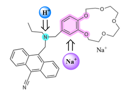
Molecular logic gate A molecule that performs a logical operation on one or more logic inputs and produces a single logic output. Modelled on logic gates, these molecules have slowly replaced the conventional silicon-based machinery. Several applications have come forth, such as water quality examination, food safety examination, metal ion detection, and pharmaceutical studies. The first example of a molecular logic gate was reported in 1993, featuring a receptor (see image) where the emission intensity could be treated as a tunable output if the concentrations of protons and sodium ions were to be considered as inputs. The first reported molecular logic gate
Molecular motor A molecule that is capable of directional rotary motion around a single or double bond and produce useful work as a result (as depicted in the image). Carbon nanotube nanomotors have also been produced. Single bond rotary motors are generally activated by chemical reactions whereas double bond rotary motors are generally fueled by light. The rotation speed of the motor can also be tuned by careful molecular design. Molecular dynamics simulation of a synthetic molecular rotor composed of three molecules in a nanopore (outer diameter 6.7 nm) at 250 K
Molecular necklace A class of mechanically interlocked molecules derived from catenanes where a large macrocycle backbone connects at least three small rings in the shape of a necklace (see image for example). A molecular necklace consisting of a large macrocycle threaded by n-1 rings (hence comprising n rings) is represented as [n]MN. The first molecular necklace was synthesized in 1992, featuring several α-cyclodextrins on a single polyethylene glycol chain backbone; the authors connected this to the idea of a "molecular abacus" proposed by Stoddart and coworkers around the same time. Several interesting applications have emerged for these molecules, such as antibacterial activity, desulfurization of fuels, and piezoelectricity. An example of a molecular necklace
Molecular propeller A molecule that can propel fluids when rotated, due to its special shape that is designed in analogy to macroscopic propellers (see schematic image on right). It has several molecular-scale blades attached at a certain pitch angle around the circumference of a nanoscale shaft. Propellers have been shown to have interesting properties, such as variations in pumping rates for hydrophilic and hydrophobic fluids. An example of a molecular propeller pumping water molecules due to its hydrophobic surface
Molecular shuttle A molecule capable of shuttling molecules or ions from one location to another. This is schematically depicted in the image on the right, where a ring (in green) can bind to either one of the yellow sites on the blue macrocyclic backbone. A common molecular shuttle consists of a rotaxane where the macrocycle can move between two sites or stations along the dumbbell backbone; controlling the properties of either site and by regulating conditions like pH can enable control over which site is selected for binding. This has led to novel applications in catalysis and drug delivery. An example of a rotaxane-based molecular shuttle
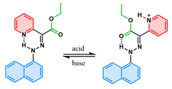
Molecular switch A molecule that can be reversibly shifted between two or more stable states in response to certain stimuli. This change of states influences the properties of the molecule according to the state it occupies at the moment. Unlike a molecular motor, any mechanical work done due to the motion in a switch is generally undone once the molecule returns to its original state unless it is part of a larger motor-like system. The image on the right shows a hydrazone-based switch that switches in response to pH changes. An example of a molecular switch
Molecular tweezers Host molecules capable of holding items between their two arms. The open cavity of the molecular tweezers binds items using non-covalent bonding including hydrogen bonding, metal coordination, hydrophobic forces, van der Waals forces, π interactions, or electrostatic effects. For instance, the image on the right depicts tweezers formed by corannulene pincers clasping a C60 fullerene molecule, termed "buckycatcher". Examples of molecular tweezers have been reported that are constructed from DNA and are considered DNA machines. An example of molecular tweezers binding a fullerene
Nanocar Single-molecule vehicles that resemble macroscopic automobiles and are important for understanding how to control molecular diffusion on surfaces. The image on the right shows an example with wheels made of fullerene molecules. The first nanocars were synthesized by James M. Tour in 2005. They had an H-shaped chassis and 4 molecular wheels (fullerenes) attached to the four corners. In 2011, Feringa and co-workers synthesized the first motorized nanocar which had molecular motors attached to the chassis as rotating wheels. The authors were able to demonstrate directional motion of the nanocar on a copper surface by providing energy from a scanning tunneling microscope tip. Later, in 2017, the world's first-ever nanocar race took place in Toulouse. A nanocar with C60 fullerenes as wheels

Biological molecular machines

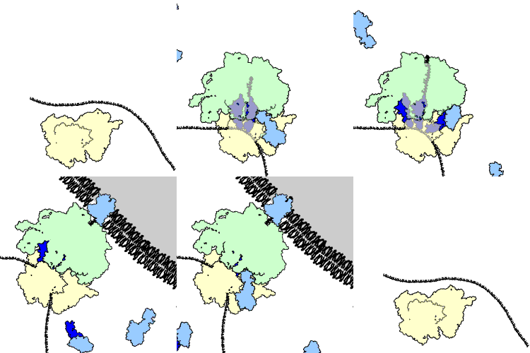
A ribosome performing the elongation and membrane targeting stages of protein translation. The ribosome is green and yellow, the tRNAs are dark blue, and the other proteins involved are light blue. The produced peptide is released into the endoplasmic reticulum. Protein domain dynamics can now be seen by neutron spin echo spectroscpoy.

Many macromolecular machines are found within cells, often in the form of multi-protein complexes. Examples of biological machines include motor proteins such as myosin, which is responsible for muscle contraction, kinesin, which moves cargo inside cells away from the nucleus along microtubules, and dynein, which moves cargo inside cells towards the nucleus and produces the axonemal beating of motile cilia and flagella. "[I]n effect, the [motile cilium] is a nanomachine composed of perhaps over 600 proteins in molecular complexes, many of which also function independently as nanomachines ... Flexible linkers allow the mobile protein domains connected by them to recruit their binding partners and induce long-range allostery via protein domain dynamics." Other biological machines are responsible for energy production, for example ATP synthase which harnesses energy from proton gradients across membranes to drive a turbine-like motion used to synthesise ATP, the energy currency of a cell. Still other machines are responsible for gene expression, including DNA polymerases for replicating DNA, RNA polymerases for producing mRNA, the spliceosome for removing introns, and the ribosome for synthesising proteins. These machines and their nanoscale dynamics are far more complex than any molecular machines that have yet been artificially constructed.

Biological machines have potential applications in nanomedicine. For example, they could be used to identify and destroy cancer cells. Molecular nanotechnology is a speculative subfield of nanotechnology regarding the possibility of engineering molecular assemblers, biological machines which could re-order matter at a molecular or atomic scale.[citation needed] Nanomedicine would make use of these nanorobots, introduced into the body, to repair or detect damages and infections, but these are considered to be far beyond current capabilities.

Research and applications

Advances in this area are inhibited by the lack of synthetic methods. In this context, theoretical modeling has emerged as a pivotal tool to understand the self-assembly or -disassembly processes in these systems.

Possible applications have been demonstrated for AMMs, including those integrated into polymericliquid crystal, and crystalline systems for varied functions. Homogenous catalysis is a prominent example, especially in areas like asymmetric synthesis, utilizing noncovalent interactions and biomimetic allosteric catalysis. AMMs have been pivotal in the design of several stimuli-responsive smart materials, such as 2D and 3D self-assembled materials and nanoparticle-based systems, for versatile applications ranging from 3D printing to drug delivery.

AMMs are gradually moving from the conventional solution-phase chemistry to surfaces and interfaces. For instance, AMM-immobilized surfaces (AMMISs) are a novel class of functional materials consisting of AMMs attached to inorganic surfaces forming features like self-assembled monolayers; this gives rise to tunable properties such as fluorescence, aggregation and drug-release activity.

Most of these "applications" remain at the proof-of-concept level. Challenges in streamlining macroscale applications include autonomous operation, the complexity of the machines, stability in the synthesis of the machines and the working conditions.

Thursday, August 21, 2025

Ribosome

From Wikipedia, the free encyclopedia
Large (red) and small (blue) subunits of a ribosome
Cell biology
Animal cell diagram

Ribosomes (/ˈrbəzm, -sm/) are macromolecular biological machines, found within all cells, that perform messenger RNA translation. Ribosomes link amino acids together in the order specified by the codons of messenger RNA molecules to form polypeptide chains. Ribosomes consist of two major components: the small and large ribosomal subunits. Each subunit consists of one or more ribosomal RNA molecules and many ribosomal proteins (r-proteins). The ribosomes and associated molecules are also known as the translational apparatus.

Overview

The sequence of DNA that encodes the sequence of the amino acids in a protein is transcribed into a messenger RNA (mRNA) chain. Ribosomes bind to the messenger RNA molecules and use the RNA's sequence of nucleotides to determine the sequence of amino acids needed to generate a protein. Amino acids are selected and carried to the ribosome by transfer RNA (tRNA) molecules, which enter the ribosome and bind to the messenger RNA chain via an anticodon stem loop. For each coding triplet (codon) in the messenger RNA, there is a unique transfer RNA that must have the exact anti-codon match, and carries the correct amino acid for incorporating into a growing polypeptide chain. Once the protein is produced, it can then fold to produce a functional three-dimensional structure.

A ribosome is made from complexes of RNAs and proteins and is therefore a ribonucleoprotein complex. In prokaryotes each ribosome is composed of small (30S) and large (50S) components, called subunits, which are bound to each other:

  1. (30S) has mainly a decoding function and is also bound to the mRNA
  2. (50S) has mainly a catalytic function and is also bound to the aminoacylated tRNAs.

The synthesis of proteins from their building blocks takes place in four phases: initiation, elongation, termination, and recycling. The start codon in all mRNA molecules has the sequence AUG. The stop codon is one of UAA, UAG, or UGA; since there are no tRNA molecules that recognize these codons, the ribosome recognizes that translation is complete. When a ribosome finishes reading an mRNA molecule, the two subunits separate and are usually broken up but can be reused. Ribosomes are a kind of enzyme, called ribozymes because the catalytic peptidyl transferase activity that links amino acids together is performed by the ribosomal RNA.

In eukaryotic cells, ribosomes are often associated with the intracellular membranes that make up the rough endoplasmic reticulum.

Ribosomes from bacteria, archaea, and eukaryotes (in the three-domain system) resemble each other to a remarkable degree, evidence of a common origin. They differ in their size, sequence, structure, and the ratio of protein to RNA. The differences in structure allow some antibiotics to kill bacteria by inhibiting their ribosomes while leaving human ribosomes unaffected. In all species, more than one ribosome may move along a single mRNA chain at one time (as a polysome), each "reading" a specific sequence and producing a corresponding protein molecule.

The mitochondrial ribosomes of eukaryotic cells are distinct from their other ribosomes. They functionally resemble those in bacteria, reflecting the evolutionary origin of mitochondria as endosymbiotic bacteria.

Discovery

Ribosomes were first observed in the mid-1950s by Romanian-American cell biologist George Emil Palade, using an electron microscope, as dense particles or granules. They were initially called Palade granules due to their granular structure. The term "ribosome" was proposed in 1958 by Howard M. Dintzis:

During the course of the symposium a semantic difficulty became apparent. To some of the participants, "microsomes" mean the ribonucleoprotein particles of the microsome fraction contaminated by other protein and lipid material; to others, the microsomes consist of protein and lipid contaminated by particles. The phrase "microsomal particles" does not seem adequate, and "ribonucleoprotein particles of the microsome fraction" is much too awkward. During the meeting, the word "ribosome" was suggested, which has a very satisfactory name and a pleasant sound. The present confusion would be eliminated if "ribosome" were adopted to designate ribonucleoprotein particles in sizes ranging from 35 to 100S.

— Albert Claude, Microsomal Particles and Protein Synthesis

Albert Claude, Christian de Duve, and George Emil Palade were jointly awarded the Nobel Prize in Physiology or Medicine, in 1974, for the discovery of the ribosome. The Nobel Prize in Chemistry 2009 was awarded to Venkatraman Ramakrishnan, Thomas A. Steitz and Ada E. Yonath for determining the detailed structure and mechanism of the ribosome.

Structure

Ribosomes assemble polymeric protein molecules, the order of which is controlled by the messenger RNA's molecule sequence.
Ribosomal RNA composition for prokaryotes and eukaryotes

The ribosome is a complex cellular machine. It is largely made up of specialized RNA known as ribosomal RNA (rRNA) as well as dozens of distinct proteins (the exact number varies slightly between species). The ribosomal proteins and rRNAs are arranged into two distinct ribosomal pieces of different sizes, known generally as the large and small subunits of the ribosome. Ribosomes consist of two subunits that fit together and work as one to translate the mRNA into a polypeptide chain during protein synthesis. Because they are formed from two subunits of non-equal size, they are slightly longer on the axis than in diameter.

Prokaryotic ribosomes

Prokaryotic ribosomes are around 20 nm (200 Å) in diameter and are composed of 65% rRNA and 35% ribosomal proteins. Eukaryotic ribosomes are between 25 and 30 nm (250–300 Å) in diameter with an rRNA-to-protein ratio that is close to 1. Crystallographic work has shown that there are no ribosomal proteins close to the reaction site for polypeptide synthesis. This suggests that the protein components of ribosomes do not directly participate in peptide bond formation catalysis, but rather that these proteins act as a scaffold that may enhance the ability of rRNA to synthesize protein.

Molecular structure of the 30S subunit from Thermus thermophilus. Proteins are shown in blue and the single RNA chain in brown.

The ribosomal subunits of prokaryotes and eukaryotes are quite similar.

The unit of measurement used to describe the ribosomal subunits and the rRNA fragments is the Svedberg unit, a measure of the rate of sedimentation in centrifugation rather than size. This accounts for why fragment names do not add up: for example, bacterial 70S ribosomes are made of 50S and 30S subunits.

Prokaryotes have 70S ribosomes, each consisting of a small (30S) and a large (50S) subunit. E. coli, for example, has a 16S RNA subunit (consisting of 1540 nucleotides) that is bound to 21 proteins. The large subunit is composed of a 5S RNA subunit (120 nucleotides), a 23S RNA subunit (2900 nucleotides) and 31 proteins.

Ribosome of E. coli (a bacterium)
ribosome subunit rRNAs r-proteins
70S 50S 23S (2904 nt) 31
5S (120 nt)
30S 16S (1542 nt) 21

Affinity label for the tRNA binding sites on the E. coli ribosome allowed the identification of A and P site proteins most likely associated with the peptidyltransferase activity; labelled proteins are L27, L14, L15, L16, L2; at least L27 is located at the donor site, as shown by E. Collatz and A.P. Czernilofsky. Additional research has demonstrated that the S1 and S21 proteins, in association with the 3′-end of 16S ribosomal RNA, are involved in the initiation of translation.

Archaeal ribosomes

Archaeal ribosomes share the same general dimensions of bacteria ones, being a 70S ribosome made up from a 50S large subunit, a 30S small subunit, and containing three rRNA chains. However, on the sequence level, they are much closer to eukaryotic ones than to bacterial ones. Every extra ribosomal protein archaea have compared to bacteria has a eukaryotic counterpart, while no such relation applies between archaea and bacteria.

Eukaryotic ribosomes

Eukaryotes have 80S ribosomes located in their cytosol, each consisting of a small (40S) and large (60S) subunit. Their 40S subunit has an 18S RNA (1900 nucleotides) and 33 proteins. The large subunit is composed of a 5S RNA (120 nucleotides), 28S RNA (4700 nucleotides), a 5.8S RNA (160 nucleotides) subunits and 49 proteins.

eukaryotic cytosolic ribosomes (R. norvegicus)
ribosome subunit rRNAs r-proteins
80S 60S 28S (4718 nt) 49
5.8S (160 nt)
5S (120 nt)
40S 18S (1874 nt) 33

During 1977, Czernilofsky published research that used affinity labeling to identify tRNA-binding sites on rat liver ribosomes. Several proteins, including L32/33, L36, L21, L23, L28/29 and L13 were implicated as being at or near the peptidyl transferase center.

Plastoribosomes and mitoribosomes

In eukaryotes, ribosomes are present in mitochondria (sometimes called mitoribosomes) and in plastids such as chloroplasts (also called plastoribosomes). They also consist of large and small subunits bound together with proteins into one 70S particle. These ribosomes are similar to those of bacteria and these organelles are thought to have originated as symbiotic bacteria. Of the two, chloroplastic ribosomes are closer to bacterial ones than mitochondrial ones are. Many pieces of ribosomal RNA in the mitochondria are shortened, and in the case of 5S rRNA, replaced by other structures in animals and fungi. In particular, Leishmania tarentolae has a minimalized set of mitochondrial rRNA. In contrast, plant mitoribosomes have both extended rRNA and additional proteins as compared to bacteria, in particular, many pentatricopetide repeat proteins.

The cryptomonad and chlorarachniophyte algae may contain a nucleomorph that resembles a vestigial eukaryotic nucleus. Eukaryotic 80S ribosomes may be present in the compartment containing the nucleomorph.

Making use of the differences

The differences between the bacterial and eukaryotic ribosomes are exploited by pharmaceutical chemists to create antibiotics that can destroy a bacterial infection without harming the cells of the infected person. Due to the differences in their structures, the bacterial 70S ribosomes are vulnerable to these antibiotics while the eukaryotic 80S ribosomes are not. Even though mitochondria possess ribosomes similar to the bacterial ones, mitochondria are not affected by these antibiotics because they are surrounded by a double membrane that does not easily admit these antibiotics into the organelle. A noteworthy counterexample is the antineoplastic antibiotic chloramphenicol, which inhibits bacterial 50S and eukaryotic mitochondrial 50S ribosomes. Ribosomes in chloroplasts, however, are different: Antibiotic resistance in chloroplast ribosomal proteins is a trait that has to be introduced as a marker, with genetic engineering.

Common properties

The various ribosomes share a core structure, which is quite similar despite the large differences in size. Much of the RNA is highly organized into various tertiary structural motifs, for example pseudoknots that exhibit coaxial stacking. The extra RNA in the larger ribosomes is in several long continuous insertions, such that they form loops out of the core structure without disrupting or changing it. All of the catalytic activity of the ribosome is carried out by the RNA; the proteins reside on the surface and seem to stabilize the structure.

High-resolution structure

Figure 4: Atomic structure of the 50S subunit from Haloarcula marismortui. Proteins are shown in blue and the two RNA chains in brown and yellow. The small patch of green in the center of the subunit is the active site.

The general molecular structure of the ribosome has been known since the early 1970s. In the early 2000s, the structure has been achieved at high resolutions, of the order of a few ångströms.

The first papers giving the structure of the ribosome at atomic resolution were published almost simultaneously in late 2000. The 50S (large prokaryotic) subunit was determined from the archaeon Haloarcula marismortui and the bacterium Deinococcus radiodurans, and the structure of the 30S subunit was determined from the bacterium Thermus thermophilus. These structural studies were awarded the Nobel Prize in Chemistry in 2009. In May 2001 these coordinates were used to reconstruct the entire T. thermophilus 70S particle at 5.5 Å resolution.

Two papers were published in November 2005 with structures of the Escherichia coli 70S ribosome. The structures of a vacant ribosome were determined at 3.5 Å resolution using X-ray crystallography. Then, two weeks later, a structure based on cryo-electron microscopy was published, which depicts the ribosome at 11–15 Å resolution in the act of passing a newly synthesized protein strand into the protein-conducting channel.

The first atomic structures of the ribosome complexed with tRNA and mRNA molecules were solved by using X-ray crystallography by two groups independently, at 2.8 Å and at 3.7 Å. These structures allow one to see the details of interactions of the Thermus thermophilus ribosome with mRNA and with tRNAs bound at classical ribosomal sites. Interactions of the ribosome with long mRNAs containing Shine-Dalgarno sequences were visualized soon after that at 4.5–5.5 Å resolution.

In 2011, the first complete atomic structure of the eukaryotic 80S ribosome from the yeast Saccharomyces cerevisiae was obtained by crystallography. The model reveals the architecture of eukaryote-specific elements and their interaction with the universally conserved core. At the same time, the complete model of a eukaryotic 40S ribosomal structure in Tetrahymena thermophila was published and described the structure of the 40S subunit, as well as much about the 40S subunit's interaction with eIF1 during translation initiation. Similarly, the eukaryotic 60S subunit structure was also determined from Tetrahymena thermophila in complex with eIF6.

Function

Ribosomes are minute particles consisting of RNA and associated proteins that function to synthesize proteins. Proteins are needed for many cellular functions, such as repairing damage or directing chemical processes. Ribosomes can be found floating within the cytoplasm or attached to the endoplasmic reticulum. Their main function is to convert genetic code into an amino acid sequence and to build protein polymers from amino acid monomers.

Ribosomes act as catalysts in two extremely important biological processes called peptidyl transfer and peptidyl hydrolysis. The "PT center is responsible for producing protein bonds during protein elongation".

In summary, ribosomes have two main functions: Decoding the message, and the formation of peptide bonds. These two functions reside in the ribosomal subunits. Each subunit is made of one or more rRNAs and many r-proteins. The small subunit (30S in bacteria and archaea, 40S in eukaryotes) has the decoding function, whereas the large subunit (50S in bacteria and archaea, 60S in eukaryotes) catalyzes the formation of peptide bonds, referred to as the peptidyl-transferase activity. The bacterial (and archaeal) small subunit contains the 16S rRNA and 21 r-proteins (Escherichia coli), whereas the eukaryotic small subunit contains the 18S rRNA and 32 r-proteins (Saccharomyces cerevisiae, although the numbers vary between species). The bacterial large subunit contains the 5S and 23S rRNAs and 34 r-proteins (E. coli), with the eukaryotic large subunit containing the 5S, 5.8S, and 25S/28S rRNAs and 46 r-proteins (S. cerevisiae; again, the exact numbers vary between species).

Translation

Ribosomes are the workplaces of protein biosynthesis, the process of translating mRNA into protein. The mRNA comprises a series of codons which are decoded by the ribosome to make the protein. Using the mRNA as a template, the ribosome traverses each codon (3 nucleotides) of the mRNA, pairing it with the appropriate amino acid provided by an aminoacyl-tRNA. Aminoacyl-tRNA contains a complementary anticodon on one end and the appropriate amino acid on the other. For fast and accurate recognition of the appropriate tRNA, the ribosome utilizes large conformational changes (conformational proofreading). The small ribosomal subunit, typically bound to an aminoacyl-tRNA containing the first amino acid methionine, binds to an AUG codon on the mRNA and recruits the large ribosomal subunit. The ribosome contains three RNA binding sites, designated A, P, and E. The A-site binds an aminoacyl-tRNA or termination release factors; the P-site binds a peptidyl-tRNA (a tRNA bound to the poly-peptide chain); and the E-site (exit) binds a free tRNA. Protein synthesis begins at a start codon AUG near the 5' end of the mRNA. mRNA binds to the P site of the ribosome first. The ribosome recognizes the start codon by using the Shine-Dalgarno sequence of the mRNA in prokaryotes and Kozak box in eukaryotes.

Although catalysis of the peptide bond involves the C2 hydroxyl of RNA's P-site adenosine in a proton shuttle mechanism, other steps in protein synthesis (such as translocation) are caused by changes in protein conformations. Since their catalytic core is made of RNA, ribosomes are classified as "ribozymes," and it is thought that they might be remnants of the RNA world.

Figure 5: Translation of mRNA (1) by a ribosome (2)(shown as small and large subunits) into a polypeptide chain (3). The ribosome begins at the start codon of RNA (AUG) and ends at the stop codon (UAG).

In Figure 5, both ribosomal subunits (small and large) assemble at the start codon (towards the 5' end of the mRNA). The ribosome uses tRNA that matches the current codon (triplet) on the mRNA to append an amino acid to the polypeptide chain. This is done for each triplet on the mRNA, while the ribosome moves towards the 3' end of the mRNA. Usually in bacterial cells, several ribosomes are working parallel on a single mRNA, forming what is called a polyribosome or polysome.

Cotranslational folding

The ribosome is known to actively participate in the protein folding. The structures obtained in this way are usually identical to the ones obtained during protein chemical refolding; however, the pathways leading to the final product may be different. In some cases, the ribosome is crucial in obtaining the functional protein form. For example, one of the possible mechanisms of folding of the deeply knotted proteins relies on the ribosome pushing the chain through the attached loop.

Addition of translation-independent amino acids

Presence of a ribosome quality control protein Rqc2 is associated with mRNA-independent protein elongation. This elongation is a result of ribosomal addition (via tRNAs brought by Rqc2) of CAT tails: ribosomes extend the C-terminus of a stalled protein with random, translation-independent sequences of alanines and threonines.

Ribosome locations

Ribosomes are classified as being either "free" or "membrane-bound".

Figure 6: A ribosome translating a protein that is secreted into the endoplasmic reticulum via protein domain dynamics.

Free and membrane-bound ribosomes differ only in their spatial distribution; they are identical in structure. Whether the ribosome exists in a free or membrane-bound state depends on the presence of an ER-targeting signal sequence on the protein being synthesized, so an individual ribosome might be membrane-bound when it is making one protein, but free in the cytosol when it makes another protein.

Ribosomes are sometimes referred to as organelles, but the use of the term organelle is often restricted to describing sub-cellular components that include a phospholipid membrane, which ribosomes, being entirely particulate, do not. For this reason, ribosomes may sometimes be described as "non-membranous organelles".

Free ribosomes

Free ribosomes can move about anywhere in the cytosol, but are excluded from the cell nucleus and other organelles. Proteins that are formed from free ribosomes are released into the cytosol and used within the cell. Since the cytosol contains high concentrations of glutathione and is, therefore, a reducing environment, proteins containing disulfide bonds, which are formed from oxidized cysteine residues, cannot be produced within it.

Membrane-bound ribosomes

When a ribosome begins to synthesize proteins needed in certain organelles, the ribosome making this protein can become "membrane-bound". In eukaryotic cells this happens in a region of the endoplasmic reticulum (ER) called the "rough ER". The newly produced polypeptide chains are inserted directly into the ER by the ribosome undertaking vectorial synthesis and are then transported to their destinations, through the secretory pathway. Bound ribosomes usually produce proteins that are used within the plasma membrane or are expelled from the cell via exocytosis.

Biogenesis

In bacterial cells, ribosomes are synthesized in the cytoplasm through the transcription of multiple ribosome gene operons. In eukaryotes, the process takes place both in the cell cytoplasm and in the nucleolus, which is a region within the cell nucleus. The assembly process involves the coordinated function of over 200 proteins in the synthesis and processing of the four rRNAs, as well as assembly of those rRNAs with the ribosomal proteins.

Origin

The ribosome may have first originated as a protoribosome, possibly containing a peptidyl transferase centre (PTC), in an RNA world, appearing as a self-replicating complex that only later evolved the ability to synthesize proteins when amino acids began to appear. Studies suggest that ancient ribosomes constructed solely of rRNA could have developed the ability to synthesize peptide bonds. In addition, evidence strongly points to ancient ribosomes as self-replicating complexes, where the rRNA in the ribosomes had informational, structural, and catalytic purposes because it could have coded for tRNAs and proteins needed for ribosomal self-replication. Hypothetical cellular organisms with self-replicating RNA but without DNA are called ribocytes (or ribocells).

As amino acids gradually appeared in the RNA world under prebiotic conditions, their interactions with catalytic RNA would increase both the range and efficiency of function of catalytic RNA molecules. Thus, the driving force for the evolution of the ribosome from an ancient self-replicating machine into its current form as a translational machine may have been the selective pressure to incorporate proteins into the ribosome's self-replicating mechanisms, so as to increase its capacity for self-replication.

Heterogeneous ribosomes

Ribosomes are compositionally heterogeneous between species and even within the same cell, as evidenced by the existence of cytoplasmic and mitochondria ribosomes within the same eukaryotic cells. Certain researchers have suggested that heterogeneity in the composition of ribosomal proteins in mammals is important for gene regulation, i.e., the specialized ribosome hypothesis. However, this hypothesis is controversial and the topic of ongoing research.

Heterogeneity in ribosome composition was first proposed to be involved in translational control of protein synthesis by Vince Mauro and Gerald Edelman. They proposed the ribosome filter hypothesis to explain the regulatory functions of ribosomes. Evidence has suggested that specialized ribosomes specific to different cell populations may affect how genes are translated. Some ribosomal proteins exchange from the assembled complex with cytosolic copies suggesting that the structure of the in vivo ribosome can be modified without synthesizing an entire new ribosome.

Certain ribosomal proteins are absolutely critical for cellular life while others are not. In budding yeast, 14/78 ribosomal proteins are non-essential for growth, while in humans this depends on the cell of study. Other forms of heterogeneity include post-translational modifications to ribosomal proteins such as acetylation, methylation, and phosphorylation. Arabidopsis,Viral internal ribosome entry sites (IRESs) may mediate translations by compositionally distinct ribosomes. For example, 40S ribosomal units without eS25 in yeast and mammalian cells are unable to recruit the CrPV IGR IRES.

Heterogeneity of ribosomal RNA modifications plays a significant role in structural maintenance and/or function and most mRNA modifications are found in highly conserved regions. The most common rRNA modifications are pseudouridylation and 2'-O-methylation of ribose.

ncRNA therapy

From Wikipedia, the free encyclopedia

A majority of the human genome is made up of non-protein coding DNA. It infers that such sequences are not commonly employed to encode for a protein. However, even though these regions do not code for protein, they have other functions and carry necessary regulatory information.They can be classified based on the size of the ncRNA. Small noncoding RNA is usually categorized as being under 200 bp in length, whereas long noncoding RNA is greater than 200bp. In addition, they can be categorized by their function within the cell; Infrastructural and Regulatory ncRNAs. Infrastructural ncRNAs seem to have a housekeeping role in translation and splicing and include species such as rRNA, tRNA, snRNA.Regulatory ncRNAs are involved in the modification of other RNAs.

Timeline of ncRNA therapeutics

RNA Classification

Long non-coding RNA

Long non-coding RNA (LncRNA) are a type of RNA which is usually defined as transcripts which are greater than 200 base-pairs in length and not translated into proteins. This limitation distinguishes lncRNA from small non-coding RNAs which encompasses microRNAs (miRNAs), small interfering RNAs (siRNAs), Piwi-interacting RNAs (piRNAs), small nucleolar RNAs (snoRNAs), and other short RNAs. Long non-coding RNAs include lincRNAs, intronic ncRNAs, circular and linear ncRNA.

Long intergenic Non-coding RNA

Long intergenic Non-coding RNA (LincRNA) is defined as RNA transcripts that are longer than 200 nucleotides. These RNAs must not have open reading frames that encode proteins. The term “intergenic” refers to the identification of these transcripts from regions of the genome that do not contain protein-encoding genes. LncRNAs also contain promoter - or enhancer-associated RNAs that are gene proximal and can be either in the sense or antisense orientation.

Circular RNA

Circular RNA (CircRNA) are a novel class of endogenous noncoding RNAs and are characterized by their covalently closed loop structures. This class of ncRNA does not have a 5’ cap or 3’ Poly A tail. It has been hypothesized that cirRNAs may function as potential molecular markers for disease diagnosis and treatment and play an important role in the initiation and progression of human diseases.

Small non-coding RNA

Small non-coding RNA (sncRNA) are a type of RNA. which is usually defined as transcripts which are lesser than 200 base-pairs in length and not translated into proteins. This limitation distinguishes sncRNA from lncRNA. This class includes but is not limited to microRNAs (miRNAs), small interfering RNAs (siRNAs), Piwi-interacting RNAs (piRNAs), small nucleolar RNAs (snoRNAs), and other short RNAs.

microRNA

microRNA (miRNA) plays an important role in regulating gene expression. Majority of miRNAs are transcribed from DNA sequences into primary miRNAs. These primary miRNAs are further processed into precursor miRNAs, and finally into mature miRNAs. The miRNAs in most cases interact with the 3’ UTR region of target to induce mRNA degradation and translational repression. Interactions of miRNAs with other regions, including the 5’ UTR, coding sequence, and gene promoters have also been reported. Under certain conditions, miRNAs are also able to activate translation or regulate transcription, but this is dependent on factors such as location of the effect. This process of interaction is very dynamic and dependent on multiple factors.

Ribosomal RNA

Ribosomal RNA (rRNA) includes non-coding RNAs that play essential roles in rRNA regulation. Ribosomal RNA (rRNA) takes part in protein synthesis. Occasional RNA molecules act catalytically, as RNA enzymes (ribozymes) or take part in protein export. The most important ribozyme is the major rRNA of the large subunit of the ribosome (28s rRNA in eukaryotes). It is now accepted that 28S rRNA catalyzes the critical step in polypeptide synthesis in addition to playing a major structural role.

Small nuclear RNA and small nucleolar RNA

Small nuclear RNA (snRNA) and small nucleolar RNA (snoRNA) are widely known to guide the nucleotide modifications and processing of rRNA. Both snRNA and snoRNA are categorized into a class of small RNA molecules that are present in the nucleus. However, they vary a lot by function. snRNA are 80-350nucletides long while snoRNA are 80-1000 nucleotides long in yeast. snRNA plays a critical role in regulating the pre-mRNA silencing. On the other hand, snoRNAs are involved in mRNA editing, modification of the rRNA and tRNA, and genome imprinting. Major function of snoRNA includes the maturation of rRNA during ribosomal formation. Small nuclear and small nucleolar RNAs are critical components of snRNPs and snoRNPs and play an essential role in the maturation of, respectively, mRNAs and rRNAs within the nucleus of eukaryotic cells. Both snRNA and snoRNA are involved in modifying RNA just after transcription. snRNA can be found in splicing speckles and Cajal bodies of the nucleus of the cell.snRNA and snoRNA requires a phosphorylated adaptor for nuclear export (PHAX) to get transported to the site of action within the nucleus.

Transfer RNA

Transfer RNA (tRNA) helps decode a messenger RNA sequence into a protein. They function at specific sites within the ribosome during translation (the process going from code to protein). Within the mRNA molecule we have three nucleotides in length codons. These codons all have a unique universal code which represents a particular amino acid. tRNAs can be classified as an adaptor molecule, being typically 76 to 90 nucleotides in length.

History

Non-coding RNA

DNA purification in 1869 by Dr. Friedrich Miescher’s, from salmon sperm and pus cells guided the scientists towards the presence of additional molecules in the cell except for proteins. Miescher identified the presence of a highly acidic molecule that he isolated from the pus cells and labeled it “nuclein”. The term was coined as the DNA isolated by Miescher was not protein and was derived from the nucleus of the cell. It wasn’t until 1944, when Oswald Avery proposed the DNA as a genetic carrier of information that the Miescher discovery was brought back to light.

Following the X-ray crystallography, by Rosalind Franklin and the determination of DNA double helix by Watson and Crick in 1953, further enhanced the understanding of DNA structure and allowed for the establishment of central dogma of molecular biology. However, one of the flaws with central dogma was the postulation that information flow proceeds from DNA to RNA to protein, which hinders the understanding of different regulatory mechanisms.

In 1955, George Palade identified the first ncRNA as a part of the large ribonucleoprotein complex (RNP). The second class of ncRNA to be discovered was transfer RNA (tRNA) in 1957. However, the first regulatory ncRNA was a microRNA discovered in 1988 from E.coli and was labeled as micF. On other hand, the first eukaryotic microRNA was discovered in C.elegans in 1993. It was derived from gene lin-4 and was identified as a small RNA molecule (as compared to longer mRNA molecules) forming stem-loop structures. This structure gets further modified to generate a shorter RNA that is complementary to the 3’UTR region of lin-14 transcript. This pathway allowed for a better understanding of different post translational gene silencing pathways. Since then, many other miRNAs have been discovered.

Detailed understanding of the mechanism behind this post translational silencing pathway was established in 2001 by Thomas Tuschl. It was discovered that the double stranded RNA gets processed into a shorter 25 nucleotides long fragment which is then modified into a short hairpin like structure by Drosha complex. The molecule is then diced by dicer enzymes into a functional double stranded RNA (dsRNA). These are then loaded onto the RISC complex which then finds and cleaves the targeted mRNA of interest in the cytoplasm.

It wasn’t until 1989 that the imprinting genes were discovered and the genome imprinting was established. The first two genomic imprinting genes were paternally expressed Igf2r and H19. These were both discovered independently in mice and were localized to chromosome 7. H19 is peculiar as it functions as a lncRNA but undergoes modifications similar to that of pre-mRNA processing such as splicing, 3’ polyadenylation and is transcribed by RNA polymerase II. This lncRNA plays a significant role in mice embryonic development and can be lethal if expressed during prenatal stages. More lncRNAs have been discovered in eukaryotes overtime. One such discovery that allowed for better understanding between H19 functions was a lncRNA called XIST (X inactive-specific transcript).

ncRNA drugs and therapy

The first ncRNA therapeutic drug approved by food and drug administration (FDA) (1998) and the European medicine agency (EMA) (1999) is called Fomivirsen or Vitravene. The target organ is the eye and works against the cytomegalovirus (CMV) retinitis in immunocompromised patients. The drug functions as an antisense oligonucleotide and binds to the complementary sequence of the mRNA that inhibits the replication of human cytomegalovirus. This therapy can also be categorized as Antisense oligonucleotide (ASO) therapy. There have been many ASO RNA therapeutics that have been approved by FDA and/or EMA over the years, but it wasn’t until 2018 that the EMA approved the drug called Patisiran/Onpattro. The drug uses ds-siRNA as a mechanism of action and is deemed effective against hereditary transthyretin amyloidosis. The mechanism specifically targets the Transthyretin (TTR) mRNA. RNA therapeutic targets are not limited to mature mRNA but have been used to target mRNA at different stages of maturation. One such example is Nusinersen (Spinaraza), it functions as an ASO and targets pre-mRNA before splicing that corresponds to Survival of motor neuron 2 gene (SMN 2). This drug therapy was approved by FDA and EMA in 2016 and 2017 respectively. There are some drugs that have been approved by FDA and not by EMA. This can be seen in the case of an ASO type therapeutics called Eteplirsen (Exondys51) which has been approved by FDA in 2016 but not by EMA. It targets pre-mRNA corresponding to Dystrophin (DMD) and works against Duchenne muscular dystrophy. There are many additional therapeutics that have been developed and are either in phase I or II of the clinical trials. Current RNA therapeutics in clinical trials range from a variety of target organs and diseases ranging from skin (potential treatment for disease such as keloid) to tumors (squamous cell lung cancer).

To date, for both the FDA and the EMA, ncRNAs are considered as "simple" medical products because of their production by chemical synthesis. When some of them, produced biologically (known as bioengineered ncRNA agents: BERAs), will be put on the market, the status of biological medical products will be applied, which could lead to inconsistencies in the legislation.

Applications

Antisense oligonucleotides

Antisense Oligonucleotide Use in ncRNA therapy

Antisense oligonucleotides (ASOs) are single-stranded DNA molecules with full complementarity to one select target mRNA and may act by blocking protein translation (via steric hindrance), causing mRNA degradation (via RNase H-cleavage) or changing pre-mRNA splicing. These short oligonucleotides have already been approved by the FDA for ten genetic disorders and many are currently in the pipeline to be approved/tested. Using oligonucleotide technology, we are now able to control protein expression via RNA interference, and are able to affect previously defined “undruggable” proteins. Even though this therapy has a lot of promise and potential, it comes with many limitations.

Compared to siRNA and microRNA, ASOs are more versatile in reducing protein expression, they have the ability to also enhance target translation. ASOs can also be customized with ease and accuracy, allowing for the targeting of virtually any mutated gene. This allows for a greater level of application in the field of precision and personalized medicine. The main challenge of ASO therapies to specific tissues and cellular uptake is what poses a great challenge and limitation. Liposomal delivery is one such way to overcome such issues. Liposomal delivery system comes with its own share of limitations. Serum proteins in the bloodstream destabilize the lipoprotein. This destabilization leads to the depletion of protein and exposing cargo to the unstable environment. This hindrance can be overcome by using PEGs (poly(ethylene glycol) . However, PEGs are not biodegradable causing them to accumulate within the body leading to adverse effects and causing hypersensitivity. In addition, multiple rounds of therapy with PEGs can lead to the formation of PEG antibodies, which can lead to lack of efficiency in preventing the rupture of the liposome that it is attached to. Using immunoliposomes it has been shown that targeting can be more specific as by using antibody’s specific to the protein of expression in that area, it results in the ASO drug directly impact the target site and nowhere else. Moreover, immunoliposomes are slow to dissociate leading to precise release of the ASO drug which they encapsulate.

LncRNA as a therapeutic approach

Long noncoding RNAs (lncRNAs) are large transcripts (more than 200 nucleotides long) that have similar mechanism of synthesis as that of mRNAs but unlike mRNAs, lncRNAs are not translated to a protein. lncRNA contains interactor elements and structural elements. Interactor elements directly interact with other nucleic acids or proteins while the structural elements indicate the ability of some lncRNAs to form secondary and/or tertiary structures. This ability of the lncRNAs to interact with nucleic acids using its interactor elements and its ability to interact with protein using its secondary structures allows it to function in a more diverse manner than other ncRNAs such as miRNA (microRNA). LncRNA has been established to play a role in gene regulation by influencing the ability of specific regions of the gene to bind to transcriptional elements and different epigenetic modifications. One such example can be seen in the case X inactive specific transcript (XIST). In humans, 46,XX females carry an extra X chromosome (155Mb of DNA) compared to 46,XY males. To overcome this dosage imbalance, one X chromosome is randomly inactivated in human females at around the 2-8 cell stage of embryo development. This inactivation is very stable across cell divisions due to epigenetic contributions both during the initial silencing and the subsequent maintenance of the inactive X chromosome (Xi). This inactivation is carried by the lncRNA, XIST. XIST is produced in cis and inactivates the X-chromosome that it has been generated from. The inactive X chromosome can be observed as a condensed heterochromatin structure called “Barr Body”. A study in 2013 utilized this ability of XIST as a potential therapeutic approach for treatment of trisomy 21. Trisomy 21 is commonly known as down syndrome and is caused due to presence of an additional copy of chromosome 21. The study was one of its kind as no other studies have been able to incorporate the XIST gene into a chromosome due to its large size. The study incorporated the XIST into one of the chromosomes 21 in the cells gathered from patients with down syndrome. The study was able to observe the inactivation of one of chromosome 21 in the form of a condensed heterochromatin and labeled it as a chromosome 21 barr body. Such experiments have shown to work in cells in the lab setting although no lncRNA based therapeutics are in clinical trials. The implications of such work can bring trisomy 21 and other chromosomal disorders in the realm of consideration for future gene therapy research.

Challenges

One of the major issues that hinders the ncRNA therapy is the stability of the single stranded RNA molecule. RNA is typically single stranded therefore slightly unstable as compared to dsDNA molecules. This however can be overcome by fabricating the single stranded RNA to double stranded RNA(dsRNA). This is quite effective as the dsRNA is more stable at room temperature and has a longer shelf life. Second major issue is the cell/tissue/organ specific targeting of the RNA molecules. Generally, this is overcome by containing the dsRNA in a lipid nanoparticle and using that as a ligand to bind to a receptor on the surface of the target cell. The lipid particles are taken into the liver cells through their specific receptors and this mechanism seems to be effective at targeting the liver cells/cancer. Another organ with a relatively easy delivery mechanism is the eye. This requires an invasive technique of directly injecting the ncRNA of interest directly into the eye. These techniques are not only invasive but also don’t ensure if all the cells in the target organ are being targeted by the ncRNA of interest. Additional issues arise once the RNA molecule enters the cell. One of the issues being the immune system. Our immune system can recognize RNA using the intracellular pathogen associated molecular pattern (PAMP) receptors and extracellular toll-like receptors (TLR). Activation of the receptors leads to a cytokine (IFNy-Interferon gamma) mediated immune response. Common applications to overcome the immune response include second generation chemical modifications. This process includes the introduction of small one at a time chemical modifications to avoid the immune response. However, there are some reports of adverse immune responses in clinical trials employing such modified reagents. There’s no fixed answer to issues with immunogenicity and ncRNA therapy. Modified adenovirus vectors have been used extensively in many clinical trials as a ncRNA delivery mechanism. In particular, adenovirus vector is considered an efficient delivery system due to its stability within live cells and non-pathogenicity. Even though viral transfections have achieved significant results in basic research, one of the issues is the non-specificity leading to off target transfections. Further research needs to be done to improve the accuracy of viral transfections for future tests and clinical trials.

ASO Guidelines

In December 2021, the FDA came up with a draft guidance for the use of ASO drug products. This draft guidance was directed towards sponsor-investigators who are developing individualized investigational antisense oligonucleotides (ASO) drug products for severely debilitating or life threatening diseases. Severely debilitating corresponds to a disease or condition that causes major irreversible morbidity. However, life-threatening is defined as the disease or condition has a likelihood of death unless the course of treatment leads to an endpoint of survival. Usually individuals that have a severely debilitating life threatening disease don't have any alternative treatment options, and their diseases will be rapidly progressing, leading to an early death and/or devastating or irreversible morbidity within a short time frame without treatment.

Drug development is usually targeted for a large number of individuals, in this case that is not possible because of the specificity of the mechanism of action of the ASO combined with the rarity of the treatment-amenable patient population. Under FDA regulations, a protocol under which an individual ASO product is administered to a human subject must be reviewed and approved by an institutional review board (IRB) before it can be administered to human subjects. When the individual is a child, additional safeguards need to be identified in order to prevent any developmental issues from occurring that may affect the life of the individual. The sponsor-investigator needs to get informed consent from the individual or from the person who is responsible for the individual. The consent needs to include a description of reasonably foreseeable risks or discomforts as part of the use of the ASO drug. The sponsor also needs to get individuals clinical and genetic diagnosis to confirm that the ASO will be beneficial. The analysis may be through gene sequencing, enzymatic analysis, biochemical testing, imaging evaluations. All results need to be included in the application. Also the sponsor needs to include evidence that establishes the role of the gene variant targeted by the ASO drug. The sponsor/investigator need to also provide evidence that the identified gene variant or variants are unique to the individual.

The guidance suggests that the starting dose should be based on available non-clinical data that has been collected from model organisms or in vitro studies and should be in correlation with other ASO drug product dosing information that is available. At the starting dose, pharmacological effects are expected. Furthermore, It is advised that a dosing escalation method be utilized. This includes the step of escalating the dodge from its initial dose based on pharmacodynamic effects and/or trial participants' response to the ASO.

In addition, protocols submitted to the FDA need to have a clear dosing plan and justification for selecting the starting dose, dosing interval, and plan for dose escalation or dose reduction based on clinical pharmacodynamic effects of the drug on the individual. Also all anticipated outcomes should be included in the drug plan when submitted to the FDA. It is extremely important for the investigators to monitor the patient closely during dose escalation. During the escalation period, adequate time should be provided in order to see therapeutic results. It is advised that the investigator not make concurrent changes to the dosing interval along with the dose without justification. The submitted plan should include a de-escalation/discontinuation plan if toxicity is observed. All drug administration needs to take place in an inpatient setting just to get a grasp of the adverse effects the drug may have. Once drug toxicity, beneficiancy and adverse effects are identified, the drug can be administered in an outpatient manner as long as the same concentration of drug is administered.

Personalized medicine

From Wikipedia, the free encyclopedia https://en.wikipedia.org/wiki/Personalized_medicine ...