Search This Blog

Tuesday, December 10, 2019

Decellularization

From Wikipedia, the free encyclopedia
 
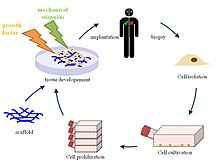
Principle of tissue engineering
 
Decellularization (also spelt decellularisation in British English) is the process used in biomedical engineering to isolate the extracellular matrix (ECM) of a tissue from its inhabiting cells, leaving an ECM scaffold of the original tissue, which can be used in artificial organ and tissue regeneration. Organ and tissue transplantation treat a variety of medical problems, ranging from end organ failure to cosmetic surgery. One of the greatest limitations to organ transplantation derives from organ rejection caused by antibodies of the transplant recipient reacting to donor antigens on cell surfaces within the donor organ. Because of unfavorable immune responses, transplant patients suffer a lifetime taking immunosuppressing medication. Stephen F. Badylak pioneered the process of decellularization at the McGowan Institute for Regenerative Medicine at the University of Pittsburgh.[2] This process creates a natural biomaterial to act as a scaffold for cell growth, differentiation and tissue development. By recellularizing an ECM scaffold with a patient’s own cells, the adverse immune response is eliminated. Nowadays, commercially available ECM scaffolds are available for a wide variety of tissue engineering. Using peracetic acid to decellularize ECM scaffolds have been found to be false and only disinfects the tissue.

With a wide variety of decellularization-inducing treatments available, combinations of physical, chemical, and enzymatic treatments are carefully monitored to ensure that the ECM scaffold maintains the structural and chemical integrity of the original tissue. Scientists can use the acquired ECM scaffold to reproduce a functional organ by introducing progenitor cells, or adult stem cells (ASCs), and allowing them to differentiate within the scaffold to develop into the desired tissue. The produced organ or tissue can be transplanted into a patient. In contrast to cell surface antibodies, the biochemical components of the ECM are conserved between hosts, so the risk of a hostile immune response is minimized. Proper conservation of ECM fibers, growth factors, and other proteins is imperative to the progenitor cells differentiating into the proper adult cells. The success of decellularization varies based on the components and density of the applied tissue and its origin. The applications to the decellularizing method of producing a biomaterial scaffold for tissue regeneration are present in cardiac, dermal, pulmonary, renal, and other types of tissues. Complete organ reconstruction is still in the early levels of development.

Process overview

Researchers are able to take the tissue from a donor or cadaver, lyse and kill the cells within the tissue without damaging the extracellular components, and finish with a product that is the natural ECM scaffold that has the same physical and biochemical functions of the natural tissue. After acquiring the ECM scaffold, scientists can recellularize the tissue with potent stem or progenitor cells that will differentiate into the original type of tissue. By removing the cells from a donor tissue, the immunogenic antibodies from the donor will be removed. The progenitor cells can be taken from the host, therefore they will not have an adverse response to the tissue. This process of decellularizing tissues and organs is still being developed, but the exact process of taking a tissue from a donor and removing all the cellular components is considered to be the decellularization process. The steps to go from a decellularized ECM scaffold to a functional organ is under the umbrella of recellularization. Because of the diverse applications of tissue in the human body, decellularization techniques have to be tailored to the specific tissue being exercised on. The researched methods of decellularization include physical, chemical, and enzymatic treatments. Though some methods are more commonly used, the exact combination of treatments is variable based on the tissue’s origin and what it is needed for.

As far as introducing the different liquidized chemicals and enzymes to an organ or tissue, perfusion and immersion decellularization techniques have been used. Perfusion decellularization is applicable when an extensive vasculature system is present in the organ or tissue. It is crucial for the ECM scaffold to be decellularized at all levels, and evenly throughout the structure. Because of this requirement, vascularized tissues can have chemicals and enzymes perfused through the present arteries, veins, and capillaries. Under this mechanism and proper physiological conditions, treatments can diffuse equally to all of the cells within the organ. The treatments can be removed through the veins at the end of the process. Cardiac and pulmonary decellularization often uses this process of decellularization to introduce the treatments because of their heavily vascularized networks. Immersion decellularization is accomplished through the submersion of a tissue in chemical and enzymatic treatments. This process is more easily accomplished than perfusion, but is limited to thin tissues with a limited vascular system.

Physical treatments

The most common physical methods used to lyse, kill, and remove cells from the matrix of a tissue through the use of temperature, force and pressure, and electrical disruption. Temperature methods are often used in a rapid freeze-thaw mechanism. By quickly freezing a tissue, microscopic ice crystals form around the plasma membrane and the cell is lysed. After lysing the cells, the tissue can be further exposed to liquidized chemicals that degrade and wash out the undesirable components. Temperature methods conserve the physical structure of the ECM scaffold, but are best handled by thick, strong tissues.

Direct force of pressure to a tissue will guarantee disruption of the ECM structure, so pressure is commonly used. Pressure decellularization involves the controlled use of hydrostatic pressure applied to a tissue or organ. This is done best at high temperatures to avoid unmonitored ice crystal formation that could damage the scaffold. Electrical disruption of the plasma membrane is another option to lyse the cells housed in a tissue or organ. By exposing a tissue to electrical pulses, micropores are formed at the plasma membrane. The cells eventually turn to death after their homeostatic electrical balance is ruined through the applied stimulus. This electrical process is documented as Non-thermal irreversible electroporation (NTIRE) and is limited to small tissues and the limited possibilities of inducing an electric current in vivo.

Chemical treatments

The proper combination of chemicals is selected for decellularization depending on the thickness, extracellular matrix composition, and intended use of the tissue or organ. For example, enzymes would not be used on a collagenous tissue because they disrupt the connective tissue fibers. However, when collagen is not present in a high concentration or needed in the tissue, enzymes can be a viable option for decellularization. The chemicals used to kill and remove the cells include acids, alkaline treatments, ionic detergents, non-ionic detergents, and zwitterionic detergents.

The ionic detergent, sodium dodecyl sulfate (SDS), is commonly used because of its high efficacy for lysing cells without significant damage to the ECM. Detergents act effectively to lyse the cell membrane and expose the contents to further degradation. After SDS lyses the cell membrane, endonucleases and exonucleases degrade the genetic contents, while other components of the cell is solubilized and washed out of the matrix. SDS is commonly used even though it has a tendency to slightly disrupt the ECM structure. Akaline and acid treatments can be effective companions with an SDS treatment due to their ability to degrade nucleic acids and solubilize cytoplasmic inclusions.

The most well known non-ionic detergent is Triton X-100, which is popular because of its ability to disrupt the interactions between lipids and between lipids and proteins. Triton X-100 does not disrupt protein-protein interactions, which is beneficial to keeping the ECM intact. EDTA is a chelating agent that binds calcium, which is a necessary component for proteins to interact with one another. By making calcium unavailable, EDTA prevents the integral proteins between cells from binding to one another. EDTA is often used with trypsin, an enzyme that acts as a protease to cleave the already existing bonds between integral proteins of neighboring cells within a tissue. Together, the EDTA-Trypsin combination make a good team for decellularizing tissues.

Enzymatic treatments

Enzymes used in decellularization treatments are used to break the bonds and interactions between nucleic acids, interacting cells through neighboring proteins, and other cellular components. Lipases, thermolysin, galactosidase, nucleases, and trypsin have all been used in the removal of cells. After a cell is lysed with a detergent, acid, physical pressure, etc., endonucleases and exonucleases can begin the degradation of the genetic material. Endonucleases cleave DNA and RNA in the middle of sequences. Benzoase, an endonuclease, produces multiple small nuclear fragments that can be further degraded and removed from the ECM scaffold. Exonucleases act at the end of DNA sequences to cleave the phosphodiester bonds and further degrade the nucleic acid sequences.

Enzymes such as trypsin act as proteases that cleave the interactions between proteins. Although trypsin can have adverse effects of collagen and elastin fibers of the ECM, using it in a time-sensitive manner controls any potential damage it could cause on the extracellular fibers. Dispase is used to prevent undesired aggregation of cells, which is beneficial in promoting their separating from the ECM scaffold. Experimentation has shown dispase to be most effective on the surface of a thin tissue, such as a lung in pulmonary tissue regeneration. To successfully remove deep cells of a tissue with dispase, mechanical agitation is often included in the process.

Collagenase is only used when the ECM scaffold product does not require an intact collagen structure. Lipases are commonly used when decellularized skin grafts are needed. Lipase acids function in decellularizing dermal tissues through delipidation and cleaving the interactions between heavily lipidized cells. The enzyme, α-galactosidase is a relevant treatment when removing the Gal epitope antigen from cell surfaces.

Applications

A natural ECM scaffold provides the necessary physical and biochemical environment to facilitate the growth and specialization of potent progenitor and stem cells. Acellular matrices have been isolated in vitro and in vivo in a number of different tissues and organs. The most applicable success from decellularized tissues has come from symmetrical tissues that have less specialization, such as bone and dermal grafts; however, research and success is ongoing at the organ level.

Acellular dermal matrices have been successful in a number of different applications. For example, skin grafts are used in cosmetic surgery and burn care. The decellularized skin graft provides mechanical support to the damaged area while supporting the development of host-derived connective tissue. Cardiac tissue has clinical success in developing human valves from natural ECM matrices.[14] A technique known as the Ross procedure uses an acellular heart valve to replace a defective valve, allowing native cells to repopulate a newly functioning valve. Decellularized allografts have been critical in bone grafts that function in bone reconstruction and replacing of deformed bones in patients. 

The limits to myocardial tissue engineering come from the ability to immediately perfuse and seed and implemented heart into a patient. Though the ECM scaffold maintains the protein and growth factors of the natural tissue, the molecular level specialization has not yet been harnessed by researchers using decellularized heart scaffolds. Better success at using a whole organ from decellularization techniques has been found in pulmonary research. Scientists have been able to regenerate whole lungs in vitro from rat lungs using perfusion-decellularization. By seeding the matrix with fetal rat lung cells, a functioning lung was produced. The in vitro-produced lung was successfully implemented into a rat, which attests to the possibilities of translating an in vitro produced organ into a patient.

Other success for decellularization has been found in small intestinal submucosa (SIS), renal, hepatic, and pancreatic engineering. Because it is a thin material, the SIS matrix can be decellularized through immersing the tissue in chemical and enzymatic treatments. Renal tissue engineering is still developing, but cadaveric kidney matrices have been able to support development of potent fetal kidney cells. Pancreatic engineering is a testament to the molecular specificity of organs. Scientists have not yet been able to produce an entirely functioning pancreas, but they have had success in producing an organ that functions at specific segments. For example, diabetes in rats was shown to decrease by seeding a pancreatic matrix at specific sites. The future applications of decellularized tissue matrix is still being discovered and is considered one of the most hopeful areas in regenerative research.
Artificial pancreas

From Wikipedia, the free encyclopedia
https://en.wikipedia.org/wiki/Artificial_pancreas
 
The artificial pancreas is a technology in development to help people with diabetes, primarily type 1, automatically and continuously control their blood glucose level by providing the substitute endocrine functionality of a healthy pancreas.

The endocrine functionality of the pancreas is provided by islet cells which produce the hormones insulin and glucagon. Artificial pancreatic technology mimics the secretion of these hormones into the bloodstream in response to the body's changing blood glucose levels. Maintaining balanced blood sugar levels is crucial to the function of the brain, liver, and kidneys. Therefore, for type 1 patients, it is necessary that the levels be kept balanced when the body cannot produce insulin itself.

The artificial pancreas is a broad term for different bio-engineering strategies currently in development to achieve these requirements. Different bio-engineering approaches under consideration include:

Approaches

Medical equipment

The medical equipment approach involves combining a continuous glucose monitor and an implanted insulin pump that can function together with a computer-controlled algorithm to replace the normal function of the pancreas. The development of continuous glucose monitors has led to the progress in artificial pancreas technology using this integrated system.

Continuous glucose monitors

Artificial pancreas feedback system
 
The original devices for use in type 1 diabetes were blood glucose meters. Continuous blood glucose monitors are one of the set of devices that make up an artificial pancreas device system, the other being an insulin pump, and a glucose meter to calibrate the device. Continuous glucose monitors are a more recent breakthrough and have begun to hit the markets for patient use after approval from the FDA. Both the traditional and the continuous monitor require manual insulin delivery or carbohydrate intake depending on the readings from the devices. While the traditional blood glucose meters require the user to prick their finger every few hours to obtain data, continuous monitors use sensors placed just under the skin on the arm or abdomen to deliver blood sugar level data to receivers or smartphone apps as often as every few minutes. The sensors can be used for up to fourteen days. A number of different continuous monitors are currently approved by the FDA.

The first continuous glucose monitor (CGM) was approved in December 2016. Developed by Dexcom, the G5 Mobile Continuous Monitoring System requires users to prick their fingers twice a day (as opposed to the typical average 8 times daily with the traditional meters) in order to calibrate the sensors. The sensors last up to seven days. The device uses Bluetooth technology to warn the user either through a handheld receiver or app on a smartphone if blood glucose levels reach below a certain point. The cost for this device excluding any co-insurance is an estimated $4,800 a year.

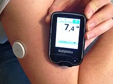
The white sensor is fixed to the upper arm and scanned with the reader. The reader is showing (top to bottom) days to replacement of sensor (11), current BG (7,4) & change (-> i.e. steady) and a diagram of the latest BG levels.
Blood glucose meter FreeStyle Libre from Abbott.
 
Abbott Laboratories' FreeStyle Libre CGM was approved in September 2017. Recently, the technology was modified to support smartphone use through the LibreLink app. This device does not require finger pricks at all and the sensor, placed on the upper arm, lasts 14 days. The estimated cost for this monitor is $1,300 a year.

Dexcom's next G6 model CGM was approved in March 2018, which can last up to ten days and does not need finger prick calibration. Like Medtronic's monitor, it can predict glucose level trends. It is compatible for integration into insulin pumps.

Closed-loop systems

Unlike the continuous sensor alone, the closed-loop system requires no user input in response to reading from the monitor; the monitor and insulin pump system automatically delivers the correct amount of hormone calculated from the readings transmitted. The system is what makes up the artificial pancreas device.

Inreda Diabetic
In collaboration with the Academic Medical Centre (AMC) in Amsterdam Inreda is developing a closed loop system with insuline and glucagon. The initiator, Robin Koops, started to develop the device in 2004 and ran the first tests on himself. After several highly successful trails it received the European EC license in 2016. The product is expected to market in the second half of 2020. A smaller improved version is scheduled for 2023. 

MiniMed 670G
In September 2016, the FDA approved the Medtronic MiniMed 670G, which was the first approved hybrid closed loop system. The device senses a diabetic person's basal insulin requirement and automatically adjusts its delivery to the body. It is made up of a continuous glucose monitor, an insulin pump, and a glucose meter for calibration. It automatically functions to modify the level of insulin delivery based off the detection of blood glucose levels by continuous monitor. It does this by sending the blood glucose data through an algorithm that analyzes and makes the subsequent adjustments. The system has two modes. Manual mode lets the user choose the rate at which basal insulin is delivered. Auto mode regulates basal insulin levels from the continuous monitor's readings every five minutes.

The device was originally available only to those aged 14 or older, and in June 2018 was approved by the FDA for use in children aged 7–14. Families have reported better sleep quality from use of the new system, as they do not have to worry about manually checking blood glucose levels during the night. The full cost of the system is $3700, but patients have the opportunity to get it for less.

Ilet Bionic Pancreas
A team at Boston University working in collaboration with Massachusetts General Hospital on a dual hormone artificial pancreas system  began clinical trials on their device called the Bionic Pancreas in 2008. In 2016, the Public Benefit Corporation Beta Bionics was formed. In conjunction with the formation of the company, Beta Bionics changed the preliminary name for their device from the Bionic Pancreas to the iLet. The device uses a closed-loop system to deliver both insulin and glucagon in response to sensed blood glucose levels. While not yet approved for public use, the 4th generation iLet prototype, presented in 2017, is around the size of an iPhone, with a touchscreen interface. It contain two chambers for both insulin and glucagon, and the device is configurable for use with only one hormone, or both. While trials continue to be run, the iLet has a projected final approval for the insulin-only system in 2020.

Current studies
Four studies on different artificial pancreas systems are being conducted starting in 2017 and going into the near future. The projects are funded by the National Institute of Diabetes and Digestive and Kidney Diseases(NIDDK), and are the final part of testing the devices before applying for approval for use. Participants in the studies are able to live their lives at home while using the devices and being monitored remotely for safety, efficacy, and a number of other factors.

The International Diabetes Closed-Loop trial, led by researchers from the University of Virginia, is testing a closed-loop system called inControl, which has a smartphone user interface. 240 people of ages 14 and up are participating for 6 months.

A full-year trial led by researchers from the University of Cambridge started in May 2017 and has enrolled an estimated 150 participants of ages 6 to 18 years. The artificial pancreas system being studied uses a smartphone and has a low glucose feature to improve glucose level control.

The International Diabetes Center in Minneapolis, Minnesota, in collaboration with Schneider Children's Medical Center in Petah Tikva, Israel, are planning a 6-month study that will begin in early 2019 and will involve 112 adolescents and young adults, ages 14 to 30. The main object of the study is to compare the current Medtronic 670G system to a new Medtronic-developed system. The new system has programming that aims to improve glucose control around mealtime, which is still a big challenge in the field.

The current 6-month study lead by the Bionic Pancreas team started in mid-2018 and enrolled 312 participants of ages 18 and above.

Physiological

The Bio-artificial pancreas: this diagram shows a cross section of bio-engineered tissue with encapsulated islet cells which deliver endocrine hormones in response to glucose.
 
The biotechnical company Defymed, based in France, is developing an implantable bio-artificial device called MailPan which features a bio-compatible membrane with selective permeability to encapsulate different cells types, including pancreatic beta cells. The implantation of the device does not require conjunctive immuno-suppressive therapy because the membrane prevents antibodies of the patient from entering the device and damaging the encapsulated cells. After being surgically implanted, the membrane sheet will be viable for years.The cells that the device holds can be produced from stem cells rather than human donors, and may also be replaced over time using input and output connections without surgery. Defymed is partially funded by JDRF, formerly known as the Juvenile Diabetes Research Foundation, but is now defined as an organization for all ages and all stages of type 1 diabetes.

In November 2018, it was announced that Defymed would partner with the Israel-based Kadimastem, a bio-pharmaceutical company developing stem-cell based regenerative therapies, to receive a two-year grant worth approximately $1.47 million for the development of a bio-artificial pancreas that would treat type 1 diabetes. Kadimastem's stem cell technology uses differentiation of human embryonic stem cells to obtain pancreatic endocrine cells. These include insulin-producing beta cells, as well as alpha cells, which produce glucagon. Both cells arrange in islet-like clusters, mimicking the structure of the pancreas. The aim of the partnership is to combine both technologies in a bio-artificial pancreas device, which releases insulin in response to blood glucose levels, to bring to clinical trial stages.

The San Diego, California based biotech company ViaCyte has also developed a product aiming to provide a solution for type 1 diabetes which uses an encapsulation device made of a semi-permeable immune reaction-protective membrane. The device contains pancreatic progenitor cells that have been differentiated from embryonic stem cells. After surgical implantation in an outpatient procedure, the cells mature into endocrine cells which arrange in islet-like clusters and mimic the function of the pancreas, producing insulin and glucagon. The technology advanced from pre-clinical studies to FDA approval for phase 1 clinical trials in 2014, and presented two-year data from the trial in June 2018. They reported that their product, called PEC-Encap, has so far been safe and well tolerated in patients at a dose below therapeutic levels. The encapsulated cells were able to survive and mature after implantation, and immune system rejection was decreased due to the protective membrane. The second phase of the trial will evaluate the efficacy of the product. ViaCyte has also been receiving financial support from JDRF on this project.

Initiatives around the globe

In the United States in 2006, JDRF (formerly the Juvenile Diabetes Research Foundation) launched a multi-year initiative to help accelerate the development, regulatory approval, and acceptance of continuous glucose monitoring and artificial pancreas technology.

Grassroots efforts to create and commercialize a fully automated artificial pancreas system have also arisen directly from patient advocates and the diabetes community. Bigfoot Biomedical, a company founded by parents of children with T1D have created algorithms and are developing a closed loop device that monitor blood sugar and appropriately provide insulin.

Monday, December 9, 2019

Neuroprosthetics

From Wikipedia, the free encyclopedia
https://en.wikipedia.org/wiki/Neuroprosthetics

Neuroprosthetics (also called neural prosthetics) is a discipline related to neuroscience and biomedical engineering concerned with developing neural prostheses. They are sometimes contrasted with a brain–computer interface, which connects the brain to a computer rather than a device meant to replace missing biological functionality.

Neural prostheses are a series of devices that can substitute a motor, sensory or cognitive modality that might have been damaged as a result of an injury or a disease. Cochlear implants provide an example of such devices. These devices substitute the functions performed by the eardrum and stapes while simulating the frequency analysis performed in the cochlea. A microphone on an external unit gathers the sound and processes it; the processed signal is then transferred to an implanted unit that stimulates the auditory nerve through a microelectrode array. Through the replacement or augmentation of damaged senses, these devices intend to improve the quality of life for those with disabilities.

These implantable devices are also commonly used in animal experimentation as a tool to aid neuroscientists in developing a greater understanding of the brain and its functioning. By wirelessly monitoring the brain's electrical signals sent out by electrodes implanted in the subject's brain, the subject can be studied without the device affecting the results. 

Accurately probing and recording the electrical signals in the brain would help better understand the relationship among a local population of neurons that are responsible for a specific function. 

Neural implants are designed to be as small as possible in order to be minimally invasive, particularly in areas surrounding the brain, eyes or cochlea. These implants typically communicate with their prosthetic counterparts wirelessly. Additionally, power is currently received through wireless power transmission through the skin. The tissue surrounding the implant is usually highly sensitive to temperature rise, meaning that power consumption must be minimal in order to prevent tissue damage.

The neuroprosthetic currently undergoing the most widespread use is the cochlear implant, with over 300,000 in use worldwide as of 2012.

History

The first known cochlear implant was created in 1957. Other milestones include the first motor prosthesis for foot drop in hemiplegia in 1961, the first auditory brainstem implant in 1977 and a peripheral nerve bridge implanted into the spinal cord of an adult rat in 1981. In 1988, the lumbar anterior root implant and functional electrical stimulation (FES) facilitated standing and walking, respectively, for a group of paraplegics.

Regarding the development of electrodes implanted in the brain, an early difficulty was reliably locating the electrodes, originally done by inserting the electrodes with needles and breaking off the needles at the desired depth. Recent systems utilize more advanced probes, such as those used in deep brain stimulation to alleviate the symptoms of Parkinson's disease. The problem with either approach is that the brain floats free in the skull while the probe does not, and relatively minor impacts, such as a low speed car accident, are potentially damaging. Some researchers, such as Kensall Wise at the University of Michigan, have proposed tethering 'electrodes to be mounted on the exterior surface of the brain' to the inner surface of the skull. However, even if successful, tethering would not resolve the problem in devices meant to be inserted deep into the brain, such as in the case of deep brain stimulation (DBS). 

Visual prosthetics

A visual prosthesis can create a sense of image by electrically stimulating neurons in the visual system. A camera would wirelessly transmit to an implant, the implant would map the image across an array of electrodes. The array of electrodes has to effectively stimulate 600-1000 locations, stimulating these optic neurons in the retina thus will create an image. The stimulation can also be done anywhere along the optic signal's path way. The optical nerve can be stimulated in order to create an image, or the visual cortex can be stimulated, although clinical tests have proven most successful for retinal implants.

A visual prosthesis system consists of an external (or implantable) imaging system which acquires and processes the video. Power and data will be transmitted to the implant wirelessly by the external unit. The implant uses the received power/data to convert the digital data to an analog output which will be delivered to the nerve via micro electrodes.

Photoreceptors are the specialized neurons that convert photons into electrical signals. They are part of the retina, a multilayer neural structure about 200 um thick that lines the back of the eye. The processed signal is sent to the brain through the optical nerve. If any part of this pathway is damaged blindness can occur.

Blindness can result from damage to the optical pathway (cornea, aqueous humor, crystalline lens, and vitreous). This can happen as a result of accident or disease. The two most common retinal degenerative diseases that result in blindness secondary to photoreceptor loss is age related macular degeneration (AMD) and retinitis pigmentosa (RP).

The first clinical trial of a permanently implanted retinal prosthesis was a device with a passive microphotodiode array with 3500 elements. This trial was implemented at Optobionics, Inc., in 2000. In 2002, Second Sight Medical Products, Inc. (Sylmar, CA) began a trial with a prototype epiretinal implant with 16 electrodes. The subjects were six individuals with bare light perception secondary to RP. The subjects demonstrated their ability to distinguish between three common objects (plate, cup, and knife) at levels statistically above chance. An active sub retinal device developed by Retina Implant GMbH (Reutlingen, Germany) began clinical trials in 2006. An IC with 1500 microphotodiodes was implanted under the retina. The microphotodiodes serve to modulate current pulses based on the amount of light incident on the photo diode.

The seminal experimental work towards the development of visual prostheses was done by cortical stimulation using a grid of large surface electrodes. In 1968 Giles Brindley implanted an 80 electrode device on the visual cortical surface of a 52-year-old blind woman. As a result of the stimulation the patient was able to see phosphenes in 40 different positions of the visual field. This experiment showed that an implanted electrical stimulator device could restore some degree of vision. Recent efforts in visual cortex prosthesis have evaluated efficacy of visual cortex stimulation in a non-human primate. In this experiment after a training and mapping process the monkey is able to perform the same visual saccade task with both light and electrical stimulation.

The requirements for a high resolution retinal prosthesis should follow from the needs and desires of blind individuals who will benefit from the device. Interactions with these patients indicate that mobility without a cane, face recognition and reading are the main necessary enabling capabilities.

The results and implications of fully functional visual prostheses are exciting. However, the challenges are grave. In order for a good quality image to be mapped in the retina a high number of micro-scale electrode arrays are needed. Also, the image quality is dependent on how much information can be sent over the wireless link. Also this high amount of information must be received and processed by the implant without much power dissipation which can damage the tissue. The size of the implant is also of great concern. Any implant would be preferred to be minimally invasive.

With this new technology, several scientists, including Karen Moxon at Drexel, John Chapin at SUNY, and Miguel Nicolelis at Duke University, started research on the design of a sophisticated visual prosthesis. Other scientists have disagreed with the focus of their research, arguing that the basic research and design of the densely populated microscopic wire was not sophisticated enough to proceed. 

Auditory prosthetics

Cochlear implants (CIs), auditory brain stem implants (ABIs), and auditory midbrain implants (AMIs) are the three main categories for auditory prostheses. CI electrode arrays are implanted in the cochlea, ABI electrode arrays stimulate the cochlear nucleus complex in the lower brain stem, and AMIs stimulates auditory neurons in the inferior colliculus. Cochlear implants have been very successful among these three categories. Today the Advanced Bionics Corporation, the Cochlear Corporation and the Med-El Corporation are the major commercial providers of cochlea implants. 

In contrast to traditional hearing aids that amplify sound and send it through the external ear, cochlear implants acquire and process the sound and convert it into electrical energy for subsequent delivery to the auditory nerve. The microphone of the CI system receives sound from the external environment and sends it to processor. The processor digitizes the sound and filters it into separate frequency bands that are sent to the appropriate tonotonic region in the cochlea that approximately corresponds to those frequencies. 

In 1957, French researchers A. Djourno and C. Eyries, with the help of D. Kayser, provided the first detailed description of directly stimulation the auditory nerve in a human subject. The individuals described hearing chirping sounds during simulation. In 1972, the first portable cochlear implant system in an adult was implanted at the House Ear Clinic. The U.S. Food and Drug Administration (FDA) formally approved the marketing of the House-3M cochlear implant in November 1984.

Improved performance on cochlear implant not only depends on understanding the physical and biophysical limitations of implant stimulation but also on an understanding of the brain's pattern processing requirements. Modern signal processing represents the most important speech information while also providing the brain the pattern recognition information that it needs. Pattern recognition in the brain is more effective than algorithmic preprocessing at identifying important features in speech. A combination of engineering, signal processing, biophysics, and cognitive neuroscience was necessary to produce the right balance of technology to maximize the performance of auditory prosthesis.

Cochlear implants have been also used to allow acquiring of spoken language development in congenitally deaf children, with remarkable success in early implantations (before 2–4 years of life have been reached). There have been about 80,000 children implanted worldwide.

The concept of combining simultaneous electric-acoustic stimulation (EAS) for the purposes of better hearing was first described by C. von Ilberg and J. Kiefer, from the Universitätsklinik Frankfurt, Germany, in 1999. That same year the first EAS patient was implanted. Since the early 2000s FDA has been involved in a clinical trial of device termed the "Hybrid" by Cochlear Corporation. This trial is aimed at examining the usefulness of cochlea implantation in patients with residual low-frequency hearing. The "Hybrid" utilizes a shorter electrode than the standard cochlea implant, since the electrode is shorter it stimulates the basil region of the cochlea and hence the high-frequency tonotopic region. In theory these devices would benefit patients with significant low-frequency residual hearing who have lost perception in the speech frequency range and hence have decreased discrimination scores.

For producing sound see Speech synthesis

Prosthetics for pain relief

The SCS (Spinal Cord Stimulator) device has two main components: an electrode and a generator. The technical goal of SCS for neuropathic pain is to mask the area of a patient's pain with a stimulation induced tingling, known as "paresthesia", because this overlap is necessary (but not sufficient) to achieve pain relief. Paresthesia coverage depends upon which afferent nerves are stimulated. The most easily recruited by a dorsal midline electrode, close to the pial surface of spinal cord, are the large dorsal column afferents, which produce broad paresthesia covering segments caudally. 

In ancient times the electrogenic fish was used as a shocker to subside pain. Healers had developed specific and detailed techniques to exploit the generative qualities of the fish to treat various types of pain, including headache. Because of the awkwardness of using a living shock generator, a fair level of skill was required to deliver the therapy to the target for the proper amount of time. (Including keeping the fish alive as long as possible) Electro analgesia was the first deliberate application of electricity. By the nineteenth century, most western physicians were offering their patients electrotherapy delivered by portable generator. In the mid-1960s, however, three things converged to ensure the future of electro stimulation.
  1. Pacemaker technology, which had it start in 1950, became available.
  2. Melzack and Wall published their gate control theory of pain, which proposed that the transmission of pain could be blocked by stimulation of large afferent fibers.
  3. Pioneering physicians became interested in stimulating the nervous system to relieve patients from pain.
The design options for electrodes include their size, shape, arrangement, number, and assignment of contacts and how the electrode is implanted. The design option for the pulse generator include the power source, target anatomic placement location, current or voltage source, pulse rate, pulse width, and number of independent channels. Programming options are very numerous (a four-contact electrode offers 50 functional bipolar combinations). The current devices use computerized equipment to find the best options for use. This reprogramming option compensates for postural changes, electrode migration, changes in pain location, and suboptimal electrode placement.

Motor prosthetics

Devices which support the function of autonomous nervous system include the implant for bladder control. In the somatic nervous system attempts to aid conscious control of movement include Functional electrical stimulation and the lumbar anterior root stimulator

Bladder control implants

Where a spinal cord lesion leads to paraplegia, patients have difficulty emptying their bladders and this can cause infection. From 1969 onwards Brindley developed the sacral anterior root stimulator, with successful human trials from the early 1980s onwards. This device is implanted over the sacral anterior root ganglia of the spinal cord; controlled by an external transmitter, it delivers intermittent stimulation which improves bladder emptying. It also assists in defecation and enables male patients to have a sustained full erection.

The related procedure of sacral nerve stimulation is for the control of incontinence in able-bodied patients.

Motor prosthetics for conscious control of movement

Researchers are currently investigating and building motor neuroprosthetics that will help restore movement and the ability to communicate with the outside world to persons with motor disabilities such as tetraplegia or amyotrophic lateral sclerosis. Research has found that the striatum plays a crucial role in motor sensory learning. This was demonstrated by an experiment in which lab rats' firing rates of the striatum was recorded at higher rates after performing a task consecutively.

To capture electrical signals from the brain, scientists have developed microelectrode arrays smaller than a square centimeter that can be implanted in the skull to record electrical activity, transducing recorded information through a thin cable. After decades of research in monkeys, neuroscientists have been able to decode neuronal signals into movements. Completing the translation, researchers have built interfaces that allow patients to move computer cursors, and they are beginning to build robotic limbs and exoskeletons that patients can control by thinking about movement.

The technology behind motor neuroprostheses is still in its infancy. Investigators and study participants continue to experiment with different ways of using the prostheses. Having a patient think about clenching a fist, for example, produces a different result than having him or her think about tapping a finger. The filters used in the prostheses are also being fine-tuned, and in the future, doctors hope to create an implant capable of transmitting signals from inside the skull wirelessly, as opposed to through a cable.

Prior to these advancements, Philip Kennedy (Emory and Georgia Tech) had an operable if somewhat primitive system which allowed an individual with paralysis to spell words by modulating their brain activity. Kennedy's device used two neurotrophic electrodes: the first was implanted in an intact motor cortical region (e.g. finger representation area) and was used to move a cursor among a group of letters. The second was implanted in a different motor region and was used to indicate the selection.

Developments continue in replacing lost arms with cybernetic replacements by using nerves normally connected to the pectoralis muscles. These arms allow a slightly limited range of motion, and reportedly are slated to feature sensors for detecting pressure and temperature.

Dr. Todd Kuiken at Northwestern University and Rehabilitation Institute of Chicago has developed a method called targeted reinnervation for an amputee to control motorized prosthetic devices and to regain sensory feedback. 

In 2002 a Multielectrode array of 100 electrodes, which now forms the sensor part of a Braingate, was implanted directly into the median nerve fibers of scientist Kevin Warwick. The recorded signals were used to control a robot arm developed by Warwick's colleague, Peter Kyberd and was able to mimic the actions of Warwick's own arm. Additionally, a form of sensory feedback was provided via the implant by passing small electrical currents into the nerve. This caused a contraction of the first lumbrical muscle of the hand and it was this movement that was perceived.

In June 2014, Juliano Pinto, a paraplegic athlete, performed the ceremonial first kick at the 2014 FIFA World Cup using a powered exoskeleton with a brain interface. The exoskeleton was developed by the Walk Again Project at the laboratory of Miguel Nicolelis, funded by the government of Brazil. Nicolelis says that feedback from replacement limbs (for example, information about the pressure experienced by a prosthetic foot touching the ground) is necessary for balance. He has found that as long as people can see the limbs being controlled by a brain interface move at the same time as issuing the command to do so, with repeated use the brain will assimilate the externally powered limb and it will start to perceive it (in terms of position awareness and feedback) as part of the body.

Amputation techniques

The MIT Biomechatronics Group has designed a novel amputation paradigm that enables biological muscles and myoelectric prostheses to interface neurally with high reliability. This surgical paradigm, termed the agonist-antagonist myoneural interface (AMI), provides the user with the ability to sense and control their prosthetic limb as an extension of their own body, rather than using a prosthetic that merely resembles an appendage. In a normal agonist-antagonist muscle pair relationship (e.g. bicep-tricep), when the agonist muscle contracts, the antagonist muscle is stretched, and vice versa, providing one with the knowledge of the position of one's limb without even having to look at it. During a standard amputation, agonist-antagonist muscles (e.g. bicep-tricep) are isolated from each other, preventing the ability to have the dynamic contract-extend mechanism that generates sensory feedback. Therefore, current amputees have no way of feeling the physical environment their prosthetic limb encounters. Moreover, with the current amputation surgery which has been in place for over 200 years, 1/3 patients undergo revision surgeries due to pain in their stumps. 

An AMI is composed of two muscles that originally shared an agonist-antagonist relationship. During the amputation surgery, these two muscles are mechanically linked together within the amputated stump. One AMI muscle pair can be created for each joint degree of freedom in a patient in order to establish control and sensation of multiple prosthetic joints. In preliminary testing of this new neural interface, patients with an AMI have demonstrated and reported greater control over the prosthesis. Additionally, more naturally reflexive behavior during stair walking was observed compared to subjects with a traditional amputation. An AMI can also be constructed through the combination of two devascularized muscle grafts. These muscle grafts (or flaps) are spare muscle that is denervated (detached from original nerves) and removed from one part of the body to be re-innervated by severed nerves found in the limb to be amputated. Through the use of regenerated muscle flaps, AMIs can be created for patients with muscle tissue that has experienced extreme atrophy or damage or for patients who are undergoing revision of an amputated limb for reasons such as neuroma pain, bone spurs, etc. 

Obstacles

Mathematical modelling

Accurate characterization of the nonlinear input/output (I/O) parameters of the normally functioning tissue to be replaced is paramount to designing a prosthetic that mimics normal biologic synaptic signals. Mathematical modeling of these signals is a complex task "because of the nonlinear dynamics inherent in the cellular/molecular mechanisms comprising neurons and their synaptic connections". The output of nearly all brain neurons are dependent on which post-synaptic inputs are active and in what order the inputs are received. (spatial and temporal properties, respectively).

Once the I/O parameters are modeled mathematically, integrated circuits are designed to mimic the normal biologic signals. For the prosthetic to perform like normal tissue, it must process the input signals, a process known as transformation, in the same way as normal tissue. 

Size

Implantable devices must be very small to be implanted directly in the brain, roughly the size of a quarter. One of the example of microimplantable electrode array is the Utah array.

Wireless controlling devices can be mounted outside of the skull and should be smaller than a pager. 

Power consumption

Power consumption drives battery size. Optimization of the implanted circuits reduces power needs. Implanted devices currently need on-board power sources. Once the battery runs out, surgery is needed to replace the unit. Longer battery life correlates to fewer surgeries needed to replace batteries. One option that could be used to recharge implant batteries without surgery or wires is being used in powered toothbrushes. These devices make use of inductive coupling to recharge batteries. Another strategy is to convert electromagnetic energy into electrical energy, as in radio-frequency identification tags. 

Biocompatibility

Cognitive prostheses are implanted directly in the brain, so biocompatibility is a very important obstacle to overcome. Materials used in the housing of the device, the electrode material (such as iridium oxide), and electrode insulation must be chosen for long term implantation. Subject to Standards: ISO 14708-3 2008-11-15, Implants for Surgery - Active implantable medical devices Part 3: Implantable neurostimulators. 

Crossing the blood–brain barrier can introduce pathogens or other materials that may cause an immune response. The brain has its own immune system that acts differently from the immune system of the rest of the body. 

Questions to answer: How does this affect material choice? Does the brain have unique phages that act differently and may affect materials thought to be biocompatible in other areas of the body? 

Data transmission

Wireless Transmission is being developed to allow continuous recording of neuronal signals of individuals in their daily life. This allows physicians and clinicians to capture more data, ensuring that short term events like epileptic seizures can be recorded, allowing better treatment and characterization of neural disease.

A small, light weight device has been developed that allows constant recording of primate brain neurons at Stanford University. This technology also enables neuroscientists to study the brain outside of the controlled environment of a lab.

Methods of data transmission must be robust and secure. Neurosecurity is a new issue. Makers of cognitive implants must prevent unwanted downloading of information or thoughts from and uploading of detrimental data to the device that may interrupt function. 

Correct implantation

Implantation of the device presents many problems. First, the correct presynaptic inputs must be wired to the correct postsynaptic inputs on the device. Secondly, the outputs from the device must be targeted correctly on the desired tissue. Thirdly, the brain must learn how to use the implant. Various studies in brain plasticity suggest that this may be possible through exercises designed with proper motivation. 

Technologies involved

Local field potentials

Local field potentials (LFPs) are electrophysiological signals that are related to the sum of all dendritic synaptic activity within a volume of tissue. Recent studies suggest goals and expected value are high-level cognitive functions that can be used for neural cognitive prostheses. Also, Rice University scientists have discovered a new method to tune the light-induced vibrations of nanoparticles through slight alterations to the surface to which the particles are attached. According to the university, the discovery could lead to new applications of photonics from molecular sensing to wireless communications. They used ultrafast laser pulses to induce the atoms in gold nanodisks to vibrate.

Automated movable electrical probes

One hurdle to overcome is the long term implantation of electrodes. If the electrodes are moved by physical shock or the brain moves in relation to electrode position, the electrodes could be recording different nerves. Adjustment to electrodes is necessary to maintain an optimal signal. Individually adjusting multi electrode arrays is a very tedious and time consuming process. Development of automatically adjusting electrodes would mitigate this problem. Anderson's group is currently collaborating with Yu-Chong Tai's lab and the Burdick lab (all at Caltech) to make such a system that uses electrolysis-based actuators to independently adjust electrodes in a chronically implanted array of electrodes.

Imaged guided surgical techniques

Image-guided surgery is used to precisely position brain implants.

Artificial organ

From Wikipedia, the free encyclopedia
https://en.wikipedia.org/wiki/Artificial_organ
 
An artificial organ is an engineered device or tissue that is implanted or integrated into a human — interfacing with living tissue — to replace a natural organ, to duplicate or augment a specific function or functions so the patient may return to a normal life as soon as possible. The replaced function does not have to be related to life support, but it often is. For example, replacement bones and joints, such as those found in hip replacements, could also be considered artificial organs.

Implied by definition, is that the device must not be continuously tethered to a stationary power supply or other stationary resources such as filters or chemical processing units. (Periodic rapid recharging of batteries, refilling of chemicals, and/or cleaning/replacing of filters would exclude a device from being called an artificial organ.) Thus, a dialysis machine, while a very successful and critically important life support device that almost completely replaces the duties of a kidney, is not an artificial organ.

Purpose

Constructing and installing artificial organs, an extremely research-intensive and expensive process initially, may entail many years of ongoing maintenance services not needed by a natural organ.:
The use of any artificial organ by humans is almost always preceded by extensive experiments with animals. Initial testing in humans is frequently limited to those either already facing death or who have exhausted every other treatment possibility. 

Examples

Artificial limbs

A prosthetic arm
 
Artificial arms and legs, or prosthetics, are intended to restore a degree of normal function to amputees. Mechanical devices that allow amputees to walk again or continue to use two hands have probably been in use since ancient times, the most notable one being the simple peg leg. Since then, the development of artificial limbs has progressed rapidly. New plastics and other materials, such as carbon fiber have allowed artificial limbs to become stronger and lighter, limiting the amount of extra energy necessary to operate the limb. Additional materials have allowed artificial limbs to look much more realistic. Prostheses can roughly be categorized as upper- and lower-extremity and can take many shapes and sizes. 

New advances in artificial limbs include additional levels of integration with the human body. Electrodes can be placed into nervous tissue, and the body can be trained to control the prosthesis. This technology has been used in both animals and humans. The prosthetic can be controlled by the brain using a direct implant or implant into various muscles.

Bladder

The two main methods for replacing bladder function involve either redirecting urine flow or replacing the bladder in situ. Standard methods for replacing the bladder involve fashioning a bladder-like pouch from intestinal tissue. As of 2017 methods to grow bladders using stem cells had been attempted in clinical research but this procedure was not part of medicine.

Brain

A diagram of a hippocampal prosthesis

Neural prostheses are a series of devices that can substitute a motor, sensory or cognitive modality that might have been damaged as a result of an injury or a disease.

Neurostimulators, including deep brain stimulators, send electrical impulses to the brain in order to treat neurological and movement disorders, including Parkinson's disease, epilepsy, treatment resistant depression, and other conditions such as urinary incontinence. Rather than replacing existing neural networks to restore function, these devices often serve by disrupting the output of existing malfunctioning nerve centers to eliminate symptoms.

Scientists in 2013 created a mini brain that developed key neurological components until the early gestational stages of fetal maturation.

Corpora cavernosa

To treat erectile dysfunction, both corpora cavernosa can be irreversibly surgically replaced with manually inflatable penile implants. This is a drastic therapeutic surgery meant only for men who suffer from complete impotence who have resisted all other treatment approaches. An implanted pump in the (groin) or (scrotum) can be manipulated by hand to fill these artificial cylinders, normally sized to be direct replacements for the natural corpora cavernosa, from an implanted reservoir in order to achieve an erection.

Testes

Men whom have sustained testicular abnormalities through birth defects or injury have been able to replace the damaged testicle with a testicular prosthesis. Although the prosthetic does not restore biological reproductive function, the device has been shown to improve mental health for these patients.

Ear

An illustration of a cochlear implant

In cases when a person is profoundly deaf or severely hard of hearing in both ears, a cochlear implant may be surgically implanted. Cochlear implants bypass most of the peripheral auditory system to provide a sense of sound via a microphone and some electronics that reside outside the skin, generally behind the ear. The external components transmit a signal to an array of electrodes placed in the cochlea, which in turn stimulates the cochlear nerve.

In the case of an outer ear trauma, a craniofacial prosthesis may be necessary.

Eye

A bionic eye

The most successful function-replacing artificial eye so far is actually an external miniature digital camera with a remote unidirectional electronic interface implanted on the retina, optic nerve, or other related locations inside the brain. The present state of the art yields only partial functionality, such as recognizing levels of brightness, swatches of color, and/or basic geometric shapes, proving the concept's potential.

Various researchers have demonstrated that the retina performs strategic image preprocessing for the brain. The problem of creating a completely functional artificial electronic eye is even more complex. Advances towards tackling the complexity of the artificial connection to the retina, optic nerve, or related brain areas, combined with ongoing advances in computer science, are expected to dramatically improve the performance of this technology. 

Heart

An artificial heart
 
Cardiovascular-related artificial organs are implanted in cases where the heart, its valves, or another part of the circulatory system is in disorder. The artificial heart is typically used to bridge the time to heart transplantation, or to permanently replace the heart in case heart transplantation is impossible. Artificial pacemakers represent another cardiovascular device that can be implanted to either intermittently augment (defibrillator mode), continuously augment, or completely bypass the natural living cardiac pacemaker as needed. Ventricular assist devices are another alternative, acting as mechanical circulatory devices that partially or completely replace the function of a failing heart, without the removal of the heart itself.

Besides these, lab-grown hearts and 3D bioprinted hearts are also being researched. Currently, scientists are limited in their ability to grow and print hearts due to difficulties in getting blood vessels and lab-made tissues to function cohesively.

Kidney

It has been reported that scientists at the University of California, San Francisco, are developing an implantable artificial kidney. As of 2018, these scientists have made significant advancements with the technology but are still identifying methods to prevent the blood clotting associated with their machine.

Liver

HepaLife is developing a bioartificial liver device intended for the treatment of liver failure using stem cells. The artificial liver is designed to serve as a supportive device, either allowing the liver to regenerate upon failure, or to bridge the patient's liver functions until transplant is available. It is only made possible by the fact that it uses real liver cells (hepatocytes), and even then, it is not a permanent substitute.

Researchers from Japan found that a mixture of human liver precursor cells (differentiated from human induced pluripotent stem cells [iPSCs]) and two other cell types can spontaneously form three-dimensional structures dubbed “liver buds.”

Lungs

An artificial lung by MC3

With some almost fully functional, artificial lungs promise to be a great success in the near future. An Ann Arbor company MC3 is currently working on this type of medical device. 

Extracorporeal membrane oxygenation (ECMO) can be used to take significant load off of the native lung tissue and heart. In ECMO, a one or more catheters are placed into the patient and a pump is used to flow blood over hollow membrane fibers, which exchange oxygen and carbon dioxide with the blood. Similar to ECMO, Extracorporeal CO2 Removal (ECCO2R) has a similar set-up, but mainly benefits the patient through carbon dioxide removal, rather than oxygenation, with the goal of allowing the lungs to relax and heal.

Ovaries

The ground work for the development of the artificial ovary was laid in the early 1990s.

Reproductive age patients who develop cancer often receive chemotherapy or radiation therapy, which damages oocytes and leads to early menopause. An artificial human ovary has been developed at Brown University with self-assembled microtissues created using novel 3-D petri dish technology. In a study funded and conducted by the NIH in 2017, scientists were successful in printing 3-D ovaries and implanting them in sterile mice. In the future, scientists hope to replicate this in larger animals as well as humans. The artificial ovary will be used for the purpose of in vitro maturation of immature oocytes and the development of a system to study the effect of environmental toxins on folliculogenesis. 

Pancreas

An artificial pancreas is used to substitute endocrine functionality of a healthy pancreas for diabetic and other patients who require it. It can be used to improve insulin replacement therapy until glycemic control is practically normal as evident by the avoidance of the complications of hyperglycemia, and it can also ease the burden of therapy for the insulin-dependent. Approaches include using an insulin pump under closed loop control, developing a bio-artificial pancreas consisting of a biocompatible sheet of encapsulated beta cells, or using gene therapy.

Thymus

An implantable machine that performs the function of a thymus does not exist. However, researchers have been able to grow a thymus from reprogrammed fibroblasts. They expressed hope that the approach could one day replace or supplement neonatal thymus transplantation.

As of 2017, researchers at UCLA developed an artificial thymus that, although not yet implantable, is capable of performing all functions of a true thymus.

Trachea

The field of artificial tracheas went through a period of high interest and excitement with the work of Paolo Macchiarini at the Karolinska Institute and elsewhere from 2008 to around 2014, with front-page coverage in newspapers and on television. Concerns were raised about his work in 2014 and by 2016 he had been fired and high level management at Karolinska had been dismissed, including people involved in the Nobel Prize.

As of 2017 engineering a trachea—a hollow tube lined with cells—had proved more challenging then originally thought; challenges include the difficult clinical situation of people who present as clinical candidates, who generally have been through multiple procedures already; creating an implant that can become fully developed and integrate with host while withstanding respiratory forces, as well as the rotational and longitudinal movement the trachea undergoes.

Enhancement

It is also possible to construct and install an artificial organ to give its possessor abilities that are not naturally occurring. Research is proceeding in areas of vision, memory, and information processing. Some current research focuses on restoring short-term memory in accident victims and long-term memory in dementia patients.

One area of success was achieved when Kevin Warwick carried out a series of experiments extending his nervous system over the internet to control a robotic hand and the first direct electronic communication between the nervous systems of two humans.

This might also include the existing practice of implanting subcutaneous chips for identification and location purposes (ex. RFID tags).

Microchips

Organ chips are devices containing hollow microvessels filled with cells simulating tissue and/or organs as a microfluidic system that can provide key chemical and electrical signal information.

This information can create various applications such as creating "human in vitro models" for both healthy and diseased organs, drug advancements in toxicity screening as well as replacing animal testing.

Using 3D cell culture techniques enables scientists to recreate the complex extracellular matrix, ECM, found in in vivo to mimic human response to drugs and human diseases. Organs on chips are used to reduce the failure rate in new drug development; microengineering these allows for a microenvironment to be modeled as an organ.

Pantheism

From Wikipedia, the free encyclopedia https://en.wikipedia.org/wiki/Pantheism     Pantheism is the philosophic...