Search This Blog

Wednesday, December 11, 2024

Bile acid

From Wikipedia, the free encyclopedia
https://en.wikipedia.org/wiki/Bile_acid

Bile acids are steroid acids found predominantly in the bile of mammals and other vertebrates. Diverse
bile acids are synthesized in the liver. Bile acids are conjugated with taurine or glycine residues to give anions called bile salts.

Primary bile acids are those synthesized by the liver. Secondary bile acids result from bacterial actions in the colon. In humans, taurocholic acid and glycocholic acid (derivatives of cholic acid) and taurochenodeoxycholic acid and glycochenodeoxycholic acid (derivatives of chenodeoxycholic acid) are the major bile salts. They are roughly equal in concentration. The salts of their 7-alpha-dehydroxylated derivatives, deoxycholic acid and lithocholic acid, are also found, with derivatives of cholic, chenodeoxycholic and deoxycholic acids accounting for over 90% of human biliary bile acids.

Bile acids comprise about 80% of the organic compounds in bile (others are phospholipids and cholesterol). An increased secretion of bile acids produces an increase in bile flow. Bile acids facilitate digestion of dietary fats and oils. They serve as micelle-forming surfactants, which encapsulate nutrients, facilitating their absorption. These micelles are suspended in the chyme before further processing. Bile acids also have hormonal actions throughout the body, particularly through the farnesoid X receptor and GPBAR1 (also known as TGR5).

Bile acid synthesis is the only manner in which humans or other mammals may excrete excess cholesterol, as the parent compound of all bile acids is cholesterol.

Structure of cholic acid showing relationship to other bile acids

Production

Primary bile acids

Bile acid synthesis occurs in liver cells, which synthesize primary bile acids (cholic acid and chenodeoxycholic acid in humans) via cytochrome P450-mediated oxidation of cholesterol in a multi-step process. Approximately 600 mg of bile salts are synthesized daily to replace bile acids lost in the feces, although, as described below, much larger amounts are secreted, reabsorbed in the gut and recycled.

The rate-limiting step in synthesis is the addition of a hydroxyl group of the 7th position of the steroid nucleus by the enzyme cholesterol 7 alpha-hydroxylase. This enzyme is down-regulated by cholic acid, up-regulated by cholesterol and is inhibited by the actions of the ileal hormone FGF15/19.

Prior to secreting any of the bile acids (primary or secondary, see below), liver cells conjugate them with either glycine or taurine, to form a total of 8 possible conjugated bile acids. These conjugated bile acids are often referred to as bile salts. The pKa of the unconjugated bile acids are between 5 and 6.5, and the pH of the duodenum ranges between 3 and 5, so when unconjugated bile acids are in the duodenum, they are almost always protonated (HA form), which makes them relatively insoluble in water. Conjugating bile acids with amino acids lowers the pKa of the bile-acid/amino-acid conjugate to between 1 and 4. Thus conjugated bile acids are almost always in their deprotonated (A-) form in the duodenum, which makes them much more water-soluble and much more able to fulfil their physiologic function of emulsifying fats.

Secondary bile acids

Once secreted into the lumen of the intestine, bile salts are modified by gut bacteria. They are partially dehydroxylated. Their glycine and taurine groups are removed to give the secondary bile acids, deoxycholic acid and lithocholic acid. Cholic acid is converted into deoxycholic acid and chenodeoxycholic acid into lithocholic acid. All four of these bile acids are recycled, in a process known as enterohepatic circulation.

Functions

Lipid digestion

As molecules with hydrophobic and hydrophilic regions, conjugated bile salts sit at the lipid/water interface and, above the right concentration, form micelles. The added solubility of conjugated bile salts aids in their function by preventing passive re-absorption in the small intestine. As a result, the concentration of bile acids/salts in the small intestine is high enough to form micelles and solubilize lipids. "Critical micellar concentration" refers to both an intrinsic property of the bile acid itself and amount of bile acid necessary to function in the spontaneous and dynamic formation of micelles. Bile acid-containing micelles aid lipases to digest lipids and bring them near the intestinal brush border membrane, which results in fat absorption.

Synthesis of bile acids is a major route of cholesterol metabolism in most species other than humans. The body produces about 800 mg of cholesterol per day and about half of that is used for bile acid synthesis producing 400–600 mg daily. Human adults secrete between 12 and 18 g of bile acids into the intestine each day, mostly after meals. The bile acid pool size is between 4–6 g, which means that bile acids are recycled several times each day. About 95% of bile acids are reabsorbed by active transport in the ileum and recycled back to the liver for further secretion into the biliary system and gallbladder. This enterohepatic circulation of bile acids allows a low rate of synthesis, only about 0.3 g/day, but with large amounts being secreted into the intestine.

Bile acids have other functions, including eliminating cholesterol from the body, driving the flow of bile to eliminate certain catabolites (including bilirubin), emulsifying fat-soluble vitamins to enable their absorption, and aiding in motility and the reduction of the bacteria flora found in the small intestine and biliary tract.

Cell signalling

Bile acids have metabolic actions in the body resembling those of hormones, acting through two specific receptors, the farnesoid X receptor and G protein-coupled bile acid receptor/TGR5. They bind less specifically to some other receptors and have been reported to regulate the activity of certain enzymes  and ion channels and the synthesis of diverse substances including endogenous fatty acid ethanolamides.

Structure and synthesis

Bile salts constitute a large family of molecules, composed of a steroid structure with four rings, a five- or eight-carbon side-chain terminating in a carboxylic acid, and several hydroxyl groups, the number and orientation of which is different among the specific bile salts. The four rings are labeled A, B, C, and D, from the farthest to the closest to the side chain with the carboxyl group. The D-ring is smaller by one carbon than the other three. The structure is commonly drawn with A at the left and D at the right. The hydroxyl groups can be in either of two configurations: either up (or out), termed beta (β; often drawn by convention as a solid line), or down, termed alpha (α; displayed as a dashed line). All bile acids have a 3-hydroxyl group, derived from the parent molecule, cholesterol, in which the 3-hydroxyl is beta.

IUPAC recommended ring lettering (left) and atom numbering (right) of the steroid skeleton. The four rings A-D form a sterane core.

The initial step in the classical pathway of hepatic synthesis of bile acids is the enzymatic addition of a 7α hydroxyl group by cholesterol 7α-hydroxylase (CYP7A1) forming 7α-hydroxycholesterol. This is then metabolised to 7α-hydroxy-4-cholesten-3-one. There are multiple steps in bile acid synthesis requiring 14 enzymes in all. These result in the junction between the first two steroid rings (A and B) being altered, making the molecule bent; in this process, the 3-hydroxyl is converted to the α orientation. The simplest 24-carbon bile acid has two hydroxyl groups at positions 3α and 7α. This is 3α,7α-dihydroxy-5β-cholan-24-oic acid, or, as more usually known, chenodeoxycholic acid. This bile acid was first isolated from the domestic goose, from which the "cheno" portion of the name was derived (Greek: χήν = goose). The 5β in the name denotes the orientation of the junction between rings A and B of the steroid nucleus (in this case, they are bent). The term "cholan" denotes a particular steroid structure of 24 carbons, and the "24-oic acid" indicates that the carboxylic acid is found at position 24, at the end of the side-chain. Chenodeoxycholic acid is made by many species, and is the prototypic functional bile acid.

An alternative (acidic) pathway of bile acid synthesis is initiated by mitochondrial sterol 27-hydroxylase (CYP27A1), expressed in liver, and also in macrophages and other tissues. CYP27A1 contributes significantly to total bile acid synthesis by catalyzing sterol side chain oxidation, after which cleavage of a three-carbon unit in the peroxisomes leads to formation of a C24 bile acid. Minor pathways initiated by 25-hydroxylase in the liver and 24-hydroxylase in the brain also may contribute to bile acid synthesis. 7α-hydroxylase (CYP7B1) generates oxysterols, which may be further converted in the liver to CDCA.

Cholic acid, 3α,7α,12α-trihydroxy-5β-cholan-24-oic acid, the most abundant bile acid in humans and many other species, was discovered before chenodeoxycholic acid. It is a tri-hydroxy-bile acid with 3 hydroxyl groups (3α, 7α and 12α). In its synthesis in the liver, 12α hydroxylation is performed by the additional action of CYP8B1. As this had already been described, the discovery of chenodeoxycholic acid (with 2 hydroxyl groups) made this new bile acid a "deoxycholic acid" in that it had one fewer hydroxyl group than cholic acid.

Deoxycholic acid is formed from cholic acid by 7-dehydroxylation, resulting in 2 hydroxyl groups (3α and 12α). This process with chenodeoxycholic acid results in a bile acid with only a 3α hydroxyl group, termed lithocholic acid (litho = stone) having been identified first in a gallstone from a calf. It is poorly water-soluble and rather toxic to cells.

Different vertebrate families have evolved to use modifications of most positions on the steroid nucleus and side-chain of the bile acid structure. To avoid the problems associated with the production of lithocholic acid, most species add a third hydroxyl group to chenodeoxycholic acid. The subsequent removal of the 7α hydroxyl group by intestinal bacteria will then result in a less toxic but still-functional dihydroxy bile acid. Over the course of vertebrate evolution, a number of positions have been chosen for placement of the third hydroxyl group. Initially, the 16α position was favored, in particular in birds. Later, this position was superseded in a large number of species selecting the 12α position. Primates (including humans) utilize 12α for their third hydroxyl group position, producing cholic acid. In mice and other rodents, 6β hydroxylation forms muricholic acids (α or β depending on the 7 hydroxyl position). Pigs have 6α hydroxylation in hyocholic acid (3α,6α,7α-trihydroxy-5β-cholanoic acid), and other species have a hydroxyl group on position 23 of the side-chain.

Many other bile acids have been described, often in small amounts, resulting from bacterial enzymatic or other modifications. The "iso-" epimers have the 3-hydroxyl group in the β position. The "allo-" epimers have the 5α configuration, which changes the relative position of the A and B rings.

Ursodeoxycholic acid was first isolated from bear bile, which has been used medicinally for centuries. Its structure resembles chenodeoxycholic acid but with the 7-hydroxyl group in the β position.

Obeticholic acid, 6α-ethyl-chenodeoxycholic acid, is a semi-synthetic bile acid with greater activity as an FXR agonist, which has been developed as a pharmaceutical agent in certain liver diseases.

Hormonal actions

Bile acids also act as steroid hormones, secreted from the liver, absorbed from the intestine and having various direct metabolic actions in the body through the nuclear receptor Farnesoid X receptor (FXR), also known by its gene name NR1H4. Another bile acid receptor is the cell membrane receptor known as G protein-coupled bile acid receptor 1 or TGR5. Many of their functions as signaling molecules in the liver and the intestines are by activating FXR, whereas TGR5 may be involved in metabolic, endocrine and neurological functions.

Regulation of synthesis

As surfactants or detergents, bile acids are potentially toxic to cells, and so their concentrations are tightly regulated. Activation of FXR in the liver inhibits synthesis of bile acids, and is one mechanism of feedback control when bile acid levels are too high. Secondly, FXR activation by bile acids during absorption in the intestine increases transcription and synthesis of FGF19, which then inhibits bile acid synthesis in the liver.

Metabolic functions

Emerging evidence associates FXR activation with alterations in triglyceride metabolism, glucose metabolism, and liver growth.

Other interactions

Bile acids bind to some other proteins in addition to their hormone receptors (FXR and TGR5) and their transporters. Among these protein targets, the enzyme N-acyl phosphatidylethanolamine-specific phospholipase D (NAPE-PLD) generates bioactive lipid amides (e.g. the endogenous cannabinoid anandamide) that play important roles in several physiological pathways including stress and pain responses, appetite, and lifespan. NAPE-PLD orchestrates a direct cross-talk between lipid amide signals and bile acid physiology.

Clinical significance

Hyperlipidemia

As bile acids are made from endogenous cholesterol, disruption of the enterohepatic circulation of bile acids will lower cholesterol. Bile acid sequestrants bind bile acids in the gut, preventing reabsorption. In so doing, more endogenous cholesterol is shunted into the production of bile acids, thereby lowering cholesterol levels. The sequestered bile acids are then excreted in the feces.

Cholestasis

Tests for bile acids are useful in both human and veterinary medicine, as they aid in the diagnosis of a number of conditions, including types of cholestasis such as intrahepatic cholestasis of pregnancy, portosystemic shunt, and hepatic microvascular dysplasia in dogs. Structural or functional abnormalities of the biliary system result in an increase in bilirubin (jaundice) and in bile acids in the blood. Bile acids are related to the itching (pruritus) which is common in cholestatic conditions such as primary biliary cirrhosis (PBC), primary sclerosing cholangitis or intrahepatic cholestasis of pregnancy. Treatment with ursodeoxycholic acid has been used for many years in these cholestatic disorders.

Gallstones

The relationship of bile acids to cholesterol saturation in bile and cholesterol precipitation to produce gallstones has been studied extensively. Gallstones may result from increased saturation of cholesterol or bilirubin, or from bile stasis. Lower concentrations of bile acids or phospholipids in bile reduce cholesterol solubility and lead to microcrystal formation. Oral therapy with chenodeoxycholic acid and/or ursodeoxycholic acid has been used to dissolve cholesterol gallstones. Stones may recur when treatment is stopped. Bile acid therapy may be of value to prevent stones in certain circumstances such as following bariatric surgery.

Bile acid diarrhea

Excess concentrations of bile acids in the colon are a cause of chronic diarrhea. It is commonly found when the ileum is abnormal or has been surgically removed, as in Crohn's disease, or cause a condition that resembles diarrhea-predominant irritable bowel syndrome (IBS-D). This condition of bile acid diarrhea/bile acid malabsorption can be diagnosed by the SeHCAT test and treated with bile acid sequestrants.

Bile acids and colon cancer

Bile acids may have some importance in the development of colorectal cancer. Deoxycholic acid (DCA) is increased in the colonic contents of humans in response to a high fat diet. In populations with a high incidence of colorectal cancer, fecal concentrations of bile acids are higher, and this association suggests that increased colonic exposure to bile acids could play a role in the development of cancer. In one particular comparison, the fecal DCA concentrations in Native Africans in South Africa (who eat a low fat diet) compared to African Americans (who eat a higher fat diet) was 7.30 vs. 37.51 nmol/g wet weight stool. Native Africans in South Africa have a low incidence rate of colon cancer of less than 1:100,000, compared to the high incidence rate for male African Americans of 72:100,000.

Experimental studies also suggest mechanisms for bile acids in colon cancer. Exposure of colonic cells to high DCA concentrations increase formation of reactive oxygen species, causing oxidative stress, and also increase DNA damage. Mice fed a diet with added DCA mimicking colonic DCA levels in humans on a high fat diet developed colonic neoplasia, including adenomas and adenocarcinomas (cancers), unlike mice fed a control diet producing one-tenth the level of colonic DCA who had no colonic neoplasia.

The effects of ursodeoxycholic acid (UDCA) in modifying the risk of colorectal cancer has been looked at in several studies, particularly in primary sclerosing cholangitis and inflammatory bowel disease, with varying results partly related to dosage. Genetic variation in the key bile acid synthesis enzyme, CYP7A1, influenced the effectiveness of UDCA in colorectal adenoma prevention in a large trial.

Dermatology

Bile acids may be used in subcutaneous injections to remove unwanted fat (see Mesotherapy). Deoxycholic acid as an injectable has received FDA approval to dissolve submental fat. Phase III trials showed significant responses although many subjects had mild adverse reactions of bruising, swelling, pain, numbness, erythema, and firmness around the treated area.

Genome instability

From Wikipedia, the free encyclopedia
https://en.wikipedia.org/wiki/Genome_instability

Genome instability (also genetic instability or genomic instability) refers to a high frequency of mutations within the genome of a cellular lineage. These mutations can include changes in nucleic acid sequences, chromosomal rearrangements or aneuploidy. Genome instability does occur in bacteria. In multicellular organisms genome instability is central to carcinogenesis, and in humans it is also a factor in some neurodegenerative diseases such as amyotrophic lateral sclerosis or the neuromuscular disease myotonic dystrophy.

The sources of genome instability have only recently begun to be elucidated. A high frequency of externally caused DNA damage can be one source of genome instability since DNA damage can cause inaccurate translesion DNA synthesis past the damage or errors in repair, leading to mutation. Another source of genome instability may be epigenetic or mutational reductions in expression of DNA repair genes. Because endogenous (metabolically-caused) DNA damage is very frequent, occurring on average more than 60,000 times a day in the genomes of human cells, any reduced DNA repair is likely an important source of genome instability.

Usual genome situation

Usually, all cells in an individual in a given species (plant or animal) show a constant number of chromosomes, which constitute what is known as the karyotype defining this species (see also List of number of chromosomes of various organisms), although some species present a very high karyotypic variability. In humans, mutations that would change an amino acid within the protein coding region of the genome occur at an average of only 0.35 per generation (less than one mutated protein per generation).

Sometimes, in a species with a stable karyotype, random variations that modify the normal number of chromosomes may be observed. In other cases, there are structural alterations (e.g., chromosomal translocations, deletions) that modify the standard chromosomal complement. In these cases, it is indicated that the affected organism presents genome instability (also genetic instability, or even chromosomic instability). The process of genome instability often leads to a situation of aneuploidy, in which the cells present a chromosomic number that is either higher or lower than the normal complement for the species.

Causes of genome instability

DNA Replication Defects

In the cell cycle, DNA is usually most vulnerable during replication. The replisome must be able to navigate obstacles such as tightly wound chromatin with bound proteins, single and double stranded breaks which can lead to the stalling of the replication fork. Each protein or enzyme in the replisome must perform its function well to result in a perfect copy of DNA. Mutations of proteins such as DNA polymerase or DNA ligase can lead to impairment of replication and lead to spontaneous chromosomal exchanges. Proteins such as Tel1 and Mec1 (ATR, ATM in humans) can detect single and double-stranded breaks and recruit factors such as Rmr3 helicase to stabilize the replication fork in order to prevent its collapse. Mutations in Tel1, Mec1, and Rmr3 helicase result in a significant increase of chromosomal recombination. ATR responds specifically to stalled replication forks and single-stranded breaks resulting from UV damage while ATM responds directly to double-stranded breaks. These proteins also prevent progression into mitosis by inhibiting the firing of late replication origins until the DNA breaks are fixed by phosphorylating CHK1 and CHK2, which results in a signaling cascade arresting the cell in S-phase. For single stranded breaks, replication occurs until the location of the break, then the other strand is nicked to form a double stranded break, which can then be repaired by Break Induced Replication or homologous recombination using the sister chromatid as an error-free template. In addition to S-phase checkpoints, G1 and G2 checkpoints exist to check for transient DNA damage which could be caused by mutagens such as UV damage. An example is the Saccharomyces pombe gene rad9 which arrests the cells in late S/G2 phase in the presence of DNA damage caused by radiation. The yeast cells with defective rad9 failed to arrest following irradiation, continued cell division, and died rapidly; the cells with wild-type rad9 successfully arrested in late S/G2 phase and remained viable. The cells that arrested were able to survive due to the increased time in S/G2 phase allowing for DNA repair enzymes to function fully.

Fragile Sites

There are hotspots in the genome where DNA sequences are prone to gaps and breaks after inhibition of DNA synthesis such as in the aforementioned checkpoint arrest. These sites are called fragile sites, and can occur commonly as naturally present in most mammalian genomes or occur rarely as a result of mutations, such as DNA-repeat expansion. Rare fragile sites can lead to genetic disease such as fragile X mental retardation syndrome, myotonic dystrophy, Friedrich's ataxia, and Huntington's disease, most of which are caused by expansion of repeats at the DNA, RNA, or protein level. Although, seemingly harmful, these common fragile sites are conserved all the way to yeast and bacteria. These ubiquitous sites are characterized by trinucleotide repeats, most commonly CGG, CAG, GAA, and GCN. These trinucleotide repeats can form into hairpins, leading to difficulty of replication. Under replication stress, such as defective machinery or further DNA damage, DNA breaks and gaps can form at these fragile sites. Using a sister chromatid as repair is not a fool-proof backup as the surrounding DNA information of the n and n+1 repeat is virtually the same, leading to copy number variation. For example, the 16th copy of CGG might be mapped to the 13th copy of CGG in the sister chromatid since the surrounding DNA is both CGGCGGCGG..., leading to 3 extra copies of CGG in the final DNA sequence.

Transcription-associated instability

In both E. coli and Saccharomyces pombe, transcription sites tend to have higher recombination and mutation rates. The coding or non-transcribed strand accumulates more mutations than the template strand. This is due to the fact that the coding strand is single-stranded during transcription, which is chemically more unstable than double-stranded DNA. During elongation of transcription, supercoiling can occur behind an elongating RNA polymerase, leading to single-stranded breaks. When the coding strand is single-stranded, it can also hybridize with itself, creating DNA secondary structures that can compromise replication. In E. coli, when attempting to transcribe GAA triplets such as those found in Friedrich's ataxia, the resulting RNA and template strand can form mismatched loops between different repeats, leaving the complementary segment in the coding strand available to form its own loops which impede replication. Furthermore, replication of DNA and transcription of DNA are not temporally independent; they can occur at the same time and lead to collisions between the replication fork and RNA polymerase complex. In S. cerevisiae, Rrm3 helicase is found at highly transcribed genes in the yeast genome, which is recruited to stabilize a stalling replication fork as described above. This suggests that transcription is an obstacle to replication, which can lead to increased stress in the chromatin spanning the short distance between the unwound replication fork and transcription start site, potentially causing single-stranded DNA breaks. In yeast, proteins act as barriers at the 3' of the transcription unit to prevent further travel of the DNA replication fork.

Increase Genetic Variability

In some portions of the genome, variability is essential to survival. One such locale is the Ig genes. In a pre-B cell, the region consists of all V, D, and J segments. During development of the B cell, a specific V, D, and J segment is chosen to be spliced together to form the final gene, which is catalyzed by RAG1 and RAG2 recombinases. Activation-Induced Cytidine Deaminase (AID) then converts cytidine into uracil. Uracil normally does not exist in DNA, and thus the base is excised and the nick is converted into a double-stranded break which is repaired by non-homologous end joining (NHEJ). This procedure is very error-prone and leads to somatic hypermutation. This genomic instability is crucial in ensuring mammalian survival against infection. V, D, J recombination can ensure millions of unique B-cell receptors; however, random repair by NHEJ introduces variation which can create a receptor that can bind with higher affinity to antigens.

In neuronal and neuromuscular disease

Of about 200 neurological and neuromuscular disorders, 15 have a clear link to an inherited or acquired defect in one of the DNA repair pathways or excessive genotoxic oxidative stress. Five of them (xeroderma pigmentosum, Cockayne's syndrome, trichothiodystrophy, Down's syndrome, and triple-A syndrome) have a defect in the DNA nucleotide excision repair pathway. Six (spinocerebellar ataxia with axonal neuropathy-1, Huntington's disease, Alzheimer's disease, Parkinson's disease, Down's syndrome and amyotrophic lateral sclerosis) seem to result from increased oxidative stress, and the inability of the base excision repair pathway to handle the damage to DNA that this causes. Four of them (Huntington's disease, various spinocerebellar ataxias, Friedreich's ataxia and myotonic dystrophy types 1 and 2) often have an unusual expansion of repeat sequences in DNA, likely attributable to genome instability. Four (ataxia-telangiectasia, ataxia-telangiectasia-like disorder, Nijmegen breakage syndrome and Alzheimer's disease) are defective in genes involved in repairing DNA double-strand breaks. Overall, it seems that oxidative stress is a major cause of genomic instability in the brain. A particular neurological disease arises when a pathway that normally prevents oxidative stress is deficient, or a DNA repair pathway that normally repairs damage caused by oxidative stress is deficient.

In cancer

In cancer, genome instability can occur prior to or as a consequence of transformation. Genome instability can refer to the accumulation of extra copies of DNA or chromosomes, chromosomal translocations, chromosomal inversions, chromosome deletions, single-strand breaks in DNA, double-strand breaks in DNA, the intercalation of foreign substances into the DNA double helix, or any abnormal changes in DNA tertiary structure that can cause either the loss of DNA, or the misexpression of genes. Situations of genome instability (as well as aneuploidy) are common in cancer cells, and they are considered a "hallmark" for these cells. The unpredictable nature of these events are also a main contributor to the heterogeneity observed among tumour cells.

It is currently accepted that sporadic tumors (non-familial ones) are originated due to the accumulation of several genetic errors. An average cancer of the breast or colon can have about 60 to 70 protein altering mutations, of which about 3 or 4 may be "driver" mutations, and the remaining ones may be "passenger" mutations Any genetic or epigenetic lesion increasing the mutation rate will have as a consequence an increase in the acquisition of new mutations, increasing then the probability to develop a tumor. During the process of tumorogenesis, it is known that diploid cells acquire mutations in genes responsible for maintaining genome integrity (caretaker genes), as well as in genes that are directly controlling cellular proliferation (gatekeeper genes). Genetic instability can originate due to deficiencies in DNA repair, or due to loss or gain of chromosomes, or due to large scale chromosomal reorganizations. Losing genetic stability will favour tumor development, because it favours the generation of mutants that can be selected by the environment.

The tumor microenvironment has an inhibitory effect on DNA repair pathways contributing to genomic instability, which promotes tumor survival, proliferation, and malignant transformation.

Low frequency of mutations without cancer

The protein coding regions of the human genome, collectively called the exome, constitutes only 1.5% of the total genome. As pointed out above, ordinarily there are only an average of 0.35 mutations in the exome per generation (parent to child) in humans. In the entire genome (including non-protein coding regions) there are only about 70 new mutations per generation in humans.

Cause of mutations in cancer

The likely major underlying cause of mutations in cancer is DNA damage. For example, in the case of lung cancer, DNA damage is caused by agents in exogenous genotoxic tobacco smoke (e.g. acrolein, formaldehyde, acrylonitrile, 1,3-butadiene, acetaldehyde, ethylene oxide and isoprene). Endogenous (metabolically-caused) DNA damage is also very frequent, occurring on average more than 60,000 times a day in the genomes of human cells (see DNA damage (naturally occurring)). Externally and endogenously caused damages may be converted into mutations by inaccurate translesion synthesis or inaccurate DNA repair (e.g. by non-homologous end joining). In addition, DNA damages can also give rise to epigenetic alterations during DNA repair. Both mutations and epigenetic alterations (epimutations) can contribute to progression to cancer.

Very frequent mutations in cancer

As noted above, about 3 or 4 driver mutations and 60 passenger mutations occur in the exome (protein coding region) of a cancer. However, a much larger number of mutations occur in the non-protein-coding regions of DNA. The average number of DNA sequence mutations in the entire genome of a breast cancer tissue sample is about 20,000. In an average melanoma tissue sample (where melanomas have a higher exome mutation frequency) the total number of DNA sequence mutations is about 80,000.

Cause of high frequency of mutations in cancer

The high frequency of mutations in the total genome within cancers suggests that, often, an early carcinogenic alteration may be a deficiency in DNA repair. Mutation rates substantially increase (sometimes by 100-fold) in cells defective in DNA mismatch repair or in homologous recombinational DNA repair. Also, chromosomal rearrangements and aneuploidy increase in humans defective in DNA repair gene BLM.

A deficiency in DNA repair itself can allow DNA damages to accumulate, and error-prone translesion synthesis past some of those damages may give rise to mutations. In addition, faulty repair of these accumulated DNA damages may give rise to epigenetic alterations or epimutations. While a mutation or epimutation in a DNA repair gene itself would not confer a selective advantage, such a repair defect may be carried along as a passenger in a cell when the cell acquires an additional mutation/epimutation that does provide a proliferative advantage. Such cells, with both proliferative advantages and one or more DNA repair defects (causing a very high mutation rate), likely give rise to the 20,000 to 80,000 total genome mutations frequently seen in cancers.

DNA repair deficiency in cancer

In somatic cells, deficiencies in DNA repair sometimes arise by mutations in DNA repair genes, but much more often are due to epigenetic reductions in expression of DNA repair genes. Thus, in a sequence of 113 colorectal cancers, only four had somatic missense mutations in the DNA repair gene MGMT, while the majority of these cancers had reduced MGMT expression due to methylation of the MGMT promoter region. Five reports, listed in the article Epigenetics (see section "DNA repair epigenetics in cancer") presented evidence that between 40% and 90% of colorectal cancers have reduced MGMT expression due to methylation of the MGMT promoter region.

Similarly, for 119 cases of colorectal cancers classified as mismatch repair deficient and lacking DNA repair gene PMS2 expression, Pms2 was deficient in 6 due to mutations in the PMS2 gene, while in 103 cases PMS2 expression was deficient because its pairing partner MLH1 was repressed due to promoter methylation (PMS2 protein is unstable in the absence of MLH1). The other 10 cases of loss of PMS2 expression were likely due to epigenetic overexpression of the microRNA, miR-155, which down-regulates MLH1.

In cancer epigenetics (see section Frequencies of epimutations in DNA repair genes), there is a partial listing of epigenetic deficiencies found in DNA repair genes in sporadic cancers. These include frequencies of between 13–100% of epigenetic defects in genes BRCA1, WRN, FANCB, FANCF, MGMT, MLH1, MSH2, MSH4, ERCC1, XPF, NEIL1 and ATM located in cancers including breast, ovarian, colorectal and head and neck. Two or three epigenetic deficiencies in expression of ERCC1, XPF and/or PMS2 were found to occur simultaneously in the majority of the 49 colon cancers evaluated. Some of these DNA repair deficiencies can be caused by epimutations in microRNAs as summarized in the MicroRNA article section titled miRNA, DNA repair and cancer.

Lymphomas as a consequence of genome instability

Cancers usually result from disruption of a tumor repressor or dysregulation of an oncogene. Knowing that B-cells experience DNA breaks during development can give insight to the genome of lymphomas. Many types of lymphoma are caused by chromosomal translocation, which can arise from breaks in DNA, leading to incorrect joining. In Burkitt's lymphoma, c-myc, an oncogene encoding a transcription factor, is translocated to a position after the promoter of the immunoglobulin gene, leading to dysregulation of c-myc transcription. Since immunoglobulins are essential to a lymphocyte and highly expressed to increase detection of antigens, c-myc is then also highly expressed, leading to transcription of its targets, which are involved in cell proliferation. Mantle cell lymphoma is characterized by fusion of cyclin D1 to the immunoglobulin locus. Cyclin D1 inhibits Rb, a tumor suppressor, leading to tumorigenesis. Follicular lymphoma results from the translocation of the immunoglobulin promoter to the Bcl-2 gene, giving rise to high levels of Bcl-2 protein, which inhibits apoptosis. DNA-damaged B-cells no longer undergo apoptosis, leading to further mutations which could affect driver genes, leading to tumorigenesis. The location of translocation in the oncogene shares structural properties of the target regions of AID, suggesting that the oncogene was a potential target of AID, leading to a double-stranded break that was translocated to the immunoglobulin gene locus through NHEJ repair.

Neoplasm

From Wikipedia, the free encyclopedia
 
Neoplasm
Other namesTumor, tumour, carcinocytes
Colectomy specimen containing a malignant neoplasm, namely an invasive example of colorectal cancer (the crater-like, reddish, irregularly shaped tumor at top-center)

A neoplasm (/ˈnplæzəm, ˈnə-/) is a type of abnormal and excessive growth of tissue. The process that occurs to form or produce a neoplasm is called neoplasia. The growth of a neoplasm is uncoordinated with that of the normal surrounding tissue, and persists in growing abnormally, even if the original trigger is removed. This abnormal growth usually forms a mass, which may be called a tumour or tumor.

ICD-10 classifies neoplasms into four main groups: benign neoplasms, in situ neoplasms, malignant neoplasms, and neoplasms of uncertain or unknown behavior. Malignant neoplasms are also simply known as cancers and are the focus of oncology.

Prior to the abnormal growth of tissue, such as neoplasia, cells often undergo an abnormal pattern of growth, such as metaplasia or dysplasia. However, metaplasia or dysplasia does not always progress to neoplasia and can occur in other conditions as well. The word neoplasm is from Ancient Greek νέος- neo 'new' and πλάσμα plasma 'formation, creation'.

Types

A neoplasm can be benign, potentially malignant, or malignant (cancer).

  • Benign tumors include uterine fibroids, osteophytes, and melanocytic nevi (skin moles). They are circumscribed and localized and do not transform into cancer.
  • Potentially-malignant neoplasms include carcinoma in situ. They are localised, and do not invade and destroy but in time, may transform into cancer.
  • Malignant neoplasms are commonly called cancer. They invade and destroy the surrounding tissue, may form metastases and, if untreated or unresponsive to treatment, will generally prove fatal.
  • Secondary neoplasm refers to any of a class of cancerous tumor that is either a metastatic offshoot of a primary tumor, or an apparently unrelated tumor that increases in frequency following certain cancer treatments such as chemotherapy or radiotherapy.
  • Rarely there can be a metastatic neoplasm with no known site of the primary cancer and this is classed as a cancer of unknown primary origin.

Clonality

Neoplastic tumors are often heterogeneous and contain more than one type of cell, but their initiation and continued growth are usually dependent on a single population of neoplastic cells. These cells are presumed to be monoclonal – that is, they are derived from the same cell, and all carry the same genetic or epigenetic anomaly – evident of clonality. For lymphoid neoplasms, e.g. lymphoma and leukemia, clonality is proven by the amplification of a single rearrangement of their immunoglobulin gene (for B cell lesions) or T cell receptor gene (for T cell lesions). The demonstration of clonality is now considered to be necessary to identify a lymphoid cell proliferation as neoplastic.

Neoplasm vs. tumor

The word tumor or tumour comes from the Latin word for swelling, which is one of the cardinal signs of inflammation. The word originally referred to any form of swelling, neoplastic or not. In modern English, tumor (non-US spelling: tumour) is used as a synonym for a neoplasm (a solid or fluid-filled cystic lesion that may or may not be formed by an abnormal growth of neoplastic cells) that appears enlarged in size. Some neoplasms do not form a tumor; these include leukemia and most forms of carcinoma in situ. Tumor is also not synonymous with cancer. While cancer is by definition malignant, a tumor can be benign, precancerous, or malignant.

The terms mass and nodule are often used synonymously with tumor. Generally speaking, however, the term tumor is used generically, without reference to the physical size of the lesion. More specifically, the term mass is often used when the lesion has a maximal diameter of at least 20 millimeters (mm) in the greatest direction, while the term nodule is usually used when the size of the lesion is less than 20 mm in its greatest dimension (25.4 mm = 1 inch).

Causes

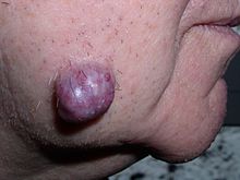
Neoplastic tumor of the cheek skin, here a benign neoplasm of the sweat glands called hidradenoma, which is not solid but is fluid-filled
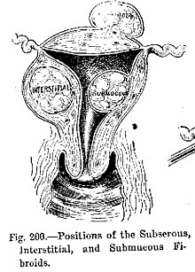
Diagram illustrating benign neoplasms, namely fibroids of the uterus

Tumors in humans occur as a result of accumulated genetic and epigenetic alterations within single cells, which cause the cell to divide and expand uncontrollably. A neoplasm can be caused by an abnormal proliferation of tissues, which can be caused by genetic mutations. Not all types of neoplasms cause a tumorous overgrowth of tissue (such as leukemia or carcinoma in situ), however similarities between neoplasmic growths and regenerative processes, e.g., dedifferentiation and rapid cell proliferation, have been pointed out.

Tumor growth has been studied using mathematics and continuum mechanics. Vascular tumors such as hemangiomas and lymphangiomas (formed from blood or lymph vessels) are thus looked at as being amalgams of a solid skeleton formed by sticky cells and an organic liquid filling the spaces in which cells can grow. Under this type of model, mechanical stresses and strains can be dealt with and their influence on the growth of the tumor and the surrounding tissue and vasculature elucidated. Recent findings from experiments that use this model show that active growth of the tumor is restricted to the outer edges of the tumor and that stiffening of the underlying normal tissue inhibits tumor growth as well.

Benign conditions that are not associated with an abnormal proliferation of tissue (such as sebaceous cysts) can also present as tumors, however, but have no malignant potential. Breast cysts (as occur commonly during pregnancy and at other times) are another example, as are other encapsulated glandular swellings (thyroid, adrenal gland, pancreas).

Encapsulated hematomas, encapsulated necrotic tissue (from an insect bite, foreign body, or other noxious mechanism), keloids (discrete overgrowths of scar tissue) and granulomas may also present as tumors.

Discrete localized enlargements of normal structures (ureters, blood vessels, intrahepatic or extrahepatic biliary ducts, pulmonary inclusions, or gastrointestinal duplications) due to outflow obstructions or narrowings, or abnormal connections, may also present as a tumor. Examples are arteriovenous fistulae or aneurysms (with or without thrombosis), biliary fistulae or aneurysms, sclerosing cholangitis, cysticercosis or hydatid cysts, intestinal duplications, and pulmonary inclusions as seen with cystic fibrosis. It can be dangerous to biopsy a number of types of tumor in which the leakage of their contents would potentially be catastrophic. When such types of tumors are encountered, diagnostic modalities such as ultrasound, CT scans, MRI, angiograms, and nuclear medicine scans are employed prior to (or during) biopsy or surgical exploration/excision in an attempt to avoid such severe complications.

Malignant neoplasms

DNA damage

The central role of DNA damage and epigenetic defects in DNA repair genes in malignant neoplasms

DNA damage is considered to be the primary underlying cause of malignant neoplasms known as cancers. Its central role in progression to cancer is illustrated in the figure in this section, in the box near the top. (The central features of DNA damage, epigenetic alterations and deficient DNA repair in progression to cancer are shown in red.) DNA damage is very common. Naturally occurring DNA damages (mostly due to cellular metabolism and the properties of DNA in water at body temperatures) occur at a rate of more than 10,000 new damages, on average, per human cell, per day. Additional DNA damages can arise from exposure to exogenous agents. Tobacco smoke causes increased exogenous DNA damage, and these DNA damages are the likely cause of lung cancer due to smoking. UV light from solar radiation causes DNA damage that is important in melanoma. Helicobacter pylori infection produces high levels of reactive oxygen species that damage DNA and contributes to gastric cancer. Bile acids, at high levels in the colons of humans eating a high fat diet, also cause DNA damage and contribute to colon cancer. Katsurano et al. indicated that macrophages and neutrophils in an inflamed colonic epithelium are the source of reactive oxygen species causing the DNA damages that initiate colonic tumorigenesis (creation of tumors in the colon). Some sources of DNA damage are indicated in the boxes at the top of the figure in this section.

Individuals with a germline mutation causing deficiency in any of 34 DNA repair genes (see article DNA repair-deficiency disorder) are at increased risk of cancer. Some germline mutations in DNA repair genes cause up to 100% lifetime chance of cancer (e.g., p53 mutations). These germline mutations are indicated in a box at the left of the figure with an arrow indicating their contribution to DNA repair deficiency.

About 70% of malignant (cancerous) neoplasms have no hereditary component and are called "sporadic cancers". Only a minority of sporadic cancers have a deficiency in DNA repair due to mutation in a DNA repair gene. However, a majority of sporadic cancers have deficiency in DNA repair due to epigenetic alterations that reduce or silence DNA repair gene expression. For example, of 113 sequential colorectal cancers, only four had a missense mutation in the DNA repair gene MGMT, while the majority had reduced MGMT expression due to methylation of the MGMT promoter region (an epigenetic alteration). Five reports present evidence that between 40% and 90% of colorectal cancers have reduced MGMT expression due to methylation of the MGMT promoter region.

Similarly, out of 119 cases of mismatch repair-deficient colorectal cancers that lacked DNA repair gene PMS2 expression, PMS2 was deficient in 6 due to mutations in the PMS2 gene, while in 103 cases PMS2 expression was deficient because its pairing partner MLH1 was repressed due to promoter methylation (PMS2 protein is unstable in the absence of MLH1). In the other 10 cases, loss of PMS2 expression was likely due to epigenetic overexpression of the microRNA, miR-155, which down-regulates MLH1.

In further examples, epigenetic defects were found at frequencies of between 13%-100% for the DNA repair genes BRCA1, WRN, FANCB, FANCF, MGMT, MLH1, MSH2, MSH4, ERCC1, XPF, NEIL1 and ATM. These epigenetic defects occurred in various cancers, including breast, ovarian, colorectal, and head and neck cancers. Two or three deficiencies in expression of ERCC1, XPF or PMS2 occur simultaneously in the majority of the 49 colon cancers evaluated by Facista et al. Epigenetic alterations causing reduced expression of DNA repair genes is shown in a central box at the third level from the top of the figure in this section, and the consequent DNA repair deficiency is shown at the fourth level.

When expression of DNA repair genes is reduced, DNA damages accumulate in cells at a higher than normal level, and these excess damages cause increased frequencies of mutation or epimutation. Mutation rates strongly increase in cells defective in DNA mismatch repair or in homologous recombinational repair (HRR).

During repair of DNA double strand breaks, or repair of other DNA damages, incompletely cleared sites of repair can cause epigenetic gene silencing. DNA repair deficiencies (level 4 in the figure) cause increased DNA damages (level 5 in the figure) which result in increased somatic mutations and epigenetic alterations (level 6 in the figure).

Field defects, normal-appearing tissue with multiple alterations (and discussed in the section below), are common precursors to development of the disordered and improperly proliferating clone of tissue in a malignant neoplasm. Such field defects (second level from bottom of figure) may have multiple mutations and epigenetic alterations.

Once a cancer is formed, it usually has genome instability. This instability is likely due to reduced DNA repair or excessive DNA damage. Because of such instability, the cancer continues to evolve and to produce sub clones. For example, a renal cancer, sampled in 9 areas, had 40 ubiquitous mutations, demonstrating tumor heterogeneity (i.e. present in all areas of the cancer), 59 mutations shared by some (but not all areas), and 29 "private" mutations only present in one of the areas of the cancer.

Field defects

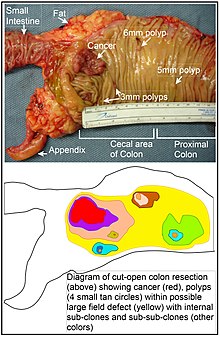
Longitudinally opened freshly resected colon segment showing a cancer and four polyps, plus a schematic diagram indicating a likely field defect (a region of tissue that precedes and predisposes to the development of cancer) in this colon segment. The diagram indicates sub-clones and sub-sub-clones that were precursors to the tumors.

Various other terms have been used to describe this phenomenon, including "field effect", "field cancerization", and "field carcinogenesis". The term "field cancerization" was first used in 1953 to describe an area or "field" of epithelium that has been preconditioned by (at that time) largely unknown processes so as to predispose it towards development of cancer. Since then, the terms "field cancerization" and "field defect" have been used to describe pre-malignant tissue in which new cancers are likely to arise.

Field defects are important in progression to cancer. However, in most cancer research, as pointed out by Rubin "The vast majority of studies in cancer research has been done on well-defined tumors in vivo, or on discrete neoplastic foci in vitro. Yet there is evidence that more than 80% of the somatic mutations found in mutator phenotype human colorectal tumors occur before the onset of terminal clonal expansion. Similarly, Vogelstein et al. point out that more than half of somatic mutations identified in tumors occurred in a pre-neoplastic phase (in a field defect), during growth of apparently normal cells. Likewise, epigenetic alterations present in tumors may have occurred in pre-neoplastic field defects.

An expanded view of field effect has been termed "etiologic field effect", which encompasses not only molecular and pathologic changes in pre-neoplastic cells but also influences of exogenous environmental factors and molecular changes in the local microenvironment on neoplastic evolution from tumor initiation to patient death.

In the colon, a field defect probably arises by natural selection of a mutant or epigenetically altered cell among the stem cells at the base of one of the intestinal crypts on the inside surface of the colon. A mutant or epigenetically altered stem cell may replace the other nearby stem cells by natural selection. Thus, a patch of abnormal tissue may arise. The figure in this section includes a photo of a freshly resected and lengthwise-opened segment of the colon showing a colon cancer and four polyps. Below the photo, there is a schematic diagram of how a large patch of mutant or epigenetically altered cells may have formed, shown by the large area in yellow in the diagram. Within this first large patch in the diagram (a large clone of cells), a second such mutation or epigenetic alteration may occur so that a given stem cell acquires an advantage compared to other stem cells within the patch, and this altered stem cell may expand clonally forming a secondary patch, or sub-clone, within the original patch. This is indicated in the diagram by four smaller patches of different colors within the large yellow original area. Within these new patches (sub-clones), the process may be repeated multiple times, indicated by the still smaller patches within the four secondary patches (with still different colors in the diagram) which clonally expand, until stem cells arise that generate either small polyps or else a malignant neoplasm (cancer).

In the photo, an apparent field defect in this segment of a colon has generated four polyps (labeled with the size of the polyps, 6mm, 5mm, and two of 3mm, and a cancer about 3 cm across in its longest dimension). These neoplasms are also indicated, in the diagram below the photo, by 4 small tan circles (polyps) and a larger red area (cancer). The cancer in the photo occurred in the cecal area of the colon, where the colon joins the small intestine (labeled) and where the appendix occurs (labeled). The fat in the photo is external to the outer wall of the colon. In the segment of colon shown here, the colon was cut open lengthwise to expose the inner surface of the colon and to display the cancer and polyps occurring within the inner epithelial lining of the colon.

If the general process by which sporadic colon cancers arise is the formation of a pre-neoplastic clone that spreads by natural selection, followed by formation of internal sub-clones within the initial clone, and sub-sub-clones inside those, then colon cancers generally should be associated with, and be preceded by, fields of increasing abnormality reflecting the succession of premalignant events. The most extensive region of abnormality (the outermost yellow irregular area in the diagram) would reflect the earliest event in formation of a malignant neoplasm.

In experimental evaluation of specific DNA repair deficiencies in cancers, many specific DNA repair deficiencies were also shown to occur in the field defects surrounding those cancers. The Table, below, gives examples for which the DNA repair deficiency in a cancer was shown to be caused by an epigenetic alteration, and the somewhat lower frequencies with which the same epigenetically caused DNA repair deficiency was found in the surrounding field defect.

Some of the small polyps in the field defect shown in the photo of the opened colon segment may be relatively benign neoplasms. Of polyps less than 10mm in size, found during colonoscopy and followed with repeat colonoscopies for 3 years, 25% were unchanged in size, 35% regressed or shrank in size while 40% grew in size.

Genome instability

Cancers are known to exhibit genome instability or a mutator phenotype. The protein-coding DNA within the nucleus is about 1.5% of the total genomic DNA. Within this protein-coding DNA (called the exome), an average cancer of the breast or colon can have about 60 to 70 protein altering mutations, of which about 3 or 4 may be "driver" mutations, and the remaining ones may be "passenger" mutations. However, the average number of DNA sequence mutations in the entire genome (including non-protein-coding regions) within a breast cancer tissue sample is about 20,000. In an average melanoma tissue sample (where melanomas have a higher exome mutation frequency) the total number of DNA sequence mutations is about 80,000. This compares to the very low mutation frequency of about 70 new mutations in the entire genome between generations (parent to child) in humans.

The high frequencies of mutations in the total nucleotide sequences within cancers suggest that often an early alteration in the field defects giving rise to a cancer (e.g. yellow area in the diagram in this section) is a deficiency in DNA repair. The large field defects surrounding colon cancers (extending to at about 10 cm on each side of a cancer) were shown by Facista et al. to frequently have epigenetic defects in 2 or 3 DNA repair proteins (ERCC1, XPF or PMS2) in the entire area of the field defect. Deficiencies in DNA repair cause increased mutation rates. A deficiency in DNA repair, itself, can allow DNA damages to accumulate, and error-prone translesion synthesis past some of those damages may give rise to mutations. In addition, faulty repair of these accumulated DNA damages may give rise to epimutations. These new mutations or epimutations may provide a proliferative advantage, generating a field defect. Although the mutations/epimutations in DNA repair genes do not, themselves, confer a selective advantage, they may be carried along as passengers in cells when the cells acquire additional mutations/epimutations that do provide a proliferative advantage.

Etymology

The term neoplasm is a synonym of tumor. Neoplasia denotes the process of the formation of neoplasms/tumors, and the process is referred to as a neoplastic process. The word neoplastic itself comes from Greek neo 'new' and plastic 'formed, molded'.

The term tumor derives from the Latin noun tumor 'a swelling', ultimately from the verb tumēre 'to swell'. In the British Commonwealth, the spelling tumour is commonly used, whereas in the U.S. the word is usually spelled tumor.

In its medical sense, tumor has traditionally meant an abnormal swelling of the flesh. The Roman medical encyclopedist Celsus (c. 30 BC–38 AD) described the four cardinal signs of acute inflammation as tumor, dolor, calor, and rubor (swelling, pain, increased heat, and redness). (His treatise, De Medicina, was the first medical book printed in 1478 following the invention of the movable-type printing press.)

In contemporary English, the word tumor is often used as a synonym for a cystic (liquid-filled) growth or solid neoplasm (cancerous or non-cancerous), with other forms of swelling often referred to as "swellings".

Related terms occur commonly in the medical literature, where the nouns tumefaction and tumescence (derived from the adjective tumescent) are current medical terms for non-neoplastic swelling. This type of swelling is most often caused by inflammation caused by trauma, infection, and other factors.

Tumors may be caused by conditions other than an overgrowth of neoplastic cells, however. Cysts (such as sebaceous cysts) are also referred to as tumors, even though they have no neoplastic cells. This is standard in medical-billing terminology (especially when billing for a growth whose pathology has yet to be determined).

Ocean temperature

From Wikipedia, the free encyclopedia https://en.wikipedia.org/wiki/Ocean_temperature Graph showing ocean tempe...