Search This Blog

Saturday, September 9, 2023

Ferrite (magnet)

From Wikipedia, the free encyclopedia
A stack of ferrite magnets

A ferrite is a ceramic material made by mixing and firing iron(III) oxide (Fe2O3, rust) with one or more additional metallic elements, such as strontium, barium, manganese, nickel, and zinc. They are ferrimagnetic, meaning they are attracted by magnetic fields and can be magnetized to become permanent magnets. Unlike other ferromagnetic materials, most ferrites are not electrically conductive, making them useful in applications like magnetic cores for transformers to suppress eddy currents. Ferrites can be divided into two families based on their resistance to being demagnetized (magnetic coercivity).

Hard ferrites have high coercivity, so are difficult to demagnetize. They are used to make permanent magnets for applications such as refrigerator magnets, loudspeakers, and small electric motors.

Soft ferrites have low coercivity, so they easily change their magnetization and act as conductors of magnetic fields. They are used in the electronics industry to make efficient magnetic cores called ferrite cores for high-frequency inductors, transformers and antennas, and in various microwave components.

Ferrite compounds are extremely low cost, being made of mostly iron oxide, and have excellent corrosion resistance. Yogoro Kato and Takeshi Takei of the Tokyo Institute of Technology synthesized the first ferrite compounds in 1930.

Composition, structure, and properties

Ferrites are usually ferrimagnetic ceramic compounds derived from iron oxides. Magnetite (Fe3O4) is a famous example. Like most of the other ceramics, ferrites are hard, brittle, and poor conductors of electricity.

Many ferrites adopt the spinel structure with the formula AB2O4, where A and B represent various metal cations, usually including iron (Fe). Spinel ferrites usually adopt a crystal motif consisting of cubic close-packed (fcc) oxides (O2−) with A cations occupying one eighth of the tetrahedral holes and B cations occupying half of the octahedral holes, i.e., A2+
B3+
2
O2−
4
.

Ferrite crystals do not adopt the ordinary spinel structure, but rather the inverse spinel structure: One eighth of the tetrahedral holes are occupied by B cations, one fourth of the octahedral sites are occupied by A cations. and the other one fourth by B cation. It is also possible to have mixed structure spinel ferrites with formula [M2+1−δFe3+δ][M2+δFe3+2−δ]O4 where δ is the degree of inversion.

The magnetic material known as "ZnFe" has the formula ZnFe2O4, with Fe3+ occupying the octahedral sites and Zn2+ occupy the tetrahedral sites, it is an example of normal structure spinel ferrite.

Some ferrites adopt hexagonal crystal structure, like barium and strontium ferrites BaFe12O19 (BaO:6Fe2O3) and SrFe12O19 (SrO:6Fe2O3).

In terms of their magnetic properties, the different ferrites are often classified as "soft", "semi-hard" or "hard", which refers to their low or high magnetic coercivity, as follows.

Soft ferrites

Various ferrite cores used to make small transformers and inductors

Ferrites that are used in transformer or electromagnetic cores contain nickel, zinc, and/or manganese compounds. Soft ferrites are not permanent magnets. They have magnetism (much like mild steel), but when the magnetic field is removed, the magnetism decreases. Soft ferrites are commonly used as transformers (to change the voltage from primary to secondary windings). As a result, soft ferrites are also called transformer ferrites.They have a low coercivity. The low coercivity means the material's magnetization can easily reverse direction without dissipating much energy (hysteresis losses), while the material's high resistivity prevents eddy currents in the core, another source of energy loss. Because of their comparatively low losses at high frequencies, they are extensively used in the cores of RF transformers and inductors in applications such as switched-mode power supplies and loopstick antennas used in AM radios.

The most common soft ferrites are:

  • Manganese-zinc ferrite (MnZn, with the formula MnaZn(1-a)Fe2O4). MnZn have higher permeability and saturation induction than NiZn.
  • Nickel-zinc ferrite (NiZn, with the formula NiaZn(1-a)Fe2O4). NiZn ferrites exhibit higher resistivity than MnZn, and are therefore more suitable for frequencies above 1 MHz.

For applications below 5 MHz, MnZn ferrites are used; above that, NiZn is the usual choice. The exception is with common mode inductors, where the threshold of choice is at 70 MHz.

Semi-hard ferrites

  • Cobalt ferrite, CoFe2O4 (CoO·Fe2O3), is in between soft and hard magnetic material and is usually classified as a semi-hard material. It is mainly used for its magnetostrictive applications like sensors and actuators  thanks to its high saturation magnetostriction (~200 ppm). CoFe2O4 has also the benefits to be rare-earth free, which makes it a good substitute for Terfenol-D. Moreover, its magnetostrictive properties can be tuned by inducing a magnetic uniaxial anisotropy. This can be done by magnetic annealing, magnetic field assisted compaction, or reaction under uniaxial pressure. This last solution has the advantage to be ultra fast (20 min) thanks to the use of spark plasma sintering. The induced magnetic anisotropy in cobalt ferrite is also beneficial to enhance the magnetoelectric effect in composite.

Hard ferrites

In contrast, permanent ferrite magnets are made of hard ferrites, which have a high coercivity and high remanence after magnetization. Iron oxide and barium or strontium carbonate are used in manufacturing of hard ferrite magnets. The high coercivity means the materials are very resistant to becoming demagnetized, an essential characteristic for a permanent magnet. They also have high magnetic permeability. These so-called ceramic magnets are cheap, and are widely used in household products such as refrigerator magnets. The maximum magnetic field B is about 0.35 tesla and the magnetic field strength H is about 30 to 160 kiloampere turns per meter (400 to 2000 oersteds). The density of ferrite magnets is about 5 g/cm3.

The most common hard ferrites are:

  • Strontium ferrite, SrFe12O19 (SrO·6Fe2O3), used in small electric motors, micro-wave devices, recording media, magneto-optic media, telecommunication and electronic industry. Strontium hexaferrite (SrFe12O19) is well known for its high coercivity due to its magnetocrystalline anisotropy. It has been widely used in industrial applications as permanent magnets and, because they can be powdered and formed easily, they are finding their applications into micro and nano-types systems such as biomarkers, bio diagnostics and biosensors.
  • Barium ferrite, BaFe12O19 (BaO·6Fe2O3), a common material for permanent magnet applications. Barium ferrites are robust ceramics that are generally stable to moisture and corrosion-resistant. They are used in e.g. loudspeaker magnets and as a medium for magnetic recording, e.g. on magnetic stripe cards.

Production

Ferrites are produced by heating a mixture of the oxides of the constituent metals at high temperatures, as shown in this idealized equation:

Fe2O3 + ZnO → ZnFe2O4

In some cases, the mixture of finely-powdered precursors is pressed into a mold. For barium and strontium ferrites, these metals are typically supplied as their carbonates, BaCO3 or SrCO3. During the heating process, these carbonates undergo calcination:

MCO3 → MO + CO2

After this step, the two oxides combine to give the ferrite. The resulting mixture of oxides undergoes sintering.

Processing

Having obtained the ferrite, the cooled product is milled to particles smaller than 2 µm, sufficiently small that each particle consists of a single magnetic domain. Next the powder is pressed into a shape, dried, and re-sintered. The shaping may be performed in an external magnetic field, in order to achieve a preferred orientation of the particles (anisotropy).

Small and geometrically easy shapes may be produced with dry pressing. However, in such a process small particles may agglomerate and lead to poorer magnetic properties compared to the wet pressing process. Direct calcination and sintering without re-milling is possible as well but leads to poor magnetic properties.

Electromagnets are pre-sintered as well (pre-reaction), milled and pressed. However, the sintering takes place in a specific atmosphere, for instance one with an oxygen shortage. The chemical composition and especially the structure vary strongly between the precursor and the sintered product.

To allow efficient stacking of product in the furnace during sintering and prevent parts sticking together, many manufacturers separate ware using ceramic powder separator sheets. These sheets are available in various materials such as alumina, zirconia and magnesia. They are also available in fine, medium and coarse particle sizes. By matching the material and particle size to the ware being sintered, surface damage and contamination can be reduced while maximizing furnace loading.

Uses

Ferrite cores are used in electronic inductors, transformers, and electromagnets where the high electrical resistance of the ferrite leads to very low eddy current losses.

Ferrites are also found as a lump in a computer cable, called a ferrite bead, which helps to prevent high frequency electrical noise (radio frequency interference) from exiting or entering the equipment; these types of ferrites are made with lossy materials to not just block (reflect), but also absorb and dissipate as heat, the unwanted higher-frequency energy.

Early computer memories stored data in the residual magnetic fields of hard ferrite cores, which were assembled into arrays of core memory. Ferrite powders are used in the coatings of magnetic recording tapes.

Ferrite particles are also used as a component of radar-absorbing materials or coatings used in stealth aircraft and in the absorption tiles lining the rooms used for electromagnetic compatibility measurements. Most common audio magnets, including those used in loudspeakers and electromagnetic instrument pickups, are ferrite magnets. Except for certain "vintage" products, ferrite magnets have largely displaced the more expensive Alnico magnets in these applications. In particular, for hard hexaferrites today the most common uses are still as permanent magnets in refrigerator seal gaskets, microphones and loud speakers, small motors for cordless appliances and in automobile applications.

Ferrite nanoparticles exhibit superparamagnetic properties.

History

Yogoro Kato and Takeshi Takei of the Tokyo Institute of Technology synthesized the first ferrite compounds in 1930. This led to the founding of TDK Corporation in 1935, to manufacture the material.

Barium hexaferrite (BaO•6Fe2O3) was discovered in 1950 at the Philips Natuurkundig Laboratorium (Philips Physics Laboratory). The discovery was somewhat accidental—due to a mistake by an assistant who was supposed to be preparing a sample of hexagonal lanthanum ferrite for a team investigating its use as a semiconductor material. On discovering that it was actually a magnetic material, and confirming its structure by X-ray crystallography, they passed it on to the magnetic research group. Barium hexaferrite has both high coercivity (170 kA/m) and low raw material costs. It was developed as a product by Philips Industries (Netherlands) and from 1952 was marketed under the trade name Ferroxdure. The low price and good performance led to a rapid increase in the use of permanent magnets.

In the 1960s Philips developed strontium hexaferrite (SrO•6Fe2O3), with better properties than barium hexaferrite. Barium and strontium hexaferrite dominate the market due to their low costs. Other materials have been found with improved properties. BaO•2(FeO)•8(Fe2O3) came in 1980, and Ba2ZnFe18O23 came in 1991.

Ataxia

From Wikipedia, the free encyclopedia
Ataxia
SpecialtyNeurology, Psychiatry
Symptoms
  • Lack of coordination
  • Slurred speech
  • Trouble eating and swallowing
  • Deterioration of fine motor skills
  • Difficulty walking
  • Gait abnormalities
  • Eye movement abnormalities
  • Tremors
  • Heart problems

Ataxia (from Greek α- [a negative prefix] + -τάξις [order] = "lack of order") is a neurological sign consisting of lack of voluntary coordination of muscle movements that can include gait abnormality, speech changes, and abnormalities in eye movements, that indicates dysfunction of parts of the nervous system that coordinate movement, such as the cerebellum.

These nervous system dysfunctions occur in several different patterns, with different results and different possible causes. Ataxia can be limited to one side of the body, which is referred to as hemiataxia. Friedreich's ataxia has gait abnormality as the most commonly presented symptom. Dystaxia is a mild degree of ataxia.

Types

Cerebellar

The term cerebellar ataxia is used to indicate ataxia due to dysfunction of the cerebellum. The cerebellum is responsible for integrating a significant amount of neural information that is used to coordinate smoothly ongoing movements and to participate in motor planning. Although ataxia is not present with all cerebellar lesions, many conditions affecting the cerebellum do produce ataxia. People with cerebellar ataxia may have trouble regulating the force, range, direction, velocity, and rhythm of muscle contractions. This results in a characteristic type of irregular, uncoordinated movement that can manifest itself in many possible ways, such as asthenia, asynergy, delayed reaction time, and dyschronometria. Individuals with cerebellar ataxia could also display instability of gait, difficulty with eye movements, dysarthria, dysphagia, hypotonia, dysmetria, and dysdiadochokinesia. These deficits can vary depending on which cerebellar structures have been damaged, and whether the lesion is bi- or unilateral.

People with cerebellar ataxia may initially present with poor balance, which could be demonstrated as an inability to stand on one leg or perform tandem gait. As the condition progresses, walking is characterized by a widened base and high stepping, as well as staggering and lurching from side to side. Turning is also problematic and could result in falls. As cerebellar ataxia becomes severe, great assistance and effort are needed to stand and walk. Dysarthria, an impairment with articulation, may also be present and is characterized by "scanning" speech that consists of slower rate, irregular rhythm, and variable volume. Also, slurring of speech, tremor of the voice, and ataxic respiration may occur. Cerebellar ataxia could result with incoordination of movement, particularly in the extremities. Overshooting (or hypermetria) occurs with finger-to-nose testing and heel to shin testing; thus, dysmetria is evident. Impairments with alternating movements (dysdiadochokinesia), as well as dysrhythmia, may also be displayed. Tremor of the head and trunk (titubation) may be seen in individuals with cerebellar ataxia.

Dysmetria is thought to be caused by a deficit in the control of interaction torques in multijoint motion. Interaction torques are created at an associated joint when the primary joint is moved. For example, if a movement required reaching to touch a target in front of the body, flexion at the shoulder would create a torque at the elbow, while extension of the elbow would create a torque at the wrist. These torques increase as the speed of movement increases and must be compensated and adjusted for to create coordinated movement. This may, therefore, explain decreased coordination at higher movement velocities and accelerations.

  • Dysfunction of the vestibulocerebellum (flocculonodular lobe) impairs balance and the control of eye movements. This presents itself with postural instability, in which the person tends to separate his/her feet upon standing, to gain a wider base and to avoid titubation (bodily oscillations tending to be forward-backward ones). The instability is, therefore, worsened when standing with the feet together, regardless of whether the eyes are open or closed. This is a negative Romberg's test, or more accurately, it denotes the individual's inability to carry out the test, because the individual feels unstable even with open eyes.
  • Dysfunction of the spinocerebellum (vermis and associated areas near the midline) presents itself with a wide-based "drunken sailor" gait (called truncal ataxia), characterised by uncertain starts and stops, lateral deviations, and unequal steps. As a result of this gait impairment, falling is a concern in patients with ataxia. Studies examining falls in this population show that 74–93% of patients have fallen at least once in the past year and up to 60% admit to fear of falling.
  • 'Dysfunction of the cerebrocerebellum' (lateral hemispheres) presents as disturbances in carrying out voluntary, planned movements by the extremities (called appendicular ataxia). These include:
    • Intention tremor (coarse trembling, accentuated over the execution of voluntary movements, possibly involving the head and eyes, as well as the limbs and torso)
    • Peculiar writing abnormalities (large, unequal letters, irregular underlining)
    • A peculiar pattern of dysarthria (slurred speech, sometimes characterised by explosive variations in voice intensity despite a regular rhythm)
    • Inability to perform rapidly alternating movements, known as dysdiadochokinesia, occurs, and could involve rapidly switching from pronation to supination of the forearm. Movements become more irregular with increases of speed.
    • Inability to judge distances or ranges of movement happens. This dysmetria is often seen as undershooting, hypometria, or overshooting, hypermetria, the required distance or range to reach a target. This is sometimes seen when a patient is asked to reach out and touch someone's finger or touch his or her own nose.
    • The rebound phenomenon, also known as the loss of the check reflex, is also sometimes seen in patients with cerebellar ataxia, for example, when patients are flexing their elbows isometrically against a resistance. When the resistance is suddenly removed without warning, the patients' arms may swing up and even strike themselves. With an intact check reflex, the patients check and activate the opposing triceps to slow and stop the movement.
    • Patients may exhibit a constellation of subtle to overt cognitive symptoms, which are gathered under the terminology of Schmahmann's syndrome.

Sensory

The term sensory ataxia is used to indicate ataxia due to loss of proprioception, the loss of sensitivity to the positions of joint and body parts. This is generally caused by dysfunction of the dorsal columns of the spinal cord, because they carry proprioceptive information up to the brain. In some cases, the cause of sensory ataxia may instead be dysfunction of the various parts of the brain that receive positional information, including the cerebellum, thalamus, and parietal lobes.

Sensory ataxia presents itself with an unsteady "stomping" gait with heavy heel strikes, as well as a postural instability that is usually worsened when the lack of proprioceptive input cannot be compensated for by visual input, such as in poorly lit environments.

Physicians can find evidence of sensory ataxia during physical examination by having patients stand with their feet together and eyes shut. In affected patients, this will cause the instability to worsen markedly, producing wide oscillations and possibly a fall; this is called a positive Romberg's test. Worsening of the finger-pointing test with the eyes closed is another feature of sensory ataxia. Also, when patients are standing with arms and hands extended toward the physician, if the eyes are closed, the patients' fingers tend to "fall down" and then be restored to the horizontal extended position by sudden muscular contractions (the "ataxic hand").

Vestibular

The term vestibular ataxia is used to indicate ataxia due to dysfunction of the vestibular system, which in acute and unilateral cases is associated with prominent vertigo, nausea, and vomiting. In slow-onset, chronic bilateral cases of vestibular dysfunction, these characteristic manifestations may be absent, and dysequilibrium may be the sole presentation.

Causes

The three types of ataxia have overlapping causes, so can either coexist or occur in isolation. Cerebellar ataxia can have many causes despite normal neuroimaging.

Focal lesions

Any type of focal lesion of the central nervous system (such as stroke, brain tumor, multiple sclerosis, inflammatory [such as sarcoidosis], and "chronic lymphocytyc inflammation with pontine perivascular enhancement responsive to steroids syndrome" [CLIPPERS]) will cause the type of ataxia corresponding to the site of the lesion: cerebellar if in the cerebellum; sensory if in the dorsal spinal cord...to include cord compression by thickened ligamentum flavum or stenosis of the boney spinal canal...(and rarely in the thalamus or parietal lobe); or vestibular if in the vestibular system (including the vestibular areas of the cerebral cortex).

Exogenous substances (metabolic ataxia)

Exogenous substances that cause ataxia mainly do so because they have a depressant effect on central nervous system function. The most common example is ethanol (alcohol), which is capable of causing reversible cerebellar and vestibular ataxia. Chronic intake of ethanol causes atrophy of the cerebellum by oxidative and endoplasmic reticulum stresses induced by thiamine deficiency.

Other examples include various prescription drugs (e.g. most antiepileptic drugs have cerebellar ataxia as a possible adverse effect), Lithium level over 1.5mEq/L, synthetic cannabinoid HU-211 ingestion and various other medical and recreational drugs (e.g. ketamine, PCP or dextromethorphan, all of which are NMDA receptor antagonists that produce a dissociative state at high doses). A further class of pharmaceuticals which can cause short term ataxia, especially in high doses, are benzodiazepines. Exposure to high levels of methylmercury, through consumption of fish with high mercury concentrations, is also a known cause of ataxia and other neurological disorders.

Radiation poisoning

Ataxia can be induced as a result of severe acute radiation poisoning with an absorbed dose of more than 30 grays.

Vitamin B12 deficiency

Vitamin B12 deficiency may cause, among several neurological abnormalities, overlapping cerebellar and sensory ataxia. Neuropsychological symptoms may include sense loss, difficulty in proprioception, poor balance, loss of sensation in the feet, changes in reflexes, dementia, and psychosis, can be reversible with treatment. Complications may include a neurological complex known as subacute combined degeneration of spinal cord, and other neurological disorders.

Hypothyroidism

Symptoms of neurological dysfunction may be the presenting feature in some patients with hypothyroidism. These include reversible cerebellar ataxia, dementia, peripheral neuropathy, psychosis and coma. Most of the neurological complications improve completely after thyroid hormone replacement therapy.

Causes of isolated sensory ataxia

Peripheral neuropathies may cause generalised or localised sensory ataxia (e.g. a limb only) depending on the extent of the neuropathic involvement. Spinal disorders of various types may cause sensory ataxia from the lesioned level below, when they involve the dorsal columns.

Non-hereditary cerebellar degeneration

Non-hereditary causes of cerebellar degeneration include chronic alcohol use disorder, head injury, paraneoplastic and non-paraneoplastic autoimmune ataxia, high altitude cerebral oedema, coeliac disease, normal pressure hydrocephalus and infectious or post-infectious cerebellitis.

Hereditary ataxias

Ataxia may depend on hereditary disorders consisting of degeneration of the cerebellum or of the spine; most cases feature both to some extent, and therefore present with overlapping cerebellar and sensory ataxia, even though one is often more evident than the other. Hereditary disorders causing ataxia include autosomal dominant ones such as spinocerebellar ataxia, episodic ataxia, and dentatorubropallidoluysian atrophy, as well as autosomal recessive disorders such as Friedreich's ataxia (sensory and cerebellar, with the former predominating) and Niemann Pick disease, ataxia-telangiectasia (sensory and cerebellar, with the latter predominating),autosomal recessive spinocerebellar ataxia-14 and abetalipoproteinaemia. An example of X-linked ataxic condition is the rare fragile X-associated tremor/ataxia syndrome or FXTAS.

Arnold–Chiari malformation (congenital ataxia)

Arnold–Chiari malformation is a malformation of the brain. It consists of a downward displacement of the cerebellar tonsils and the medulla through the foramen magnum, sometimes causing hydrocephalus as a result of obstruction of cerebrospinal fluid outflow.

Succinic semialdehyde dehydrogenase deficiency

Succinic semialdehyde dehydrogenase deficiency is an autosomal-recessive gene disorder where mutations in the ALDH5A1 gene results in the accumulation of gamma-Hydroxybutyric acid (GHB) in the body. GHB accumulates in the nervous system and can cause ataxia as well as other neurological dysfunction.

Wilson's disease

Wilson's disease is an autosomal-recessive gene disorder whereby an alteration of the ATP7B gene results in an inability to properly excrete copper from the body. Copper accumulates in the nervous system and liver and can cause ataxia as well as other neurological and organ impairments.

Gluten ataxia

Gluten ataxia is an autoimmune disease triggered by the ingestion of gluten. Early diagnosis and treatment with a gluten-free diet can improve ataxia and prevent its progression. The effectiveness of the treatment depends on the elapsed time from the onset of the ataxia until diagnosis, because the death of neurons in the cerebellum as a result of gluten exposure is irreversible. It accounts for 40% of ataxias of unknown origin and 15% of all ataxias. Less than 10% of people with gluten ataxia present any gastrointestinal symptom and only about 40% have intestinal damage. This entity is classified into primary auto-immune cerebellar ataxias (PACA).

Potassium pump

Malfunction of the sodium-potassium pump may be a factor in some ataxias. The Na+
-K+
pump has been shown to control and set the intrinsic activity mode of cerebellar Purkinje neurons. This suggests that the pump might not simply be a homeostatic, "housekeeping" molecule for ionic gradients; but could be a computational element in the cerebellum and the brain. Indeed, an ouabain block of Na+
-K+
pumps in the cerebellum of a live mouse results in it displaying ataxia and dystonia. Ataxia is observed for lower ouabain concentrations, dystonia is observed at higher ouabain concentrations.

Cerebellar ataxia associated with anti-GAD antibodies

Antibodies against the enzyme glutamic acid decarboxylase (GAD: enzyme changing glutamate into GABA) cause cerebellar deficits. The antibodies impair motor learning and cause behavioral deficits. GAD antibodies related ataxia is part of the group called immune-mediated cerebellar ataxias. The antibodies induce a synaptopathy. The cerebellum is particularly vulnerable to autoimmune disorders. Cerebellar circuitry has capacities to compensate and restore function thanks to cerebellar reserve, gathering multiple forms of plasticity. LTDpathies gather immune disorders targeting long-term depression (LTD), a form of plasticity.

Diagnosis

  • Imaging studies - A CT scan or MRI of the brain might help determine potential causes. An MRI can sometimes show shrinkage of the cerebellum and other brain structures in people with ataxia. It may also show other treatable findings, such as a blood clot or benign tumour, that could be pressing on the cerebellum.
  • Lumbar puncture (spinal tap) - A needle is inserted into the lower back (lumbar region) between two lumbar vertebrae to obtain a sample of cerebrospinal fluid for testing.
  • Genetic testing - Determines whether the mutation that causes one of the hereditary ataxic conditions is present. Tests are available for many but not all of the hereditary ataxias.

Treatment

The treatment of ataxia and its effectiveness depend on the underlying cause. Treatment may limit or reduce the effects of ataxia, but it is unlikely to eliminate them entirely. Recovery tends to be better in individuals with a single focal injury (such as stroke or a benign tumour), compared to those who have a neurological degenerative condition. A review of the management of degenerative ataxia was published in 2009. A small number of rare conditions presenting with prominent cerebellar ataxia are amenable to specific treatment and recognition of these disorders is critical. Diseases include vitamin E deficiency, abetalipoproteinemia, cerebrotendinous xanthomatosis, Niemann–Pick type C disease, Refsum's disease, glucose transporter type 1 deficiency, episodic ataxia type 2, gluten ataxia, glutamic acid decarboxylase ataxia. Novel therapies target the RNA defects associated with cerebellar disorders, using in particular anti-sense oligonucleotides.

The movement disorders associated with ataxia can be managed by pharmacological treatments and through physical therapy and occupational therapy to reduce disability. Some drug treatments that have been used to control ataxia include: 5-hydroxytryptophan (5-HTP), idebenone, amantadine, physostigmine, L-carnitine or derivatives, trimethoprim/sulfamethoxazole, vigabatrin, phosphatidylcholine, acetazolamide, 4-aminopyridine, buspirone, and a combination of coenzyme Q10 and vitamin E.

Physical therapy requires a focus on adapting activity and facilitating motor learning for retraining specific functional motor patterns. A recent systematic review suggested that physical therapy is effective, but there is only moderate evidence to support this conclusion. The most commonly used physical therapy interventions for cerebellar ataxia are vestibular habituation, Frenkel exercises, proprioceptive neuromuscular facilitation (PNF), and balance training; however, therapy is often highly individualized and gait and coordination training are large components of therapy.

Current research suggests that, if a person is able to walk with or without a mobility aid, physical therapy should include an exercise program addressing five components: static balance, dynamic balance, trunk-limb coordination, stairs, and contracture prevention. Once the physical therapist determines that the individual is able to safely perform parts of the program independently, it is important that the individual be prescribed and regularly engage in a supplementary home exercise program that incorporates these components to further improve long term outcomes. These outcomes include balance tasks, gait, and individual activities of daily living. While the improvements are attributed primarily to changes in the brain and not just the hip or ankle joints, it is still unknown whether the improvements are due to adaptations in the cerebellum or compensation by other areas of the brain.

Decomposition, simplification, or slowing of multijoint movement may also be an effective strategy that therapists may use to improve function in patients with ataxia. Training likely needs to be intense and focused—as indicated by one study performed with stroke patients experiencing limb ataxia who underwent intensive upper limb retraining. Their therapy consisted of constraint-induced movement therapy which resulted in improvements of their arm function. Treatment should likely include strategies to manage difficulties with everyday activities such as walking. Gait aids (such as a cane or walker) can be provided to decrease the risk of falls associated with impairment of balance or poor coordination. Severe ataxia may eventually lead to the need for a wheelchair. To obtain better results, possible coexisting motor deficits need to be addressed in addition to those induced by ataxia. For example, muscle weakness and decreased endurance could lead to increasing fatigue and poorer movement patterns.

There are several assessment tools available to therapists and health care professionals working with patients with ataxia. The International Cooperative Ataxia Rating Scale (ICARS) is one of the most widely used and has been proven to have very high reliability and validity. Other tools that assess motor function, balance and coordination are also highly valuable to help the therapist track the progress of their patient, as well as to quantify the patient's functionality. These tests include, but are not limited to:

  • The Berg Balance Scale
  • Tandem Walking (to test for Tandem gaitability)
  • Scale for the Assessment and Rating of Ataxia (SARA)
  • tapping tests – The person must quickly and repeatedly tap their arm or leg while the therapist monitors the amount of dysdiadochokinesia.
  • finger-nose testing – This test has several variations including finger-to-therapist's finger, finger-to-finger, and alternate nose-to-finger.

Other uses

The term "ataxia" is sometimes used in a broader sense to indicate lack of coordination in some physiological process. Examples include optic ataxia (lack of coordination between visual inputs and hand movements, resulting in inability to reach and grab objects) and ataxic respiration (lack of coordination in respiratory movements, usually due to dysfunction of the respiratory centres in the medulla oblongata). Optic ataxia may be caused by lesions to the posterior parietal cortex, which is responsible for combining and expressing positional information and relating it to movement. Outputs of the posterior parietal cortex include the spinal cord, brain stem motor pathways, pre-motor and pre-frontal cortex, basal ganglia and the cerebellum. Some neurons in the posterior parietal cortex are modulated by intention. Optic ataxia is usually part of Balint's syndrome, but can be seen in isolation with injuries to the superior parietal lobule, as it represents a disconnection between visual-association cortex and the frontal premotor and motor cortex.

Infectious mononucleosis

From Wikipedia, the free encyclopedia
 
Infectious mononucleosis
Other namesGlandular fever, Pfeiffer's disease, Filatov's disease, kissing disease
Swollen lymph nodes in the neck of a person with infectious mononucleosis
SpecialtyInfectious disease
SymptomsFever, sore throat, enlarged lymph nodes in the neck, fatigue
ComplicationsSwelling of the liver or spleen
Duration2–4 weeks
CausesEpstein–Barr virus (EBV) usually spread via saliva
Diagnostic methodBased on symptoms and blood tests
TreatmentDrinking enough fluids, getting sufficient rest, pain medications such as paracetamol (acetaminophen) and ibuprofen
Frequency45 per 100,000 per year (U.S.)

Infectious mononucleosis (IM, mono), also known as glandular fever, is an infection usually caused by the Epstein–Barr virus (EBV). Most people are infected by the virus as children, when the disease produces few or no symptoms. In young adults, the disease often results in fever, sore throat, enlarged lymph nodes in the neck, and fatigue. Most people recover in two to four weeks; however, feeling tired may last for months. The liver or spleen may also become swollen, and in less than one percent of cases splenic rupture may occur.

While usually caused by the Epstein–Barr virus, also known as human herpesvirus 4, which is a member of the herpesvirus family, a few other viruses may also cause the disease. It is primarily spread through saliva but can rarely be spread through semen or blood. Spread may occur by objects such as drinking glasses or toothbrushes or through a cough or sneeze. Those who are infected can spread the disease weeks before symptoms develop. Mono is primarily diagnosed based on the symptoms and can be confirmed with blood tests for specific antibodies. Another typical finding is increased blood lymphocytes of which more than 10% are atypical. The monospot test is not recommended for general use due to poor accuracy.

There is no vaccine for EBV, though promising vaccine research results exist. Infection can be prevented by not sharing personal items or saliva with an infected person. Mono generally improves without any specific treatment. Symptoms may be reduced by drinking enough fluids, getting sufficient rest, and taking pain medications such as paracetamol (acetaminophen) and ibuprofen.

Mononucleosis most commonly affects those between the ages of 15 and 24 years in the developed world. In the developing world, people are more often infected in early childhood when there are fewer symptoms. In those between 16 and 20 it is the cause of about 8% of sore throats. About 45 out of 100,000 people develop infectious mono each year in the United States. Nearly 95% of people have had an EBV infection by the time they are adults. The disease occurs equally at all times of the year. Mononucleosis was first described in the 1920s and is colloquially known as "the kissing disease".

Signs and symptoms

Main symptoms of infectious mononucleosis
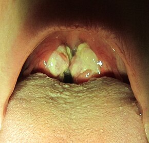
Exudative pharyngitis in a person with infectious mononucleosis
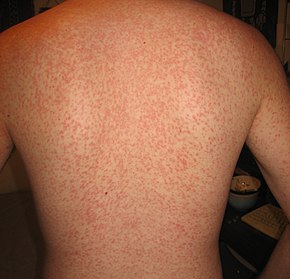
Cross reaction rash
Rash from using penicillin while infected with IM
Maculopapular rash from amoxicillin use during EBV infection

The signs and symptoms of infectious mononucleosis vary with age.

Children

Before puberty, the disease typically only produces flu-like symptoms, if any at all. When found, symptoms tend to be similar to those of common throat infections (mild pharyngitis, with or without tonsillitis).

Adolescents and young adults

In adolescence and young adulthood, the disease presents with a characteristic triad:

  • Fever – usually lasting 14 days; often mild
  • Sore throat – usually severe for 3–5 days, before resolving in the next 7–10 days.
  • Swollen glands –  mobile; usually located around the back of the neck (posterior cervical lymph nodes) and sometimes throughout the body.

Another major symptom is feeling tired. Headaches are common, and abdominal pains with nausea or vomiting sometimes also occur. Symptoms most often disappear after about 2–4 weeks. However, fatigue and a general feeling of being unwell (malaise) may sometimes last for months. Fatigue lasts more than one month in an estimated 28% of cases. Mild fever, swollen neck glands and body aches may also persist beyond 4 weeks. Most people are able to resume their usual activities within 2–3 months.

The most prominent sign of the disease is often the pharyngitis, which is frequently accompanied by enlarged tonsils with pus—an exudate similar to that seen in cases of strep throat. In about 50% of cases, small reddish-purple spots called petechiae can be seen on the roof of the mouth. Palatal enanthem can also occur, but is relatively uncommon.

A small minority of people spontaneously present a rash, usually on the arms or trunk, which can be macular (morbilliform) or papular. Almost all people given amoxicillin or ampicillin eventually develop a generalized, itchy maculopapular rash, which however does not imply that the person will have adverse reactions to penicillins again in the future. Occasional cases of erythema nodosum and erythema multiforme have been reported. Seizures may also occasionally occur.

Complications

Spleen enlargement is common in the second and third weeks, although this may not be apparent on physical examination. Rarely the spleen may rupture. There may also be some enlargement of the liver. Jaundice occurs only occasionally.

It generally gets better on its own in people who are otherwise healthy. When caused by EBV, infectious mononucleosis is classified as one of the Epstein–Barr virus–associated lymphoproliferative diseases. Occasionally the disease may persist and result in a chronic infection. This may develop into systemic EBV-positive T cell lymphoma.

Older adults

Infectious mononucleosis mainly affects younger adults. When older adults do catch the disease, they less often have characteristic signs and symptoms such as the sore throat and lymphadenopathy. Instead, they may primarily experience prolonged fever, fatigue, malaise and body pains. They are more likely to have liver enlargement and jaundice. People over 40 years of age are more likely to develop serious illness.

Incubation period

The exact length of time between infection and symptoms is unclear. A review of the literature made an estimate of 33–49 days. In adolescents and young adults, symptoms are thought to appear around 4–6 weeks after initial infection. Onset is often gradual, though it can be abrupt. The main symptoms may be preceded by 1–2 weeks of fatigue, feeling unwell and body aches.

Cause

Epstein–Barr virus

About 90% of cases of infectious mononucleosis are caused by the Epstein–Barr virus, a member of the Herpesviridae family of DNA viruses. It is one of the most commonly found viruses throughout the world. Contrary to common belief, the Epstein–Barr virus is not highly contagious. It can only be contracted through direct contact with an infected person's saliva, such as through kissing or sharing toothbrushes. About 95% of the population has been exposed to this virus by the age of 40, but only 15–20% of teenagers and about 40% of exposed adults actually develop infectious mononucleosis.

Cytomegalovirus

About 5–7% of cases of infectious mononucleosis is caused by human cytomegalovirus (CMV), another type of herpes virus. This virus is found in body fluids including saliva, urine, blood, and tears. A person becomes infected with this virus by direct contact with infected body fluids. Cytomegalovirus is most commonly transmitted through kissing and sexual intercourse. It can also be transferred from an infected mother to her unborn child. This virus is often "silent" because the signs and symptoms cannot be felt by the person infected. However, it can cause life-threatening illness in infants, people with HIV, transplant recipients, and those with weak immune systems. For those with weak immune systems, cytomegalovirus can cause more serious illnesses such as pneumonia and inflammations of the retina, esophagus, liver, large intestine, and brain. Approximately 90% of the human population has been infected with cytomegalovirus by the time they reach adulthood, but most are unaware of the infection. Once a person becomes infected with cytomegalovirus, the virus stays in their body fluids throughout the person's lifetime.

Transmission

Epstein–Barr virus infection is spread via saliva, and has an incubation period of four to seven weeks. The length of time that an individual remains contagious is unclear, but the chances of passing the illness to someone else may be the highest during the first six weeks following infection. Some studies indicate that a person can spread the infection for many months, possibly up to a year and a half.

Pathophysiology

The virus replicates first within epithelial cells in the pharynx (which causes pharyngitis, or sore throat), and later primarily within B cells (which are invaded via their CD21). The host immune response involves cytotoxic (CD8-positive) T cells against infected B lymphocytes, resulting in enlarged, atypical lymphocytes (Downey cells).

When the infection is acute (recent onset, instead of chronic), heterophile antibodies are produced.

Cytomegalovirus, adenovirus and Toxoplasma gondii (toxoplasmosis) infections can cause symptoms similar to infectious mononucleosis, but a heterophile antibody test will test negative and differentiate those infections from infectious mononucleosis.

Mononucleosis is sometimes accompanied by secondary cold agglutinin disease, an autoimmune disease in which abnormal circulating antibodies directed against red blood cells can lead to a form of autoimmune hemolytic anemia. The cold agglutinin detected is of anti-i specificity.

Diagnosis

Infectious mononucleosis, peripheral smear, high power showing reactive lymphocytes
Splenomegaly due to mononucleosis resulting in a subcapsular hematoma

Diagnostic modalities for infectious mononucleosis include:

Physical examination

The presence of an enlarged spleen, and swollen posterior cervical, axillary, and inguinal lymph nodes are the most useful to suspect a diagnosis of infectious mononucleosis. On the other hand, the absence of swollen cervical lymph nodes and fatigue are the most useful to dismiss the idea of infectious mononucleosis as the correct diagnosis. The insensitivity of the physical examination in detecting an enlarged spleen means it should not be used as evidence against infectious mononucleosis. A physical examination may also show petechiae in the palate.

Heterophile antibody test

The heterophile antibody test, or monospot test, works by agglutination of red blood cells from guinea pigs, sheep and horses. This test is specific but not particularly sensitive (with a false-negative rate of as high as 25% in the first week, 5–10% in the second, and 5% in the third). About 90% of diagnosed people have heterophile antibodies by week 3, disappearing in under a year. The antibodies involved in the test do not interact with the Epstein–Barr virus or any of its antigens.

The monospot test is not recommended for general use by the CDC due to its poor accuracy.

Serology

Serologic tests detect antibodies directed against the Epstein–Barr virus. Immunoglobulin G (IgG), when positive, mainly reflects a past infection, whereas immunoglobulin M (IgM) mainly reflects a current infection. EBV-targeting antibodies can also be classified according to which part of the virus they bind to:

  • Viral capsid antigen (VCA):
  • Anti-VCA IgM appear early after infection, and usually, disappear within 4 to 6 weeks.
  • Anti-VCA IgG appears in the acute phase of EBV infection, reaches a maximum at 2 to 4 weeks after onset of symptoms and thereafter declines slightly and persists for the rest of a person’s life.
  • Early antigen (EA)
  • Anti-EA IgG appears in the acute phase of illness and disappears after 3 to 6 months. It is associated with having an active infection. Yet, 20% of people may have antibodies against EA for years despite having no other sign of infection.
  • EBV nuclear antigen (EBNA)
  • Antibody to EBNA slowly appears 2 to 4 months after the onset of symptoms and persists for the rest of a person’s life.

When negative, these tests are more accurate than the heterophile antibody test in ruling out infectious mononucleosis. When positive, they feature similar specificity to the heterophile antibody test. Therefore, these tests are useful for diagnosing infectious mononucleosis in people with highly suggestive symptoms and a negative heterophile antibody test.

Other tests

Differential diagnosis

About 10% of people who present a clinical picture of infectious mononucleosis do not have an acute Epstein–Barr-virus infection. A differential diagnosis of acute infectious mononucleosis needs to take into consideration acute cytomegalovirus infection and Toxoplasma gondii infections. Because their management is much the same, it is not always helpful, or possible, to distinguish between Epstein–Barr-virus mononucleosis and cytomegalovirus infection. However, in pregnant women, differentiation of mononucleosis from toxoplasmosis is important, since it is associated with significant consequences for the fetus.

Acute HIV infection can mimic signs similar to those of infectious mononucleosis, and tests should be performed for pregnant women for the same reason as toxoplasmosis.

People with infectious mononucleosis are sometimes misdiagnosed with a streptococcal pharyngitis (because of the symptoms of fever, pharyngitis and adenopathy) and are given antibiotics such as ampicillin or amoxicillin as treatment.

Other conditions from which to distinguish infectious mononucleosis include leukemia, tonsillitis, diphtheria, common cold and influenza (flu).

Treatment

Infectious mononucleosis is generally self-limiting, so only symptomatic or supportive treatments are used. The need for rest and return to usual activities after the acute phase of the infection may reasonably be based on the person's general energy levels. Nevertheless, in an effort to decrease the risk of splenic rupture experts advise avoidance of contact sports and other heavy physical activity, especially when involving increased abdominal pressure or the Valsalva maneuver (as in rowing or weight training), for at least the first 3–4 weeks of illness or until enlargement of the spleen has resolved, as determined by a treating physician.

Medications

Paracetamol (acetaminophen) and NSAIDs, such as ibuprofen, may be used to reduce fever and pain. Prednisone, a corticosteroid, while used to try to reduce throat pain or enlarged tonsils, remains controversial due to the lack of evidence that it is effective and the potential for side effects. Intravenous corticosteroids, usually hydrocortisone or dexamethasone, are not recommended for routine use but may be useful if there is a risk of airway obstruction, a very low platelet count, or hemolytic anemia.

Antiviral agents act by inhibiting viral DNA replication. There is little evidence to support the use of antivirals such as aciclovir and valacyclovir although they may reduce initial viral shedding. Antivirals are expensive, risk causing resistance to antiviral agents, and (in 1% to 10% of cases) can cause unpleasant side effects. Although antivirals are not recommended for people with simple infectious mononucleosis, they may be useful (in conjunction with steroids) in the management of severe EBV manifestations, such as EBV meningitis, peripheral neuritis, hepatitis, or hematologic complications.

Although antibiotics exert no antiviral action they may be indicated to treat bacterial secondary infections of the throat, such as with streptococcus (strep throat). However, ampicillin and amoxicillin are not recommended during acute Epstein–Barr virus infection as a diffuse rash may develop.

Observation

Splenomegaly is a common symptom of infectious mononucleosis and health care providers may consider using abdominal ultrasonography to get insight into the enlargement of a person's spleen. However, because spleen size varies greatly, ultrasonography is not a valid technique for assessing spleen enlargement and should not be used in typical circumstances or to make routine decisions about fitness for playing sports.

Prognosis

Serious complications are uncommon, occurring in less than 5% of cases:

Once the acute symptoms of an initial infection disappear, they often do not return. But once infected, the person carries the virus for the rest of their life. The virus typically lives dormant in B lymphocytes. Independent infections of mononucleosis may be contracted multiple times, regardless of whether the person is already carrying the virus dormant. Periodically, the virus can reactivate, during which time the person is again infectious, but usually without any symptoms of illness. Usually, a person with IM has few, if any, further symptoms or problems from the latent B lymphocyte infection. However, in susceptible hosts under the appropriate environmental stressors, the virus can reactivate and cause vague physical symptoms (or may be subclinical), and during this phase, the virus can spread to others.

History

The characteristic symptomatology of infectious mononucleosis does not appear to have been reported until the late nineteenth century. In 1885, the renowned Russian pediatrician Nil Filatov reported an infectious process he called "idiopathic adenitis" exhibiting symptoms that correspond to infectious mononucleosis, and in 1889 a German balneologist and pediatrician, Emil Pfeiffer, independently reported similar cases (some of lesser severity) that tended to cluster in families, for which he coined the term Drüsenfieber ("glandular fever").

The word mononucleosis has several senses, but today it usually is used in the sense of infectious mononucleosis, which is caused by EBV.

The term "infectious mononucleosis" was coined in 1920 by Thomas Peck Sprunt and Frank Alexander Evans in a classic clinical description of the disease published in the Bulletin of the Johns Hopkins Hospital, entitled "Mononuclear leukocytosis in reaction to acute infection (infectious mononucleosis)". A lab test for infectious mononucleosis was developed in 1931 by Yale School of Public Health Professor John Rodman Paul and Walls Willard Bunnell based on their discovery of heterophile antibodies in the sera of persons with the disease. The Paul-Bunnell Test or PBT was later replaced by the heterophile antibody test.

The Epstein–Barr virus was first identified in Burkitt's lymphoma cells by Michael Anthony Epstein and Yvonne Barr at the University of Bristol in 1964. The link with infectious mononucleosis was uncovered in 1967 by Werner and Gertrude Henle at the Children's Hospital of Philadelphia, after a laboratory technician handling the virus contracted the disease: comparison of serum samples collected from the technician before and after the onset revealed development of antibodies to the virus.

Yale School of Public Health epidemiologist Alfred E. Evans confirmed through testing that mononucleosis was transmitted mainly through kissing, leading to it being referred to colloquially as "the kissing disease".

Operator (computer programming)

From Wikipedia, the free encyclopedia https://en.wikipedia.org/wiki/Operator_(computer_programmin...